

Strategies for Bovine Respiratory Disease (BRD) Diagnosis and Prognosis: A Comprehensive Overview

Abstract
:Simple Summary
Abstract
1. Introduction
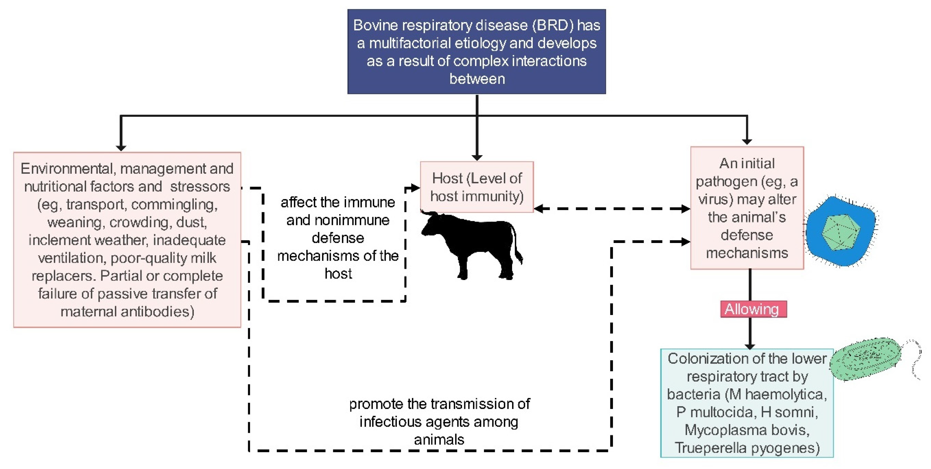
2. Overview of BRD
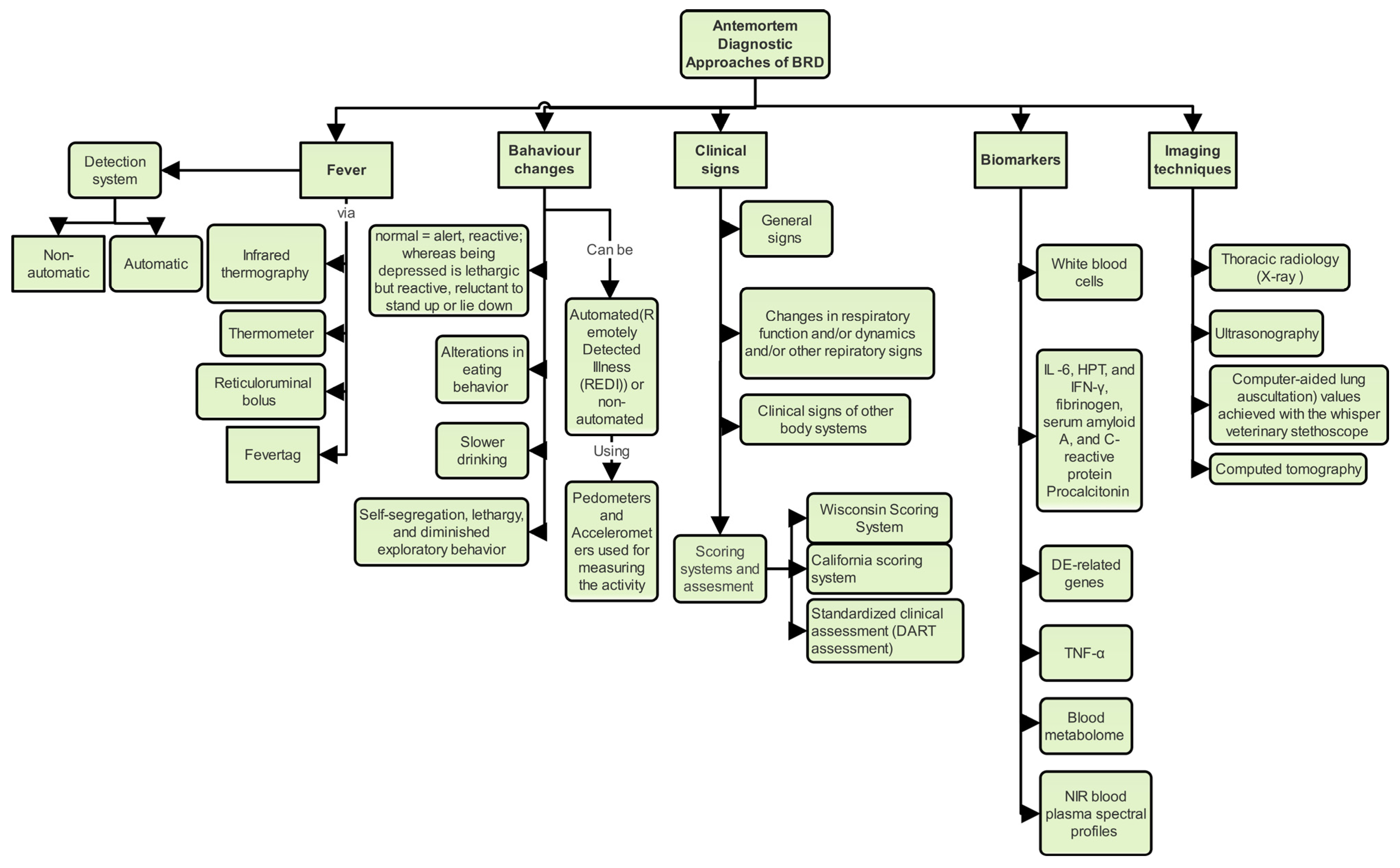
3. Clinical and Behavioral Tools BRD Diagnosis and Prognosis
3.1. Clinical Signs, Clinical Scoring and Clinical Signs Assessment
3.1.1. Overview of Clinical Signs Resulting from BRD
3.1.2. Clinical Scoring and Clinical Signs Assessment in BRD Diagnosis and Prognosis
3.2. Temperature Detection as a Tool for BRD Diagnosis, Prediction, and Prognosis
3.2.1. Fever as a General Non-Specific Sign of BRD and Its Causes
3.2.2. Methods for Detecting Fever in BRD
3.2.3. Fever as a Method for Detecting and Diagnosing Clinical and Subclinical BRD
3.3. Behavioral Changes in BRD Diagnosis and Prognosis
3.3.1. Alertness as a Behavior Change in BRD Diagnosis
3.3.2. Energy and Feeding, Drinking Behavior, and Other Behavior Changes during BRD
3.3.3. Lameness as a Behavior Change in BRD Diagnosis
3.3.4. Some Prospective Studies in Behavior Changes in BRD Diagnosis and Prognosis
3.3.5. Behavior Changes and BRD Treatment by Antibiotics and NSAIDs
3.4. Necropsy as a BRD Gold Standard Diagnostic Test
4. Imaging Techniques for BRD Diagnosis and Prognosis
4.1. Thoracic Radiography
4.2. Thoracic Ultrasonography of the Chest for BRD Diagnosis, Prognosis, and Treatment Follow-Up
4.2.1. TUS in BRD Diagnosis and Prognosis: Sensitivity, Specificity, and Probes Used
4.2.2. TUS as a Tool for Treatment Follow-Up and the Disease’s Recovery
4.2.3. The Pros and Cons of TUS as a Promising Tool for BRD Diagnosis and Prognosis Research

| Objective | Study Design | Sample Size | Date of the Published Study | Location | Key Findings | Reference |
|---|---|---|---|---|---|---|
| Assessing the diagnostic and prognostic utility of lung ultrasonography in BRD | longitudinal design | 600 | 2023 | Austria | High sensitivity (86%) and specificity (78%) | [163] |
| Assessing TUS’s diagnostic and prognostic value in initial BRD cases | Prospective longitudinal study | 174 | 2012 | Canada | TUS proved valuable in targeted populations of animals with prolonged respiratory disease. | [29] |
| Improving BRD detection in experimental infection using TUS | Experimental Study | 62 | 2021 | USA |
| [164] |
| Assessing the impact of lung consolidation detected via TUS on health and growth outcomes in BRD | Longitudinal Study | 221 | 2023 | Iran | The sensitivity ranges from 86% to 94% and the specificity ranges from 98% to 100%. Additionally, one or more consolidation episodes resulted in a significantly lower ADG. | [165] |
| Assessing subclinical lung lesions in Holstein calves using TUS and bronchoalveolar lavage fluid analysis. | Prospective study | 25 | 2015 | Canada | TUS had a 94% sensitivity and 100% specificity in detecting lung lesions related to subclinical BRD in healthy calves. | [111] |
| Evaluating the accuracy and inter-rater reliability of lung auscultation among bovine practitioners relative to TUS findings | Diagnostic test study | 49 | 2019 | Netherlands | TUS is the most accurate for practical use in the field | [166] |
| Assessing the association of clinical respiratory signs and lung lesions detected via TUS with growth performance in pre-weaned dairy calves | Retrospective cohort study | 53 | 2021 | Ireland | TUS found lung consolidation in healthy calves, and severe lung lesions affected their pre-weaning growth. | [67] |
| Assessing the efficacy of TUS for diagnosing BRD in preweaned dairy calves | Longitudinal study | 60 | 2023 | USA | The sensitivity and specificity of TUS vary depending on the scoring method used. | [167] |
| Evaluating the efficacy of lung ultrasonography and clinical assessments in monitoring BRD in fattening bulls during restocking and post-treatment with tulathromycin and ketoprofen | case time-series analysis | 60 | 2022 | Italy | TUS presents a higher sensitivity (80–94%) and specificity (94–100%) than clinical scoring to detect lung lesions. It is a non-invasive and cost-effective tool for BRD early diagnosis and for monitoring treatment efficacy. | [51] |
4.3. Focus Lung Ultrasonography as a Tool for Evaluating Lung Lesions in BRD
5. Molecular and Sample-Based Methods for BRD Diagnosis and Prognosis
5.1. Molecular Diagnostics
5.2. Diagnosing BRD through White and Red Blood Cells as Well as Blood Plasma
5.3. Transcriptomic and Metabolic Profiling in BRD Diagnosis
5.4. Acute Phase Proteins in BRD Diagnosis and Prognosis
6. Automated and Predictive Methods for BRD Diagnosis and Prognosis
6.1. Automated Behavior Detection
Role of Accelerometers in Automated BRD Diagnosis
6.2. Early Disease Detection Systems
6.3. Machine Learning and Modeling Approaches for BRD Diagnosis and Prognosis
7. Utility of Diagnostic Techniques in Prognosticating BRD Outcomes
8. Conclusions
Author Contributions
Funding
Data Availability Statement
Conflicts of Interest
References
- Ellis, J.A. The Immunology of the Bovine Respiratory Disease Complex. Vet. Clin. N. Am. Food Anim. Pract. 2001, 17, 535–550. [Google Scholar] [CrossRef]
- Taylor The Epidemiology of Bovine Respiratory Disease: What Is the Evidence for Predisposing Factors? Can. Vet. J. 2010, 51, 1095.
- McGill, J.L.; Sacco, R.E. The Immunology of Bovine Respiratory Disease: Recent Advancements. Vet. Clin. N. Am. Food Anim. Pract. 2020, 36, 333–348. [Google Scholar] [CrossRef]
- Johnson, K.K.; Pendell, D.L. Market Impacts of Reducing the Prevalence of Bovine Respiratory Disease in United States Beef Cattle Feedlots. Front. Vet. Sci. 2017, 4, 189. [Google Scholar] [CrossRef]
- Myer, P.R.; McDaneld, T.G.; Kuehn, L.A.; DeDonder, K.D.; Apley, M.D.; Capik, S.F.; Lubbers, B.V.; Harhay, G.P.; Harhay, D.M.; Keele, J.W.; et al. Classification of 16S rRNA Reads Is Improved Using a Niche-Specific Database Constructed by near-Full Length Sequencing. PLoS ONE 2020, 15, e0235498. [Google Scholar] [CrossRef] [PubMed]
- Brumbaugh, G.W. Will Antimicrobial Resistance of BRD Pathogens Impact BRD Management in the Future? Anim. Health Res. Rev. 2014, 15, 175–177. [Google Scholar] [CrossRef] [PubMed]
- Klima, C.L.; Zaheer, R.; Cook, S.R.; Booker, C.W.; Hendrick, S.; Alexander, T.W.; McAllister, T.A. Pathogens of Bovine Respiratory Disease in North American Feedlots Conferring Multidrug Resistance via Integrative Conjugative Elements. J. Clin. Microbiol. 2014, 52, 438–448. [Google Scholar] [CrossRef] [PubMed]
- Cameron, A.; McAllister, T.A. Antimicrobial Usage and Resistance in Beef Production. J. Anim. Sci. Biotechnol. 2016, 7, 68. [Google Scholar] [CrossRef]
- Amat, S. Bovine Respiratory Disease in Feedlot Cattle: Antimicrobial Resistance in Bovine Respiratory Bacterial Pathogens and Alternative Antimicrobial Approaches. In Bacterial Cattle Diseases; IntechOpen: London, UK, 2019. [Google Scholar] [CrossRef]
- Andrés-Lasheras, S.; Jelinski, M.; Zaheer, R.; McAllister, T.A. Bovine Respiratory Disease: Conventional to Culture-Independent Approaches to Studying Antimicrobial Resistance in North America. Antibiotics 2022, 11, 487. [Google Scholar] [CrossRef] [PubMed]
- HAMDY, A.H.; TRAPP, A.L.; GALE, C.; KING, N.B. Experimental Transmission of Shipping Fever in Calves. Am. J. Vet. Res. 1963, 24, 287–294. [Google Scholar]
- Griffin, D.; Chengappa, M.M.; Kuszak, J.; McVey, D.S. Bacterial Pathogens of the Bovine Respiratory Disease Complex. Vet. Clin. N. Am. Food Anim. Pract. 2010, 26, 381–394. [Google Scholar] [CrossRef]
- Gaudino, M.; Nagamine, B.; Ducatez, M.F.; Meyer, G. Understanding the Mechanisms of Viral and Bacterial Coinfections in Bovine Respiratory Disease: A Comprehensive Literature Review of Experimental Evidence. Can. J. Comp. Med. 2022, 53, 70. [Google Scholar] [CrossRef] [PubMed]
- Buczinski, S.; Pardon, B. Bovine Respiratory Disease Diagnosis: What Progress Has Been Made in Clinical Diagnosis? Vet. Clin. N. Am. Food Anim. Pract. 2020, 36, 399–423. [Google Scholar] [CrossRef] [PubMed]
- Ferraro, S.; Fecteau, G.; Dubuc, J.; Francoz, D.; Rousseau, M.; Roy, J.-P.; Buczinski, S. Scoping Review on Clinical Definition of Bovine Respiratory Disease Complex and Related Clinical Signs in Dairy Cows. J. Dairy Sci. 2021, 104, 7095–7108. [Google Scholar] [CrossRef]
- Apley, M. Bovine Respiratory Disease: Pathogenesis, Clinical Signs, and Treatment in Lightweight Calves. Vet. Clin. N. Am. Food Anim. Pract. 2006, 22, 399–411. [Google Scholar] [CrossRef]
- Pardon, B.; Buczinski, S. Bovine Respiratory Disease Diagnosis: What Progress Has Been Made in Infectious Diagnosis? J. Agric. Res. Econ. 2020, 36, 425–444. [Google Scholar] [CrossRef]
- Booker, C.W.; Lubbers, B.V. Bovine Respiratory Disease Treatment Failure: Impact and Potential Causes. Vet. Clin. Food Anim. Pract. 2020, 36, 487–496. [Google Scholar] [CrossRef]
- Cummings, D.B.; Meyer, N.F.; Step, D.L. Bovine Respiratory Disease Considerations in Young Dairy Calves. Vet. Clin. N. Am. Food Anim. Pract. 2022, 38, 93–105. [Google Scholar] [CrossRef]
- Buczinski, S.; Faure, C.; Jolivet, S.; Abdallah, A. Evaluation of Inter-Observer Agreement When Using a Clinical Respiratory Scoring System in Pre-Weaned Dairy Calves. N. Z. Vet. J. 2016, 64, 243–247. [Google Scholar] [CrossRef]
- Buczinski, S.; Boccardo, A.; Pravettoni, D. Clinical Scores in Veterinary Medicine: What Are the Pitfalls of Score Construction, Reliability, and Validation? A General Methodological Approach Applied in Cattle. Animals 2021, 11, 3244. [Google Scholar] [CrossRef]
- Love, W.J.; Lehenbauer, T.W.; Kass, P.H.; Van Eenennaam, A.L.; Aly, S.S. Development of a Novel Clinical Scoring System for On-Farm Diagnosis of Bovine Respiratory Disease in Pre-Weaned Dairy Calves. PeerJ 2014, 2, e238. [Google Scholar] [CrossRef]
- Love, W.J.; Lehenbauer, T.W.; Van Eenennaam, A.L.; Drake, C.M.; Kass, P.H.; Farver, T.B.; Aly, S.S. Sensitivity and Specificity of On-Farm Scoring Systems and Nasal Culture to Detect Bovine Respiratory Disease Complex in Preweaned Dairy Calves. J. VET Diagn. Invest. 2016, 28, 119–128. [Google Scholar] [CrossRef]
- Buczinski, S.; Fecteau, G.; Dubuc, J.; Francoz, D. Validation of a Clinical Scoring System for Bovine Respiratory Disease Complex Diagnosis in Preweaned Dairy Calves Using a Bayesian Framework. Prev. Vet. Med. 2018, 156, 102–112. [Google Scholar] [CrossRef]
- Maier, G.U.; Rowe, J.D.; Lehenbauer, T.W.; Karle, B.M.; Williams, D.R.; Champagne, J.D.; Aly, S.S. Development of a Clinical Scoring System for Bovine Respiratory Disease in Weaned Dairy Calves. J. Dairy Sci. 2019, 102, 7329–7344. [Google Scholar] [CrossRef] [PubMed]
- Aich, P.; Babiuk, L.A.; Potter, A.A.; Griebel, P. Biomarkers for Prediction of Bovine Respiratory Disease Outcome. OMICS J. Integr. Biol. 2009, 13, 199–209. [Google Scholar] [CrossRef] [PubMed]
- Babkine, M.; Blond, L. Ultrasonography of the Bovine Respiratory System and Its Practical Application. Vet. Clin. N. Am. Food Anim. Pract. 2009, 25, 633–649. [Google Scholar] [CrossRef] [PubMed]
- Bannikov, G.A.; Hinds, C.A.; Rajala-Schultz, P.J.; Premanandan, C.; Rings, D.M.; Lakritz, J. Serum Haptoglobin-Matrix Metalloproteinase 9 (Hp-MMP 9) Complex as a Biomarker of Systemic Inflammation in Cattle. Vet. Immunol. Immunopathol. 2011, 139, 41–49. [Google Scholar] [CrossRef] [PubMed]
- Abutarbush, S.M.; Pollock, C.M.; Wildman, B.K.; Perrett, T.; Schunicht, O.C.; Fenton, R.K.; Hannon, S.J.; Vogstad, A.R.; Jim, G.K.; Booker, C.W. Evaluation of the Diagnostic and Prognostic Utility of Ultrasonography at First Diagnosis of Presumptive Bovine Respiratory Disease. Can. J. Vet. Res. 2012, 76, 23–32. [Google Scholar] [PubMed]
- Adams, E.A.; Buczinski, S. Short Communication: Ultrasonographic Assessment of Lung Consolidation Postweaning and Survival to the First Lactation in Dairy Heifers. J. Dairy Sci. 2016, 99, 1465–1470. [Google Scholar] [CrossRef] [PubMed]
- Kovačić, M.; Marković, D.; Maslovarić, I.; Obrenović, S.; Grujić-Milanović, J.; Arsić, A.; Milanović, Z.; Savić, O.; Fratrić, N.; Ilić, V. Serum Proteins and Lipids in Mild Form of Calf Bronchopneumonia: Candidates for Reliable Biomarkers. Acta Vet. 2017, 67, 201–221. [Google Scholar] [CrossRef]
- Buczinski, S.; Buathier, C.; Bélanger, A.M.; Michaux, H.; Tison, N.; Timsit, E. Inter-Rater Agreement and Reliability of Thoracic Ultrasonographic Findings in Feedlot Calves, with or without Naturally Occurring Bronchopneumonia. J. Vet. Intern. Med. 2018, 32, 1787–1792. [Google Scholar] [CrossRef] [PubMed]
- Maurer, D.; Koziel, J.; Engelken, T.; Cooper, V.; Funk, J. Detection of Volatile Compounds Emitted from Nasal Secretions and Serum: Towards Non-Invasive Identification of Diseased Cattle Biomarkers. Separations 2018, 5, 18. [Google Scholar] [CrossRef]
- Moisá, S.J.; Aly, S.S.; Lehenbauer, T.W.; Love, W.J.; Rossitto, P.V.; Van Eenennaam, A.L.; Trombetta, S.C.; Bortoluzzi, E.M.; Hulbert, L.E. Association of Plasma Haptoglobin Concentration and Other Biomarkers with Bovine Respiratory Disease Status in Pre-Weaned Dairy Calves. J. Vet. Diagn. Invest. 2019, 31, 40–46. [Google Scholar] [CrossRef]
- Berman, J.; Masseau, I.; Fecteau, G.; Buczinski, S.; Francoz, D. Comparison between Thoracic Ultrasonography and Thoracic Radiography for the Detection of Thoracic Lesions in Dairy Calves Using a Two-Stage Bayesian Method. Prev. Vet. Med. 2020, 184, 105153. [Google Scholar] [CrossRef] [PubMed]
- Juge, A.E.; Hall, N.J.; Richeson, J.T.; Daigle, C.L. Using Canine Olfaction to Detect Bovine Respiratory Disease: A Pilot Study. Front. Vet. Sci. 2022, 9, 902151. [Google Scholar] [CrossRef] [PubMed]
- Kamel, M.S.; El-Sayed, A.A.; Munds, R.A.; Verma, M.S. Interactions between Humans and Dogs during the COVID-19 Pandemic: Recent Updates and Future Perspectives. Animals 2023, 13, 524. [Google Scholar] [CrossRef]
- Stokstad, M.; Klem, T.B.; Myrmel, M.; Oma, V.S.; Toftaker, I.; Østerås, O.; Nødtvedt, A. Using Biosecurity Measures to Combat Respiratory Disease in Cattle: The Norwegian Control Program for Bovine Respiratory Syncytial Virus and Bovine Coronavirus. Front. Vet. Sci. 2020, 7, 167. [Google Scholar] [CrossRef]
- Peel, D.S. The Effect of Market Forces on Bovine Respiratory Disease. J. Agric. Res. Econ. 2020, 36, 497–508. [Google Scholar] [CrossRef]
- Sanderson, M.W.; Dargatz, D.A.; Wagner, B.A. Risk Factors for Initial Respiratory Disease in United States’ Feedlots Based on Producer-Collected Daily Morbidity Counts. Can. Vet. J. 2008, 49, 373–378. [Google Scholar]
- Hay, K.E.; Morton, J.M.; Schibrowski, M.L.; Clements, A.C.A.; Mahony, T.J.; Barnes, T.S. Associations between Prior Management of Cattle and Risk of Bovine Respiratory Disease in Feedlot Cattle. Prev. Vet. Med. 2016, 127, 37–43. [Google Scholar] [CrossRef]
- Padalino, B.; Cirone, F.; Zappaterra, M.; Tullio, D.; Ficco, G.; Giustino, A.; Ndiana, L.A.; Pratelli, A. Factors Affecting the Development of Bovine Respiratory Disease: A Cross-Sectional Study in Beef Steers Shipped From France to Italy. Front. Vet. Sci. 2021, 8, 627894. [Google Scholar] [CrossRef] [PubMed]
- Lehenbauer, T.W. Control of BRD in Large Dairy Calf Populations. Anim. Health Res. Rev. 2014, 15, 184–185. [Google Scholar] [CrossRef] [PubMed]
- Cirone, F.; Padalino, B.; Tullio, D.; Capozza, P.; Lo Surdo, M.; Lanave, G.; Pratelli, A. Prevalence of Pathogens Related to Bovine Respiratory Disease Before and After Transportation in Beef Steers: Preliminary Results. Animals 2019, 9, 1093. [Google Scholar] [CrossRef]
- Coetzee, J.F.; Cernicchiaro, N.; Sidhu, P.K.; Kleinhenz, M.D. Association between Antimicrobial Drug Class Selection for Treatment and Retreatment of Bovine Respiratory Disease and Health, Performance, and Carcass Quality Outcomes in Feedlot Cattle. J. Anim. Sci. 2020, 98, skaa109. [Google Scholar] [CrossRef] [PubMed]
- Centeno-Martinez, R.E.; Glidden, N.; Mohan, S.; Davidson, J.L.; Fernández-Juricic, E.; Boerman, J.P.; Schoonmaker, J.; Pillai, D.; Koziol, J.; Ault, A.; et al. Identification of Bovine Respiratory Disease through the Nasal Microbiome. Anim. Microbiome 2022, 4, 15. [Google Scholar] [CrossRef]
- Zhang, M.; Hill, J.E.; Fernando, C.; Alexander, T.W.; Timsit, E.; van der Meer, F.; Huang, Y. Respiratory Viruses Identified in Western Canadian Beef Cattle by Metagenomic Sequencing and Their Association with Bovine Respiratory Disease. Transbound. Emerg. Dis. 2019, 66, 1379–1386. [Google Scholar] [CrossRef]
- Fulton, R.W. Viruses in Bovine Respiratory Disease in North America: Knowledge Advances Using Genomic Testing. J. Agric. Res. Econ. 2020, 36, 321–332. [Google Scholar] [CrossRef]
- Abell, K.M.; Theurer, M.E.; Larson, R.L.; White, B.J.; Apley, M. A Mixed Treatment Comparison Meta-Analysis of Metaphylaxis Treatments for Bovine Respiratory Disease in Beef Cattle. J. Anim. Sci. 2017, 95, 626. [Google Scholar] [CrossRef]
- Toaff-Rosenstein, R.L.; Gershwin, L.J.; Zanella, A.J.; Tucker, C.B. The Sickness Response in Steers with Induced Bovine Respiratory Disease before and after Treatment with a Non-Steroidal Anti-Inflammatory Drug. Appl. Anim. Behav. Sci. 2016, 181, 49–62. [Google Scholar] [CrossRef]
- Fiore, E.; Lisuzzo, A.; Beltrame, A.; Contiero, B.; Gianesella, M.; Schiavon, E.; Tessari, R.; Morgante, M.; Mazzotta, E. Lung Ultrasonography and Clinical Follow-Up Evaluations in Fattening Bulls Affected by Bovine Respiratory Disease (BRD) during the Restocking Period and after Tulathromycin and Ketoprofen Treatment. Animals 2022, 12, 994. [Google Scholar] [CrossRef]
- Lowie, T.; Van Leenen, K.; Jourquin, S.; Pas, M.L.; Bokma, J.; Pardon, B. Differences in the Association of Cough and Other Clinical Signs with Ultrasonographic Lung Consolidation in Dairy, Veal, and Beef Calves. J. Dairy Sci. 2022, 105, 6111–6124. [Google Scholar] [CrossRef]
- Ng, T.F.F.; Kondov, N.O.; Deng, X.; van Eenennaam, A.; Neibergs, H.L.; Delwart, E. A Metagenomics and Case-Control Study to Identify Viruses Associated with Bovine Respiratory Disease. J. Virol. 2015, 89, 5340–5349. [Google Scholar] [CrossRef] [PubMed]
- Radostits, O.M.; Gay, C.C.; Hinchcliff, K.W.; Constable, P.D. (Eds.) Veterinary Medicine E-Book, 10th ed.; Saunders Ltd.: Nottingham, UK, 2006. [Google Scholar]
- Stanton, A.L.; Kelton, D.F.; LeBlanc, S.J.; Wormuth, J.; Leslie, K.E. The Effect of Respiratory Disease and a Preventative Antibiotic Treatment on Growth, Survival, Age at First Calving, and Milk Production of Dairy Heifers. J. Dairy Sci. 2012, 95, 4950–4960. [Google Scholar] [CrossRef]
- Virtala, A.-M.K.; Mechor, G.D.; Gröhn, Y.T.; Erb, H.N. The Effect of Calfhood Diseases on Growth of Female Dairy Calves During the First 3 Months of Life in New York State. J. Dairy Sci. 1996, 79, 1040–1049. [Google Scholar] [CrossRef]
- Waltner-Toews, D.; Martin, S.W.; Meek, A.H. The Effect of Early Calfhood Health Status on Survivorship and Age at First Calving. Can. J. Vet. Res. 1986, 50, 314–317. [Google Scholar]
- Sivula, N.J.; Ames, T.R.; Marsh, W.E.; Werdin, R.E. Descriptive Epidemiology of Morbidity and Mortality in Minnesota Dairy Heifer Calves. Prev. Vet. Med. 1996, 27, 155–171. [Google Scholar] [CrossRef]
- Cramer, M.C.; Ollivett, T.L. Growth of Preweaned, Group-Housed Dairy Calves Diagnosed with Respiratory Disease Using Clinical Respiratory Scoring and Thoracic Ultrasound-A Cohort Study. J. Dairy Sci. 2019, 102, 4322–4331. [Google Scholar] [CrossRef] [PubMed]
- Timsit, E.; Dendukuri, N.; Schiller, I.; Buczinski, S. Diagnostic Accuracy of Clinical Illness for Bovine Respiratory Disease (BRD) Diagnosis in Beef Cattle Placed in Feedlots: A Systematic Literature Review and Hierarchical Bayesian Latent-Class Meta-Analysis. Prev. Vet. Med. 2016, 135, 67–73. [Google Scholar] [CrossRef]
- McGuirk, S.M. Disease Management of Dairy Calves and Heifers. Vet. Clin. N. Am. Food Anim. Pract. 2008, 24, 139–153. [Google Scholar] [CrossRef]
- Timsit, E.; Assié, S.; Quiniou, R.; Seegers, H.; Bareille, N. Early Detection of Bovine Respiratory Disease in Young Bulls Using Reticulo-Rumen Temperature Boluses. Vet. J. 2011, 190, 136–142. [Google Scholar] [CrossRef]
- Schroeder, M.E.; Bounpheng, M.A.; Rodgers, S.; Baker, R.J.; Black, W.; Naikare, H.; Velayudhan, B.; Sneed, L.; Szonyi, B.; Clavijo, A. Development and Performance Evaluation of Calf Diarrhea Pathogen Nucleic Acid Purification and Detection Workflow. J. Vet. Diagn. Investig. 2012, 24, 945–953. [Google Scholar] [CrossRef] [PubMed]
- Decaris, N.; Buczinski, S.; Tárdon, D.I.C.; Camargo, L.; Schllemer, N.R.; Hagen, S.C.F.; Woolums, A.R.; Gomes, V. Diagnostic Accuracy of Wisconsin and California Scoring Systems to Detect Bovine Respiratory Disease in Preweaning Dairy Calves under Subtropical Environmental Conditions. J. Dairy Sci. 2022, 105, 7750–7763. [Google Scholar] [CrossRef] [PubMed]
- White, B.J.; Renter, D.G. Bayesian Estimation of the Performance of Using Clinical Observations and Harvest Lung Lesions for Diagnosing Bovine Respiratory Disease in Post-Weaned Beef Calves. J. Vet. Diagn. Invest. 2009, 21, 446–453. [Google Scholar] [CrossRef]
- Wolfger, B.; Timsit, E.; White, B.J.; Orsel, K. A Systematic Review of Bovine Respiratory Disease Diagnosis Focused on Diagnostic Confirmation, Early Detection, and Prediction of Unfavorable Outcomes in Feedlot Cattle. Vet. Clin. N. Am. Food Anim. Pract. 2015, 31, 351–365. [Google Scholar] [CrossRef]
- Cuevas-Gómez, I.; McGee, M.; Sánchez, J.M.; O’Riordan, E.; Byrne, N.; McDaneld, T.; Earley, B. Association between Clinical Respiratory Signs, Lung Lesions Detected by Thoracic Ultrasonography and Growth Performance in Pre-Weaned Dairy Calves. Ir. Vet. J. 2021, 74, 7. [Google Scholar] [CrossRef]
- Berman, J.; Francoz, D.; Abdallah, A.; Dufour, S.; Buczinski, S. Evaluation of Inter-Rater Agreement of the Clinical Signs Used to Diagnose Bovine Respiratory Disease in Individually Housed Veal Calves. J. Dairy Sci. 2021, 104, 12053–12065. [Google Scholar] [CrossRef] [PubMed]
- Grissett, G.P.; White, B.J.; Larson, R.L. Structured Literature Review of Responses of Cattle to Viral and Bacterial Pathogens Causing Bovine Respiratory Disease Complex. J. Vet. Intern. Med. 2015, 29, 770–780. [Google Scholar] [CrossRef]
- Eberhart, N.L.; Storer, J.M.; Caldwell, M.; Saxton, A.M.; Krawczel, P.D. Behavioral and Physiologic Changes in Holstein Steers Experimentally Infected with Mannheimia Haemolytica. Am. J. Vet. Res. 2017, 78, 1056–1064. [Google Scholar] [CrossRef]
- Naylor, J.M.; Streeter, R.M.; Torgerson, P. Factors Affecting Rectal Temperature Measurement Using Commonly Available Digital Thermometers. Res. Vet. Sci. 2012, 92, 121–123. [Google Scholar] [CrossRef]
- Timsit, E.; Bareille, N.; Seegers, H.; Lehebel, A.; Assié, S. Visually Undetected Fever Episodes in Newly Received Beef Bulls at a Fattening Operation: Occurrence, Duration, and Impact on Performance1,2. J. Anim. Sci. 2011, 89, 4272–4280. [Google Scholar] [CrossRef]
- Montanholi, Y.R.; Lim, M.; Macdonald, A.; Smith, B.A.; Goldhawk, C.; Schwartzkopf-Genswein, K.; Miller, S.P. Technological, Environmental and Biological Factors: Referent Variance Values for Infrared Imaging of the Bovine. J. Anim. Sci. Biotechnol. 2015, 6, 27. [Google Scholar] [CrossRef]
- Mahendran, S.A. Use of Fever Detection in Combination with Thoracic Ultrasonography to Identify Respiratory Disease, and Compare Treatments of Antimicrobials and NSAID: A Randomised Study in Dairy Calves. Vet. Rec. Open 2020, 7, e000415. [Google Scholar] [CrossRef]
- Schaefer, A.L.; Cook, N.J.; Bench, C.; Chabot, J.B.; Colyn, J.; Liu, T.; Okine, E.K.; Stewart, M.; Webster, J.R. The Non-Invasive and Automated Detection of Bovine Respiratory Disease Onset in Receiver Calves Using Infrared Thermography. Res. Vet. Sci. 2012, 93, 928–935. [Google Scholar] [CrossRef]
- Lowe, G.; McCane, B.; Sutherland, M.; Waas, J.; Schaefer, A.; Cox, N.; Stewart, M. Automated Collection and Analysis of Infrared Thermograms for Measuring Eye and Cheek Temperatures in Calves. Animals 2020, 10, 292. [Google Scholar] [CrossRef] [PubMed]
- Schaefer, A.L.; Cook, N.J.; Church, J.S.; Basarab, J.; Perry, B.; Miller, C.; Tong, A.K.W. The Use of Infrared Thermography as an Early Indicator of Bovine Respiratory Disease Complex in Calves. Res. Vet. Sci. 2007, 83, 376–384. [Google Scholar] [CrossRef] [PubMed]
- Costa, J.H.C.; Cantor, M.C.; Neave, H.W. Symposium Review: Precision Technologies for Dairy Calves and Management Applications. J. Dairy Sci. 2021, 104, 1203–1219. [Google Scholar] [CrossRef] [PubMed]
- Flattot, E.A.-L.; Batterham, T.R.; Timsit, E.; White, B.J.; McMeniman, J.P.; Ward, M.P.; González, L.A. Evaluation of Reticulorumen Temperature Boluses for the Diagnosis of Subclinical Cases of Bovine Respiratory Disease in Feedlot Cattle. J. Anim. Sci. 2021, 99, skab337. [Google Scholar] [CrossRef] [PubMed]
- Theurer, M.E.; Amrine, D.E.; White, B.J. Remote Noninvasive Assessment of Pain and Health Status in Cattle. Vet. Clin. N. Am. Food Anim. Pract. 2013, 29, 59–74. [Google Scholar] [CrossRef] [PubMed]
- Engler, M.; Defoor, P.; King, C.; Gleghorn, J. The Impact of Bovine Respiratory Disease: The Current Feedlot Experience. Anim. Health Res. Rev. 2014, 15, 126–129. [Google Scholar] [CrossRef]
- Noffsinger, T.; Brattain, K.; Quakenbush, G.; Taylor, G. Field Results from Whisper® Stethoscope Studies. Anim. Health Res. Rev. 2014, 15, 142–144. [Google Scholar] [CrossRef]
- Buczinski, S.; Forté, G.; Francoz, D.; Bélanger, A.M. Comparison of Thoracic Auscultation, Clinical Score, and Ultrasonography as Indicators of Bovine Respiratory Disease in Preweaned Dairy Calves. J. Vet. Intern. Med. 2014, 28, 234–242. [Google Scholar] [CrossRef]
- Rabeling, B.; Rehage, J.; Döpfer, D.; Scholz, H. Ultrasonographic Findings in Calves with Respiratory Disease. Vet. Rec. 1998, 143, 468–471. [Google Scholar] [CrossRef] [PubMed]
- Wolfger, B.; Schwartzkopf-Genswein, K.S.; Barkema, H.W.; Pajor, E.A.; Levy, M.; Orsel, K. Feeding Behavior as an Early Predictor of Bovine Respiratory Disease in North American Feedlot Systems1. J. Anim. Sci. 2015, 93, 377–385. [Google Scholar] [CrossRef] [PubMed]
- Ollivett, T.L.; Buczinski, S. On-Farm Use of Ultrasonography for Bovine Respiratory Disease. Vet. Clin. N. Am. Food Anim. Pract. 2016, 32, 19–35. [Google Scholar] [CrossRef] [PubMed]
- Cramer, M.C.; Ollivett, T.L.; Stanton, A.L. Associations of Behavior-Based Measurements and Clinical Disease in Preweaned, Group-Housed Dairy Calves. J. Dairy Sci. 2016, 99, 7434–7443. [Google Scholar] [CrossRef] [PubMed]
- Cramer, M.C.; Stanton, A.L. Associations between Health Status and the Probability of Approaching a Novel Object or Stationary Human in Preweaned Group-Housed Dairy Calves. J. Dairy Sci. 2015, 98, 7298–7308. [Google Scholar] [CrossRef] [PubMed]
- Cramer, C.; Ollivett, T.L. Behavior Assessment and Applications for BRD Diagnosis: Preweaned Dairy Calves. Anim. Health Res. Rev. 2020, 21, 188–191. [Google Scholar] [CrossRef] [PubMed]
- Buczinski, S.; L Ollivett, T.; Dendukuri, N. Bayesian Estimation of the Accuracy of the Calf Respiratory Scoring Chart and Ultrasonography for the Diagnosis of Bovine Respiratory Disease in Pre-Weaned Dairy Calves. Prev. Vet. Med. 2015, 119, 227–231. [Google Scholar] [CrossRef]
- Hart, B.L. Biological Basis of the Behavior of Sick Animals. Neurosci. Biobehav. Rev. 1988, 12, 123–137. [Google Scholar] [CrossRef]
- White, B.J.; Anderson, D.E.; Renter, D.G.; Larson, R.L.; Mosier, D.A.; Kelly, L.L.; Theurer, M.E.; Robért, B.D.; Walz, M.L. Clinical, Behavioral, and Pulmonary Changes in Calves Following Inoculation with Mycoplasma Bovis. Am. J. Vet. Res. 2012, 73, 490–497. [Google Scholar] [CrossRef]
- Cramer, M.C.; Proudfoot, K.L.; Ollivett, T.L. Short Communication: Behavioral Attitude Scores Associated with Bovine Respiratory Disease Identified Using Calf Lung Ultrasound and Clinical Respiratory Scoring. J. Dairy Sci. 2019, 102, 6540–6544. [Google Scholar] [CrossRef]
- Proudfoot, K.L.; Jensen, M.B.; Weary, D.M.; Keyserlingk, M.A.G. Dairy Cows Seek Isolation at Calving and When Ill. J. Dairy Sci. 2014, 97, 2731–2739. [Google Scholar] [CrossRef] [PubMed]
- Puig, A.; Ruiz, M.; Bassols, M.; Fraile, L.; Armengol, R. Technological Tools for the Early Detection of Bovine Respiratory Disease in Farms. Animals 2022, 12, 2623. [Google Scholar] [CrossRef] [PubMed]
- Quimby, W.F.; Sowell, B.F.; Bowman, J.G.P.; Branine, M.E.; Hubbert, M.E.; Sherwood, H.W. Application of Feeding Behaviour to Predict Morbidity of Newly Received Calves in a Commercial Feedlot. Can. J. Anim. Sci. 2001, 81, 315–320. [Google Scholar] [CrossRef]
- Smith, J.L.; Vanzant, E.S.; Carter, C.N.; Jackson, C.B. Discrimination of Healthy versus Sick Steers by Means of Continuous Remote Monitoring of Animal Activity. Am. J. Vet. Res. 2015, 76, 739–744. [Google Scholar] [CrossRef] [PubMed]
- Jackson, K.S.; Carstens, G.E.; Tedeschi, L.O.; Pinchak, W.E. Changes in Feeding Behavior Patterns and Dry Matter Intake before Clinical Symptoms Associated with Bovine Respiratory Disease in Growing Bulls. J. Anim. Sci. 2016, 94, 1644–1652. [Google Scholar] [CrossRef]
- Cramer, C.; Proudfoot, K.; Ollivett, T. Automated Feeding Behaviors Associated with Subclinical Respiratory Disease in Preweaned Dairy Calves. Animals 2020, 10, 988. [Google Scholar] [CrossRef] [PubMed]
- Krehbiel, C.R. Bovine Respiratory Disease Influences on Nutrition and Nutrient Metabolism. Vet. Clin. N. Am. Food Anim. Pract. 2020, 36, 361–373. [Google Scholar] [CrossRef] [PubMed]
- Carroll, J.A.; Forsberg, N.E. Influence of Stress and Nutrition on Cattle Immunity. Vet. Clin. N. Am. Food Anim. Pract. 2007, 23, 105–149. [Google Scholar] [CrossRef]
- Richeson, J.T.; Samuelson, K.L.; Tomczak, D.J. BEEF SPECIES-RUMINANT NUTRITION CACTUS BEEF SYMPOSIUM: Energy and Roughage Levels in Cattle Receiving Diets and Impacts on Health, Performance, and Immune Responses1. J. Anim. Sci. 2019, 97, 3596–3604. [Google Scholar] [CrossRef]
- Richeson, J.T. Behavior Assessment and Applications for BRD Diagnosis: Beef. Anim. Health Res. Rev. 2020, 21, 192–195. [Google Scholar] [CrossRef]
- Knauer, W.A.; Godden, S.M.; Dietrich, A.; James, R.E. The Association between Daily Average Feeding Behaviors and Morbidity in Automatically Fed Group-Housed Preweaned Dairy Calves. J. Dairy Sci. 2017, 100, 5642–5652. [Google Scholar] [CrossRef]
- Moya, D.; Silasi, R.; McAllister, T.A.; Genswein, B.; Crowe, T.; Marti, S.; Schwartzkopf-Genswein, K.S. Use of Pattern Recognition Techniques for Early Detection of Morbidity in Receiving Feedlot Cattle. J. Anim. Sci. 2015, 93, 3623–3638. [Google Scholar] [CrossRef] [PubMed]
- Lukas, J.M.; Reneau, J.K.; Linn, J.G. Water Intake and Dry Matter Intake Changes as a Feeding Management Tool and Indicator of Health and Estrus Status in Dairy Cows. J. Dairy Sci. 2008, 91, 3385–3394. [Google Scholar] [CrossRef] [PubMed]
- Cantor, M.C.; Costa, J.H.C. Daily Behavioral Measures Recorded by Precision Technology Devices May Indicate Bovine Respiratory Disease Status in Preweaned Dairy Calves. J. Dairy Sci. 2022, 105, 6070–6082. [Google Scholar] [CrossRef] [PubMed]
- Haines, D.M.; Martin, K.M.; Clark, E.G.; Jim, G.K.; Janzen, E.D. The Immunohistochemical Detection of Mycoplasma Bovis and Bovine Viral Diarrhea Virus in Tissues of Feedlot Cattle with Chronic, Unresponsive Respiratory Disease and/or Arthritis. Can. Vet. J. 2001, 42, 857–860. [Google Scholar]
- Caswell, J.L.; Bateman, K.G.; Cai, H.Y.; Castillo-Alcala, F. Mycoplasma Bovis in Respiratory Disease of Feedlot Cattle. J. Agric. Res. Econ. 2010, 26, 365–379. [Google Scholar] [CrossRef]
- Davis-Unger, J.; Schwartzkopf-Genswein, K.S.G.; Pajor, E.A.; Hendrick, S.; Marti, S.; Dorin, C.; Orsel, K. Prevalence and Lameness-Associated Risk Factors in Alberta Feedlot Cattle. Transl. Anim. Sci. 2019, 3, 595–606. [Google Scholar] [CrossRef]
- Ollivett, T.L.; Caswell, J.L.; Nydam, D.V.; Duffield, T.; Leslie, K.E.; Hewson, J.; Kelton, D. Thoracic Ultrasonography and Bronchoalveolar Lavage Fluid Analysis in Holstein Calves with Subclinical Lung Lesions. J. Vet. Intern. Med. 2015, 29, 1728–1734. [Google Scholar] [CrossRef]
- Borderas, T.F.; de Passillé, A.M.; Rushen, J. Behavior of Dairy Calves after a Low Dose of Bacterial Endotoxin. J. Anim. Sci. 2008, 86, 2920–2927. [Google Scholar] [CrossRef]
- Elam, N.A.; Thomson, D.; Gleghorn, J.F. Effects of Long-or Short-Term Exposure to a Calf Identified as Persistently Infected with Bovine Viral Diarrhea Virus on Feedlot Performance of Freshly Weaned, Transport-Stressed Beef Heifers. J. Anim. Sci. 2008, 86, 1917–1924. [Google Scholar] [CrossRef]
- Smith, D.R.; Ponce, C.H.; DiLorenzo, N.; Quinn, M.J.; May, M.L.; MacDonald, J.C.; Luebbe, M.K.; Bondurant, R.G.; Galyean, M.L. Effects of Dietary Concentration of Wet Distillers Grains on Performance by Newly Received Beef Cattle, in Vitro Gas Production and Volatile Fatty Acid Concentrations, and in Vitro Dry Matter Disappearance. J. Anim. Sci. 2013, 91, 2836–2845. [Google Scholar]
- van Engen, N.K.; Engelken, T.J.; Lockard, C.G.; Lakritz, J.; Cernicchiaro, N.; Wilson, B.K.; Krehbiel, C.R.; Coetzee, J.F. The Effects of Pretransportation or Arrival Meloxicam Administration to Calves Entering the Feedlot on Morbidity, Biomarkers, Performance, and Carcass Characteristics. Transl. Anim. Sci. 2019, 3, 620–632. [Google Scholar] [CrossRef]
- Cantor, M.C.; Renaud, D.L.; Neave, H.W.; Costa, J.H.C. Feeding Behavior and Activity Levels Are Associated with Recovery Status in Dairy Calves Treated with Antimicrobials for Bovine Respiratory Disease. Sci. Rep. 2022, 12, 4854. [Google Scholar] [CrossRef] [PubMed]
- Moore, S.J.; O’Dea, M.A.; Perkins, N.; Barnes, A.; O’Hara, A.J. Mortality of Live Export Cattle on Long-Haul Voyages: Pathologic Changes and Pathogens. J. Vet. Diagn. Investig. Off. Publ. Am. Assoc. Vet. Lab. Diagn. Inc. 2014, 26, 252–265. [Google Scholar] [CrossRef]
- Ollivett, T.L.; Hewson, J.; Shubotz, R.; Caswell, J. Ultrasonographic Progression of Lung Consolidation after Experimental Infection with Mannheimia Haemolytica in Holstein Bull Calves. In Proceedings of the American Association of Bovine Practitioners Annual Conference, Milwaulkee, WI, USA, 19–21 September 2013; p. 147. [Google Scholar]
- Panciera, R.J.; Confer, A.W. Pathogenesis and Pathology of Bovine Pneumonia. J. Agric. Res. Econ. 2010, 26, 191–214. [Google Scholar] [CrossRef]
- Peroni, J.F.; Horner, N.T.; Robinson, N.E.; Stick, J.A. Equine Thoracoscopy: Normal Anatomy and Surgical Technique. Equine Vet. J. 2001, 33, 231–237. [Google Scholar] [CrossRef] [PubMed]
- Monnet, E. Interventional Thoracoscopy in Small Animals. Vet. Clin. N. Am. Small Aimal Pract. 2009, 39, 965–975. [Google Scholar] [CrossRef]
- Perez-Villalobos, N.; Espinosa-Crespo, I.; Sampayo-Cabrera, J.; González-Martín, J.-V.; Gonzalez-Bulnes, A.; Astiz, S. Thoracoscopy as a Safe and Effective Technique for Exploring Calves Affected with Bovine Respiratory Disease. J. Anim. Sci. Technol. 2017, 59, 1–10. [Google Scholar] [CrossRef]
- Bortoluzzi, E.M.; Schmidt, P.H.; Brown, R.E.; Jensen, M.; Mancke, M.R.; Larson, R.L.; Lancaster, P.A.; White, B.J. Image Classification and Automated Machine Learning to Classify Lung Pathologies in Deceased Feedlot Cattle. Vet. Sci. 2023, 10, 113. [Google Scholar] [CrossRef]
- Tegtmeier, C.; Uttenthal, A.; Friis, N.F.; Jensen, N.E.; Jensen, H.E. Pathological and Microbiological Studies on Pneumonic Lungs from Danish Calves. Zentralblatt Fur Veterinarmedizin. Reihe B. J. Vet. Medicine. Ser. B 1999, 46, 693–700. [Google Scholar] [CrossRef] [PubMed]
- Gagea, M.I.; Bateman, K.G.; van Dreumel, T.; McEwen, B.J.; Carman, S.; Archambault, M.; Shanahan, R.A.; Caswell, J.L. Diseases and Pathogens Associated with Mortality in Ontario Beef Feedlots. J. Vet. Diagn. Investig. Off. Publ. Am. Assoc. Vet. Lab. Diagn. Inc. 2006, 18, 18–28. [Google Scholar] [CrossRef] [PubMed]
- McConnel, C.S.; Nelson, D.D.; Burbick, C.R.; Buhrig, S.M.; Wilson, E.A.; Klatt, C.T.; Moore, D.A. Clarifying Dairy Calf Mortality Phenotypes through Postmortem Analysis. J. Dairy Sci. 2019, 102, 4415–4426. [Google Scholar] [CrossRef] [PubMed]
- Dorso, L.; Rouault, M.; Barbotin, C.; Chartier, C.; Assié, S. Infectious Bovine Respiratory Diseases in Adult Cattle: An Extensive Necropsic and Etiological Study. Animals 2021, 11, 2280. [Google Scholar] [CrossRef] [PubMed]
- Snowder, G.D.; van Vleck, L.D.; Cundiff, L.V.; Bennett, G.L. Bovine Respiratory Disease in Feedlot Cattle: Environmental, Genetic, and Economic Factors. J. Anim. Sci. 2006, 84, 1999–2008. [Google Scholar] [CrossRef] [PubMed]
- Brscic, M.; Leruste, H.; Heutinck, L.F.M.; Bokkers, E.A.M.; Wolthuis-Fillerup, M.; Stockhofe, N.; Gottardo, F.; Lensink, B.J.; Cozzi, G.; van Reenen, C.G. Prevalence of Respiratory Disorders in Veal Calves and Potential Risk Factors. J. Dairy Sci. 2012, 95, 2753–2764. [Google Scholar] [CrossRef]
- Saif, L.J. Bovine Respiratory Coronavirus. J. Agric. Res. Econ. 2010, 26, 349–364. [Google Scholar] [CrossRef]
- Shin, T.; Acland, H. Tissue Distribution of Bovine Viral Diarrhea Virus Antigens in Persistently Infected Cattle. J. Vet. Sci. 2001, 2, 81–84. [Google Scholar] [CrossRef]
- Weinstock, D.; Bhudevi, B.; Castro, A.E. Single-Tube Single-Enzyme Reverse Transcriptase PCR Assay for Detection of Bovine Viral Diarrhea Virus in Pooled Bovine Serum. J. Clin. Microbiol. 2001, 39, 343–346. [Google Scholar] [CrossRef] [PubMed]
- Gogolewski, R.P.; Schaefer, D.C.; Wasson, S.K.; Corbeil, R.R.; Corbeil, L.B. Pulmonary Persistence of Haemophilus Somnus in the Presence of Specific Antibody. J. Clin. Microbiol. 1989, 27, 1767–1774. [Google Scholar] [CrossRef] [PubMed]
- Geertsema, R.S.; Zekarias, B.; La Franco Scheuch, L.; Worby, C.; Russo, R.; Gershwin, L.J.; Herdman, D.S.; Lo, K.; Corbeil, L.B. IbpA DR2 Subunit Immunization Protects Calves against Histophilus Somni Pneumonia. Vaccine 2011, 29, 4805–4812. [Google Scholar] [CrossRef] [PubMed]
- O’Toole, D.; Sondgeroth, K.S. Histophilosis as a Natural Disease. In Histophilus somni. Biology, Molecular Basis of Pathogenesis, and Host Immunity; Inzana, T.J., Ed.; Current Topics in Microbiology and Immunology; Springer International Publishing: Cham, Switzerland, 2016; pp. 15–48. ISBN 978-3-319-29556-5. [Google Scholar]
- Magstadt, D.R.; Schuler, A.M.; Coetzee, J.F.; Krull, A.C.; O’Connor, A.M.; Cooper, V.L.; Engelken, T.J. Treatment History and Antimicrobial Susceptibility Results for Mannheimia Haemolytica, Pasteurella Multocida, and Histophilus Somni Isolates from Bovine Respiratory Disease Cases Submitted to the Iowa State University Veterinary Diagnostic Laboratory from 2013 to 2015. J. Vet. Diagn. Investig. 2018, 30, 99–104. [Google Scholar] [CrossRef]
- Sarchet, J.J.; Pollreisz, J.H.; Bechtol, D.T.; Blanding, M.; Saltman, R.L.; Taube, P.C. Limitations of Bacterial Culture, Viral PCR, and Tulathromycin Susceptibility Test Methods from Upper Respiratory Tract Samples in Predicting the Outcome of Tulathromycin Control or Treatment of Bovine Respiratory Disease in High Risk Feeder Heifers. PLoS ONE 2022, 17, e0247213. [Google Scholar] [CrossRef]
- Thomsen, P.T.; Dahl-Pedersen, K.; Jensen, H.E. Necropsy as a Means to Gain Additional Information about Causes of Dairy Cow Deaths. J. Dairy Sci. 2012, 95, 5798–5803. [Google Scholar] [CrossRef]
- Küker, S.; Faverjon, C.; Furrer, L.; Berezowski, J.; Posthaus, H.; Rinaldi, F.; Vial, F. The Value of Necropsy Reports for Animal Health Surveillance. BMC Vet. Res. 2018, 14, 191. [Google Scholar] [CrossRef]
- Biesheuvel, M.M.; van Schaik, G.; Meertens, N.M.; Peperkamp, N.H.; van Engelen, E.; van Garderen, E. Emergence of Fatal Mannheimia Haemolytica Infections in Cattle in the Netherlands. Veterinary journal 2021, 268, 105576. [Google Scholar] [CrossRef]
- Schiller, I.; van Smeden, M.; Hadgu, A.; Libman, M.; Reitsma, J.B.; Dendukuri, N. Bias Due to Composite Reference Standards in Diagnostic Accuracy Studies. Stat. Med. 2015, 35, 1454–1470. [Google Scholar] [CrossRef]
- Branscum, A.J.; Gardner, I.A.; Johnson, W.O. Estimation of Diagnostic-Test Sensitivity and Specificity through Bayesian Modeling. Prev. Vet. Med. 2005, 68, 145–163. [Google Scholar] [CrossRef]
- Pringle, J.K.; Viel, L.; Shewen, P.E.; Willoughby, R.A.; Martin, S.W.; Valli, V.E. Bronchoalveolar Lavage of Cranial and Caudal Lung Regions in Selected Normal Calves: Cellular, Microbiological, Immunoglobulin, Serological and Histological Variables. Can. J. Vet. Res. 1988, 52, 239–248. [Google Scholar]
- Fowler, J.; Stieger-Vanegas, S.M.; Vanegas, J.A.; Bobe, G.; Poulsen, K.P. Comparison of Thoracic Radiography and Computed Tomography in Calves with Naturally Occurring Respiratory Disease. Front. Vet. Sci. 2017, 4, 101. [Google Scholar] [CrossRef] [PubMed]
- Booker, C.W.; Jim, G.K.; Grimson, T.M.; Hill, K.T.; Wildman, B.K.; Nickell, J.N. Association between Computer-Aided Lung Auscultation and Treatment Failure Risk in Calves Treated for Respiratory Disease. Can. Vet. J. 2021, 62, 511–514. [Google Scholar]
- Buczinski, S.; Forté, G.; Bélanger, A.M. Short Communication: Ultrasonographic Assessment of the Thorax as a Fast Technique to Assess Pulmonary Lesions in Dairy Calves with Bovine Respiratory Disease. J. Dairy Sci. 2013, 96, 4523–4528. [Google Scholar] [CrossRef]
- Mahmoud, A.E.; Fathy, A.; Ahmed, E.A.; Ali, A.O.; Abdelaal, A.M.; El-Maghraby, M.M. Ultrasonographic Diagnosis of Clinical and Subclinical Bovine Respiratory Disease in Holstein Calves. Vet. World 2022, 15, 1932–1942. [Google Scholar] [CrossRef]
- Teixeira, A.G.V.; McArt, J.A.A.; Bicalho, R.C. Thoracic Ultrasound Assessment of Lung Consolidation at Weaning in Holstein Dairy Heifers: Reproductive Performance and Survival. J. Dairy Sci. 2017, 100, 2985–2991. [Google Scholar] [CrossRef]
- Dunn, T.R.; Ollivett, T.L.; Renaud, D.L.; Leslie, K.E.; LeBlanc, S.J.; Duffield, T.F.; Kelton, D.F. The Effect of Lung Consolidation, as Determined by Ultrasonography, on First-Lactation Milk Production in Holstein Dairy Calves. J. Dairy Sci. 2018, 101, 5404–5410. [Google Scholar] [CrossRef]
- Timsit, E.; Tison, N.; Booker, C.W.; Buczinski, S. Association of Lung Lesions Measured by Thoracic Ultrasonography at First Diagnosis of Bronchopneumonia with Relapse Rate and Growth Performance in Feedlot Cattle. J. Vet. Intern. Med. 2019, 33, 1540–1546. [Google Scholar] [CrossRef]
- Berman, J.; Francoz, D.; Dufour, S.; Buczinski, S. Bayesian Estimation of Sensitivity and Specificity of Systematic Thoracic Ultrasound Exam for Diagnosis of Bovine Respiratory Disease in Pre-Weaned Calves. Prev. Vet. Med. 2019, 162, 38–45. [Google Scholar] [CrossRef]
- Zeineldin, M.M.; El-Raof, Y.M.A.; El-attar, H.A.; Ghanem, M.M. Lung Ultrasonography and Computer-Aided Scoring System as a Diagnostic Aid for Bovine Respiratory Disease in Feedlot Cattle. Glob. Vet. 2016, 17, 588. [Google Scholar]
- Baxter-Smith, K.; More, J.; Hyde, R. Use of Thoracic Ultrasound on Scottish Dairy Cattle Farms to Support the Diagnosis and Treatment of Bovine Respiratory Disease in Calves. Vet. Rec. 2021, 190, e939. [Google Scholar] [CrossRef]
- Reynolds, J.; Brennan, M. Is Thoracic Ultrasound More Efficient than the Wisconsin Calf Scoring System for the Detection of Pneumonia in Calves? Vet. Rec. 2021, 189, 73–75. [Google Scholar] [CrossRef] [PubMed]
- Jung, C.; Bostedt, H. Thoracic Ultrasonography Technique in Newborn Calves and Description of Normal and Pathological Findings. Vet. Radiol. Ultrasound 2004, 45, 331–335. [Google Scholar] [CrossRef]
- Hussein, H.A.; Binici, C.; Staufenbiel, R. Comparative Evaluation of Ultrasonography with Clinical Respiratory Score in Diagnosis and Prognosis of Respiratory Diseases in Weaned Dairy Buffalo and Cattle Calves. J. Anim. Sci. Technol. 2018, 60, 29. [Google Scholar] [CrossRef] [PubMed]
- Celestino, M.L.; Fernandes, L.; Menta, P.R.; Paiva, D.; Ribeiro, T.L.; Silva, T.; Bilby, T.R.; Neves, R.C.; Ballou, M.A.; Machado, V.S. The Effect of Metaphylactic Use of Tildipirosin for the Control of Respiratory Disease in Long-Distance Transported Dairy Calves. Front. Vet. Sci. 2020, 7, 632. [Google Scholar] [CrossRef]
- Amrine, D.E.; White, B.J.; Larson, R.L.; Mosier, D.A. Pulmonary Lesions and Clinical Disease Response to Mannheimia Haemolytica Challenge 10 Days Following Administration of Tildipirosin or Tulathromycin. J. Anim. Sci. 2014, 92, 311–319. [Google Scholar] [CrossRef]
- Confer, A.W.; Snider, T.A.; Taylor, J.D.; Montelongo, M.; Sorensen, N.J. Clinical Disease and Lung Lesions in Calves Experimentally Inoculated with Histophilus Somni Five Days after Metaphylactic Administration of Tildipirosin or Tulathromycin. Am. J. Vet. Res. 2016, 77, 358–366. [Google Scholar] [CrossRef] [PubMed]
- van Leenen, K.; Jouret, J.; Demeyer, P.; van Driessche, L.; Cremer, L.; Masmeijer, C.; Boyen, F.; Deprez, P.; Pardon, B. Associations of Barn Air Quality Parameters with Ultrasonographic Lung Lesions, Airway Inflammation and Infection in Group-Housed Calves. Prev. Vet. Med. 2020, 181, 105056. [Google Scholar] [CrossRef] [PubMed]
- van Leenen, K.; van Driessche, L.; Cremer, L.; Masmeijer, C.; Boyen, F.; Deprez, P.; Pardon, B. Comparison of Bronchoalveolar Lavage Fluid Bacteriology and Cytology in Calves Classified Based on Combined Clinical Scoring and Lung Ultrasonography. Prev. Vet. Med. 2020, 176, 104901. [Google Scholar] [CrossRef]
- Rhodes, V.; Ryan, E.G.; Hayes, C.J.; McAloon, C.; O’Grady, L.; Hoey, S.; Mee, J.F.; Pardon, B.; Earley, B.; McAloon, C.G. Diagnosis of Respiratory Disease in Preweaned Dairy Calves Using Sequential Thoracic Ultrasonography and Clinical Respiratory Scoring: Temporal Transitions and Association with Growth Rates. J. Dairy Sci. 2021, 104, 11165–11175. [Google Scholar] [CrossRef]
- Hoffelner, J.; Peinhopf-Petz, W.; Wittek, T. Diagnostic and Prognostic Value of Clinical Scoring and Lung Ultrasonography to Assess Pulmonary Lesions in Veal Calves. Animals 2023, 13, 3464. [Google Scholar] [CrossRef]
- Porter, M.M.; McDonald, P.O.; Slate, J.R.; Kreuder, A.J.; McGill, J.L. Use of Thoracic Ultrasonography to Improve Disease Detection in Experimental BRD Infection. Front. Vet. Sci. 2021, 8, 763972. [Google Scholar] [CrossRef]
- Sáadatnia, A.; Mohammadi, G.R.; Azizzadeh, M.; Mirshahi, A.; Mohieddini, A.A.; Buczinski, S. Effect of Ultrasonographic Lung Consolidation on Health and Growth in Dairy Calves: A Longitudinal Study. J. Dairy Sci. 2023, 106, 8047–8059. [Google Scholar] [CrossRef]
- Pardon, B.; Buczinski, S.; Deprez, P.R. Accuracy and Inter-Rater Reliability of Lung Auscultation by Bovine Practitioners When Compared with Ultrasonographic Findings. Vet. Rec. 2019, 185, 109. [Google Scholar] [CrossRef]
- Hinnant, H.; Elder, L.; Claus-Walker, R.; Mandella, C.; Slanzon, G.; Parrish, L.; Trombetta, S.; McConnel, C. Comparative Diagnoses of Respiratory Disease in Preweaned Dairy Calves Using Sequential Thoracic Ultrasonography and Clinical Respiratory Scoring. Aust. Vet. J. 2023. [Google Scholar] [CrossRef]
- Pravettoni, D.; Buczinski, S.; Sala, G.; Ferrulli, V.; Bianchi, F.; Boccardo, A. Short Communication: Diagnostic Accuracy of Focused Lung Ultrasonography as a Rapid Method for the Diagnosis of Respiratory Disease in Dairy Calves. J. Dairy Sci. 2021, 104, 4929–4935. [Google Scholar] [CrossRef]
- Blakebrough-Hall, C.; Dona, A.; D’occhio, M.J.; McMeniman, J.; González, L.A. Diagnosis of Bovine Respiratory Disease in Feedlot Cattle Using Blood 1H NMR Metabolomics. Sci. Rep. 2020, 10, 115. [Google Scholar] [CrossRef]
- Goto, Y.; Fukunari, K.; Suzuki, T. Multiplex RT-qPCR Application in Early Detection of Bovine Respiratory Disease in Healthy Calves. Viruses 2023, 15. [Google Scholar] [CrossRef]
- Barnewall, R.J.; Marsh, I.B.; Quinn, J.C. Meta-Analysis of qPCR for Bovine Respiratory Disease Based on MIQE Guidelines. Front. Mol. Biosci. 2022, 9, 902401. [Google Scholar] [CrossRef] [PubMed]
- Chai, J.; Capik, S.F.; Kegley, B.; Richeson, J.T.; Powell, J.G.; Zhao, J. Bovine Respiratory Microbiota of Feedlot Cattle and Its Association with Disease. Vet. Res. 2022, 53, 4. [Google Scholar] [CrossRef] [PubMed]
- Fulton Laboratory Test Descriptions for Bovine Respiratory Disease Diagnosis and Their Strengths and Weaknesses: Gold Standards for Diagnosis, Do They Exist? Can. Vet. J. 2012, 53, 754.
- Oliveira, B.B.; Veigas, B.; Baptista, P.V. Isothermal Amplification of Nucleic Acids: The Race for the Next “Gold Standard”. Front. Sens. 2021, 2, 752600. [Google Scholar] [CrossRef]
- Mohan, S.; Pascual-Garrigos, A.; Brouwer, H.; Pillai, D.; Koziol, J.; Ault, A.; Schoonmaker, J.; Johnson, T.; Verma, M.S. Loop-Mediated Isothermal Amplification for the Detection of Pasteurella Multocida, Mannheimia Haemolytica, and Histophilus Somni in Bovine Nasal Samples. ACS Agric. Sci. Technol. 2021, 1, 100–108. [Google Scholar] [CrossRef]
- Pascual-Garrigos, A.; Maruthamuthu, M.K.; Ault, A.; Davidson, J.L.; Rudakov, G.; Pillai, D.; Koziol, J.; Schoonmaker, J.P.; Johnson, Y.; Verma, M.S. On-Farm Colorimetric Detection of Pasteurella Multocida, Mannheimia Haemolytica, and Histophilus Somni in Crude Bovine Nasal Samples TI2-Veterinary Research. Vet. Res. 2021, 52, 126. [Google Scholar] [CrossRef]
- Davidson, J.L.; Wang, J.; Maruthamuthu, M.K.; Dextre, A.; Pascual-Garrigos, A.; Mohan, S.; Putikam, S.V.S.; Osman, F.O.I.; McChesney, D.; Seville, J.; et al. A Paper-Based Colorimetric Molecular Test for SARS-CoV-2 in Saliva. Biosens. Bioelectron. X 2021, 9, 100076. [Google Scholar] [CrossRef]
- Wang, J.; Dextre, A.; Pascual-Garrigos, A.; Davidson, J.L.; Maruthamuthu, M.K.; McChesney, D.; Seville, J.; Verma, M.S. Fabrication of a Paper-Based Colorimetric Molecular Test for SARS-CoV-2. MethodsX 2021, 8, 101586. [Google Scholar] [CrossRef]
- Gardner, I.A.; Greiner, M. Receiver-Operating Characteristic Curves and Likelihood Ratios: Improvements over Traditional Methods for the Evaluation and Application of Veterinary Clinical Pathology Tests. Vet. Clin. Pathol. 2006, 35, 8–17. [Google Scholar] [CrossRef]
- Baruch, J.; Cernicchiaro, N.; Cull, C.A.; Lechtenberg, K.F.; Nickell, J.S.; Renter, D.G. Assessment of Bovine Respiratory Disease Progression in Calves Challenged with Bovine Herpesvirus 1 and Mannheimia Haemolytica Using Point-of-Care and Laboratory-Based Blood Leukocyte Differential Assays. Transl. Anim. Sci. 2021, 5, txab200. [Google Scholar] [CrossRef]
- Santos-Rivera, M.; Woolums, A.; Thoresen, M.; Blair, E.; Jefferson, V.; Meyer, F.; Vance, C.K. Profiling Mannheimia Haemolytica Infection in Dairy Calves Using near Infrared Spectroscopy (NIRS) and Multivariate Analysis (MVA). Sci. Rep. 2021, 11, 1392. [Google Scholar] [CrossRef] [PubMed]
- Richeson, J.T.; Pinedo, P.J.; Kegley, E.B.; Powell, J.G.; Gadberry, M.S.; Beck, P.A.; Falkenberg, S.M. Association of Hematologic Variables and Castration Status at the Time of Arrival at a Research Facility with the Risk of Bovine Respiratory Disease in Beef Calves. J. Am. Vet. Med. Assoc. 2013, 243, 1035–1041. [Google Scholar] [CrossRef] [PubMed]
- Roland, L.; Drillich, M.; Iwersen, M. Hematology as a Diagnostic Tool in Bovine Medicine. J. Vet. Diagn. Investig. 2014, 26, 592–598. [Google Scholar] [CrossRef] [PubMed]
- Tizioto, P.C.; Kim, J.; Seabury, C.M.; Schnabel, R.D.; Gershwin, L.J.; van Eenennaam, A.L.; Toaff-Rosenstein, R.; Neibergs, H.L.; Taylor, J.F. Immunological Response to Single Pathogen Challenge with Agents of the Bovine Respiratory Disease Complex: An RNA-Sequence Analysis of the Bronchial Lymph Node Transcriptome. PLoS ONE 2015, 10, e0131459. [Google Scholar] [CrossRef] [PubMed]
- Johnston, D.; Earley, B.; McCabe, M.S.; Lemon, K.; Duffy, C.; McMenamy, M.; Cosby, S.L.; Kim, J.; Blackshields, G.; Taylor, J.F.; et al. Experimental Challenge with Bovine Respiratory Syncytial Virus in Dairy Calves: Bronchial Lymph Node Transcriptome Response. Sci. Rep. 2019, 9, 14736. [Google Scholar] [CrossRef] [PubMed]
- Rai, A.N.; Epperson, W.B.; Nanduri, B. Application of Functional Genomics for Bovine Respiratory Disease Diagnostics. Bioinform. Biol. Insights 2015, 9, 13–23. [Google Scholar] [CrossRef] [PubMed]
- Chen, J.; Yang, C.; Tizioto, P.C.; Huang, H.; Lee, M.O.K.; Payne, H.R.; Lawhon, S.D.; Schroeder, F.; Taylor, J.F.; Womack, J.E. Expression of the Bovine NK-Lysin Gene Family and Activity against Respiratory Pathogens. PLoS ONE 2016, 11, e0158882. [Google Scholar] [CrossRef] [PubMed]
- Behura, S.K.; Tizioto, P.C.; Kim, J.; Grupioni, N.V.; Seabury, C.M.; Schnabel, R.D.; Gershwin, L.J.; van Eenennaam, A.L.; Toaff-Rosenstein, R.; Neibergs, H.L.; et al. Tissue Tropism in Host Transcriptional Response to Members of the Bovine Respiratory Disease Complex. Sci. Rep. 2017, 7, 17938. [Google Scholar] [CrossRef] [PubMed]
- Gershwin, L.J.; van Eenennaam, A.L.; Anderson, M.L.; McEligot, H.A.; Shao, M.X.; Toaff-Rosenstein, R.; Taylor, J.F.; Neibergs, H.L.; Womack, J. Single Pathogen Challenge with Agents of the Bovine Respiratory Disease Complex. PLoS ONE 2015, 10, e0142479. [Google Scholar] [CrossRef] [PubMed]
- Sun, H.-Z.; Srithayakumar, V.; Jiminez, J.; Jin, W.; Hosseini, A.; Raszek, M.; Orsel, K.; Le Guan, L.; Plastow, G. Longitudinal Blood Transcriptomic Analysis to Identify Molecular Regulatory Patterns of Bovine Respiratory Disease in Beef Cattle. Genomics 2020, 112, 3968–3977. [Google Scholar] [CrossRef]
- Jiminez, J.; Timsit, E.; Orsel, K.; van der Meer, F.; Le Guan, L.; Plastow, G. Whole-Blood Transcriptome Analysis of Feedlot Cattle With and Without Bovine Respiratory Disease. Front. Genet. 2021, 12, 627623. [Google Scholar] [CrossRef] [PubMed]
- Johnston, D.; Earley, B.; McCabe, M.S.; Kim, J.; Taylor, J.F.; Lemon, K.; Duffy, C.; McMenamy, M.; Cosby, S.L.; Waters, S.M. Messenger RNA Biomarkers of Bovine Respiratory Syncytial Virus Infection in the Whole Blood of Dairy Calves. Sci. Rep. 2021, 11, 9392. [Google Scholar] [CrossRef]
- Scott, M.A.; Woolums, A.R.; Swiderski, C.E.; Perkins, A.D.; Nanduri, B.; Smith, D.R.; Karisch, B.B.; Epperson, W.B.; Blanton, J.R. Comprehensive At-Arrival Transcriptomic Analysis of Post-Weaned Beef Cattle Uncovers Type I Interferon and Antiviral Mechanisms Associated with Bovine Respiratory Disease Mortality. PLoS ONE 2021, 16, e0250758. [Google Scholar] [CrossRef]
- Scott, M.A.; Woolums, A.R.; Swiderski, C.E.; Perkins, A.D.; Nanduri, B.; Smith, D.R.; Karisch, B.B.; Epperson, W.B.; Blanton, J.R. Whole Blood Transcriptomic Analysis of Beef Cattle at Arrival Identifies Potential Predictive Molecules and Mechanisms That Indicate Animals That Naturally Resist Bovine Respiratory Disease. PLoS ONE 2020, 15, e0227507. [Google Scholar] [CrossRef]
- Scott, M.A.; Woolums, A.R.; Swiderski, C.E.; Perkins, A.D.; Nanduri, B.; Smith, D.R.; Karisch, B.B.; Epperson, W.B.; Blanton, J.R. Multipopulational Transcriptome Analysis of Post-Weaned Beef Cattle at Arrival Further Validates Candidate Biomarkers for Predicting Clinical Bovine Respiratory Disease. Sci. Rep. 2021, 11, 23877. [Google Scholar] [CrossRef]
- Scott, M.A.; Woolums, A.R.; Swiderski, C.E.; Thompson, A.C.; Perkins, A.D.; Nanduri, B.; Karisch, B.B.; Goehl, D.R. Use of nCounter mRNA Profiling to Identify At-Arrival Gene Expression Patterns for Predicting Bovine Respiratory Disease in Beef Cattle. BMC Vet. Res. 2022, 18, 77. [Google Scholar] [CrossRef]
- Casas, E.; Falkenberg, S.M.; Dassanayake, R.P.; Register, K.B.; Neill, J.D. MicroRNA Profiles for Different Tissues from Calves Challenged with Mycoplasma Bovis or Challenged with Mycoplasma Bovis and Bovine Viral Diarrhea Virus. PLoS ONE 2022, 17, e0271581. [Google Scholar] [CrossRef]
- O’Donoghue, S.; Earley, B.; Johnston, D.; McCabe, M.S.; Kim, J.W.; Taylor, J.F.; Duffy, C.; Lemon, K.; McMenamy, M.; Cosby, S.L.; et al. Whole Blood Transcriptome Analysis in Dairy Calves Experimentally Challenged with Bovine Herpesvirus 1 (BoHV-1) and Comparison to a Bovine Respiratory Syncytial Virus (BRSV) Challenge. Front. Genet. 2023, 14, 1092877. [Google Scholar] [CrossRef]
- Elder, L.A.; Hinnant, H.R.; Mandella, C.M.; Claus-Walker, R.A.; Parrish, L.M.; Slanzon, G.S.; McConnel, C.S. Differential Gene Expression in Peripheral Leukocytes of Pre-Weaned Holstein Heifer Calves with Respiratory Disease. PLoS ONE 2023, 18, e0285876. [Google Scholar] [CrossRef]
- Green, M.M.; Woolums, A.R.; Karisch, B.B.; Harvey, K.M.; Capik, S.F.; Scott, M.A. Influence of the At-Arrival Host Transcriptome on Bovine Respiratory Disease Incidence during Backgrounding. Vet. Sci. 2023, 10, 211. [Google Scholar] [CrossRef]
- Basoglu, A.; Baspinar, N.; Tenori, L.; Vignoli, A.; Yildiz, R. Plasma Metabolomics in Calves with Acute Bronchopneumonia. Metabolomics 2016, 12, 1–10. [Google Scholar] [CrossRef]
- Blakebrough-Hall, C.; Hick, P.; González, L.A. Predicting Bovine Respiratory Disease Outcome in Feedlot Cattle Using Latent Class Analysis. J. Anim. Sci. 2020, 98, skaa381. [Google Scholar] [CrossRef]
- Eichie, F.O.; Taiwo, G.; Idowu, M.; Sidney, T.; Treon, E.; Ologunagba, D.; Leal, Y.; Ogunade, I.M. Effects of Bovine Respiratory Disease on the Plasma Metabolome of Beef Steers during the Receiving Period. Front. Vet. Sci. 2023, 10, 1239651. [Google Scholar] [CrossRef] [PubMed]
- Santos-Rivera, M.; Fitzkee, N.C.; Hill, R.A.; Baird, R.E.; Blair, E.; Thoresen, M.; Woolums, A.R.; Meyer, F.; Vance, C.K. NMR-Based Metabolomics of Plasma from Dairy Calves Infected with Two Primary Causal Agents of Bovine Respiratory Disease (BRD). Sci. Rep. 2023, 13, 2671. [Google Scholar] [CrossRef] [PubMed]
- Marcato, F.; van den Brand, H.; Kemp, B.; van Reenen, K. Evaluating Potential Biomarkers of Health and Performance in Veal Calves. Front. Vet. Sci. 2018, 5, 133. [Google Scholar] [CrossRef]
- Huzzey, J.M.; Duffield, T.F.; LeBlanc, S.J.; Veira, D.M.; Weary, D.M.; Keyserlingk, M.A.G. Short Communication: Haptoglobin as an Early Indicator of Metritis. J. Dairy Sci. 2009, 92, 621–625. [Google Scholar] [CrossRef]
- ŞAHİNDURAN, Ş.; DÖRTKARDEŞ, A.B. Determination of Serum Amyloid A, Haptoglobin and Hepcidin Levels in Calves with Endemic Viral Pneumonia. Ank. Üniversitesi Vet. Fakültesi Derg. 2020, 67, 127–131. [Google Scholar] [CrossRef]
- Heegaard, P.M.H.; Godson, D.L.; Toussaint, M.J.M.; Tjørnehøj, K.; Larsen, L.E.; Viuff, B.; Rønsholt, L. The Acute Phase Response of Haptoglobin and Serum Amyloid A (SAA) in Cattle Undergoing Experimental Infection with Bovine Respiratory Syncytial Virus. Vet. Immunol. Immunopathol. 2000, 77, 151–159. [Google Scholar] [CrossRef] [PubMed]
- Grell, S.N.; Tjørnehøj, K.; Larsen, L.E.; Heegaard, P.M.H. Marked Induction of IL-6, Haptoglobin and IFNgamma Following Experimental BRSV Infection in Young Calves. Vet. Immunol. Immunopathol. 2005, 103, 235–245. [Google Scholar] [CrossRef]
- Wernicki, A.; Urban-Chmiel, R.; Puchalski, A.; Dec, M. Evaluation of the Influence of Transport and Adaptation Stress on Chosen Immune and Oxidative Parameters and Occurrence of Respiratory Syndrome in Feedlot Calves. Bull. Vet. Inst. Pulawy 2014, 58, 111–116. [Google Scholar] [CrossRef]
- Chitko-McKown, C.G.; Bennett, G.L.; Kuehn, L.A.; DeDonder, K.D.; Apley, M.D.; Harhay, G.P.; Clawson, M.L.; Workman, A.M.; White, B.J.; Larson, R.L.; et al. Cytokine and Haptoglobin Profiles From Shipping Through Sickness and Recovery in Metaphylaxis- or Un-Treated Cattle. Front. Vet. Sci. 2021, 8, 611927. [Google Scholar] [CrossRef]
- Tsukano, K.; Fukuda, T.; Ikeda, K.; Sato, K.; Suzuki, K. Serum Iron Concentration Is Candidate Inflammatory Marker for Respiratory Diseases in Beef Cows. J. Vet. Med. Sci. 2021, 83, 824–828. [Google Scholar] [CrossRef]
- Buczinski, S.; Rademacher, R.D.; Tripp, H.M.; Edmonds, M.; Johnson, E.G.; Dufour, S. Assessment of L-Lactatemia as a Predictor of Respiratory Disease Recognition and Severity in Feedlot Steers. Prev. Vet. Med. 2015, 118, 306–318. [Google Scholar] [CrossRef]
- Haddadi, S.; Koziel, J.A.; Engelken, T.J. Analytical Approaches for Detection of Breath VOC Biomarkers of Cattle Diseases -A Review. Anal. Chim. Acta 2022, 1206, 339565. [Google Scholar] [CrossRef]
- Li, J.; Zhu, Y.; Shoemake, B.; Liu, B.; Adkins, P.; Wallace, L. A Systematic Review of the Utility of Biomarkers as Aids in the Early Diagnosis and Outcome Prediction of Bovine Respiratory Disease Complex in Feedlot Cattle. J. Vet. Diagn. Invest. 2022, 34, 577–586. [Google Scholar] [CrossRef]
- Abdallah, A.; Hewson, J.; Francoz, D.; Selim, H.; Buczinski, S. Systematic Review of the Diagnostic Accuracy of Haptoglobin, Serum Amyloid A, and Fibrinogen versus Clinical Reference Standards for the Diagnosis of Bovine Respiratory Disease. J. Vet. Intern. Med. 2016, 30, 1356–1368. [Google Scholar] [CrossRef]
- Bonelli, F.; Meucci, V.; Divers, T.J.; Boccardo, A.; Pravettoni, D.; Meylan, M.; Belloli, A.G.; Sgorbini, M. Plasma Procalcitonin Concentration in Healthy Calves and Those with Septic Systemic Inflammatory Response Syndrome. Vet. J. 2018, 234, 61–65. [Google Scholar] [CrossRef]
- El-Deeb, W.; Elsohaby, I.; Fayez, M.; Mkrtchyan, H.V.; El-Etriby, D.; ElGioushy, M. Use of Procalcitonin, Neopterin, Haptoglobin, Serum Amyloid A and Proinflammatory Cytokines in Diagnosis and Prognosis of Bovine Respiratory Disease in Feedlot Calves under Field Conditions. Acta Trop. 2020, 204, 105336. [Google Scholar] [CrossRef]
- Windeyer, M.C.; Leslie, K.E.; Godden, S.M.; Hodgins, D.C.; Lissemore, K.D.; LeBlanc, S.J. Factors Associated with Morbidity, Mortality, and Growth of Dairy Heifer Calves up to 3 Months of Age. Prev. Vet. Med. 2014, 113, 231–240. [Google Scholar] [CrossRef] [PubMed]
- Kalaeva, E.; Kalaev, V.; Efimova, K.; Chernitskiy, A.; Safonov, V. Protein Metabolic Changes and Nucleolus Organizer Regions Activity in the Lymphocytes of Neonatal Calves during the Development of Respiratory Diseases. Vet. World 2019, 12, 1657–1667. [Google Scholar] [CrossRef]
- Fales-Williams, A.J.; Gallup, J.M.; Ramírez-Romero, R.; Brogden, K.A.; Ackermann, M.R. Increased Anionic Peptide Distribution and Intensity during Progression and Resolution of Bacterial Pneumonia. Clin. Diagn. Lab. Immunol. 2002, 9, 28–32. [Google Scholar] [CrossRef] [PubMed]
- Murakami, Y.; Tsukano, K.; Hirata, H.; Suzuki, K. Evaluation of Blood Serum Iron Concentration as an Alternative Biomarker for Inflammation in Dairy Cows. Biol. Trace Elem. Res. 2023, 201, 4710–4717. [Google Scholar] [CrossRef] [PubMed]
- Senthilkumaran, C.; Clark, M.E.; Abdelaziz, K.; Bateman, K.G.; MacKay, A.; Hewson, J.; Caswell, J.L. Increased Annexin A1 and A2 Levels in Bronchoalveolar Lavage Fluid Are Associated with Resistance to Respiratory Disease in Beef Calves. Vet. Res. 2013, 44, 24. [Google Scholar] [CrossRef]
- Morenikeji, O.B.; Strutton, E.; Wallace, M.; Bernard, K.; Yip, E.; Thomas, B.N. Dissecting Transcription Factor-Target Interaction in Bovine Coronavirus Infection. Microorganisms 2020, 8, 1323. [Google Scholar] [CrossRef]
- Taxis, T.M.; Bauermann, F.V.; Ridpath, J.F.; Casas, E. Circulating MicroRNAs in Serum from Cattle Challenged with Bovine Viral Diarrhea Virus. Front. Genet. 2017, 8, 91. [Google Scholar] [CrossRef] [PubMed]
- Blakebrough-Hall, C.R.; Dona, A.; D’occhio, M.; McMeniman, J.; Gonzalez, L. PSII-1 Blood Metabolomics for Biomarker Discovery and the Diagnosis of Bovine Respiratory Disease. J. Anim. Sci. 2019, 97, 238. [Google Scholar] [CrossRef]
- Martin, M.; Kleinhenz, M.D.; Montgomery, S.R.; Blasi, D.A.; Almes, K.M.; Baysinger, A.K.; Coetzee, J.F. Assessment of Diagnostic Accuracy of Biomarkers to Assess Lung Consolidation in Calves with Induced Bacterial Pneumonia Using Receiver Operating Characteristic Curves. J. Anim. Sci. 2022, 100, skab368. [Google Scholar] [CrossRef] [PubMed]
- Joshi, V.; Gupta, V.K.; Bhanuprakash, A.G.; Mandal, R.S.K.; Dimri, U.; Ajith, Y. Haptoglobin and Serum Amyloid A as Putative Biomarker Candidates of Naturally Occurring Bovine Respiratory Disease in Dairy Calves. Microb. Pathog. 2018, 116, 33–37. [Google Scholar] [CrossRef] [PubMed]
- McGuirk, S.M.; Peek, S.F. Timely Diagnosis of Dairy Calf Respiratory Disease Using a Standardized Scoring System. Anim. Health Res. Rev. 2014, 15, 145–147. [Google Scholar] [CrossRef]
- Hixson, C.L.; Krawczel, P.D.; Caldwell, J.M.; Miller-Cushon, E.K. Behavioral Changes in Group-Housed Dairy Calves Infected with Mannheimia Haemolytica. J. Dairy Sci. 2018, 101, 10351–10360. [Google Scholar] [CrossRef]
- White, B.J.; Amrine, D.E.; Goehl, D.R. Determination of Value of Bovine Respiratory Disease Control Using a Remote Early Disease Identification System Compared with Conventional Methods of Metaphylaxis and Visual Observations1. J. Anim. Sci. 2015, 93, 4115–4122. [Google Scholar] [CrossRef]
- White, B.J.; Goehl, D.R.; Amrine, D.E.; Booker, C.; Wildman, B.; Perrett, T. Bayesian Evaluation of Clinical Diagnostic Test Characteristics of Visual Observations and Remote Monitoring to Diagnose Bovine Respiratory Disease in Beef Calves. Prev. Vet. Med. 2016, 126, 74–80. [Google Scholar] [CrossRef]
- Schirmann, K.; Chapinal, N.; Weary, D.M.; Heuwieser, W.; Keyserlingk, M.A.G. Rumination and Its Relationship to Feeding and Lying Behavior in Holstein Dairy Cows. J. Dairy Sci. 2012, 95, 3212–3217. [Google Scholar] [CrossRef]
- Sowell, B.F.; Branine, M.E.; Bowman, J.G.; Hubbert, M.E.; Sherwood, H.E.; Quimby, W. Feeding and Watering Behavior of Healthy and Morbid Steers in a Commercial Feedlot. J. Anim. Sci. 1999, 77, 1105. [Google Scholar] [CrossRef]
- Bowen, J.M.; Haskell, M.J.; Miller, G.A.; Mason, C.S.; Bell, D.J.; Duthie, C.-A. Early Prediction of Respiratory Disease in Preweaning Dairy Calves Using Feeding and Activity Behaviors. J. Dairy Sci. 2021, 104, 12009–12018. [Google Scholar] [CrossRef]
- Sutherland, M.A.; Lowe, G.L.; Huddart, F.J.; Waas, J.R.; Stewart, M. Measurement of Dairy Calf Behavior Prior to Onset of Clinical Disease and in Response to Disbudding Using Automated Calf Feeders and Accelerometers. J. Dairy Sci. 2018, 101, 8208–8216. [Google Scholar] [CrossRef]
- Hanzlicek, G.A.; White, B.J.; Mosier, D.; Renter, D.G.; Anderson, D.E. Serial Evaluation of Physiologic, Pathological, and Behavioral Changes Related to Disease Progression of Experimentally Induced Mannheimia Haemolytica Pneumonia in Postweaned Calves. Am. J. Vet. Res. 2010, 71, 359–369. [Google Scholar] [CrossRef]
- Bayne, J.E.; Walz, P.H.; Passler, T.; White, B.J.; Theurer, M.E.; van Santen, E. Use of Three-Dimensional Accelerometers to Evaluate Behavioral Changes in Cattle Experimentally Infected with Bovine Viral Diarrhea Virus. Am. J. Vet. Res. 2016, 77, 589–596. [Google Scholar] [CrossRef]
- Tizard, I. Sickness Behavior, Its Mechanisms and Significance. Anim. Health Res. Rev. 2008, 9, 87–99. [Google Scholar] [CrossRef]
- Challacombe, J.F.; Duncan, A.J.; Brettin, T.S.; Bruce, D.; Chertkov, O.; Detter, J.C.; Han, C.S.; Misra, M.; Richardson, P.; Tapia, R.; et al. Complete Genome Sequence of Haemophilus Somnus (Histophilus Somni) Strain 129Pt and Comparison to Haemophilus Ducreyi 35000HP and Haemophilus Influenzae Rd. J. Bacteriol. 2007, 189, 1890–1898. [Google Scholar] [CrossRef]
- Wise, K.S.; Calcutt, M.J.; Foecking, M.F.; Röske, K.; Madupu, R.; Methé, B.A. Complete Genome Sequence of Mycoplasma Bovis Type Strain PG45 (ATCC 25523). Infect. Immun. 2011, 79, 982–983. [Google Scholar] [CrossRef]
- Ferran, A.A.; Toutain, P.-L.; Bousquet-Mélou, A. Impact of Early versus Later Fluoroquinolone Treatment on the Clinical; Microbiological and Resistance Outcomes in a Mouse-Lung Model of Pasteurella Multocida Infection. Vet. Microbiol. 2011, 148, 292–297. [Google Scholar] [CrossRef] [PubMed]
- Neibergs, H.; Zanella, R.; Casas, E.; Snowder, G.D.; Wenz, J.; Neibergs, J.S.; Moore, D. Loci on Bos Taurus Chromosome 2 and Bos Taurus Chromosome 26 Are Linked with Bovine Respiratory Disease and Associated with Persistent Infection of Bovine Viral Diarrhea Virus. J. Anim. Sci. 2011, 89, 907–915. [Google Scholar] [CrossRef]
- Neibergs, H.L.; Seabury, C.M.; Wojtowicz, A.J.; Wang, Z.; Scraggs, E.; Kiser, J.N.; Neupane, M.; Womack, J.E.; van Eenennaam, A.; Hagevoort, G.R. Susceptibility Loci Revealed for Bovine Respiratory Disease Complex in Pre-Weaned Holstein Calves. BMC Genom. 2014, 15, 1–19. [Google Scholar] [CrossRef]
- Zanella, R.; Casas, E.; Snowder, G.; Neibergs, H.L. Fine Mapping of Loci on BTA2 and BTA26 Associated with Bovine Viral Diarrhea Persistent Infection and Linked with Bovine Respiratory Disease in Cattle. Front. Genet. 2011, 2, 82. [Google Scholar] [CrossRef]
- Saatchi, M.; Schnabel, R.D.; Taylor, J.F.; Garrick, D.J. Large-Effect Pleiotropic or Closely Linked QTL Segregate within and across Ten US Cattle Breeds. BMC Genom. 2014, 15, 1–17. [Google Scholar] [CrossRef]
- Belaid, M.A.; Rodriguez-Prado, M.; Chevaux, E.; Calsamiglia, S. The Use of an Activity Monitoring System for the Early Detection of Health Disorders in Young Bulls. Animals 2019, 9, 924. [Google Scholar] [CrossRef]
- Marchesini, G.; Mottaran, D.; Contiero, B.; Schiavon, E.; Segato, S.; Garbin, E.; Tenti, S.; Andrighetto, I. Use of Rumination and Activity Data as Health Status and Performance Indicators in Beef Cattle during the Early Fattening Period. Vet. J. 2018, 231, 41–47. [Google Scholar] [CrossRef] [PubMed]
- Theurer, M.E.; Anderson, D.E.; White, B.J.; Miesner, M.D.; Mosier, D.A.; Coetzee, J.F.; Lakritz, J.; Amrine, D.E. Amrine Effect of Mannheimia Haemolytica Pneumonia on Behavior and Physiologic Responses of Calves during High Ambient Environmental Temperatures. J. Anim. Sci. 2013, 91, 3917–3929. [Google Scholar] [CrossRef] [PubMed]
- Richeson, J.T.; Lawrence, T.E.; White, B.J. Using Advanced Technologies to Quantify Beef Cattle Behavior. Transl. Anim. Sci. 2018, 2, 223–229. [Google Scholar] [CrossRef] [PubMed]
- Cantor, M.C.; Casella, E.; Silvestri, S.; Renaud, D.L.; Costa, J.H.C. Using Machine Learning and Behavioral Patterns Observed by Automated Feeders and Accelerometers for the Early Indication of Clinical Bovine Respiratory Disease Status in Preweaned Dairy Calves. Front. Anim. Sci. 2022, 3, 852359. [Google Scholar] [CrossRef]
- Ramezani Gardaloud, N.; Guse, C.; Lidauer, L.; Steininger, A.; Kickinger, F.; Öhlschuster, M.; Auer, W.; Iwersen, M.; Drillich, M.; Klein-Jöbstl, D. Early Detection of Respiratory Diseases in Calves by Use of an Ear-Attached Accelerometer. Animals 2022, 12, 1093. [Google Scholar] [CrossRef] [PubMed]
- Wottlin, L.R.; Carstens, G.E.; Kayser, W.C.; Pinchak, W.E.; Pinedo, P.J.; Richeson, J.T. Efficacy of Statistical Process Control Procedures to Monitor Deviations in Physical Behavior for Preclinical Detection of Bovine Respiratory Disease in Feedlot Cattle. Livest. Sci. 2021, 248, 104488. [Google Scholar] [CrossRef]
- Swartz, T.H.; Findlay, A.N.; Petersson-Wolfe, C.S. Short Communication: Automated Detection of Behavioral Changes from Respiratory Disease in Pre-Weaned Calves. J. Dairy Sci. 2017, 100, 9273–9278. [Google Scholar] [CrossRef]
- Knauer, W.A.; Godden, S.M.; Rendahl, A.K.; Endres, M.I.; Crooker, B.A. The Effect of Individual versus Pair Housing of Dairy Heifer Calves during the Preweaning Period on Measures of Health, Performance, and Behavior up to 16 Weeks of Age. J. Dairy Sci. 2021, 104, 3495–3507. [Google Scholar] [CrossRef]
- Wisnieski, L.; Amrine, D.E.; Renter, D.G. Predictive Modeling of Bovine Respiratory Disease Outcomes in Feedlot Cattle: A Narrative Review. Livest. Sci. 2021, 251, 104666. [Google Scholar] [CrossRef]
- Rojas, H.A.; White, B.J.; Amrine, D.E.; Larson, R.L. Predicting Bovine Respiratory Disease Risk in Feedlot Cattle in the First 45 Days Post Arrival. Pathogens 2022, 11, 442. [Google Scholar] [CrossRef]
- Thompson, P.N.; Stone, A.; Schultheiss, W.A. Use of Treatment Records and Lung Lesion Scoring to Estimate the Effect of Respiratory Disease on Growth during Early and Late Finishing Periods in South African Feedlot Cattle. J. Anim. Sci. 2006, 84, 488–498. [Google Scholar] [CrossRef]
- CCAC CCAC Guidelines on: The Care and Use of Farm Animals in Research, Teaching, and Testing. CCAC Guidelines on: The Care and Use of Farm Animals in Research, Teaching, and Testing. 2009, 91. Available online: https://ccac.ca/Documents/Standards/Guidelines/Farm_Animals.pdf (accessed on 13 February 2024).
- Theurer, M.E.; White, B.J.; Larson, R.L.; Schroeder, T.C. A Stochastic Model to Determine the Economic Value of Changing Diagnostic Test Characteristics for Identification of Cattle for Treatment of Bovine Respiratory Disease. J. Anim. Sci. 2015, 93, 1398. [Google Scholar] [CrossRef]
- Theurer, M.E.; White, B.J.; Larson, R.L.; Holstein, K.K.; Amrine, D.E. Relationship between Rectal Temperature at First Treatment for Bovine Respiratory Disease Complex in Feedlot Calves and the Probability of Not Finishing the Production Cycle. J. Am. Vet. Med. Assoc. 2014, 245, 1279–1285. [Google Scholar] [CrossRef] [PubMed]
- Petersen, H.H.; Nielsen, J.P.; Heegaard, P.M.H. Application of Acute Phase Protein Measurements in Veterinary Clinical Chemistry. Vet. Res. 2004, 35, 163–187. [Google Scholar] [CrossRef] [PubMed]
- Godson, D.L.; Campos, M.; Attah-Poku, S.K.; Redmond, M.J.; Cordeiro, D.M.; Sethi, M.S.; Harland, R.J.; Babiuk, L.A. Serum Haptoglobin as an Indicator of the Acute Phase Response in Bovine Respiratory Disease. Vet. Immunol. Immunopathol. 1996, 51, 277–292. [Google Scholar] [CrossRef]
- Young, C.R.; Wittum, T.E.; Stanker, L.H.; Perino, L.J.; Griffin, D.D.; Littledike, E.T. Serum Haptoglobin Concentrations in a Population of Feedlot Cattle. Am. J. Vet. Res. 1996, 57, 138–141. [Google Scholar] [CrossRef] [PubMed]
- Wittum, T.E.; Young, C.R.; Stanker, L.H.; Griffin, D.D.; Perino, L.J.; Littledike, E.T. Haptoglobin Response to Clinical Respiratory Tract Disease in Feedlot Cattle. Am. J. Vet. Res. 1996, 57, 646–649. [Google Scholar] [CrossRef] [PubMed]
- Step, D.L.; Krehbiel, C.R.; DePra, H.A.; Cranston, J.J.; Fulton, R.W.; Kirkpatrick, J.G.; Gill, D.R.; Payton, M.E.; Montelongo, M.A.; Confer, A.W. Effects of Commingling Beef Calves from Different Sources and Weaning Protocols during a Forty-Two-Day Receiving Period on Performance and Bovine Respiratory Disease. J. Anim. Sci. 2008, 86, 3146–3158. [Google Scholar] [CrossRef]
- Babcock, A.H.; White, B.J.; Dritz, S.S.; Thomson, D.U.; Renter, D.G. Feedlot Health and Performance Effects Associated with the Timing of Respiratory Disease Treatment. J. Anim. Sci. 2009, 87, 314–327. [Google Scholar] [CrossRef] [PubMed]
- Delabouglise, A.; James, A.; Valarcher, J.-F.; Hagglünd, S.; Raboisson, D.; Rushton, J. Linking Disease Epidemiology and Livestock Productivity: The Case of Bovine Respiratory Disease in France. PLoS ONE 2017, 12, e0189090. [Google Scholar] [CrossRef] [PubMed]
- Murray, G.M.; More, S.J.; Sammin, D.; Casey, M.J.; McElroy, M.C.; O’Neill, R.G.; Byrne, W.J.; Earley, B.; Clegg, T.A.; Ball, H.; et al. Pathogens, Patterns of Pneumonia, and Epidemiologic Risk Factors Associated with Respiratory Disease in Recently Weaned Cattle in Ireland. J. Vet. Diagn. Investig. 2017, 29, 20–34. [Google Scholar] [CrossRef] [PubMed]
- Dutta, E.; Loy, J.D.; Deal, C.A.; Wynn, E.L.; Clawson, M.L.; Clarke, J.; Wang, B. Development of a Multiplex Real-Time PCR Assay for Predicting Macrolide and Tetracycline Resistance Associated with Bacterial Pathogens of Bovine Respiratory Disease. Pathogens 2021, 10, 64. [Google Scholar] [CrossRef]
- White, B.J.; Hanzlicek, G.; Sanderson, M.W.; Anderson, D.E.; Larson, R.L. Mollicutes Species and Mycoplasma Bovis Prevalence and Association with Health Outcomes in Beef Feeder Calves at Arrival and Initial Treatment for Bovine Respiratory Disease. Can. Vet. J. 2010, 51, 1016–1018. [Google Scholar]


| Sample Size Included in the Study | Diagnostic Methods | Key Findings | Reference |
|---|---|---|---|
| 25 BRD, 10 healthy | Clinical signs, PCR | Low TIBC, TS, serum Fe; high ferritin in infected animals | [201] |
| 12 BRSV challenged, 6 controls | Transcriptomic analysis | 281 DE genes with fold change >2 | [192] |
| 162 healthy beef calves | Clinical signs, gel electrophoresis, mass spectrometry | Lowered proteins post transportation/weaning (lowered annexins A1, A2, calcyphosin, peroxiredoxin I, macrophage capping protein, superoxide dismutase and dihydrodiol dehydrogenase 3) | [223] |
| 477 preweaned dairy calves | TUS, auscultation | Lower Hp in controls; 82% specificity for BRD | [34] |
| 69 BRD infected, 20 healthy | Visual, clinical exams | Predictive biomarkers for BRD treatment response (PCT, IL-8, NP, IF-γ, Hp and IL-1β) | [218] |
| Not specified | In silico tools | Potential BCV diagnostic markers identified (AREB6, YY1, NKX2, and LMO2) | [224] |
| 297 (149 BRD, 148 non-BRD) | Multiple diagnostics; metabolomics assessment | Blood metabolomics accuracy: varying Acc rates for methods (Acc = 0.85) compared to VD, However, (Acc = 0.65) to TD, Compared to LAD score (Acc = 0.61), and LLD (Acc = 0.71). | [169] |
| 50 BRD, 10 healthy | NMR metabolomics analysis | Metabolite alterations in diseased calves (Alterations in metabolites, increases in phenylalanine, 2-methyl glutarate, phosphatidylcholine, and decreases in dimethylsulfone, ethanol, acetate, propionate, free cholesterol, allantoin, cholesterol (–C18)) | [201] |
| Four male Holstein calves control and five challenged) | microRNA analysis | Potential BVDV exposure biomarkers identified (Bta-miR-423-5p or bta-miR-151-3p) | [225] |
| 200 samples (100 serum, 100 nasal swabs) | Volatile organic compounds analysis | Different compounds in ‘normal’ vs. ‘sick’ cattle samples; Four for nasal swabs, while five differed in their serum samples, with phenol being the most common | [33] |
| 54 weaned calves (Healthy, moderate, severe BRD) | LDH activities, serum cTnI concentrations, clinical index scores | Severe BRD calves exhibited elevated LDH activities, serum cTnI concentrations, neutrophilia, lymphopenia, and increased WBC counts. Clinical index scores determined disease severity. | [33] |
| 20 animals | Proteomic and metabonomic studies; viral challenge model | Proteomic studies identified APPs (HP and apolipoprotein AI) linked to viral respiratory infections. Metabonomics and elemental analyses detected biomarkers for disease outcome (glucose and lactate) and viral infection (LDL, glucose, phosphorus, iron, and valine). Significant serum proteome changes occurred on day 4 postviral infection compared to preinfection (day 0) samples, after BHV-1 infection challenge and M. haemolytica. | [26] |
| 300 steers (148 with BRD, 152 without) | Metabolomics analysis; various diagnostic methods compared | VD, VCD, and LLD were diagnostic methods for BRD. In the validation dataset, metabolomics showed high accuracy in detecting BRD using VD (Acc = 0.85) and VCD (Acc = 0.81), but lower accuracy using LLD (Acc = 0.74). | [226] |
| 26 induced BRD | Clinical signs, biomarker analysis for lung lesions | Multiple biomarkers and clinical signs analysis post-experimental infection. | [227] |
| 482 heifers | Biomarker analysis (HP, SAA, LPO, Alb, R-GSH, SOD, and CAT) | Oxidative stress association with APPs in calves with naturally occurring BRD. | [228] |
| 43 animals (25 with BRD, 18 without) | RNA sequencing of whole blood | Distinct immune response profile in animals with BRD; potential diagnostic use of blood transcriptome. In infection and inflammation DE genes up, and maintenance genes and metabolic and cell growth down. | [191] |
| 864 blood samples (300 Entry, 466 Pulled, 98 Close-out) | Biomarker identification at different stages | Prediction of sick cattle at entry by specific biomarkers including IFIT3, IFI6, MX1, ISG15, and OAS2. | [190] |
| 48 cattle within first 28 days of arrival (24 with BRD, 24 healthy) | mRNA sequencing; stratified by antimicrobial use | Biomarkers to predict BRD severity and occurrence; gene expression associated with disease progression and severity differentiation. Six genes (CFB, MARCO, ALOX15, MCF2L, SLC18A2, LOC100335828) predict BRD severity and development from arrival blood samples.These genes fall into two groups:
| [195] |
Disclaimer/Publisher’s Note: The statements, opinions and data contained in all publications are solely those of the individual author(s) and contributor(s) and not of MDPI and/or the editor(s). MDPI and/or the editor(s) disclaim responsibility for any injury to people or property resulting from any ideas, methods, instructions or products referred to in the content. |
© 2024 by the authors. Licensee MDPI, Basel, Switzerland. This article is an open access article distributed under the terms and conditions of the Creative Commons Attribution (CC BY) license (https://creativecommons.org/licenses/by/4.0/).
Share and Cite
Kamel, M.S.; Davidson, J.L.; Verma, M.S. Strategies for Bovine Respiratory Disease (BRD) Diagnosis and Prognosis: A Comprehensive Overview. Animals 2024, 14, 627. https://doi.org/10.3390/ani14040627
Kamel MS, Davidson JL, Verma MS. Strategies for Bovine Respiratory Disease (BRD) Diagnosis and Prognosis: A Comprehensive Overview. Animals. 2024; 14(4):627. https://doi.org/10.3390/ani14040627
Chicago/Turabian StyleKamel, Mohamed S., Josiah Levi Davidson, and Mohit S. Verma. 2024. "Strategies for Bovine Respiratory Disease (BRD) Diagnosis and Prognosis: A Comprehensive Overview" Animals 14, no. 4: 627. https://doi.org/10.3390/ani14040627