
The Occurrence and Meta-Analysis of Investigations on Sarcocystis Infection among Ruminants (Ruminantia) in Mainland China

Abstract
:Simple Summary
Abstract
1. Introduction
2. Materials and Methods
2.1. Search Strategy and Inclusion Criteria
2.2. Data Extraction
2.3. Statistical Analysis
3. Results
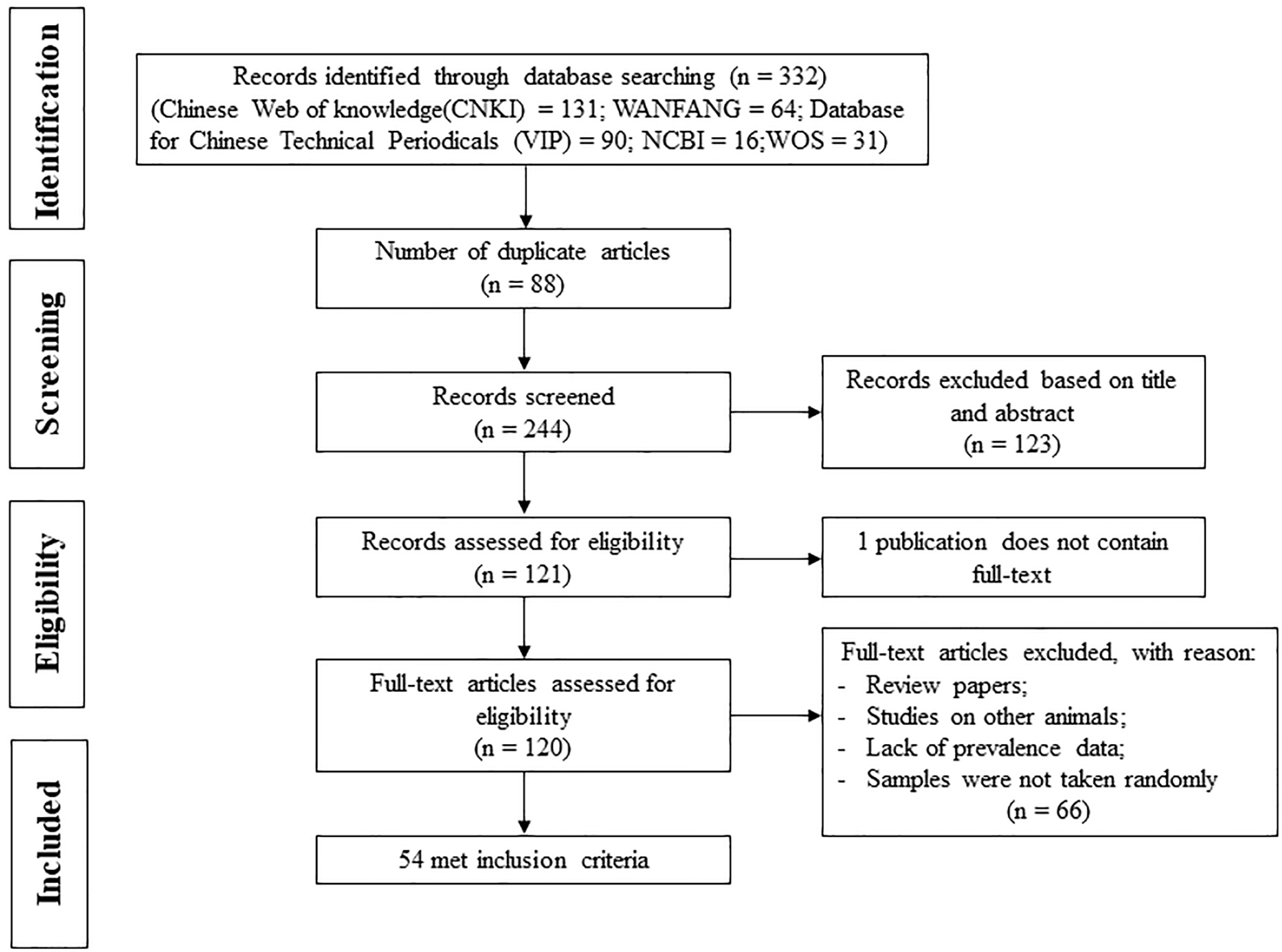
3.1. Study Selection and Data Extraction
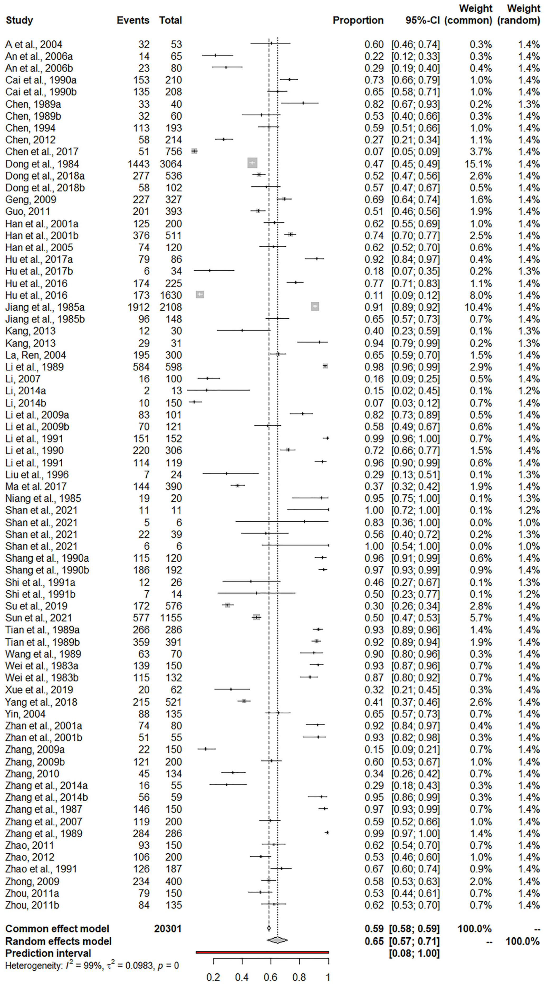
3.2. Prevalence of Sarcocystis in Ruminants in China
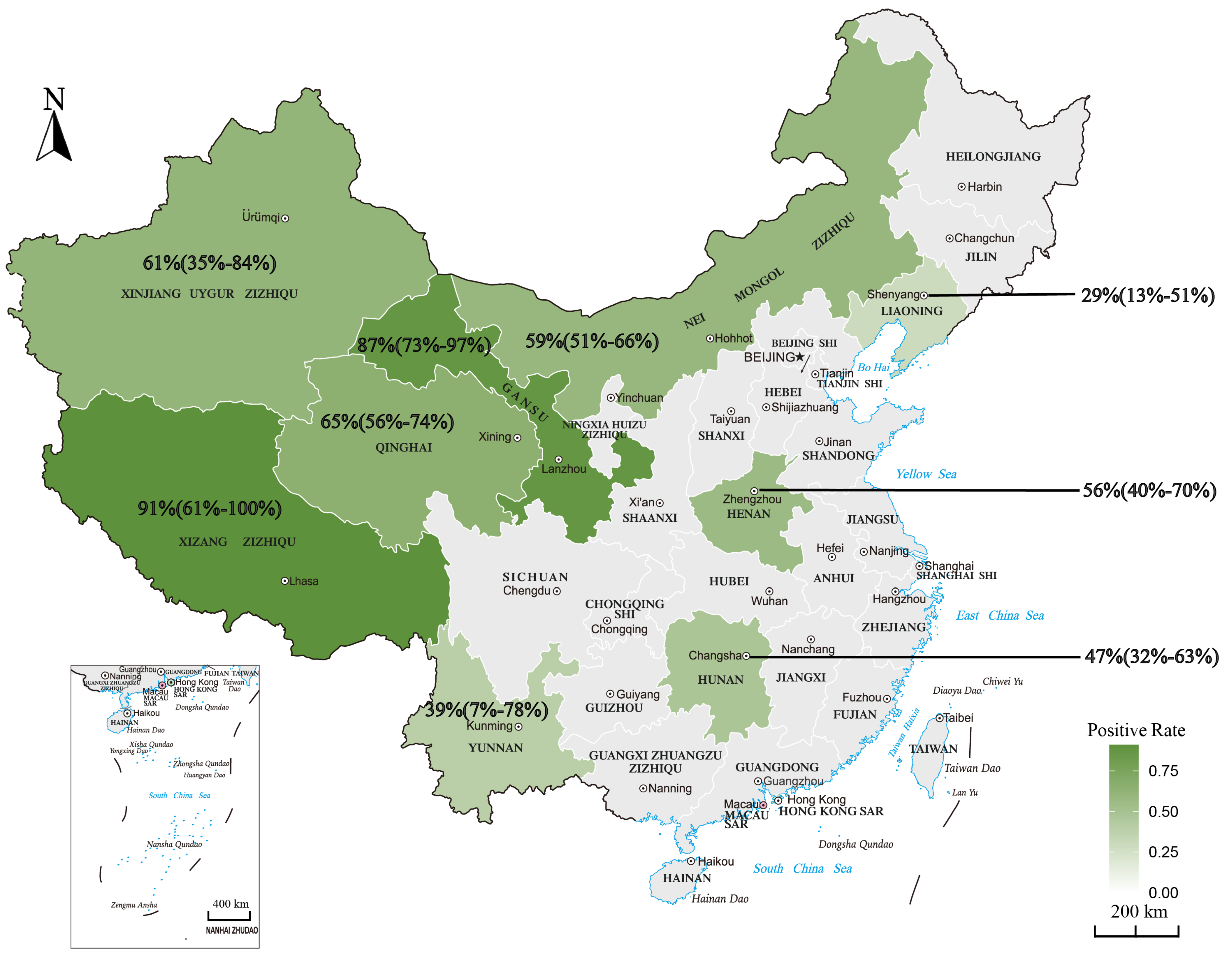
3.3. Subgroup Analysis
4. Discussion
5. Conclusions
Supplementary Materials
Author Contributions
Funding
Institutional Review Board Statement
Informed Consent Statement
Data Availability Statement
Conflicts of Interest
References
- Dubey, J.P.; Calero-Bernal, R.; Rosenthal, B.M.; Speer, C.A.; Fayer, R. Sarcocystis of Animals and Humans, 2nd ed.; CRC Press: Boca Raton, FL, USA, 2016; pp. 1–256. [Google Scholar]
- Dubey, J.P.; Moré, G.; van Wilpe, E.; Calero-Bernal, R.; Verma, S.K.; Schares, G. Sarcocystis rommeli, n. sp. (Apicomplexa: Sarcocystidae) from cattle (Bos taurus) and its differentiation from Sarcocystis hominis. J. Eukaryot. Microbiol. 2016, 63, 62–68. [Google Scholar] [CrossRef]
- Dubey, J.P.; van Wilpe, E.; Calero-Bernal, R.; Verma, S.K.; Fayer, R. Sarcocystis heydorni, n. sp. (Apicomplexa: Sarcocystidae) with cattle (Bos taurus) and human (Homo sapiens) cycle. Parasitol. Res. 2015, 114, 4143–4147. [Google Scholar] [CrossRef] [PubMed]
- Rosenthal, B.M. Zoonotic Sarcocystis. Res. Vet. Sci. 2021, 136, 151–157. [Google Scholar] [CrossRef] [PubMed]
- Rubiola, S.; Civera, T.; Panebianco, F.; Vercellino, D.; Chiesa, F. Molecular detection of cattle Sarcocystis spp. in North-West Italy highlights their association with bovine eosinophilic myositis. Parasit. Vectors 2021, 23, 223. [Google Scholar] [CrossRef] [PubMed]
- Dong, H.; Lu, Y.Y.; Yang, Y.R. Epidemiology and classification of Sarcocystis in sheep and goat. Chin. J. Zoonoses 2017, 33, 828–836. (In Chinese) [Google Scholar]
- Dong, H.; Su, R.J.; Wang, Y.H.; Tong, Z.X.; Zhang, L.X.; Yang, Y.R.; Hu, J.J. Sarcocystis species in wild and domestic sheep (Ovis ammon and Ovis aries) from China. BMC Vet. Res. 2018, 14, 377. [Google Scholar] [CrossRef]
- Ying, Z.; Zhu, Z.F.; Yang, X.; Liu, J.; Liu, Q. Prevalence and associated risk factors of Neospora caninum infection among cattle in mainland China: A systematic review and meta-analysis. Prev. Vet. Med. 2022, 201, 105593. [Google Scholar] [CrossRef]
- Moher, D.; Shamseer, L.; Clarke, M.; Ghersi, D.; Liberati, A.; Petticrew, M.; Shekelle, P.; Stewart, L.A.; PRISMA-P Group. Preferred reporting items for systematic review and meta-analysis protocols (PRISMA-P) 2015 statement. Syst. Rev. 2015, 4, 1. [Google Scholar] [CrossRef] [Green Version]
- Ah, R.Z.; Tan, S.K. Investigation on Sarcocystis infection in Tibetan sheep in Gangcha Country, Qinghai Province. Chin. Q.H. J. Ani. Vet. Sci. 2004, 34, 19. (In Chinese) [Google Scholar]
- An, N.; Xin, D.; Shan, B.; Yu, Z.X. Detection of Sarcocystis in yak by microscopy. Chin. Diary Cattl. 2006, 12, 34–35. (In Chinese) [Google Scholar]
- Cai, J.Z.; Zhao, M.Y. Investigation on the infection of Taenia and Sarcocystis in cattle and sheep in Tole Pasture. Chin. Q.H. J. Ani. Vet. Sci. 1990, 2, 35–36. (In Chinese) [Google Scholar]
- Chen, H.Z. Investigation on Sarcocystis of swine, cattle and sheep in South Henan. Chin. J. Vet. Med. 1989, 7, 26. (In Chinese) [Google Scholar]
- Chen, T.J. Post-mortem quarantine investigation and treatment of Sarcocystis in slaughtered camels. Chin. J. Meat Hygiene. 1994, 10, 13. (In Chinese) [Google Scholar]
- Chen, L. Investigation on Sarcocystis infection in commercial yaks in Chengduo County, Qinghai Province. Chi. Ani. Hus. Vet. Med. 2012, 44, 111–112. (In Chinese) [Google Scholar]
- Chen, X.W.; Wen, T.; Hu, J.J.; Liu, T.T.; Esch, G.W.; Liang, Y.; Li, H.L.; Huang, S. Sarcocystis dehongensis n. sp. (Apicomplexa: Sarcocystidae) from water buffalo (Bubalus bubalis) in China. Parasitol. Res. 2017, 116, 2145–2150. [Google Scholar] [CrossRef]
- Dong, S.Z.; Liu, S.G. Investigation on Sarcocystis of goats in Gansu Province. Chin. J. Vet. Sci. Tech. 1984, 10, 37–39. (In Chinese) [Google Scholar]
- Geng, T.Y. Investigation on Sarcocystis infection of yaks in Xinghai, Qinghai Province. Chin. J. Vet. Med. 2009, 45, 49. (In Chinese) [Google Scholar]
- Guo, Z.P. Investigation on Sarcocystis of sheep carcass in Dilan Country, Qinghai Province. Chi. Ani. H. Insp. 2011, 28, 44. (In Chinese) [Google Scholar]
- Han, X.M.; Wu, X.T.; Liu, B.R.; Liu, P.Y.; Tang, X.Y. Examination of zoonotic parasitic diseases in livestock in Xining, Qinghai Province. Chin. J. Zoonoses 2001, 17, 123–124. (In Chinese) [Google Scholar]
- Han, L.; Yan, Z.L. Investigation on Sarcocystis infection in sheep in six counties of Qinghai Province. Chin. Ani. H. Insp. 2005, 22, 41. (In Chinese) [Google Scholar]
- Hu, J.J.; Huang, S.; Wen, T.; Esch, G.W.; Liang, Y.; Li, H.L. Sarcocystis spp. in domestic sheep in Kunming City, China: Prevalence, morphology, and molecular characteristics. Parasite. 2017, 24, 30. [Google Scholar] [CrossRef] [PubMed] [Green Version]
- Hu, J.J.; Huang, S.; Wen, T.; Esch, G.W.; Liang, Y.; Li, H.L. Morphology, molecular characteristics, and demonstration of a definitive host for Sarcocystis rommeli from Cattle (Bos taurus) in China. J. Parasitol. 2017, 103, 471–476. [Google Scholar] [CrossRef] [PubMed]
- Hu, J.J.; Liu, T.T.; Liu, Q.; Esch, G.W.; Chen, J.Q.; Huang, S.; Wen, T. Prevalence, morphology, and molecular characteristics of Sarcocystis spp. in domestic goats (Capra hircus) from Kunming, China. Parasitol. Res. 2016, 115, 3973–3981. [Google Scholar] [CrossRef]
- Hu, J.J.; Wen, T.; Chen, X.W.; Liu, T.T.; Esch, G.W.; Huang, S. Prevalence, morphology, and molecular characterization of Sarcocystis heydorni sarcocysts from cattle (Bos taurus) in China. J. Parasitol. 2016, 102, 545–548. [Google Scholar] [CrossRef] [PubMed]
- Jiang, Y.P.; Liu, J.R.; Miu, L.W.; Li, Z.D.; Luo, Q.G.; Liu, C.S. Investigation on the prevalence of Sarcocystis in sheep in Shihezi area of Xinjiang Province. Chin. J. Vet. Sci. Tech. 1985, 12, 20–22. (In Chinese) [Google Scholar]
- Kang, M.; Li, Y.; Shi, W.H.; Xi, J.G.H. Study on the infection of Sarcocystis in the diaphragm of livestock in Hualong Country, Qinghai Province. J. Anhui Agri. Sci. 2013, 41, 1154–1156. (In Chinese) [Google Scholar]
- La, M.J.; Ren, Q.C. Investigation on Sarcocystis infection of Tibetan mutton in Maqin Country, Qinghai Province. Chin. J. Vet. Sci. 2004, 40, 50–51. (In Chinese) [Google Scholar]
- Li, O.; Xu, T.D.; Li, W.; Sai, Q. Investigation on Sarcocystis of sheep in Qinghai Province. Chin. Q.H. J. Ani. Vet. Sci. 1989, 2, 12–13. (In Chinese) [Google Scholar]
- Li, F.Y. Investigation of Sarcocystis infection in sheep in Angssuto town, Qinghai Province. Chin. Q.H. J. Ani. Vet. Sci. 2007, 4, 33. (In Chinese) [Google Scholar]
- Li, S.S. Investigation report on Sarcocystis infection of slaughtered cattle in market. Chin. Q.H. J. Vet. Ani. Vet. Sci. 2014, 44, 26. (In Chinese) [Google Scholar]
- Li, C.H.; Cai, J.Z. Investigation of Sarocystis in yaks and sheep in some areas of Qinghai Province. Chin. J. Vet. Sci. 2009, 45, 33–35. (In Chinese) [Google Scholar]
- Li, W.; Li, O.; Li, Z.N.; Jiang, Y.S.; Shi, H.N.; Yun, S.M. Investigation on Sarcocystis infection of sheep on a farm in Qinghai Province. Chin. Q.H. J. Ani. Vet. Sci. 1991, 1, 18–19. (In Chinese) [Google Scholar]
- Li, X.W.; Wang, Y.P.; Li, W.L. Investigation of Sarcocystis infection in swine and beef. J. Henan Agri. Sci. 1990, 5, 27–28. (In Chinese) [Google Scholar]
- Li, C.Y.; Yun, S.L. Report on inspection of Sarcocystis in sheep carcass. Chin. Q.H. J. Ani. Vet. Sci. 1991, 2, 25. (In Chinese) [Google Scholar]
- Liu, X.G.; Yu, J.L.; Zhu, R.; Yan, B.G.; Shen, J.Y. Investigation of Sarcocystis infection in sheep. Chin. J. Meat Hygiene 1996, 10, 10–11. (In Chinese) [Google Scholar]
- Ma, D.D.; Cai, J.Z.; Li, C.H.; Lei, M.T.; Sun, J. Investigation of Sarcocystis infection in yaks in Qinghai Province. Chin. Ani. Hus. Vet. Med. 2017, 49, 149–152. (In Chinese) [Google Scholar]
- Niang, J.X.; Cai, D.; Yan, Q.L. Investigation of Sarcocystis infection in yaks in Huangnan Area, Qinghai Province. Chin. Q.H. J. Ani. Vet. Sci. 1985, 4, 30–31. (In Chinese) [Google Scholar]
- Shan, Q.L.M.; Zhu, C.G.; Ji, R.Y.; Shi, B.; Tang, W.Q.; Suo, L.Q.Y.; Xia, C.Y. Species identification and evolutionary analysis of Sarcocystis from cattle and sheep in Tibet. Chin. Herb. Sci. 2021, 5, 27–30. (In Chinese) [Google Scholar]
- Shang, H.Z.; Zhu, X.W.; Qi, Y.X.; Gao, L.Y. Investigation on Sarcocystis infection of cattle and sheep in Tianjun Country, Qinghai Province. Chin. Q.H. J. Ani. Vet. Sci. 1990, 2, 34. (In Chinese) [Google Scholar]
- Shi, Y.L. Investigation on Sarcocystis infection of water buffalo and cattle in western Hunan. Chin. Hunan Ani. Hus. Vet. Med. 1991, 2, 34. (In Chinese) [Google Scholar]
- Su, X.X.; Xie, C.Y.; Kang, M. Investigation on Sarcocystis infection of Tibetan sheep in Gonghe Country, Qinghai Province. Chin. J. Vet. Sci. 2019, 12, 52–54. (In Chinese) [Google Scholar]
- Sun, Y.L.; Ju, J.L.; Su, X.X.; Xie, C.Y.; Li, Y.; Kang, M. Infection survey and morphological characteristics of Sarcocystis spp. in naturally infected Tibetan sheep from Qinghai in northwestern China. Parasitol. Int. 2021, 80, 102219. [Google Scholar] [CrossRef] [PubMed]
- Tian, W.F.; Jia, J.; Liang, S.Z.; Niu, H.F. Investigation on Sarcocystis infection of cattle and sheep in Gannan Tibetan Autonomous Prefecture. Chin. Gansu Ani. Hus. Vet. Med. 1989, 2, 16. (In Chinese) [Google Scholar]
- Wang, G.L.; Xu, M. Investigation of Sarcocystis infection in cattle in Burqin Country, Xinjiang. Xinjiang J. Ani. Hus. 1989, 2, 36. (In Chinese) [Google Scholar]
- Wei, T.; Zhang, P.C.; Han, M.C.; Cai, D.; Liu, Q.H. Investigation on Sarcocystis infection of yak in Zeku Country, Qinghai Province. Chin. J. Trad. Vet. Sci. 1983, 2, 11–15. (In Chinese) [Google Scholar]
- Wei, T.; Zhang, P.C.; Dong, M.X.; Sun, Y.J.; Li, W.Y. Investigation on Sarcocystis infection of yaks in Tianzhu, Qinghai Province. Chin. Yak. 1983, 1, 41–44. (In Chinese) [Google Scholar]
- Xue, R.; Yan, W.C.; Qian, W.F.; Wang, T.Q.; Zhang, M.; Wei, Z.G.; Han, L.F.; He, B.; Dou, J.C. Prevalence and molecular characterization of Sarcocystis infections of retail beef products from central China. Acta Trop. 2019, 190, 339–343. [Google Scholar] [CrossRef]
- Yang, Y.R.; Dong, H.; Su, R.J.; Wang, Y.H.; Wang, R.H.; Jiang, Y.B.; Tong, Z.X. High prevalence of Sarcocystis spp. infections in cattle (Bos taurus) from central China. Parasitol. Int. 2018, 67, 800–804. [Google Scholar] [CrossRef]
- Yin, H. Examination of Sarcocystis infection in sheep carcasses in Xunhua Country, Qinghai Province. Chin. J. Vet. Parasitol. 2004, 12, 64. (In Chinese) [Google Scholar]
- Zhan, S.X.; Zhao, C.C.; Qi, H.Y.; Jia, Y.Z.; Zhang, Y.H. Investigation of Sarcocystis infection in sheep in Delhi City, Qinghai Province. Chin. Ani. H. Insp. 2001, 1, 28–29. (In Chinese) [Google Scholar]
- Zhang, C.W. Investigation on Sarcocystis infection of cattle and sheep in Hualong Country, Qinghai Province. Chin. Mod. Agri. Sci. Tech. 2009, 3, 217. (In Chinese) [Google Scholar]
- Zhang, G.L. Investigation on Sarcocystis infection of commercial yaks in Ulan Country, Qinghai Province. Chin. Contemp. Ani. Hus. 2009, 11, 20–21. (In Chinese) [Google Scholar]
- Zhang, H.B.; Cang, N.G.; Hou, H.M.; Ah, S.P.; Zhou, M.; Ka, D.Z.; Sun, Y.L.; Kang, M. Investigation of Sarcocystis infection in the diaphragm of cattle and sheep in Zeku Country, Qinghai Province. Chin. Ani. Hus. Vet. Med. 2014, 46, 138. (In Chinese) [Google Scholar]
- Zhang, P.C.; Dong, M.X.; Wei, T.; Wang, X.Y.; Xiao, H.; Yang, J.R.; Liang, S.Z. Investigation on Sarcocystis of yak in Luqu Country, Gansu Province. Chin. J. Trad. Vet. Sci. 1987, 6, 18–20. (In Chinese) [Google Scholar]
- Zhang, G.C.; Wei, L.; Zhang, Y.C.; Yang, J.Y.; Feng, Z.F.; Bai, Y.Y.; Bao, Z.X.; Wang, L.P.; Han, X.Y. Investigation of Sarcocystis infection in sheep in Datong Country, Qinghai Province. Chin. J. Vet. Parasitol. 2007, 15, 38–39. (In Chinese) [Google Scholar]
- Zhang, X.J.; Yang, X.C. Investigation and morphological observation on Sarcocystis of yak in Yushu, Qinghai Province. Chin. Q.H. J. Ani. Vet. Sci. 1989, 6, 20–22. (In Chinese) [Google Scholar]
- Zhao, Y.L. Investigation on Sarcocystis infection of commercial sheep in Huzhu Country, Qinghai Province. Chin. Q.H. J. Ani. Vet. Sci. 2011, 41, 59. (In Chinese) [Google Scholar]
- Zhao, G. Investigation on the Sarcocystis infection of Tibetan sheep in Heka Town, Xinghai Country. Chin. Ani. Hus. Vet. Med. 2012, 44, 106. (In Chinese) [Google Scholar]
- Zhao, W.S.; Yi, G.; Wu, B.; Bu, D.; Cai, R.J.; Ma, W.Z. Investigation on Sarcocystis infection of yak in Dari Country, Qinghai Province. Chin. Q.H. J. Vet. Sci. 1991, 3, 19. (In Chinese) [Google Scholar]
- Zhong, W.D. Investigation on Sarcocystis infection of sheep in Minhe Country. Chin. Ani. H. Insp. 2009, 9, 50. (In Chinese) [Google Scholar]
- Zhou, W.Y. Sampling survey and analysis of Sarcocystis in commercial livestock in Lenghu area, Qinghai Province. Chin. Ani. H. Insp. 2011, 28, 51–52. (In Chinese) [Google Scholar]
- R Core Team. R: A Language and Environment for Statistical Computing; R Foundation for Statistical Computing: Vienna, Austria, 2021; Available online: https://www.R-project.org (accessed on 31 March 2021).
- DerSimonian, R.; Laird, N. Meta-analysis in clinical trials revisited. Contemp. Clin. Trials. 2015, 45, 139–145. [Google Scholar] [CrossRef] [PubMed] [Green Version]
- Anvari, D.; Narouei, E.; Hosseini, M.; Narouei, M.R.; Daryani, A.; Shariatzadeh, S.A.; Pagheh, A.S.; Gholami, S.; Sarvi, S.; Sargazi, D.; et al. Sarcocystosis in ruminants of Iran, as neglected food-borne disease: A systematic review and meta-analysis. Acta Parasitol. 2022, 65, 555–568. [Google Scholar] [CrossRef] [PubMed]
- Tenter, A.M. Current research on Sarcocystis species of domestic animals. Int. J. Parasitol. 1995, 25, 1311–1330. [Google Scholar] [CrossRef] [PubMed]
- Zeng, H.; Van Damme, I.; Kabi, T.W.; Šoba, B.; Gabriël, S. Sarcocystis species in bovine carcasses from a Belgian abattoir: A cross-sectional study. Parasit. Vectors. 2021, 14, 271. [Google Scholar] [CrossRef] [PubMed]
- van Bree, F.P.J.; Bokken, G.C.A.M.; Mineur, R.; Franssen, F.; Opsteegh, M.; van der Giessen, J.W.B.; Lipman, L.J.A.; Overgaauw, P.A.M. Zoonotic bacteria and parasites found in raw meat-based diets for cats and dogs. Vet. Rec. 2018, 182, 50. [Google Scholar] [CrossRef]
- Wouda, W.; Snoep, J.J.; Dubey, J.P. Eosinophilic myositis due to Sarcocystis hominis in a beef cow. J. Comp. Pathol. 2006, 135, 249–253. [Google Scholar] [CrossRef]
- Fukuyo, M.; Battsetseg, G.; Byambaa, B. Prevalence of Sarcocystis infection in meat-producing animals in Mongolia. Southeast Asian J. Trop. Med. Public Health 2002, 33, 490–495. [Google Scholar]
- Bittencourt, M.V.; Meneses, I.D.; Ribeiro-Andrade, M.; de Jesus, R.F.; de Araújo, F.R.; Gondim, L.F. Sarcocystis spp. in sheep and goats: Frequency of infection and species identification by morphological, ultrastructural, and molecular tests in Bahia, Brazil. Parasitol Res. 2016, 115, 1683–1689. [Google Scholar] [CrossRef]
- El-Morsey, A.; Abdo, W.; Zaid, A.A.A.; Sorour, S.S.G. Morphologic and molecular identification of three macroscopic Sarcocystis species infecting domestic sheep (Ovis aries) and cattle (Bos taurus) in Egypt. Parasitol. Res. 2021, 120, 637–654. [Google Scholar] [CrossRef]
- Wang, H.B.; Zan, L.S.; Zhang, Y.Y. Profiling of the yak skeletal muscle tissue gene expression and comparison with the domestic cattle by genome array. Animal 2014, 8, 28–35. [Google Scholar] [CrossRef] [PubMed] [Green Version]
- Yang, X.; Li, Y.; Wang, Y.; Wang, J.; Lai, P.; Li, Y.; Song, J.; Qi, M.; Zhao, G. Molecular characterization of Blastocystis sp. In Camelus bactrianus in Northwestern China. Animals 2021, 11, 3016. [Google Scholar] [CrossRef] [PubMed]
- Page, M.J.; McKenzie, J.E.; Bossuyt, P.M.; Boutron , I.; Hoffmann, T.C.; Mulrow, C.D.; Shamseer, L.; Tetzlaff, J.M.; Akl, E.A.; Brennan, S.E.; et al. The PRISMA 2020 statement: An updated guideline for reporting systematic reviews. BMJ 2021, 372:n71. [Google Scholar] [CrossRef]


| First Author, Year | Host | Province | Period of Study | Detection Method | Positive Samples/Total Samples (%) | Reference |
|---|---|---|---|---|---|---|
| A et al., 2004 | Tibetan sheep | Qinghai | 2003 | Macroscopy, LM | 32/53 (60) | [10] |
| An et al., 2006 | Yaks | Xinjiang | 2001 | LM | 14/65 (22) | [11] |
| An et al., 2006 | Yaks | Xinjiang | 2004 | LM | 23/80 (29) | [11] |
| Cai et al., 1990 | Yaks | Qinghai | 1988 | Macroscopy, LM | 153/210 (73) | [12] |
| Cai et al., 1990 | Tibetan sheep | Qinghai | 1988 | Macroscopy, LM | 135/208 (65) | [12] |
| Chen, 1989 | Cattle | Henan | 1985 | Macroscopy, LM, H&E | 33/40 (82) | [13] |
| Chen, 1989 | Sheep | Henan | 1985 | Macroscopy, LM, H&E | 32/60 (53) | [13] |
| Chen, 1994 | Camel | Inner Mongolia | 1992 | Macroscopy, LM | 113/193 (59) | [14] |
| Chen, 2012 | Yaks | Qinghai | 2011 | LM | 58/214 (27) | [15] |
| Chen et al., 2017 | Water buffalo | Yunnan | n.s. | LM, TEM, PCR | 51/756 (7) | [16] |
| Dong et al., 1984 | Goats | Gansu | n.s. | LM; Pepsin digestion examination | 1443/3064 (47) | [17] |
| Dong et al., 2018 | Sheep | Henan | 2014–2017 | LM; Pepsin digestion examination; PCR | 277/536 (52) | [7] |
| Dong et al., 2018 | Sheep | Xinjiang | 2014–2017 | LM; Pepsin digestion examination; PCR | 58/102 (57) | [7] |
| Geng, 2009 | Yaks | Qinghai | 2008 | LM | 227/327 (69) | [18] |
| Guo, 2011 | Sheep | Qinghai | 2011 | Macroscopy, LM | 201/393 (51) | [19] |
| Han et al., 2001 | Yaks | Qinghai | n.s. | LM | 125/200 (62) | [20] |
| Han et al., 2001 | Sheep | Qinghai | n.s. | LM | 376/511 (74) | [20] |
| Han et al., 2005 | Sheep | Qinghai | n.s. | LM | 74/120 (62) | [21] |
| Hu et al., 2017 | Sheep | Yunnan | n.s. | LM, TEM, PCR | 79/86 (92) | [22] |
| Hu et al., 2017 | Cattle | Yunnan | n.s. | LM, TEM, PCR | 6/34 (18) | [23] |
| Hu et al., 2016 | Goats | Yunnan | 2014–2015 | LM, TEM, PCR | 174/225 (77) | [24] |
| Hu et al., 2016 | Cattle | Yunnan | 2014–2015 | LM, TEM, PCR | 173/1630 (11) | [25] |
| Jiang et al., 1985a | Sheep | Xinjiang | n.s. | LM; Pepsin digestion examination | 1912/2108 (91) | [26] |
| Jiang et al., 1985b | Sheep | Xinjiang | n.s. | LM; Pepsin digestion examination | 96/148 (65) | [26] |
| Kang, 2013 | Yaks | Qinghai | n.s. | LM | 12/30 (40) | [27] |
| Kang, 2013 | Sheep | Qinghai | n.s. | LM | 29/31 (94) | [27] |
| La et al., 2014 | Tibetan sheep | Qinghai | 2003 | LM | 195/300 (65) | [28] |
| Li et al., 1989 | Sheep | Qinghai | 1986 | LM | 584/598 (98) | [29] |
| Li, 2007 | Sheep | Qinghai | n.s. | Macroscopy, LM | 16/100 (16) | [30] |
| Li, 2014 | Goats | Qinghai | 2011 | Macroscopy, LM | 2/13 (15) | [31] |
| Li, 2014 | Sheep | Qinghai | 2011 | Macroscopy, LM | 10/150 (7) | [31] |
| Li et al., 2009 | Sheep | Qinghai | 2006–2007 | LM | 83/101 (82) | [32] |
| Li et al., 2009 | Yaks | Qinghai | 2006–2007 | LM | 70/121 (58) | [32] |
| Li et al., 1991 | Sheep | Qinghai | 1989 | Macroscopy, LM | 151/152 (99) | [33] |
| Li et al., 1990 | Cattle | Henan | n.s. | LM; Pepsin digestion examination, H&E | 220/306 (72) | [34] |
| Li et al., 1991 | Sheep | Qinghai | 1990 | Macroscopy, LM | 114/119 (96) | [35] |
| Liu et al., 1996 | Sheep | Liaoning | n.s. | Macroscopy, LM | 7/24 (29) | [36] |
| Ma et al., 2017 | Yaks | Qinghai | n.s. | LM | 144/390 (37) | [37] |
| Niang et al., 1985 | Yaks | Qinghai | 1984 | LM | 19/20 (95) | [38] |
| Shan et al., 2021 | Goats | Tibet | 2019 | Macroscopy, LM | 11/11 (100) | [39] |
| Shan et al., 2021 | Sheep | Tibet | 2019 | Macroscopy, LM | 5/6 (83) | [39] |
| Shan et al., 2021 | Yaks | Tibet | 2019 | Macroscopy, LM | 22/39 (56) | [39] |
| Shan et al., 2021 | Cattle | Tibet | 2019 | Macroscopy, LM | 6/6 (100) | [39] |
| Shang et al., 1990 | Yaks | Qinghai | 1989 | LM | 115/120 (96) | [40] |
| Shang et al., 1990 | Sheep | Qinghai | 1989 | LM | 186/192 (97) | [40] |
| Shi et al., 1991 | Cattle | Hunan | 1990 | Macroscopy, LM | 12/26 (46) | [41] |
| Shi et al., 1991 | Water buffalo | Hunan | 1990 | Macroscopy, LM | 7/14 (50) | [41] |
| Su et al., 2019 | Tibetan sheep | Qinghai | 2017 | Macroscopy, LM | 172/576 (30) | [42] |
| Sun et al., 2021 | Tibetan sheep | Qinghai | 2017–2018 | LM, H&E, TEM, PCR | 577/1155 (50) | [43] |
| Tian et al., 1989 | Yaks | Gansu | 1985 | Macroscopy, LM | 266/286 93) | [44] |
| Tian et al., 1989 | Sheep | Gansu | 1985 | Macroscopy, LM | 359/391 (92) | [44] |
| Wang et al., 1989 | Cattle | Xinjiang | 1988 | LM | 63/70 (90) | [45] |
| Wei et al., 1983 | Yaks | Gansu | 1982 | LM | 139/150 (93) | [46] |
| Wei et al., 1983 | Yaks | Gansu | 1981 | LM; Pepsin digestion examination | 115/132 (87) | [47] |
| Xue et al., 2019 | Cattle | Henan | 2017 | LM, H&E, PCR | 20/62 (32) | [48] |
| Yang et al., 2018 | Cattle | Henan | 2014–2016 | LM; Pepsin digestion examination, TEM, PCR | 215/521 (41) | [49] |
| Yin, 2004 | Sheep | Qinghai | 2003 | Macroscopy, LM | 88/135 (65) | [50] |
| Zhan et al., 2001 | Goats | Qinghai | 2000 | Macroscopy, LM | 74/80 (92) | [51] |
| Zhan et al., 2001 | Sheep | Qinghai | 2000 | Macroscopy, LM | 51/55 (93) | [51] |
| Zhang, 2009 | Yaks | Qinghai | n.s. | Macroscopy, LM | 22/150 (15) | [52] |
| Zhang, 2009 | Sheep | Qinghai | n.s. | Macroscopy, LM | 121/200 (60) | [52] |
| Zhang, 2010 | Yaks | Qinghai | 2008 | LM | 45/134 (34) | [53] |
| Zhang et al., 2014 | Yaks | Qinghai | n.s. | Macroscopy, LM | 16/55 (29) | [54] |
| Zhang et al., 2014 | Sheep | Qinghai | n.s. | Macroscopy, LM | 56/59 (95) | [54] |
| Zhang et al., 1987 | Yaks | Gansu | 1986 | Macroscopy, LM | 146/150 (97) | [55] |
| Zhang et al., 2007 | Sheep | Qinghai | 2005–2006 | LM | 119/200 (59) | [56] |
| Zhang et al., 1989 | Yaks | Qinghai | 1986 | H&E | 284/286 (99) | [57] |
| Zhao, 2011 | Sheep | Qinghai | 2010 | LM | 93/150 (62) | [58] |
| Zhao, 2012 | Tibetan sheep | Qinghai | 2010 | LM | 106/200 (53) | [59] |
| Zhao et al., 1991 | Yaks | Qinghai | 1988 | Macroscopy, LM | 126/187 (67) | [60] |
| Zhong, 2009 | Sheep | Qinghai | 2007 | LM | 234/400 (58) | [61] |
| Zhou, 2011a | Sheep | Qinghai | 2010 | LM | 79/150 (53) | [62] |
| Zhou, 2011b | Goats | Qinghai | 2010 | LM | 84/135 (62) | [62] |
| Conversion Form | W | p |
|---|---|---|
| PRAW | 0.9351 | 0.001641 |
| PLN | 0.8341 | 1.38 × 10⁻⁷ |
| PLOGIT | NaN | NA |
| PAS | 0.96757 | 0.05696 |
| PFT | 0.96458 | 0.03809 |
| Subgroup Variable | No. Studies | No. Positive/ No. Tested | Prevalence (95% CI) | Heterogeneity | ||
|---|---|---|---|---|---|---|
| Q | p-Value | I2 (%) | ||||
| Region | ||||||
| Northeast China | 1 | 7/24 | 29% (13–51%) | 0.00 | - | - |
| Central China | 8 | 816/1565 | 54% (42–66%) | 104.73 | <0.01 | 93% |
| Northwest China | 55 | 10,205/15,919 | 67% (59–75%) | 4972.27 | <0.01 | 99% |
| Southwest China | 9 | 527/2793 | 64% (32–91%) | 898.07 | <0.01 | 99% |
| Host | ||||||
| Camel | 1 | 113/193 | 59% (51–66%) | 0.00 | - | - |
| Cattle | 9 | 748/2695 | 56% (31–79%) | 836.64 | <0.01 | 99% |
| Goats | 6 | 1788/3528 | 71% (40–93%) | 209.62 | <0.01 | 98% |
| Sheep | 34 | 6707/9769 | 69% (59–78%) | 2928.83 | <0.01 | 99% |
| Water buffalo | 2 | 58/770 | 24% (0–72%) | 15.02 | <0.01 | 93% |
| Yaks | 21 | 2141/3346 | 64% (50–78%) | 1473.82 | <0.01 | 99% |
| Publish year | ||||||
| <2005 | 33 | 7771/10,588 | 85% (77–90%) | 2738.77 | <0.01 | 98% |
| 2005–2010 | 12 | 1048/1998 | 46% (33–60%) | 337.88 | <0.01 | 96% |
| 2011–2015 | 12 | 746/1580 | 50% (30–70%) | 352.37 | <0.01 | 94% |
| >2016 | 16 | 1990/6135 | 52% (33–71%) | 1551.60 | <0.01 | 98% |
| Total | 73 | 11,555/20,301 | 65% (57–72%) | 8755.27 | <0.01 | 99% |
Disclaimer/Publisher’s Note: The statements, opinions and data contained in all publications are solely those of the individual author(s) and contributor(s) and not of MDPI and/or the editor(s). MDPI and/or the editor(s) disclaim responsibility for any injury to people or property resulting from any ideas, methods, instructions or products referred to in the content. |
© 2022 by the authors. Licensee MDPI, Basel, Switzerland. This article is an open access article distributed under the terms and conditions of the Creative Commons Attribution (CC BY) license (https://creativecommons.org/licenses/by/4.0/).
Share and Cite
Zhu, Z.; Ying, Z.; Feng, Z.; Liu, Q.; Liu, J. The Occurrence and Meta-Analysis of Investigations on Sarcocystis Infection among Ruminants (Ruminantia) in Mainland China. Animals 2023, 13, 149. https://doi.org/10.3390/ani13010149
Zhu Z, Ying Z, Feng Z, Liu Q, Liu J. The Occurrence and Meta-Analysis of Investigations on Sarcocystis Infection among Ruminants (Ruminantia) in Mainland China. Animals. 2023; 13(1):149. https://doi.org/10.3390/ani13010149
Chicago/Turabian StyleZhu, Zifu, Zhu Ying, Zixuan Feng, Qun Liu, and Jing Liu. 2023. "The Occurrence and Meta-Analysis of Investigations on Sarcocystis Infection among Ruminants (Ruminantia) in Mainland China" Animals 13, no. 1: 149. https://doi.org/10.3390/ani13010149