Article

Antibiotics Resistance and Adhesive Properties of Clinical Staphylococcus aureus Isolated from Wound Infections

by
Khulood Fahad Alabbosh
1,*,
Tarek Zmantar
2,
Abdulrahman S. Bazaid
3,
Mejdi Snoussi
1,4 and
Emira Noumi
1,4,*
1
Department of Biology, College of Science, University of Hail, Hail 2440, Saudi Arabia
2
Laboratory of Analysis, Treatment, Valorization of Environmental, and Product Pollutants, Faculty of Pharmacy of Monastir, University of Monastir, Monastir 5000, Tunisia
3
Department of Medical Laboratory Science, College of Applied Medical Sciences, University of Hail, Hail 55476, Saudi Arabia
4
Laboratory of Genetics, Biodiversity and Valorization of Bio-Resources (LR11ES41), Higher Institute of Biotechnology of Monastir, University of Monastir, Avenue Tahar Haddad, BP74, Monastir 5000, Tunisia
*
Authors to whom correspondence should be addressed.
Microorganisms 2023, 11(5), 1353; https://doi.org/10.3390/microorganisms11051353
Submission received: 26 April 2023 / Revised: 16 May 2023 / Accepted: 19 May 2023 / Published: 22 May 2023
(This article belongs to the Special Issue Biology and Pathogenesis of Staphylococcus Infection 2.0)

Abstract

:
Staphylococcus aureus (S. aureus) is a ubiquitous pathogen responsible for several severe infections. This study aimed to investigate the adhesive properties and antibiotic resistance among clinical S. aureus isolated from Hail Hospital Province, Kingdom of Saudi Arabia (KSA), using molecular approaches. This study was conducted according to the ethical committee at Hail’s guidelines on twenty-four S. aureus isolates. A polymerase chain reaction (PCR) was performed to identify genes encoding the β-lactamase resistance (blaZ), methicillin resistance (mecA), fluoroquinolone resistance (norA), nitric oxide reductase (norB), fibronectin (fnbA and fnbB), clumping factor (clfA) and intracellular adhesion factors (icaA and icaD). This qualitative study tested adhesion based on exopolysaccharide production on Congo red agar (CRA) medium and biofilm formation on polystyrene by S. aureus strains. Among 24 isolates, the cna and blaz were the most prevalent (70.8%), followed by norB (54.1%), clfA (50.0%), norA (41.6%), mecA and fnbB (37.5%) and fnbA (33.3%). The presence of icaA/icaD genes was demonstrated in almost all tested strains in comparison to the reference strain, S. aureus ATCC 43300. The phenotypic study of adhesion showed that all tested strains had moderate biofilm-forming capacity on polystyrene and represented different morphotypes on a CRA medium. Five strains among the twenty-four harbored the four genes of resistance to antibiotics (mecA, norA, norB and blaz). Considering the genes of adhesion (cna, clfA, fnbA and fnbB), these genes were present in 25% of the tested isolates. Regarding the adhesive properties, the clinical isolates of S. aureus formed biofilm on polystyrene, and only one strain (S17) produced exopolysaccharides on Congo red agar. All these results contribute to an understanding that the pathogenesis of clinical S. aureus isolates is due to their antibiotic resistance and adhesion to medical material.

1. Introduction

Epidemiological surveys in clinical settings have revealed a significant increase in hospital infections [1,2]. This rise is linked to the use of immunosuppressive medications in treatment. Staphylococcus is the most common genus among the microorganisms involved [3]. Several Staphylococcus species are now responsible for severe infections with high morbidity and mortality [4]. The Staphylococcus genus contains 47 species and 24 subspecies, 17 of which are found in humans [2]. Its other species can be found in animals or food [5]. Three species are primarily responsible for human pathology among those isolated in humans: Staphylococcus aureus, Staphylococcus epidermidis, and Staphylococcus saprophyticus [6]. Of these, S. aureus is prevalent. These species are frequently found on human skin and in the nostrils. They are both commensal bacteria and important human pathogens. They are involved in various pathologies, including 1 to 5% of community infections and up to 30% of hospital infections [7].
Staphylococcus aureus is a bacterium that can infect both humans and animals. It can also result in food poisoning. Other sources of contamination include hospital equipment and surfaces and the food industry [4,8]. This germ has recently gained notoriety due to its resistance to antimicrobials, particularly methicillin (β-Lactamines). The presence of the exact clone of S. aureus in the anterior part of the nose and on the skin is frequently detected in colonized people, indicating an endogenous source of bacteria that can cause infections [4] or spread to other patients [9,10]. Hospital staff represent a second reservoir for S. aureus which can be transmitted to patients [11]. S. aureus strains’ high pathogenicity is related to their ability to resist multiple antimicrobials and adapt to changing environmental conditions [12]. This species’ virulence is associated with numerous virulence factors encoded by genes on the chromosome or plasmid 13, as well as with the combined action of various bacterial surface components [11,12]. This bacterial genus’ pathogenicity is due to its ability to adhere to host cells [13].
The formation of a biofilm contributes to the resistance to the immune defenses and antimicrobial agents [14]. Many genes are involved in adhesion and biofilm formation [15]. The ica ADBC gene, which produces biofilm formation by PIA, is found in all strains of S. aureus. Stress conditions such as anaerobic conditions, extreme temperature, ethanol, and antimicrobials regulate the ica gene. The ica ADBC locus is required for Staphylococcus biofilm formation [16]. Other proteins, such as FnbpA and Fnbp B, are required for biofilm formation. Indeed, there are two main FnBPs in S. aureus, FnBPA, and FnBPB. They are encoded by two closely related genes, fnbA, and fnbB, respectively [17].
Due to the increase in the rate of nosocomial infections with S. aureus strains and the prevalence of the multidrug-resistant strains, we aimed in this study to look into the biofilm potency and antibiotic resistance profile, and to explain theses virulence factors by the study of the distribution of the genes responsible for these mechanisms in clinical S. aureus strain genomes.

2. Materials and Methods

2.1. Tested Strains and Culture Conditions

This study was carried out by the Ethics Committee at Hail Affairs (reference: H-08-L-074). Patient privacy and data confidentiality were protected following the specifications of Helsinki Declaration. Patients developing wound infections were the subjects of this study, which was conducted in March 2021. For this, swab samples were collected from the depths of the wound using a sterile cotton swab under aseptic conditions. The isolates were transferred into a sterile nutrient broth in a test tube and were delivered to the microbiology laboratory at King Khalid Hospital in Hail, Saudi Arabia within 1 h. Twenty-four (24) S. aureus strains were studied in total.
Blood and MacConkey agar plates (Bio-rad, Marnes-la-Coquette, France) were used to plate wound samples, which were then incubated at 37 °C for 24 h. Subculturing on mannitol salt agar was used to confirm the purity of the suspected S. aureus isolates (Bio-rad, Marnes-la-Coquette, France). Catalase, coagulase, and DNase enzymes were identified during the strain identification process. The reference strain of S. aureus ATCC 43300 was used as a quality control.

2.2. Study of Exopolysaccharide Production

As previously described, the slime production of the ability pathogenic bacteria was evaluated by culturing each bacterium on Congo red agar (CRA) [18,19]. After aerobic incubation at 37 °C for 24 h, the results were interpreted as follows: very black and black colonies were considered to be regular slime-producing strains. Almost black, very red, red, and Bordeaux-colored colonies were classified as non-slime-producing strains [20,21].

2.3. Quantitative Biofilm Production Assay by S. aureus Cells

A semi-quantitative technique performed using 96-well polystyrene plates (Nunc, Roskilde, Denmark) was used to produce biofilm by S. aureus strains in trypticase soya broth (TSB, Bio-rad, Marnes-la-Coquette, France) using the crystal violet 1% (CV) staining assay [22,23]. Bacterial cells were cultured for 24 h at 37 °C in TSB supplemented with 2% glucose (w/v). The optical density after the CV staining was measured at 570 nm (OD570) and the biofilm formation was interpreted as highly positive (OD570 ≥ 1), having low positivity (0.1 ≤ OD570 < 1), and negative (OD570 < 0.1) [24].

2.4. Detection of ica A and ica D loci, cna, fnbA, fnbB and clfA Adhesins Genes

The inoculated bacterial strains of S. aureus were incubated for 18 to 24 h at 37 °C in nutrient broth for bacterial DNA extraction. First, pure colonies were suspended in 1 mL of a Tris–EDTA (TE) solution. The cell suspension was washed by centrifugation at 13,200 rpm for 5 min and the pellet was suspended in 200 μL TE, vortexed, and then heated at 95 °C for 10 min. A final centrifugation was performed at 13,200 rpm for 5 min and the bacterial DNA supernatant was stored at −20 °C.
The detection of icaA and icaD genes in the tested strains’ genomes was also performed using the protocol described previously [25]. All the primers used, the PCR conditions, and the amplicon size are reported in Table 1. The strain of S. aureus ATCC 43300 was used as a positive control. The PCR conditions of cna, fnbA, and fnbB genes were accomplished as described elsewhere [24,26]. The clfA gene (1000 bp) responsible for the binding to fibrinogen was amplified by PCR according to the protocol previously described [27]. All PCR primers sequences are listed in Table 1.

2.5. PCR Amplification of Efflux Pump Genes

Amplification conditions are summarized in Table 2. The annealing temperature was about 45 °C for nor A and 53 °C for nor B. The strain of S. aureus ATCC 43300 was used as a positive control.

2.6. Detection of mec A, blaZ, norA and norB Genes

Molecular detection of mec A and bla Z genes was performed using the forward and reverse primers presented in Table 2. PCR assessments were performed according to the specifications of Geha et al. [28] and Martineau et al. [29], respectively. The strain of S. aureus ATCC 43300 was used as a positive control.

2.7. Study of Antimicrobial Susceptibility Profile

The study of antibacterial resistance profiles was performed according to the protocol previously described by Bazaid et al. [30], using a BD Phoenix™ M50 instrument (Becton, Dickinson and Co., Franklin Lakes, NJ, USA). Twenty-two (22) antibiotics were tested: gentamicin, cefoxitin, cefotaxime, ceftaroline, ampicillin, penicillin G, oxacillin, daptomycin, trimethoprim, teicoplanin, vancomycin, clindamycin, erythromycin, linezolid, mupirocin, nitrofurantoin, ciprofloxacin, levofloxacin, moxifloxacin, rifampin, tetracycline, and tigecycline. The obtained data were analyzed and interpreted according to the guidelines of the Clinical Laboratory Standards Institute (CLSI).

3. Results

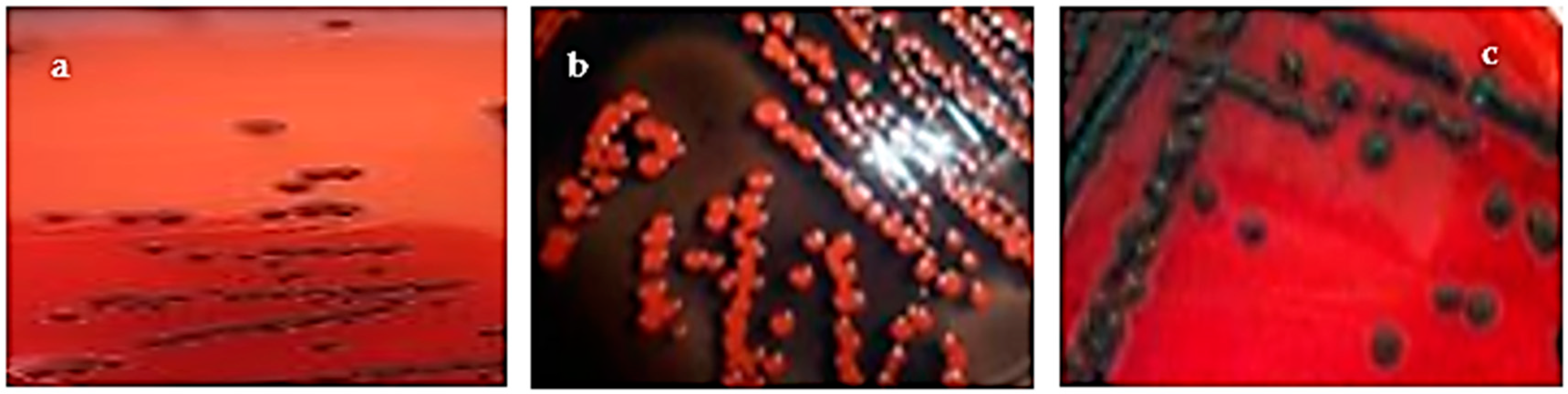
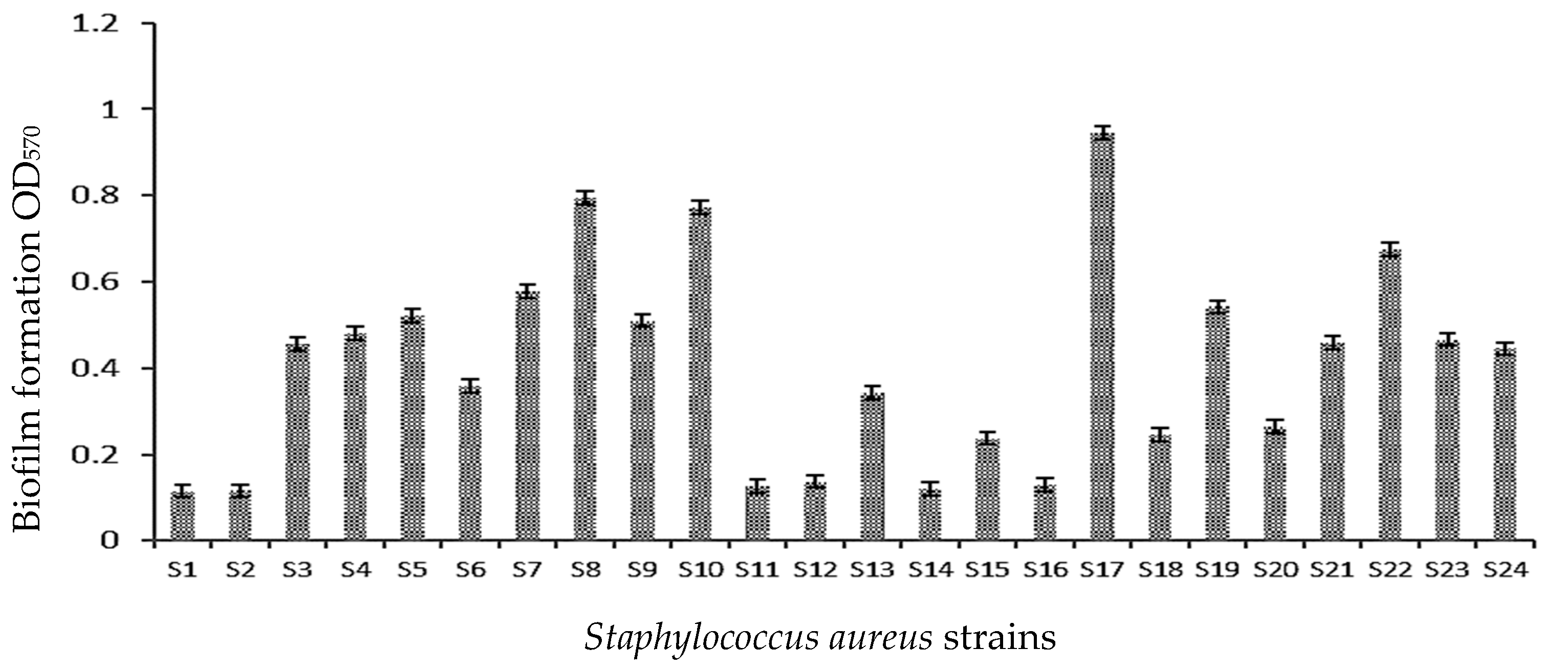
3.1. Qualitative and Quantitative Study of Biofilm Formation

The capacity of clinical S. aureus strains to produce biofilm was estimated qualitatively by the culture on the CRA medium and quantitatively by the ability to create a biofilm on a polystyrene surface. In addition, three morph types were defined according to their color on CRA were obtained: non-slime-producing S. aureus strains characterized by Bordeaux (70.8%) and almost black colonies (25.0%). In contrast, a black colony characterizing slime-positive bacteria was obtained only for strain S17 of S. aureus (4.1%) (Figure 1, Table 3).
The main results showed that all S. aureus tested strains (also S17 very black on CRA) exhibited moderate biofilm formation on polystyrene (0.1 < OD570 < 1) compared to the reference strain S. aureus ATCC 43300 (OD570 = 1.89 ± 0.13) (Figure 2, Table 3).
According to our results, 19 S. aureus strains (79.1%) were positive for both icaA (188 bp) and icaD (198 bp) genes encoding the intracellular adhesins A and D compared to the positive control strain of S. aureus ATCC 43300 for both tested genes (Supplementary Material Figure S1, Table 3). In addition, all the primers used in the experiment exhibited specificity, with a single band. Therefore, only five clinical strains (20.8%) were expected from this correlation (Table 3).

3.2. Distribution of Adhesion Genes

This study emphasized four biofilm-related genes encoding fibronectin-binding proteins A and B (fnbA and fnbB), bound coagulase (clfA), and collagen adhesin gene (cna) involved in S. aureus cell attachment and multiplication. All S. aureus strains expressed biofilm genes. As shown in Table 4, the fnbA (259 bp) and fnbB (523 bp) encoding fibronectin were detected in 33.3% and 37.5%, respectively, of the total strains (Supplementary Material Figure S2). The clfA (288 bp) and cna (192 bp) genes encoding collagen were amplified in 50.0% and 70.8% of the total tested strains (Supplementary Material Figure S3).
As shown in Table 4, six S. aureus strains (S1, S8, S9, S13, S20, and S22) among 24 (25.0%) have the four tested genes (cna, clfA, fnbA, and fnbB) in their genomes compared to the reference strain S. aureus ATCC 43330.

3.3. Distribution of Antibiotic Resistance Genes

The β-lactamases blaZ gene (blaZ), methicillin resistance determinant (mecA), fluoroquinolones resistance gene (norA), and nitric oxide reductase (norB) gene were studied for all S. aureus isolates. All these results are presented in Supplementary Material Figure S4 and Table 5.
All the genomes studied in this work had blaz (70.8%), norB (54.1%), norA (41.6%), and mecA (37.5%) genes. The reference strain of S. aureus ATCC 43300 and the clinical isolates (S1, S8, S9, S23, and S24) had in common the four genes present in their genomes (Table 5).
Based on the results of the phenotypic profile of antibacterial resistance, the clinical strains of S. aureus S12, S22, and S2 were more resistant to the tested antibiotics, with percentages of resistance of 63.6%, 54.5%, and 45.4%, respectively. Compared to the more sensitive strains, S10 was very susceptible to the action of the antibiotics (0.9% of resistance), followed by isolate S17 (9.1% of resistance) (Supplementary Material Table S1).

4. Discussion

The skin surface can be affected by several microorganisms that cause wound infection. S. aureus is a pathogenic bacterium that is resistant to penicillin due to the production of β-lactamase and which contributes to the inhibition of its antibacterial activity. S. aureus causes many problems in hospitals and is usually resistant to antimicrobials [3,32].
In the present study, antibiotic susceptibility results were reported to be influenced by various factors, such as the expression of the antimicrobials resistance genes. Our results showed that the blaZ gene was widely spread among S. aureus strains (70.83%), followed by norA, norB, and mecA genes. The norB gene has a single antimicrobial resistance mechanism. At the same time, mecA and blaZ possess several antibiotic resistance mechanisms [3,17,32].
Efflux pump complexes and resistance-conferring antibiotic subunits, as well as protein(s) (norA and norB), are the most prevalent mechanisms of antibiotic resistance. Other instruments include the antibiotic resistance gene or operon (mecA and blaZ) and antibiotic inactivation enzyme (SAT4 and mphC) [31].
S. aureus strains express resistance genes from external sources [33,34]. This can be natural or due to antimicrobial abuse and misuse, leading to chromosomal mutation and antibiotic selection. Antibiotic-resistant strains are receiving significant consideration in the contemporary era [26]. Resistance to many antimicrobial agents causes critical problems in treating S. aureus infection [35,36]. For example, methicillin resistance contributes to the inhibition of the synthesis of the cell wall.
S. aureus strains can live in biofilms in their natural environment, where planktonic cells proliferate and accumulate in multilayer cells. This structure can protect microorganisms from the action of antimicrobials, extracellular enzymes, and stress factors [24].
The results of the biofilm formation using the crystal violet method showed that all tested S. aureus strains formed a biofilm on the polystyrene. Mathur et al. [37] demonstrated that 14.47% of isolates from blood, medical devices, and skin surfaces formed a strong biofilm, 39.4% formed a moderate biofilm, and 46% of the strains were not biofilm producers [37].
The ability of clinical S. aureus isolates to produce exopolysaccharides on CRA plates showed that only one strain was able to produce mucus compared to the results obtained by Arciola et al., in which 57.5% of the isolates had mucus [20].
Bacterial biofilm formation is encoded by adhesions responsible for the pathogenicity of S. aureus strains [20]. Therefore, in this study, we focused on detecting genes coding for them.
The ica A and ica D genes responsible for the synthesis of polysaccharide intercellular adhesin (PIA) were detected in all strains. The presence of icaA/icaD genes was detected in 19/24 S. aureus strains (79.16%). Arciola et al. [26] found that 60.86% of S. aureus strains harbor icaA and icaD genes. In previous research, it has been demonstrated that 98% of clinical isolates of S. aureus have icaA, and 96% have icaD [38].
In the study of Rohde et al. [25], all tested S. aureus strains, including slime-negative isolates, expressed the icaA gene. This was contrary to the research of Arciola et al. [26], who correlate the presence of icaA/icaD genes to the production of exopolysaccharides.
Adhesin genes (cna, fnbA, fnbB and clfA) were expressed in six S. aureus strains (25%). This demonstrated that clfA and clfB proteins were implicated in the pathogenesis of S. aureus, conferring endocarditis, bacteremia, or pyonephrosis [20]. Our results are similar to those reported by Arciola et al. [26], who noted that 84/191 clinical S. aureus strains (44%) expressed two adhesins genes (fnbA and cna). The cna gene was detected in 70.83% of the tested strains in the present work.
Several virulence factors of S. aureus, including fibronectin-binding (fnbA and fnbB), responsible of bacterial adhesion have been largely described [38,39].

5. Conclusions

This work provides new information on S. aureus isolated from wound surfaces. In fact, S. aureus strains are biofilm producers. They show high percentages of genes responsible for its adhesion and resistance to antimicrobials. These genes can make strains capable of colonizing many human organs. In further work, we envisage studying the effect of natural and synthetic compounds on the biofilm and antimicrobial resistance of S. aureus strains using molecular and in silico approaches.

Supplementary Materials

The following supporting information can be downloaded at: https://www.mdpi.com/article/10.3390/microorganisms11051353/s1, Figure S1: Agarose gel electrophoresis of PCR amplification of icaA (188 bp) and icaD (198 bp) locus. Lane 1: 100 pb DNA molecular size marker; Lanes 3-5: PCR amplification of icaA; Lanes 6-8: PCR amplification of icaD. Lane 2: Negative control; Lanes 3 to 8: PCR amplicons obtained with DNA of S. aureus. Lane 3: S. aureus ATCC 43300, Lane 4: S1; Lane 5: S3; Lane 6: S4; Lane 7: S8; Lane 8: S10. Figure S2: Agarose gel electrophoresis of PCR amplification of fnbA gene (191 bp) and fnbB gene (201 bp). Lane 1: 100 bp DNA molecular size marker; Lane 2: negative control; Lanes 3-5: PCR amplification of fnbA gene; Lanes 6-7: PCR amplification of fnbB gene; Lane 3 to 7: PCR amplicons obtained with DNA amplification of S. aureus; Lane 3: S. aureus ATCC 43300; Lane 4: S8; Lane 5: S13; Lane 6: S. aureus ATCC 43300; Lane 7: S20. Figure S3: Agarose gel electrophoresis of polymerase chain reaction (PCR) amplification cna gene (192 bp) and clfA gene (1kb). Lane 1, 100 bp DNA molecular size marker. Lane 2 negative control; Lanes 3-6 PCR amplification of cna gene, lanes 7-8 PCR amplification of clfA gene. Lane 3 to 8 PCR amplicons obtained with DNA amplification of S. aureus. Lane 3, S. aureus ATCC 43300; Lane 4, S1; Lane 5, S6; Lane 6, S15; Lane 7, S. aureus ATCC 43300; Lane 8, S9. Figure S4: Agarose gel electrophoresis of polymerase chain reaction (PCR) amplification of mecA (140 pb), norA (620 bp), norB (213 bp) and blaz (172 bp) genes. Lane 1: 100 bp DNA molecular size marker; Lanes 2-3 PCR amplification of mecA gene. Lanes 4-5 PCR amplification of norA gene. Lanes 6-7 PCR amplification of norB gene and lane 8 PCR amplification of blaz gene. Lanes 2: S. aureus ATCC 43300; Lanes 3 to 8 PCR amplicons obtained with DNA amplification of S. aureus. Lane 2: S1; Lane 3: S3; Lane 4: S8; Lane 5: S9; Lane 6: S6; Lane 7: S11; Lane 8: S23. Table S1: Study of antimicrobial susceptibility profile of clinical S. aureus strains.

Author Contributions

Conceptualization, E.N., M.S and K.F.A.; methodology, K.F.A., E.N. and T.Z.; software, M.S. and A.S.B.; validation, K.F.A., E.N. and M.S.; formal analysis, E.N. and M.S.; investigation, K.F.A. and E.N.; resources, A.S.B.; data curation, K.F.A., E.N. and M.S.; writing—original draft preparation, K.F.A., E.N. and T.Z.; writing—review and editing, M.S.; visualization, K.F.A. and E.N.; supervision, K.F.A.; project administration, K.F.A. and E.N.; funding acquisition, K.F.A. All authors have read and agreed to the published version of the manuscript.

Funding

This research has been funded by Scientific Research Deanship at University of Ha’il—Saudi Arabia through project number BA-2209.

Data Availability Statement

Not applicable.

Acknowledgments

This research has been funded by Scientific Research Deanship at University of Ha’il—Saudi Arabia through project number BA-2209.

Conflicts of Interest

There are no conflict of interest.

References

  1. Rezia, R.A.; Vijendra, R.; Gopi, A. Uropathogens causing urinary tract infection in adults in a tertiary care hospital. J. Pharmacol. Pharmacother. 2020, 11, 119–124. [Google Scholar]
  2. Savini, V. Pet-to-Man Travelling Staphylococci: A World in Progress; Academic Press: New York, NY, USA, 2018. [Google Scholar]
  3. Hindy, J.R.; Quintero-Martinez, J.A.; Lee, A.T.; Scott, C.G.; Gerberi, D.J.; Mahmood, M.; DeSimone, D.C.; Baddour, L.M. Incidence Trends and Epidemiology of Staphylococcus aureus Bacteremia: A Systematic Review of Population-Based Studies. Cureus 2022, 29, e25460. [Google Scholar] [CrossRef] [PubMed]
  4. Laupland, K.B.; Steele, L.; Pasquill, K.; Parfitt, E.C. Staphylococcus aureus bloodstream infection: Secular changes associated with the implementation of a de novo clinical infectious diseases service in a Canadian population. Int. J. Infect. Dis. 2021, 104, 45–49. [Google Scholar] [CrossRef] [PubMed]
  5. Sakwinska, O.; Giddey, M.; Moreillon, M.; Morisset, D.; Waldvogel, A.; Moreillon, P. Staphylococcus aureus host range and human-bovine host shift. Appl. Environ. Microbiol. 2011, 77, 5908–5915. [Google Scholar] [CrossRef]
  6. Vos, P.; Garrity, G.; Jones, D.; Krieg, N.R.; Ludwig, W.; Rainey, F.A.; Schleifer, K.-H.; Whitman, W.B. Bergey’s Manual of Systematic Bacteriology: Volume 3: The Firmicutes; Springer Science & Business Media: Berlin/Heidelberg, Germany, 2011; Volume 3. [Google Scholar]
  7. Gong, C.; Guan, W.; Liu, X.; Zheng, Y.; Li, Z.; Zhang, Y.; Zhu, S.; Jiang, H.; Cui, Z.; Wu, S. Biomimetic Bacteriophage-Like Particles Formed from Probiotic Extracts and NO Donors for Eradicating Multidrug-Resistant Staphylococcus aureus. Adv. Mater. 2022, 34, e2206134. [Google Scholar] [CrossRef]
  8. Morar, A.; Ban-Cucerzan, A.; Herman, V.; Tîrziu, E.; Sallam, K.I.; Abd-Elghany, S.M.; Imre, K. Multidrug resistant coagulase-positive Staphylococcus aureus and their enterotoxins detection in traditional cheeses marketed in Banat Region, Romania. Antibiotics 2021, 10, 1458. [Google Scholar] [CrossRef]
  9. Onyango, L.A.; Alreshidi, M.M. Adaptive metabolism in staphylococci: Survival and persistence in environmental and clinical settings. J. Pathog. 2018, 2018, 1092632. [Google Scholar] [CrossRef]
  10. Yarovoy, J.Y.; Monte, A.A.; Knepper, B.C.; Young, H.L. Epidemiology of Community-Onset Staphylococcus aureus Bacteremia. West J. Emerg. Med. 2019, 20, 438–442. [Google Scholar] [CrossRef]
  11. Vuong, C.; Kocianova, S.; Voyich, J.M.; Yao, Y.; Fischer, E.R.; DeLeo, F.R.; Otto, M. A crucial role for exopolysaccharide modification in bacterial biofilm formation, immune evasion, and virulence. J. Biol. Chem. 2004, 279, 54881–54886. [Google Scholar] [CrossRef]
  12. Foster, T.J.; Geoghegan, J.A.; Ganesh, V.K.; Höök, M. Adhesion, invasion and evasion: The many functions of the surface proteins of Staphylococcus aureus. Nat. Rev. Microbiol. 2014, 12, 49–62. [Google Scholar] [CrossRef]
  13. Blaiotta, G.; Pennacchia, C.; Villani, F.; Ricciardi, A.; Tofalo, R.; Parente, E. Diversity and dynamics of communities of coagulase-negative staphylococci in traditional fermented sausages. J. Appl. Microbiol. 2004, 97, 271–284. [Google Scholar] [CrossRef] [PubMed]
  14. Donlan, R.M.; Costerton, J.W. Biofilms: Survival mechanisms of clinically relevant microorganisms. Clin. Microbiol. Rev. 2002, 15, 167–193. [Google Scholar] [CrossRef] [PubMed]
  15. Kot, B.; Sytykiewicz, H.; Sprawka, I. Expression of the biofilm-associated genes in methicillin-resistant Staphylococcus aureus in biofilm and planktonic conditions. Int. J. Mol. Sci. 2018, 19, 3487. [Google Scholar] [CrossRef] [PubMed]
  16. Foster, T.J. Surface Proteins of Staphylococcus aureus. Microbiol. Spectr. 2019, 7, 4. [Google Scholar] [CrossRef]
  17. Murai, M.; Moriyama, H.; Hata, E.; Takeuchi, F.; Amemura-Maekawa, J. Variation and association of fibronectin-binding protein genes fnbA and fnbB in Staphylococcus aureus Japanese isolates. Microbiol. Immunol. 2016, 60, 312–325. [Google Scholar] [CrossRef]
  18. Snoussi, M.; Noumi, E.; Cheriaa, J.; Usai, D.; Sechi, L.A.; Zanetti, S.; Bakhrouf, A. Adhesive properties of environmental Vibrio alginolyticus strains to biotic and abiotic surfaces. New Microbiol. 2008, 31, 489–500. [Google Scholar] [PubMed]
  19. Freeman, D.; Falkiner, F.; Keane, C. New method for detecting slime production by coagulase negative staphylococci. J. Clin. Pathol. 1989, 42, 872–874. [Google Scholar] [CrossRef] [PubMed]
  20. Arciola, C.R.; Campoccia, D.; Gamberini, S.; Cervellati, M.; Donati, E.; Montanaro, L. Detection of slime production by means of an optimised Congo red agar plate test based on a colourimetric scale in Staphylococcus epidermidis clinical isolates genotyped for ica locus. Biomaterials 2002, 23, 4233–4239. [Google Scholar] [CrossRef]
  21. Ziebuhr, W.; Krimmer, V.; Rachid, S.; Lößner, I.; Götz, F.; Hacker, J. A novel mechanism of phase variation of virulence in Staphylococcus epidermidis: Evidence for control of the polysaccharide intercellular adhesin synthesis by alternating insertion and excision of the insertion sequence element IS256. Mol. Microbiol. 1999, 32, 345–356. [Google Scholar] [CrossRef]
  22. Knobloch, J.K.-M.; Bartscht, K.; Sabottke, A.; Rohde, H.; Feucht, H.-H.; Mack, D. Biofilm formation by Staphylococcus epidermidis depends on functional RsbU, an activator of the sigB operon: Differential activation mechanisms due to ethanol and salt stress. J. Bacteriol. 2001, 183, 2624–2633. [Google Scholar] [CrossRef]
  23. Christensen, G.D.; Simpson, W.; Younger, J.; Baddour, L.; Barrett, F.; Melton, D.; Beachey, E. Adherence of coagulase-negative staphylococci to plastic tissue culture plates: A quantitative model for the adherence of staphylococci to medical devices. J. Clin. Microbiol. 1985, 22, 996–1006. [Google Scholar] [CrossRef]
  24. Chaieb, K.; Chehab, O.; Zmantar, T.; Rouabhia, M.; Mahdouani, K.; Bakhrouf, A. In vitro effect of pH and ethanol on biofilm formation by clinical ica-positive Staphylococcus epidermidis strains. Ann. Microbiol. 2007, 57, 431–437. [Google Scholar] [CrossRef]
  25. Rohde, H.; Knobloch, J.K.; Horstkotte, M.A.; Mack, D. Correlation of Staphylococcus aureus icaADBC genotype and biofilm expression phenotype. J. Clin. Microbiol. 2001, 39, 4595–4596. [Google Scholar] [CrossRef] [PubMed]
  26. Arciola, C.R.; Campoccia, D.; Gamberini, S.; Baldassarri, L.; Montanaro, L. Prevalence of cna fnbA and fnbB adhesin genes among Staphylococcus aureus isolates from orthopedic infections associated to different types of implant. FEMS Microbiol. Lett. 2005, 246, 81–86. [Google Scholar] [CrossRef] [PubMed]
  27. McDevitt, D.; Francois, P.; Vaudaux, P.; Foster, T. Identification of the ligand-binding domain of the surface-located fibrinogen receptor (clumping factor) of Staphylococcus aureus. Mol. Microbiol. 1995, 16, 895–907. [Google Scholar] [CrossRef] [PubMed]
  28. Geha, D.J.; Uhl, J.R.; Gustaferro, C.A.; Persing, D.H. Multiplex PCR for identification of methicillin-resistant staphylococci in the clinical laboratory. J. Clin. Microbiol. 1994, 32, 1768–1772. [Google Scholar] [CrossRef] [PubMed]
  29. Martineau, F.; Picard, F.J.; Lansac, N.; Ménard, C.; Roy, P.H.; Ouellette, M.; Bergeron, M.G. Correlation between the resistance genotype determined by multiplex PCR assays and the antibiotic susceptibility patterns of Staphylococcus aureus and Staphylococcus epidermidis. Antimicrob. Agents Chemother. 2000, 44, 231–238. [Google Scholar] [CrossRef]
  30. Bazaid, A.S.; Saeed, A.; Alrashidi, A.; Alrashidi, A.; Alshaghdali, K.; Hammam, A.S.; Alreshidi, T.; Alshammary, M.; Alarfaj, A.; Thallab, R. Antimicrobial surveillance for bacterial uropathogens in Ha’il, Saudi Arabia: A Five-year multicenter retrospective study. Infect. Drug Resist. 2021, 14, 1455–1465. [Google Scholar] [CrossRef]
  31. Otarigho, B.; Falade, M.O. Analysis of antibiotics resistant genes in different strains of Staphylococcus aureus. Bioinformation 2018, 14, 113. [Google Scholar] [CrossRef]
  32. Otto, M. MRSA virulence and spread. Cell. Microbiol. 2012, 14, 1513–1521. [Google Scholar] [CrossRef]
  33. Cue, D.; Lei, M.G.; Lee, C.Y. Genetic regulation of the intercellular adhesion locus in staphylococci. Front. Cell. Infect. Microbiol. 2012, 2, 38. [Google Scholar] [CrossRef]
  34. Montanaro, L.; Arciola, C.; Borsetti, E.; Collamati, S.; Baldassarri, L. Detection of fibronectin-binding protein genes in staphylococcal strains from peri-prosthesis infections. New Microbiol. 1999, 22, 331–336. [Google Scholar]
  35. He, L.; Zhang, F.; Jian, Y.; Lv, H.; Hamushan, M.; Liu, J.; Liu, Y.; Wang, H.; Tang, J.; Han, P.; et al. Key role of quorum-sensing mutations in the development of Staphylococcus aureus clinical device-associated infection. Clin. Transl. Med. 2022, 12, e801. [Google Scholar] [CrossRef] [PubMed]
  36. Said-Salim, B.; Dunman, P.; McAleese, F.; Macapagal, D.; Murphy, E.; McNamara, P.; Arvidson, S.; Foster, T.; Projan, S.; Kreiswirth, B. Global regulation of Staphylococcus aureus genes by Rot. J. Bacteriol. 2003, 185, 610–619. [Google Scholar] [CrossRef] [PubMed]
  37. Mathur, T.; Singhal, S.; Khan, S.; Upadhyay, D.; Fatma, T.; Rattan, A. Detection of biofilm formation among the clinical isolates of staphylococci: An evaluation of three different screening methods. Indian J. Med. Microbiol. 2006, 24, 25–29. [Google Scholar] [CrossRef] [PubMed]
  38. Piechota, M.; Kot, B.; Frankowska-Maciejewska, A.; Grużewska, A.; Woźniak-Kosek, A. Biofilm formation by methicillin-resistant and methicillin-sensitive Staphylococcus aureus strains from hospitalized patients in Poland. BioMed Res. Int. 2018, 2018, 4657396. [Google Scholar] [CrossRef]
  39. Foster, T.J. The MSCRAMM family of cell-wall-a.nchored surface proteins of gram-positive cocci. Trends Microbiol. 2019, 27, 927–941. [Google Scholar] [CrossRef]
Figure 1. Different morphotypes obtained on Congo red agar based on the color obtained. (a) Bordeaux; (b) almost black; (c) very black (strain S17).
Microorganisms 11 01353 g001
Figure 2. Biofilm formation (OD570) by S. aureus strains on 96-well polystyrene plates.
Microorganisms 11 01353 g002
Table 1. Primers used for detection of adhesion genes.
GenePrimer Sequence 5′-3′Amplicon Size (bp)References
icaAACACTTGCTGGCGCAGTCAA
TCTGGAACCAACATCCAACA
188[27]
icaDATGGTCAAGCCCAGACAGAG
AGTATTTTCAATGTTTAAAGCAA
198[27]
cnaAAAGCGTTGCCTAGTGGAGA
AGTGCCTTCCCAAACCTTTT
192[28]
fnbAGATACAAACCCAGGTGGTGG
TGTGCTTGACCATGCTCTTC
191[28]
fnbBTGTGCTTGACCATGCTCTTC
AGTTGATGTCGCGCTGTATG
201[28]
clfACCGGATCCGTAGCTGCAGATGCACC
GCTCTAGATCACTCATCAGGTTGTTCAGG
1000[29]
Table 2. Primers used for the detection of genes encoding antimicrobials resistance.
GenePrimer Sequence 5′-3′Amplicon Size (bp)References
mecAAACAGGTGAATTATTAGCACTTGTAAG
ATTGCTGTTAATATTTTTTGAGTTGA
140[30]
norATTCACCAAGCCATCAAAAAG
CTTGCCTTTCTCCAGCAATA
620[31]
norBAGCGCGTTGTCTATCTTTCC
GCAGGTGGTCTTGCTGATAA
213[31]
blaZACTTCAACACCTGCTGCTTTC
TGACCACTTTTATCAGCAACC
172[32]
Table 3. Correlation between phenotypic and genotypic adhesive properties of clinical S. aureus strains.
StrainsOD570 ± SDBiofilm ProductionPhenotype on CRASlimeica A/icaD Gene
S. aureus ATCC 433001.89 ± 0.13High biofilmVery BlackS+icaA+/icaD +
S10.11 ± 0.01Moderate biofilmBordeauxS−icaA+/icaD +
S20.11 ± 0.01Moderate biofilmAlmost blackS−icaA+/icaD +
S30.45 ± 0.01Moderate biofilmBordeauxS−icaA+/icaD +
S40.48 ± 0.04Moderate biofilmAlmost blackS−icaA+/icaD +
S50.52 ± 0.01Moderate biofilmBordeauxS−icaA+/icaD +
S60.35 ± 0.03Moderate biofilmAlmost blackS−icaA+/icaD +
S70.57 ± 0.04Moderate biofilmBordeauxS−icaA+/icaD +
S80.79 ± 0.01Moderate biofilmAlmost blackS−icaA+/icaD +
S90.51 ± 0.01Moderate biofilmBordeauxS−icaA+/icaD +
S100.77 ± 0.02Moderate biofilmAlmost blackS−icaA+/icaD +
S110.12 ± 0.01Moderate biofilmBordeauxS−icaA −/icaD −
S120.13 ± 0.01Moderate biofilmBordeauxS−icaA −/icaD −
S130.34 ± 0.01Moderate biofilmBordeauxS−icaA+/icaD +
S140.12 ± 0.01Moderate biofilmBordeauxS−icaA −/icaD −
S150.23 ± 0.01Moderate biofilmBordeauxS−icaA+/icaD +
S160.13 ± 0.03Moderate biofilmAlmost blackS−icaA −/icaD −
S170.94 ± 0.04Moderate biofilmVery BlackS+icaA+/icaD +
S180.24 ± 0.02Moderate biofilmBordeauxS−icaA −/icaD −
S190.54 ± 0.01Moderate biofilmBordeauxS−icaA+/icaD +
S200.26 ± 0.03Moderate biofilmBordeauxS−icaA+/icaD +
S210.45 ± 0.01Moderate biofilmBordeauxS−icaA+/icaD +
S220.67 ± 0.02Moderate biofilmBordeauxS−icaA+/icaD +
S230.46 ± 0.02Moderate biofilmBordeauxS−icaA+/icaD +
S240.44 ± 0.02Moderate biofilmBordeauxS−icaA+/icaD +
% of positivity 8.33%83.33%
S+: slime producer; S−: slime non producer.
Table 4. Distribution of adhesion genes cna, clfA, fnbA and fnbB in S. aureus strains genome.
StrainscnaclfAfnbAfnbB% of the Presence
of the Four Genes
S. aureus ATCC 43300++++100%
S1++++100%
S20%
S3+++75%
S4++50%
S5++50%
S6+25%
S70%
S8++++100%
S9++++100%
S10+25%
S110%
S120%
S13++++100%
S140%
S15+25%
S160%
S17++50%
S18++50%
S19+25%
S20++++100%
S21++50%
S22++++100%
S23++50%
S24+++75%
% of positivity75%54.16%37.5%41.66%
Table 5. Distribution of antibiotic resistance genes mec A, norA, norB and blaz in S. aureus strains genome.
StrainsmecAnorAnorBblaZ% of the Presence
of the Four Genes
S. aureus ATCC++++100%
S1++++100%
S20%
S3++50%
S4++50%
S5++50%
S6+++75%
S7+25%
S8++++100%
S9++++100%
S100%
S11++50%
S12+25%
S13++50%
S14+25%
S15++50%
S16+25%
S170%
S180%
S19+25%
S20+++75%
S21+++75%
S22+++75%
S23++++100%
S24++++100%
% of positivity41.66%45.83%58.33%75%
Disclaimer/Publisher’s Note: The statements, opinions and data contained in all publications are solely those of the individual author(s) and contributor(s) and not of MDPI and/or the editor(s). MDPI and/or the editor(s) disclaim responsibility for any injury to people or property resulting from any ideas, methods, instructions or products referred to in the content.

