
Comprehensive Approaches for the Search and Characterization of Staphylococcins
 ,
,
Abstract
:1. Introduction
2. Classification and Niche of Staphylococcus
2.1. Staphylococci in Skin/Nasal Microbiota of Humans

| Bacteriocin | Producer (Strain) | Origin | Activity against * | Classification a | References | |
|---|---|---|---|---|---|---|
| Gram (+) | Gram (−) | |||||
| Staphylococcin C55 | S. aureus (C55) | Human skin | S. aureus, streptococci, pneumococci, Corynebacterium, Enterococcus | Neisseria | Class II | [35,36] |
| Staphylococcin BacR1 | S. aureus (UT0007) S. aureus (UT0002) | Clinical | Staphylococcus, Streptococcus, Corynebacterium, Enterococcus, Bacillus | Neisseria, Haemophilus, Moraxella, Bordetella, Pasteurella | BLIS | [58] |
| Aureocin A70 | S. aureus (A70) | Milk | Listeria monocytogenes, Staphylococcus | − | Class II | [71,72] |
| Aureocin 4181 | S. aureus (4181) | Bovine mastitis | Staphylococcus, Streptococcus | − | ClassII | [73] |
| Aureocin A53 | S. aureus (A53) | Milk | Lactic acid bacteria, L. monocytogenes, S. aureus, Mycobacterium bovis | − | Class II | [74] |
| Aureocin 215FN | S. aureus (215FN) | Cow nare | Corynebacterium, Streptococcus, L. monocytogenes, Bacillus, Lactobacillus | − | BLIS | [75,76] |
| Staphylococcin 414 | S. aureus (414) | Turkey | Staphylococcus, Micrococcus, Bacillus, Lactobacillus, Streptococcus | − | BLIS | [77] |
| Staphylococcin 462 | S. aureus (462) | Mink | S. aureus | − | BLIS | [78] |
| Staphylococcin IYS2 | S. aureus (IYS2) | Human saliva | S. aureus, Streptococcus, Propionibacterium, L. monocytogenes, Corynebacterium, Actinomyces | − | BLIS | [59] |
| Staphylococcin Au-26 | S. aureus (26) | Human vagine | Staphylococcus, Lactobacillus, Micrococcus, Streptococcus | Neisseria | BLIS | [60] |
| Bac 1829 | S. aureus (KSI1829) | Laboratory isolate S. aureus (RN4220) | S. aureus, Streptococcus, Enterococcus, Corynebacterium | Haemophilus, Moraxella, Bordetella, Pasteurella | BLIS | [67] |
| Bac 201 | S. aureus (AB201) | Wound | Staphylococcus, Streptococcus, Enterococcus | Neisseria, Acinetobacter | BLIS | [61] |
| Staphylococcin 188 | S. aureus (188) | Clinical | Staphylococcus, Micrococcus, Streptococcus, Corynebacterium, Mycobacterium tuberculosis | Escherichia coli, Salmonella, Shigella, | BLIS | [62] |
| Staphylococcin D91 | S. aureus (D91) | Clinical | Staphylococcus, Streptococcus | Proteus, E. coli, Pseudomonas | BLIS | [63] |
| BacCH91 | S. aureus (CH-91) | Poultry (DSM26258) | Staphylococcus, Streptococcus, Micrococcus | − | Class I | [79] |
| Bsa | S. aureus (MW2) | MRSA community-acquired (ST8, ST80) | Staphylococcus, Micrococcus | − | Class I | [37,38] |
| Aureocyclicin 4185 | S. aureus (4185) | Bovine mastitis | Listeria monocytogenes, Micrococcus, Bacillus | − | Class IV | [80] |
| Hyicin/Agneticin 3682 | S. hycius/S. agnetis (3682) | Bovine milk | Staphylococcus, Listeria, Streptococcus | − | Class I | [81] |
| Hyicin/Agneticin 4244 | S. hycius/S. agnetis (4244) | Bovine mastitis | Staphylococcus, Listeria Anti-biofilm against S. aureus | − | Sactipeptide | [82] |
| BacSp222 | S. pseudintermedius (222) | Dog skin | Staphylococcus, Micrococcus, Streptococcus, Bacillus | + | Class II | [83] |
| Bacteriocin | Producer (Strain) | Origin | Activity against * | Classification a | References | |
|---|---|---|---|---|---|---|
| Gram (+) | Gram (−) | |||||
| Capidermicin | S. capitis (CIT060) | Human skin | Bacillus, Enterococcus, Lactococcus, Micrococcus, Staphylococcus | − | Class II | [8] |
| Endopeptidase ALE-1 | S. capitis (EPk1) | Clinical sample | + | − | Class III | [39] |
| NisinJ | S. capitis (APC2923) | Human skin | Listeria, Lactobacillus, Staphylococcus, Streptococcus, Corynebacterium, Enterococcus | − | Class I | [32,40] |
| TE8 | S. capitis (TE8) | Human skin | S. aureus | − | BLIS | [64] |
| Nukacin L217 | S. chromogenes (L217) | Bovine teat apices | Staphylococcus, Streptococcus | − | Class I | [84] |
| Staphylococcin T (StT) | S. cohnii (T) | Healthy human | Staphylococcus, Streptococcus, Micrococcus, Listeria | Neisseria | Class I | [48] |
| NukacinIVK45 | S. epidermidis (IVK45) | Human nasal | Micrococcus, Corynebacterium, Streptococcus, Dolosigranulum pigrum | − | Class I | [41] |
| Pep5 | S. epidermidis (5) | Clinical | Staphylococcus, Micrococcus, Corynebacterium | − | Class I | [42,43,44] |
| Epicidin 280 | S. epidermidis (BN 280) | Clinical | Staphylococcus | − | Class I | [57] |
| Epilancin K7 | S. epidermidis (K7) | Laboratory strain | + | − | Class I | [70] |
| Epidermin | S. epidermidis (Tü 3298) | Clinical | + | − | Class I | [52,53,54,55,56] |
| Epidermicin NI01 | S. epidermidis (224) | Clinical | S. aureus, Enterococcus Anti-biofilm against S. epidermidis | − | Class II | [45] |
| Epilancin 15X | S. epidermidis (15X154) | Clinical | Staphylococcus, Enterococcus | − | Class I | [46,47] |
| Staphylococcin 1580 | S. epidermidis (1580) | Laboratory strain | Staphylococcus, Streptococcus, Bacillus, Corynebacterium, Listeria, Acinetobacter | − | BLIS | [66] |
| Micrococcin P1 | S. equorum (WS 2733) | Cheese | S. aureus, Enterococcus, Listeria | − | Thiopeptide | [85] |
| Gallidermin | S. gallinarum F16/P57 Tü3928 | Chicken | Propionibacterium, Staphylococcus, Streptococcus, Micrococcus Anti-biofilm against S. aureus | Neisseria, Moraxella | Class I | [86,87,88,89,90] |
| Hominicin | S. hominis (MBBL 2-9) | Healthy human | S. aureus, Micrococcus, Bacillus, Lactobacillus | + | Class I | [49,50] |
| Nukacin KQU-131 | S. hominis (KQU-131) | Thai fermented fish Pla-ra | Lactic acid bacteria, Micrococcus, Bacillus | − | Class I | [91] |
| Hogocidin-α Hogocodin-β | S. hominis (A9) | Human skin | S. aureus | − | BLIS | [34] |
| Homicorcin | S. hominis (MBL_AB63) | Seeds | Staphylococcus, Micrococcus luteus, Bacillus subtilis, Lactococcus lactis | Class I | [92] | |
| Lugdunin | S. lugdunensis (N920143) | Human nasal | S. aureus, Enterococcus | − | NRPs | [51] |
| Nukacin 3299 Simulancin 3299 | S. simulans (3299) S. simulans (Ec105) | Bovine mastitis | Staphylococcus, S. agalactiae, Corynebacterium | − | Class I | [93] |
| Lysostaphin | S. simulans biovar staphyIolyticus (ATCC1362) | NRRL B-2628 | Staphylococcus | − | Class III | [68,69] |
| Warnericin RB4 | S. warneri (RB4) | Rice | Thermo-acidophiles, Alicyclobacillus, Micrococcus | − | Class I | [94] |
| Warnericin RK | S. warneri (RK) | Environmental | + | Legionella | BLIS | [65] |
| SWLP1 | S. warneri (DSM 16081) | Human skin | + | − | Class I | [19] |
| Nukacin ISK-1 | S. warneri (ISK-1) | Fermented rice bran “Nukadoko” | Staphylococcus, Streptococcus, Micrococcus, Lactococcus, Bacillus | − | Class I | [95,96,97,98,99] |
2.2. Staphylococci in Skin/Nasal Microbiota of Animals
2.3. Staphylococcus in Food
3. The Rationale for Exploring Bacteriocin-Producing Staphylococci: Beneficial and Functional Properties
4. Bacteriocins: Promising Antimicrobial Substances
4.1. Staphylococcins: Classes and Diversities
4.2. Biochemical and Genetic Characterization of Staphylococcins
5. Bacteriocin Detection and Characterization Methods
5.1. Phenotypic Methods
5.2. Genotypic Methods
5.3. Protein Methods
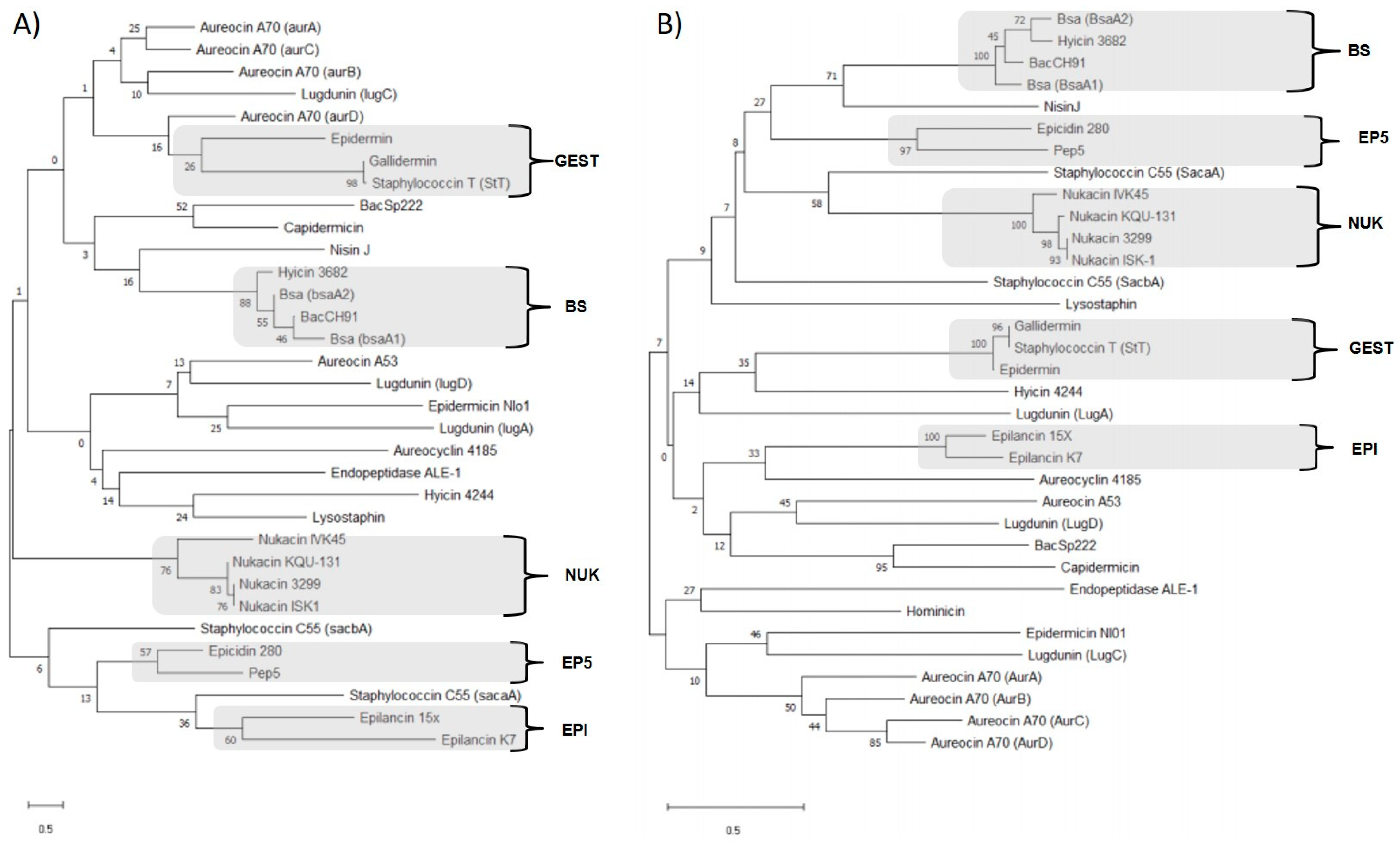
5.4. Universal Nucleotide and Amino Acid-Based Staphylococcin Phylogenetics
6. Applications of Bacteriocin-Producing Staphylococcus Isolates or Their Staphylococcins
7. Emerging Concerns Associated with the Use of Staphylococcins
8. Conclusions
Author Contributions
Funding
Institutional Review Board Statement
Informed Consent Statement
Data Availability Statement
Conflicts of Interest
References
- World Health Organization. WHO Publishes List of Bacteria for Which New Antibiotics Are Urgently Needed. WHO Jt. News Release. 2017. Available online: https://www.who.int/news/item/27-02-2017-who-publishes-list-ofbacteria-for-which-new-antibiotics-are-urgently-needed (accessed on 13 January 2023).
- Antoñanzas, F.; Lozano, C.; Torres, C. Economic Features of Antibiotic Resistance: The Case of Methicillin-Resistant Staphylococcus aureus. Pharmacoeconomics 2015, 33, 285–325. [Google Scholar] [CrossRef]
- Wang, C.-H.; Hsieh, Y.-H.; Powers, Z.M.; Kao, C.-Y. Defeating Antibiotic-Resistant Bacteria: Exploring Alternative Therapies for a Post-Antibiotic Era. Int. J. Mol. Sci. 2020, 21, 1061. [Google Scholar] [CrossRef] [PubMed]
- Anyaegbunam, N.J.; Anekpo, C.C.; Anyaegbunam, Z.K.G.; Doowuese, Y.; Chinaka, C.B.; Odo, O.J.; Sharndama, H.C.; Okeke, O.P.; Mba, I.E. The Resurgence of Phage-Based Therapy in the Era of Increasing Antibiotic Resistance: From Research Progress to Challenges and Prospects. Microbiol. Res. 2022, 264, 127155. [Google Scholar] [CrossRef] [PubMed]
- Mba, I.E.; Nweze, E.I. Application of Nanotechnology in the Treatment of Infectious Diseases: An Overview. In Nanotechnology for Infectious Diseases; Hameed, S., Rehman, S., Eds.; Springer: Singapore, 2022; pp. 25–51. [Google Scholar] [CrossRef]
- Mba, I.E.; Nweze, E.I. Antimicrobial Peptides Therapy: An Emerging Alternative for Treating Drug-Resistant Bacteria. Yale J. Biol. Med. 2022, 95, 445–463. [Google Scholar] [PubMed]
- Soltani, S.; Hammami, R.; Cotter, P.D.; Rebuffat, S.; Said, L.B.; Gaudreau, H.; Bédard, F.; Biron, E.; Drider, D.; Fliss, I. Bacteriocins as a New Generation of Antimicrobials: Toxicity Aspects and Regulations. FEMS Microbiol. Rev. 2021, 45, fuaa039. [Google Scholar] [CrossRef]
- Lynch, D.; O’Connor, P.M.; Cotter, P.D.; Hill, C.; Field, D.; Begley, M. Identification and Characterisation of Capidermicin, a Novel Bacteriocin Produced by Staphylococcus capitis. PLoS ONE 2019, 14, e0223541. [Google Scholar] [CrossRef]
- Riley, M.A.; Wertz, J.E. Bacteriocins: Evolution, Ecology, and Application. Annu. Rev. Microbiol. 2002, 56, 117–137. [Google Scholar] [CrossRef]
- Dobson, A.; Cotter, P.D.; Ross, R.P.; Hill, C. Bacteriocin Production: A Probiotic Trait? Appl. Environ. Microbiol. 2012, 78, 1–6. [Google Scholar] [CrossRef]
- Krismer, B.; Weidenmaier, C.; Zipperer, A.; Peschel, A. The Commensal Lifestyle of Staphylococcus aureus and Its Interactions with the Nasal Microbiota. Nat. Rev. Microbiol. 2017, 15, 675–687. [Google Scholar] [CrossRef]
- Heilbronner, S.; Krismer, B.; Brötz-Oesterhelt, H.; Peschel, A. The Microbiome-Shaping Roles of Bacteriocins. Nat. Rev. Microbiol. 2021, 19, 726–739. [Google Scholar] [CrossRef]
- von Eiff, C.; Peters, G.; Heilmann, C. Pathogenesis of Infections Due to Coagulase negative Staphylococci. Lancet Infect. Dis. 2002, 2, 677–685. [Google Scholar] [CrossRef] [PubMed]
- Schmidt, V.M.; Williams, N.J.; Pinchbeck, G.; Corless, C.E.; Shaw, S.; McEwan, N.; Dawson, S.; Nuttall, T. Antimicrobial Resistance and Characterisation of Staphylococci Isolated from Healthy Labrador Retrievers in the United Kingdom. BMC Vet. Res. 2014, 10, 17. [Google Scholar] [CrossRef] [PubMed]
- Schulz, J.; Friese, A.; Klees, S.; Tenhagen, B.A.; Fetsch, A.; Rösler, U.; Hartung, J. Longitudinal Study of the Contamination of Air and of Soil Surfaces in the Vicinity of Pig Barns by Livestock-Associated Methicillin-Resistant Staphylococcus aureus. Appl. Environ. Microbiol. 2012, 78, 5666–5671. [Google Scholar] [CrossRef] [PubMed]
- Khusro, A.; Aarti, C. Metabolic heterogeneity and techno-functional attributes of fermented foods-associated coagulase-negative staphylococci. Food Microbiol. 2022, 105, 104028. [Google Scholar] [CrossRef]
- Bastos, M.; Ceotto, H.; Coelho, M.; Nascimento, J. Staphylococcal Antimicrobial Peptides: Relevant Properties and Potential Biotechnological Applications. CPB 2009, 10, 38–61. [Google Scholar] [CrossRef]
- Newstead, L.L.; Varjonen, K.; Nuttall, T.; Paterson, G.K. Staphylococcal-Produced Bacteriocins and Antimicrobial Peptides: Their Potential as Alternative Treatments for Staphylococcus aureus Infections. Antibiotics 2020, 9, 40. [Google Scholar] [CrossRef]
- de Freire Bastos, M.D.C.; Miceli de Farias, F.; Carlin Fagundes, P.; Varella Coelho, M.L. Staphylococcins: An Update on Antimicrobial Peptides Produced by Staphylococci and Their Diverse Potential Applications. Appl. Microbiol. Biotechnol. 2020, 104, 10339–10368. [Google Scholar] [CrossRef]
- Fernández-Fernández, R.; Abdullahi, I.N.; González-Azcona, C.; Ulloa, A.; Martínez, A.; García-Vela, S.; Höfle, U.; Zarazaga, M.; Lozano, C.; Torres, C. Detection of antimicrobial producing Staphylococcus from migratory birds: Potential role in nasotracheal microbiota modulation. Front. Microbiol. 2023, 14, 1144975. [Google Scholar] [CrossRef]
- Fernández-Fernández, R.; Lozano, C.; Eguizábal, P.; Ruiz-Ripa, L.; Martínez-Álvarez, S.; Abdullahi, I.N.; Zarazaga, M.; Torres, C. Bacteriocin-like Inhibitory Substances in Staphylococci of Different Origins and Species with Activity against Relevant Pathogens. Front. Microbiol. 2022, 13, 870510. [Google Scholar] [CrossRef]
- Fernández-Fernández, R.; Lozano, C.; Ruiz-Ripa, L.; Robredo, B.; Azcona-Gutiérrez, J.M.; Alonso, C.A.; Aspiroz, C.; Zarazaga, M.; Torres, C. Antimicrobial Resistance and Antimicrobial Activity of Staphylococcus lugdunensis Obtained from Two Spanish Hospitals. Microorganisms 2022, 10, 1480. [Google Scholar] [CrossRef]
- Becker, K.; Heilmann, C.; Peters, G. Coagulase-Negative Staphylococci. Clin. Microbiol. Rev. 2014, 27, 870–926. [Google Scholar] [CrossRef] [PubMed]
- Kluytmans, J.A.J.W. Methicillin-Resistant Staphylococcus aureus in Food Products: Cause for Concern or Case for Complacency? Clin. Microbiol. Infect. 2010, 16, 11–15. [Google Scholar] [CrossRef]
- Gómez-Sanz, E.; Torres, C.; Ceballos, S.; Lozano, C.; Zarazaga, M. Clonal Dynamics of Nasal Staphylococcus aureus and Staphylococcus pseudintermedius in Dog-Owning Household Members. Detection of MSSA ST398. PLoS ONE 2013, 8, e69337. [Google Scholar] [CrossRef]
- Pantosti, A. Methicillin-Resistant Staphylococcus aureus Associated with Animals and Its Relevance to Human Health. Front. Microbio. 2012, 3, 127. [Google Scholar] [CrossRef] [PubMed]
- Bannoehr, J.; Guardabassi, L. Staphylococcus pseudintermedius in the Dog: Taxonomy, Diagnostics, Ecology, Epidemiology and Pathogenicity: Staphylococcus Pseudintermedius in Dogs. Vet. Dermatol. 2012, 23, 253-e52. [Google Scholar] [CrossRef]
- Edslev, S.M.; Olesen, C.M.; Nørreslet, L.B.; Ingham, A.C.; Iversen, S.; Lilje, B.; Clausen, M.-L.; Jensen, J.S.; Stegger, M.; Agner, T.; et al. Staphylococcal Communities on Skin Are Associated with Atopic Dermatitis and Disease Severity. Microorganisms 2021, 9, 432. [Google Scholar] [CrossRef] [PubMed]
- von Eiff, C.; Becker, K.; Machka, K.; Stammer, H.; Peters, G. Nasal Carriage as a Source of Staphylococcus aureus Bacteremia. N. Engl. J. Med. 2001, 344, 11–16. [Google Scholar] [CrossRef]
- Laux, C.; Peschel, A.; Krismer, B. Staphylococcus aureus Colonization of the Human Nose and Interaction with Other Microbiome Members. Microbiol. Spectr. 2019, 7, 7.2.34. [Google Scholar] [CrossRef]
- Abdullahi, I.N.; Fernández-Fernández, R.; Juárez-Fernández, G.; Martínez-Álvarez, S.; Eguizábal, P.; Zarazaga, M.; Lozano, C.; Torres, C. Wild Animals Are Reservoirs and Sentinels of Staphylococcus aureus and MRSA Clones: A Problem with “One Health” Concern. Antibiotics 2021, 10, 1556. [Google Scholar] [CrossRef]
- O’Sullivan, J.N.; Rea, M.C.; O’Connor, P.M.; Hill, C.; Ross, R.P. Human Skin Microbiota Is a Rich Source of Bacteriocin-Producing Staphylococci That Kill Human Pathogens. FEMS Microbiol. Ecol. 2019, 95, fiy241. [Google Scholar] [CrossRef]
- Hardy, B.L.; Bansal, G.; Hewlett, K.H.; Arora, A.; Schaffer, S.D.; Kamau, E.; Bennett, J.W.; Merrell, D.S. Antimicrobial Activity of Clinically Isolated Bacterial Species against Staphylococcus aureus. Front. Microbiol. 2020, 10, 2977. [Google Scholar] [CrossRef] [PubMed]
- Nakatsuji, T.; Chen, T.H.; Narala, S.; Chun, K.A.; Two, A.M.; Yun, T.; Shafiq, F.; Kotol, P.F.; Bouslimani, A.; Melnik, A.V.; et al. Antimicrobials from Human Skin Commensal Bacteria Protect against Staphylococcus aureus and Are Deficient in Atopic Dermatitis. Sci. Transl. Med. 2017, 9, eaah4680. [Google Scholar] [CrossRef] [PubMed]
- Navaratna, M.A.D.B.; Sahl, H.-G.; Tagg, J.R. Identification of Genes Encoding Two-Component Lantibiotic Production in Staphylococcus aureus C55 and Other Phage Group II S. aureus Strains and Demonstration of an Association with the Exfoliative Toxin B Gene. Infect. Immun. 1999, 67, 4268–4271. [Google Scholar] [CrossRef] [PubMed]
- Morriss, D.M.; Lawson, J.W.; Rogolsky, M. Effect of a Staphylococcin on Neisseria gonorrhoeae. Antimicrob. Agents Chemother. 1978, 14, 218–223. [Google Scholar] [CrossRef] [PubMed]
- Daly, K.M.; Upton, M.; Sandiford, S.K.; Draper, L.A.; Wescombe, P.A.; Jack, R.W.; O’Connor, P.M.; Rossney, A.; Götz, F.; Hill, C.; et al. Production of the Bsa Lantibiotic by Community-Acquired Staphylococcus aureus Strains. J. Bacteriol. 2010, 192, 1131–1142. [Google Scholar] [CrossRef] [PubMed]
- Baba, T.; Takeuchi, F.; Kuroda, M.; Yuzawa, H.; Aoki, K.; Oguchi, A.; Nagai, Y.; Iwama, N.; Asano, K.; Naimi, T.; et al. Genome and Virulence Determinants of High Virulence Community-Acquired MRSA. Lancet 2002, 359, 1819–1827. [Google Scholar] [CrossRef]
- Sugai, M.; Fujiwara, T.; Akiyama, T.; Ohara, M.; Komatsuzawa, H.; Inoue, S.; Suginaka, H. Purification and Molecular Characterization of Glycylglycine Endopeptidase Produced by Staphylococcus capitis EPK1. J. Bacteriol. 1997, 179, 1193–1202. [Google Scholar] [CrossRef]
- O’Sullivan, J.N.; O’Connor, P.M.; Rea, M.C.; O’Sullivan, O.; Walsh, C.J.; Healy, B.; Mathur, H.; Field, D.; Hill, C.; Ross, R.P. Nisin J, a Novel Natural Nisin Variant, Is Produced by Staphylococcus capitis Sourced from the Human Skin Microbiota. J. Bacteriol. 2020, 202, e00639-19. [Google Scholar] [CrossRef]
- Janek, D.; Zipperer, A.; Kulik, A.; Krismer, B.; Peschel, A. High Frequency and Diversity of Antimicrobial Activities Produced by Nasal Staphylococcus Strains against Bacterial Competitors. PLoS Pathog. 2016, 12, e1005812. [Google Scholar] [CrossRef]
- Weil, H.-P.; Beck-Sickinger, A.G.; Metzger, J.; Stevanovic, S.; Jung, G.; Josten, M.; Sahl, H.-G. Biosynthesis of the Lantibiotic Pep5. Isolation and Characterization of a Prepeptide Containing Dehydroamino Acids. Eur. J. Biochem. 1990, 194, 217–223. [Google Scholar] [CrossRef]
- Meyer, C.; Bierbaum, G.; Heidrich, C.; Reis, M.; Suling, J.; Iglesias-Wind, M.I.; Kempter, C.; Molitor, E.; Sahl, H.-G. Nucleotide Sequence of the Lantibiotic Pep5 Biosynthetic Gene Cluster and Functional Analysis of PepP and PepC. Evidence for a Role of PepC in Thioether Formation. Eur. J. Biochem. 1995, 232, 478–489. [Google Scholar] [CrossRef] [PubMed]
- Kaletta, C.; Entian, K.-D.; Kellner, R.; Jung, G.; Reis, M.; Sahl, H.-G. Pep5, a New Lantibiotic: Structural Gene Isolation and Prepeptide Sequence. Arch. Microbiol. 1989, 152, 16–19. [Google Scholar] [CrossRef] [PubMed]
- Sandiford, S.; Upton, M. Identification, Characterization, and Recombinant Expression of Epidermicin NI01, a Novel Unmodified Bacteriocin Produced by Staphylococcus epidermidis That Displays Potent Activity against Staphylococci. Antimicrob. Agents Chemother. 2012, 56, 1539–1547. [Google Scholar] [CrossRef] [PubMed]
- Velásquez, J.E.; Zhang, X.; van der Donk, W.A. Biosynthesis of the Antimicrobial Peptide Epilancin 15X and Its N-Terminal Lactate. Chem. Biol. 2011, 18, 857–867. [Google Scholar] [CrossRef]
- Ekkelenkamp, M.B.; Hanssen, M.; Danny Hsu, S.-T.; de Jong, A.; Milatovic, D.; Verhoef, J.; van Nuland, N.A.J. Isolation and Structural Characterization of Epilancin 15X, a Novel Lantibiotic from a Clinical Strain of Staphylococcus epidermidis. FEBS Lett. 2005, 579, 1917–1922. [Google Scholar] [CrossRef]
- Furmanek, B.; Kaczorowski, T.; Bugalski, R.; Bielawski, K.; Bogdanowicz, J.; Podhajska, A.A. Identification, Characterization and Purification of the Lantibiotic Staphylococcin T, a Natural Gallidermin Variant. J. Appl. Microbiol. 1999, 87, 856–866. [Google Scholar] [CrossRef]
- Kim, P.I.; Sohng, J.K.; Sung, C.; Joo, H.-S.; Kim, E.-M.; Yamaguchi, T.; Park, D.; Kim, B.-G. Characterization and Structure Identification of an Antimicrobial Peptide, Hominicin, Produced by Staphylococcus hominis MBBL 2–9. Biochem. Biophys. Res. Commun. 2010, 399, 133–138. [Google Scholar] [CrossRef]
- Sung, C.; Kim, B.-G.; Kim, S.; Joo, H.-S.; Kim, P.I. Probiotic Potential of Staphylococcus hominis MBBL 2–9 as Anti-Staphylococcus aureus Agent Isolated from the Vaginal Microbiota of a Healthy Woman. J. Appl. Microbiol. 2010, 108, 908–916. [Google Scholar] [CrossRef]
- Zipperer, A.; Konnerth, M.C.; Laux, C.; Berscheid, A.; Janek, D.; Weidenmaier, C.; Burian, M.; Schilling, N.A.; Slavetinsky, C.; Marschal, M.; et al. Human Commensals Producing a Novel Antibiotic Impair Pathogen Colonization. Nature 2016, 535, 511–516. [Google Scholar] [CrossRef]
- Schnell, N.; Entian, K.-D.; Schneider, U.; Götz, F.; Zähner, H.; Kellner, R.; Jung, G. Prepeptide Sequence of Epidermin, a Ribosomally Synthesized Antibiotic with Four Sulphide-Rings. Nature 1988, 333, 276–278. [Google Scholar] [CrossRef]
- Schnell, N.; Engelke, G.; Augustin, J.; Rosenstein, R.; Ungermann, V.; Gotz, F.; Entian, K.-D. Analysis of Genes Involved in the Biosynthesis of Lantibiotic Epidermin. Eur. J. Biochem. 1992, 204, 57–68. [Google Scholar] [CrossRef] [PubMed]
- Peschel, A.; Augustin, J.; Kupke, T.; Stevanovic, S.; Götz, F. Regulation of Epidermin Biosynthetic Genes by EpiQ. Mol. Microbiol. 1993, 9, 31–39. [Google Scholar] [CrossRef] [PubMed]
- Augustin, J.; Rosenstein, R.; Wieland, B.; Schneider, U.; Schnell, N.; Engelke, G.; Entian, K.-D.; Gotz, F. Genetic Analysis of Epidermin Biosynthetic Genes and Epidermin-Negative Mutants of Staphylococcus epidermidis. Eur. J. Biochem. 1992, 204, 1149–1154. [Google Scholar] [CrossRef] [PubMed]
- Allgaier, H.; Jung, G.; Werner, R.G.; Schneider, U.; Zahner, H. Epidermin: Sequencing of a Heterodet Tetracyclic 21-Peptide Amide Antibiotic. Eur. J. Biochem. 1986, 160, 9–22. [Google Scholar] [CrossRef] [PubMed]
- Heidrich, C.; Pag, U.; Josten, M.; Metzger, J.; Jack, R.W.; Bierbaum, G.; Jung, G.; Sahl, H.-G. Isolation, Characterization, and Heterologous Expression of the Novel Lantibiotic Epicidin 280 and Analysis of Its Biosynthetic Gene Cluster. Appl. Environ. Microbiol. 1998, 64, 3140–3146. [Google Scholar] [CrossRef]
- Crupper, S.S.; Gies, A.J.; Iandolo, J.J. Purification and Characterization of Staphylococcin BacR1, a Broad-Spectrum Bacteriocin. Appl. Environ. Microbiol. 1997, 63, 4185–4190. [Google Scholar] [CrossRef]
- Nakamura, T.; Yamazaki, N.; Taniguchi, H.; Fujimura, S. Production, Purification, and Properties of a Bacteriocin from Staphylococcus aureus Isolated from Saliva. Infect. Immun. 1983, 39, 609–614. [Google Scholar] [CrossRef]
- Scott, J.C.; Sahl, H.-G.; Carne, A.; Tagg, J.R. Lantibiotic-Mediated Anti-Lactobacillus Activity of a Vaginal Staphylococcus aureus Isolate. FEMS Microbiol. Lett. 1992, 93, 97–102. [Google Scholar] [CrossRef]
- Iqbal, A.; Ahmed, S.; Ali, S.A.; Rasool, S.A. Isolation and Partial Characterization of Bac201: A Plasmid-Associated Bacteriocin-like Inhibitory Substance From Staphylococcus aureus AB201. J. Basic Microbiol. 1999, 39, 325–336. [Google Scholar] [CrossRef]
- Saeed, S.; Ahmad, S.; Rasool, S.A. Antimicrobial Spectrum, Production and Mode of Action of Staphylococcin 188 Produced by Staphylococcus aureus 188. Pak. J. Pharm. Sci. 2004, 17, 1–8. [Google Scholar]
- Kader, O.A.; Sahl, H.-G.; Brandis, H. Isolation and Mode of Action of a Staphylococcin-like Substance Active against Gram-Positive and Gram-Negative Bacteria. Microbiology 1984, 130, 2291–2300. [Google Scholar] [CrossRef] [PubMed]
- Kumar, R.; Jangir, P.K.; Das, J.; Taneja, B.; Sharma, R. Genome Analysis of Staphylococcus capitis TE8 Reveals Repertoire of Antimicrobial Peptides and Adaptation Strategies for Growth on Human Skin. Sci. Rep. 2017, 7, 10447. [Google Scholar] [CrossRef] [PubMed]
- Héchard, Y.; Ferraz, S.; Bruneteau, E.; Steinert, M.; Berjeaud, J.-M. Isolation and Characterization of a Staphylococcus warneri Strain Producing an Anti-Legionella Peptide. FEMS Microbiol. Lett. 2005, 252, 19–23. [Google Scholar] [CrossRef] [PubMed]
- Jetten, A.M.; Vogels, G.D.; de Windt, F. Production and Purification of a Staphylococcus epidermidis Bacteriocin. J. Bacteriol. 1972, 112, 235–242. [Google Scholar] [CrossRef] [PubMed]
- Crupper, S.S.; Iandolo, J.J. Purification and Partial Characterization of a Novel Antibacterial Agent (Bac1829) Produced by Staphylococcus aureus KSI1829. Appl. Environ. Microbiol. 1996, 62, 3171–3175. [Google Scholar] [CrossRef]
- Thumm, G.; Gotz, F. Studies on Prolysostaphin Processing and Characterization of the Lysostaphin Immunity Factor (Lif) of Staphylococcus simulans biovar staphylolyticus. Mol. Microbiol. 1997, 23, 1251–1255. [Google Scholar] [CrossRef]
- Recsei, P.A.; Gruss, A.D.; Novick, R.P. Cloning, Sequence, and Expression of the Lysostaphin Gene from Staphylococcus simulans. Proc. Natl. Acad. Sci. USA 1987, 84, 1127–1131. [Google Scholar] [CrossRef]
- Kamp, M.; Hooven, H.W.; Konings, R.N.H.; Bierbaum, G.; Sahl, H.-G.; Kuipers, O.P.; Siezen, R.J.; Vos, W.M.; Hilbers, C.W.; Ven, F.J.M. Elucidation of the Primary Structure of the Lantibiotic Epilancin K7 from Staphylococcus epidermidis K7. Cloning and Characterisation of the Epilancin-K7-Encoding Gene and NMR Analysis of Mature Epilancin K7. Eur. J. Biochem. 1995, 230, 587–600. [Google Scholar] [CrossRef]
- Netz, D.J.A.; Sahl, H.-G.; Marcolino, R.; dos Santos Nascimento, J.; de Oliveira, S.S.; Soares, M.B.; do Carmo de Freire Bastos, M. Molecular Characterisation of Aureocin A70, a Multi-Peptide Bacteriocin Isolated from Staphylococcus aureus. Edited by M. Yaniv. J. Mol. Biol. 2001, 311, 939–949. [Google Scholar] [CrossRef]
- Varella Coelho, M.L.; Ceotto, H.; Madureira, D.J.; Nes, I.F.; de Freire Bastos, M.D.C. Mobilization Functions of the Bacteriocinogenic Plasmid PRJ6 of Staphylococcus aureus. J. Microbiol. 2009, 47, 327–336. [Google Scholar] [CrossRef]
- Marques-Bastos, S.L.; Varella Coelho, M.L.; Ceotto-Vigoder, H.; Carlin Fagundes, P.; Silva Almeida, G.; Brede, D.A.; Nes, I.F.; Vasconcelos de Paiva Brito, M.A.; de Freire Bastos, M.D.C. Molecular Characterization of Aureocin 4181: A Natural N-Formylated Aureocin A70 Variant with a Broad Spectrum of Activity. Braz. J. Microbiol. 2020, 51, 1527–1538. [Google Scholar] [CrossRef] [PubMed]
- Netz, D.J.A.; Pohl, R.; Beck-Sickinger, A.G.; Selmer, T.; Pierik, A.J.; de Freire Bastos, M.D.C.; Sahl, H.-G. Biochemical Characterisation and Genetic Analysis of Aureocin A53, a New, Atypical Bacteriocin from Staphylococcus aureus. J. Mol. Biol. 2002, 319, 745–756. [Google Scholar] [CrossRef] [PubMed]
- de Oliveira, S.S.; Póvoa, D.C.; dos Santos Nascimento, J.; Do SV Pereira, M.; De Siqueira, J.P., Jr.; de Freire Bastos, M.D.C. Antimicrobial Substances Produced by Staphylococcus aureus Strains Isolated from Cattle in Brazil. Lett. Appl. Microbiol. 1998, 27, 229–234. [Google Scholar] [CrossRef] [PubMed]
- Nascimento, J.S.; Ceotto, H.; Nascimento, S.B.; Giambiagi-deMarval, M.; Santos, K.R.N.; Bastos, M.C.F. Bacteriocins as Alternative Agents for Control of Multiresistant Staphylococcal Strains. Lett. Appl. Microbiol. 2006, 42, 215–221. [Google Scholar] [CrossRef] [PubMed]
- Gagliano, V.J.; Hinsdill, R.D. Characterization of a Staphylococcus aureusaureus Bacteriocin. J. Bacteriol. 1970, 104, 117–125. [Google Scholar] [CrossRef]
- Hale, E.M.; Hinsdill, R.D. Characterization of a Bacteriocin from Staphylococcus aureus Strain 462. Antimicrob. Agents Chemother. 1973, 4, 634–640. [Google Scholar] [CrossRef]
- Wladyka, B.; Wielebska, K.; Wloka, M.; Bochenska, O.; Dubin, G.; Dubin, A.; Mak, P. Isolation, Biochemical Characterization, and Cloning of a Bacteriocin from the Poultry-Associated Staphylococcus aureus Strain CH-91. Appl. Microbiol. Biotechnol. 2013, 97, 7229–7239. [Google Scholar] [CrossRef]
- Potter, A.; Ceotto, H.; Coelho, M.L.V.; Guimarães, A.J.; de Freire Bastos, M.D.C. The Gene Cluster of Aureocyclicin 4185: The First Cyclic Bacteriocin of Staphylococcus aureus. Microbiology 2014, 160, 917–928. [Google Scholar] [CrossRef]
- Carlin Fagundes, P.; Nascimento de Sousa Santos, I.; Silva Francisco, M.; Mattos Albano, R.; de Freire Bastos, M.D.C. Genetic and Biochemical Characterization of Hyicin 3682, the First Bacteriocin Reported for Staphylococcus hyicus. Microbiol. Res. 2017, 198, 36–46. [Google Scholar] [CrossRef]
- de Souza Duarte, A.F.; Ceotto-Vigoder, H.; Barrias, E.S.; Souto-Padrón, T.C.B.S.; Nes, I.F.; de Freire Bastos, M.D.C. Hyicin 4244, the First Sactibiotic Described in Staphylococci, Exhibits an Anti-Staphylococcal Biofilm Activity. Int. J. Antimicrob. Agents 2018, 51, 349–356. [Google Scholar] [CrossRef]
- Wladyka, B.; Piejko, M.; Bzowska, M.; Pieta, P.; Krzysik, M.; Mazurek, Ł.; Guevara-Lora, I.; Bukowski, M.; Sabat, A.J.; Friedrich, A.W.; et al. A Peptide Factor Secreted by Staphylococcus pseudintermedius Exhibits Properties of Both Bacteriocins and Virulence Factors. Sci. Rep. 2015, 5, 14569. [Google Scholar] [CrossRef] [PubMed]
- Braem, G.; Stijlemans, B.; Van Haken, W.; De Vliegher, S.; De Vuyst, L.; Leroy, F. Antibacterial Activities of Coagulase-Negative Staphylococci from Bovine Teat Apex Skin and Their Inhibitory Effect on Mastitis-Related Pathogens. J. Appl. Microbiol. 2014, 116, 1084–1093. [Google Scholar] [CrossRef] [PubMed]
- Carnio, M.C.; Höltzel, A.; Rudolf, M.; Henle, T.; Jung, G.; Scherer, S. The macrocyclic peptide antibiotic micrococcin P(1) is secreted by the food-borne bacterium Staphylococcus equorum WS 2733 and inhibits Listeria monocytogenes on soft cheese. Appl. Environ. Microbiol. 2000, 66, 2378–2384. [Google Scholar] [CrossRef] [PubMed]
- Peschel, A.; Schnell, N.; Hille, M.; Entian, K.-D.; Götz, F. Secretion of the Lantibiotics Epidermin and Gallidermin: Sequence Analysis of the Genes GdmT and GdmH, Their Influence on Epidermin Production and Their Regulation by EpiQ. Mol. Gen. Genet. 1997, 254, 312–318. [Google Scholar] [CrossRef]
- Schnell, N.; Entian, K.-D.; Götz, F.; Hörner, T.; Kellner, R.; Jung, G. Structural Gene Isolation and Prepeptide Sequence of Gallidermin, a New Lanthionine Containing Antibiotic. FEMS Microbiol. Lett. 1989, 58, 263–267. [Google Scholar] [CrossRef]
- Bengtsson, T.; Lönn, J.; Khalaf, H.; Palm, E. The Lantibiotic Gallidermin Acts Bactericidal against Staphylococcus Epidermidis and Staphylococcus aureus and Antagonizes the Bacteria-induced Proinflammatory Responses in Dermal Fibroblasts. MicrobiologyOpen 2018, 7, e00606. [Google Scholar] [CrossRef]
- Freund, S.; Jung, G.; Gutbrod, O.; Foikers, G.; Gibbons, W.A.; Allgaier, H.; Werner, R. The Solution Structure of the Lantibiotic Gallidermin. Biopolymers 1991, 31, 803–811. [Google Scholar] [CrossRef]
- Kellner, R.; Jung, G.; Horner, T.; Zahner, H.; Schnell, N.; Entian, K.-D.; Gotz, F. Gallidermin: A New Lanthionine-Containing Polypeptide Antibiotic. Eur. J. Biochem. 1988, 177, 53–59. [Google Scholar] [CrossRef]
- Wilaipun, P.; Zendo, T.; Okuda, K.; Nakayama, J.; Sonomoto, K. Identification of the Nukacin KQU-131, a New Type-A(II) Lantibiotic Produced by Staphylococcus hominis KQU-131 Isolated from Thai Fermented Fish Product (Pla-Ra). Biosci. Biotechnol. Biochem. 2008, 72, 2232–2235. [Google Scholar] [CrossRef]
- Aftab Uddin, M.; Akter, S.; Ferdous, M.; Haidar, B.; Amin, A.; Shofiul Islam Molla, A.H.M.; Khan, H.; Islam, M.R. A Plant Endophyte Staphylococcus hominis Strain MBL_AB63 Produces a Novel Lantibiotic, Homicorcin and a Position One Variant. Sci. Rep. 2021, 11, 11211. [Google Scholar] [CrossRef]
- Ceotto, H.; Holo, H.; da Costa, K.F.S.; dos Santos Nascimento, J.; Salehian, Z.; Nes, I.F.; de Freire Bastos, M.D.C. Nukacin 3299, a Lantibiotic Produced by Staphylococcus simulans 3299 Identical to Nukacin ISK-1. Vet. Microbiol. 2010, 146, 124–131. [Google Scholar] [CrossRef] [PubMed]
- Minamikawa, M.; Kawai, Y.; Inoue, N.; Yamazaki, K. Purification and Characterization of Warnericin RB4, Anti-Alicyclobacillus Bacteriocin, Produced by Staphylococcus warneri RB4. Curr. Microbiol. 2005, 51, 22–26. [Google Scholar] [CrossRef] [PubMed]
- Sashihara, T.; Kimura, H.; Higuchi, T.; Adachi, A.; Matsusaki, H.; Sonomoto, K.; Ishizaki, A. A Novel Lantibiotic, Nukacin ISK-1, of Staphylococcus warneri ISK-1: Cloning of the Structural Gene and Identification of the Structure. Biosci. Biotechnol. Biochem. 2000, 64, 2420–2428. [Google Scholar] [CrossRef] [PubMed]
- Roy, U.; Islam, M.R.; Nagao, J.; Iida, H.; Mahin, A.-A.; Li, M.; Zendo, T.; Nakayama, J.; Sonomoto, K. Bactericidal Activity of Nukacin ISK-1: An Alternative Mode of Action. Biosci. Biotechnol. Biochem. 2014, 78, 1270–1273. [Google Scholar] [CrossRef]
- Kimura, H.; Matsusaki, H.; Sashihara, T.; Sonomoto, K.; Ishizaki, A. Purification and Partial Identification of Bacteriocin ISK-1, a New Lantibiotic Produced by Pediococcus sp. ISK-1. Biosci. Biotechnol. Biochem. 1998, 62, 2341–2345. [Google Scholar] [CrossRef]
- Aso, Y.; Sashihara, T.; Nagao, J.; Kanemasa, Y.; Koga, H.; Hashimoto, T.; Higuchi, T.; Adachi, A.; Nomiyama, H.; Ishizaki, A.; et al. Characterization of a Gene Cluster of Staphylococcus warneri ISK-1 Encoding the Biosynthesis of and Immunity to the Lantibiotic, Nukacin ISK-1. Biosci. Biotechnol. Biochem. 2004, 68, 1663–1671. [Google Scholar] [CrossRef]
- Aso, Y.; Koga, H.; Sashihara, T.; Nagao, J.; Kanemasa, Y.; Nakayama, J.; Sonomoto, K. Description of Complete DNA Sequence of Two Plasmids from the Nukacin ISK-1 Producer, Staphylococcus warneri ISK-1. Plasmid 2005, 53, 164–178. [Google Scholar] [CrossRef]
- Petinaki, E.; Spiliopoulou, I. Methicillin-Resistant Staphylococcus aureus among Companion and Food-Chain Animals: Impact of Human Contacts. Clin. Microbiol. Infect. 2012, 18, 626–634. [Google Scholar] [CrossRef]
- van Cleef, B.A.; Verkade, E.J.M.; Wulf, M.W.; Buiting, A.G.; Voss, A.; Huijsdens, X.W.; van Pelt, W.; Mulders, M.N.; Kluytmans, J.A. Prevalence of Livestock-Associated MRSA in Communities with High Pig-Densities in The Netherlands. PLoS ONE 2010, 5, e9385. [Google Scholar] [CrossRef]
- Ceballos, S.; Aspiroz, C.; Ruiz-Ripa, L.; Reynaga, E.; Azcona-Gutiérrez, J.M.; Rezusta, A.; Seral, C.; Antoñanzas, F.; Torres, L.; López, C.; et al. Epidemiology of MRSA CC398 in Hospitals Located in Spanish Regions with Different Pig-Farming Densities: A Multicentre Study. J. Antimicrob. Chemother. 2019, 74, 2157–2161. [Google Scholar] [CrossRef]
- Strube, M.L.; Hansen, J.E.; Rasmussen, S.; Pedersen, K. A Detailed Investigation of the Porcine Skin and Nose Microbiome Using Universal and Staphylococcus Specific Primers. Sci. Rep. 2018, 8, 12751. [Google Scholar] [CrossRef] [PubMed]
- Schlattmann, A.; von Lützau, K.; Kaspar, U.; Becker, K. The Porcine Nasal Microbiota with Particular Attention to Livestock-Associated Methicillin-Resistant Staphylococcus aureus in Germany—A Culturomic Approach. Microorganisms 2020, 8, 514. [Google Scholar] [CrossRef] [PubMed]
- Slifierz, M.J.; Friendship, R.M.; Weese, J.S. Longitudinal Study of the Early-Life Fecal and Nasal Microbiotas of the Domestic Pig. BMC Microbiol. 2015, 15, 184. [Google Scholar] [CrossRef]
- Abdullahi, I.N.; Lozano, C.; Simon, C.; Latorre-Fernandez, J.; Zarazaga, M.; Torres, C. Nasal Staphylococci Community of Healthy Pigs and Pig-Farmers in Aragon (Spain). Predominance and within-Host Resistome Diversity in MRSA-CC398 and MSSA-CC9 Lineages. One Health 2023, 16, 100505. [Google Scholar] [CrossRef]
- Espinosa-Góngora, C.; Larsen, N.; Schønning, K.; Fredholm, M.; Guardabassi, L. Differential Analysis of the Nasal Microbiome of Pig Carriers or Non-Carriers of Staphylococcus aureus. PLoS ONE 2016, 11, e0160331. [Google Scholar] [CrossRef] [PubMed]
- Verstappen, K.M.; Willems, E.; Fluit, A.C.; Duim, B.; Martens, M.; Wagenaar, J.A. Staphylococcus aureus Nasal Colonization Differs among Pig Lineages and Is Associated with the Presence of Other Staphylococcal Species. Front. Vet. Sci. 2017, 4, 97. [Google Scholar] [CrossRef]
- Abdullahi, I.N.; Juárez-Fernández, G.; Höfle, Ú.; Cardona-Cabrera, T.; Mínguez, D.; Pineda-Pampliega, J.; Lozano, C.; Zarazaga, M.; Torres, C. Nasotracheal Microbiota of Nestlings of Parent White Storks with Different Foraging Habits in Spain. EcoHealth 2023. [Google Scholar] [CrossRef]
- Ruiz-Ripa, L.; Alcalá, L.; Simón, C.; Gómez, P.; Mama, O.M.; Rezusta, A.; Zarazaga, M.; Torres, C. Diversity of Staphylococcus aureus Clones in Wild Mammals in Aragon, Spain, with Detection of MRSA ST130-MecC in Wild Rabbits. J. Appl. Microbiol. 2019, 127, 284–291. [Google Scholar] [CrossRef]
- Mama, O.M.; Ruiz-Ripa, L.; Fernández-Fernández, R.; González-Barrio, D.; Ruiz-Fons, J.F.; Torres, C. High Frequency of Coagulase-Positive Staphylococci Carriage in Healthy Wild Boar with Detection of MRSA of Lineage ST398-T011. FEMS Microbiol. Lett. 2019, 366, fny292. [Google Scholar] [CrossRef]
- Ruiz-Ripa, L.; Gómez, P.; Alonso, C.A.; Camacho, M.C.; Ramiro, Y.; de la Puente, J.; Fernández-Fernández, R.; Quevedo, M.Á.; Blanco, J.M.; Báguena, G.; et al. Frequency and Characterization of Antimicrobial Resistance and Virulence Genes of Coagulase-Negative Staphylococci from Wild Birds in Spain. Detection of Tst-Carrying S. sciuri Isolates. Microorganisms 2020, 8, 1317. [Google Scholar] [CrossRef]
- Sousa, M.; Silva, N.; Igrejas, G.; Sargo, R.; Benito, D.; Gómez, P.; Lozano, C.; Manageiro, V.; Torres, C.; Caniça, M.; et al. Genetic Diversity and Antibiotic Resistance Among Coagulase-Negative Staphylococci Recovered from Birds of Prey in Portugal. Microb. Drug Resist. 2016, 22, 727–730. [Google Scholar] [CrossRef] [PubMed]
- Mama, O.M.; Ruiz-Ripa, L.; Lozano, C.; González-Barrio, D.; Ruiz-Fons, J.F.; Torres, C. High Diversity of Coagulase Negative Staphylococci Species in Wild Boars, with Low Antimicrobial Resistance Rates but Detection of Relevant Resistance Genes. Comp. Immunol. Microbiol. Infect. Dis. 2019, 64, 125–129. [Google Scholar] [CrossRef] [PubMed]
- Rasmussen, S.L.; Larsen, J.; van Wijk, R.E.; Jones, O.R.; Berg, T.B.; Angen, Ø.; Larsen, A.R. European Hedgehogs (Erinaceus Europaeus) as a Natural Reservoir of Methicillin-Resistant Staphylococcus aureusaureus Carrying mecC in Denmark. PLoS ONE 2019, 14, e0222031. [Google Scholar] [CrossRef] [PubMed]
- Bengtsson, B.; Persson, L.; Ekström, K.; Unnerstad, H.E.; Uhlhorn, H.; Börjesson, S. High Occurrence of mecC-MRSA in Wild Hedgehogs (Erinaceus europaeus) in Sweden. Vet. Microbiol. 2017, 207, 103–107. [Google Scholar] [CrossRef]
- Sahin-Tóth, J.; Albert, E.; Juhász, A.; Ghidán, Á.; Juhász, J.; Horváth, A.; Steward, M.C.; Dobay, O. Prevalence of Staphylococcus aaureus in Wild Hedgehogs (Erinaceus europaeus) and First Report of MecC-MRSA in Hungary. Sci. Total Environ. 2022, 815, 152858. [Google Scholar] [CrossRef]
- Zhang, Y.; Agidi, S.; LeJeune, J.T. Diversity of Staphylococcal Cassette Chromosome in Coagulase-Negative Staphylococci from Animal Sources: Diversity of SCC in CoNS. J. Appl. Microbiol. 2009, 107, 1375–1383. [Google Scholar] [CrossRef]
- de Martino, L.; Lucido, M.; Mallardo, K.; Facello, B.; Mallardo, M.; Iovane, G.; Pagnini, U.; Tufano, M.A.; Catalanotti, P. Methicillin-Resistant Staphylococci Isolated from Healthy Horses and Horse Personnel in Italy. J. Vet. Diagn. Investig. 2010, 22, 77–82. [Google Scholar] [CrossRef]
- Ugwu, C.C.; Gomez-Sanz, E.; Agbo, I.C.; Torres, C.; Chah, K.F. Characterization of Mannitol-Fermenting Methicillin-Resistant Staphylococci Isolated from Pigs in Nigeria. Braz. J. Microbiol. 2015, 46, 885–892. [Google Scholar] [CrossRef]
- Gómez-Sanz, E.; Torres, C.; Lozano, C.; Sáenz, Y.; Zarazaga, M. Detection and Characterization of Methicillin-Resistant Staphylococcus pseudintermedius in Healthy Dogs in La Rioja, Spain. Comp. Immunol. Microbiol. Infect. Dis. 2011, 34, 447–453. [Google Scholar] [CrossRef]
- van Duijkeren, E.; Catry, B.; Greko, C.; Moreno, M.A.; Pomba, M.C.; Pyorala, S.; Ruzauskas, M.; Sanders, P.; Threlfall, E.J.; Torren-Edo, J.; et al. Review on Methicillin-Resistant Staphylococcus pseudintermedius. J. Antimicrob. Chemother. 2011, 66, 2705–2714. [Google Scholar] [CrossRef]
- Tress, B.; Dorn, E.S.; Suchodolski, J.S.; Nisar, T.; Ravindran, P.; Weber, K.; Hartmann, K.; Schulz, B.S. Bacterial Microbiome of the Nose of Healthy Dogs and Dogs with Nasal Disease. PLoS ONE 2017, 12, e0176736. [Google Scholar] [CrossRef] [PubMed]
- Ihrke, P.J.; Schwartzman, R.M.; McGinley, K.; Horwitz, L.N.; Marples, R.R. Microbiology of Normal and Seborrheic Canine Skin. Am. J. Vet. Res. 1978, 39, 1487–1489. [Google Scholar] [PubMed]
- Gómez-Sanz, E.; Ceballos, S.; Ruiz-Ripa, L.; Zarazaga, M.; Torres, C. Clonally Diverse Methicillin and Multidrug Resistant Coagulase Negative Staphylococci Are Ubiquitous and Pose Transfer Ability Between Pets and Their Owners. Front. Microbiol. 2019, 10, 485. [Google Scholar] [CrossRef] [PubMed]
- O’Neill, A.M.; Worthing, K.A.; Kulkarni, N.; Li, F.; Nakatsuji, T.; McGrosso, D.; Mills, R.H.; Kalla, G.; Cheng, J.Y.; Norris, J.M.; et al. Antimicrobials from a Feline Commensal Bacterium Inhibit Skin Infection by Drug-Resistant S. pseudintermedius. eLife 2021, 10, e66793. [Google Scholar] [CrossRef] [PubMed]
- Irlinger, F. Safety Assessment of Dairy Microorganisms: Coagulase-Negative Staphylococci. Int. J. Food Microbiol. 2008, 126, 302–310. [Google Scholar] [CrossRef]
- Okafor, A.C.; Ogbo, F.C.; Akharaiyi, F.C.; Oladeinde, H.B. First Report of the Presence of Enterotoxin Gene in Coagulase-Negative Staphylococci Recovered from Meat of Snails (Achatina achatina). J. Vet. Res. 2022, 66, 161–165. [Google Scholar] [CrossRef]
- Widerström, M.; Wiström, J.; Sjöstedt, A.; Monsen, T. Coagulase-Negative Staphylococci: Update on the Molecular Epidemiology and Clinical Presentation, with a Focus on Staphylococcus epidermidis and Staphylococcus saprophyticus. Eur. J. Clin. Microbiol. Infect. Dis. 2012, 31, 7–20. [Google Scholar] [CrossRef]
- Van Reckem, E.; De Vuyst, L.; Weckx, S.; Leroy, F. Next-Generation Sequencing to Enhance the Taxonomic Resolution of the Microbiological Analysis of Meat and Meat-Derived Products. Curr. Opin. Food Sci. 2021, 37, 58–65. [Google Scholar] [CrossRef]
- Chen, X.; Mi, R.; Qi, B.; Xiong, S.; Li, J.; Qu, C.; Qiao, X.; Chen, W.; Wang, S. Effect of Proteolytic Starter Culture Isolated from Chinese Dong Fermented Pork (Nanx Wudl) on Microbiological, Biochemical and Organoleptic Attributes in Dry Fermented Sausages. Food Sci. Hum. Wellness 2021, 10, 13–22. [Google Scholar] [CrossRef]
- Hammes, W.P. Metabolism of Nitrate in Fermented Meats: The Characteristic Feature of a Specific Group of Fermented Foods. Food Microbiol. 2012, 29, 151–156. [Google Scholar] [CrossRef]
- Lee, J.-H.; Heo, S.; Jeong, D.-W. Genomic Insights into Staphylococcus equorum KS1039 as a Potential Starter Culture for the Fermentation of High-Salt Foods. BMC Genom. 2018, 19, 136. [Google Scholar] [CrossRef] [PubMed]
- Vermassen, A.; Talon, R.; Leroy, S. Ferritin, an Iron Source in Meat for Staphylococcus xylosus? Int. J. Food Microbiol. 2016, 225, 20–26. [Google Scholar] [CrossRef] [PubMed]
- Byrd, A.L.; Belkaid, Y.; Segre, J.A. The Human Skin Microbiome. Nat. Rev. Microbiol. 2018, 16, 143–155. [Google Scholar] [CrossRef]
- Jeong, D.-W.; Lee, B.; Her, J.-Y.; Lee, K.-G.; Lee, J.-H. Safety and Technological Characterization of Coagulase-Negative Staphylococci Isolates from Traditional Korean Fermented Soybean Foods for Starter Development. Int. J. Food Microbiol. 2016, 236, 9–16. [Google Scholar] [CrossRef] [PubMed]
- Leroy, S.; Vermassen, A.; Ras, G.; Talon, R. Insight into the Genome of Staphylococcus xylosus, a Ubiquitous Species Well Adapted to Meat Products. Microorganisms 2017, 5, 52. [Google Scholar] [CrossRef] [PubMed]
- Vermassen, A.; de la Foye, A.; Loux, V.; Talon, R.; Leroy, S. Transcriptomic Analysis of Staphylococcus xylosus in the Presence of Nitrate and Nitrite in Meat Reveals Its Response to Nitrosative Stress. Front. Microbiol. 2014, 5, 691. [Google Scholar] [CrossRef] [PubMed]
- Müller, A.; Fogarassy, G.; Bajac, A.; Weiss, J.; Weiss, A.; Schmidt, H. Selection of Staphylococcus carnosus Strains Based on in Vitro Analysis of Technologically Relevant Physiological Activities. Ann. Microbiol. 2016, 66, 479–487. [Google Scholar] [CrossRef]
- Stavropoulou, D.A.; De Vuyst, L.; Leroy, F. Nonconventional Starter Cultures of Coagulase-Negative Staphylococci to Produce Animal-Derived Fermented Foods, a SWOT Analysis. J. Appl. Microbiol. 2018, 125, 1570–1586. [Google Scholar] [CrossRef]
- Zaman, M.Z.; Abu Bakar, F.; Jinap, S.; Bakar, J. Novel Starter Cultures to Inhibit Biogenic Amines Accumulation during Fish Sauce Fermentation. Int. J. Food Microbiol. 2011, 145, 84–91. [Google Scholar] [CrossRef]
- Flores, M.; Olivares, A. Flavor. In Handbook of Fermented Meat and Poultry; Toldrá, F., Hui, Y.H., Astiasarán, I., Sebranek, J.G., Talon, R., Eds.; Wiley: Hoboken, NJ, USA, 2014; pp. 217–225. [Google Scholar] [CrossRef]
- Sánchez Mainar, M.; Matheuse, F.; De Vuyst, L.; Leroy, F. Effects of Glucose and Oxygen on Arginine Metabolism by Coagulase-Negative Staphylococci. Food Microbiol. 2017, 65, 170–178. [Google Scholar] [CrossRef]
- Pantůček, R.; Sedláček, I.; Indráková, A.; Vrbovská, V.; Mašlaňová, I.; Kovařovic, V.; Švec, P.; Králová, S.; Krištofová, L.; Kekláková, J.; et al. Staphylococcus edaphicus sp. Nov., Isolated in Antarctica, Harbors the MecC Gene and Genomic Islands with a Suspected Role in Adaptation to Extreme Environments. Appl. Environ. Microbiol. 2018, 84, e01746-17. [Google Scholar] [CrossRef] [PubMed]
- Polikanov, Y.S.; Aleksashin, N.A.; Beckert, B.; Wilson, D.N. The Mechanisms of Action of Ribosome-Targeting Peptide Antibiotics. Front. Mol. Biosci. 2018, 5, 48. [Google Scholar] [CrossRef] [PubMed]
- James, R.; Kleanthous, C.; Moore, G.R. The Biology of E Colicins: Paradigms and Paradoxes. Microbiology 1996, 142, 1569–1580. [Google Scholar] [CrossRef]
- Bierbaum, G.; Sahl, H.-G. Lantibiotics: Mode of Action, Biosynthesis and Bioengineering. CPB 2009, 10, 2–18. [Google Scholar] [CrossRef] [PubMed]
- Nissen-Meyer, J.; Rogne, P.; Oppegard, C.; Haugen, H.; Kristiansen, P. Structure-Function Relationships of the Non-Lanthionine-Containing Peptide (Class II) Bacteriocins Produced by Gram-Positive Bacteria. CPB 2009, 10, 19–37. [Google Scholar] [CrossRef]
- Heng, N.C.K.; Wescombe, P.A.; Burton, J.P.; Jack, R.W.; Tagg, J.R. The Diversity of Bacteriocins in Gram-Positive Bacteria. In Bacteriocins; Riley, M.A., Chavan, M.A., Eds.; Springer: Berlin/Heidelberg, Germany, 2007; pp. 45–92. [Google Scholar] [CrossRef]
- van Belkum, M.J.; Martin-Visscher, L.A.; Vederas, J.C. Structure and Genetics of Circular Bacteriocins. Trends Microbiol. 2011, 19, 411–418. [Google Scholar] [CrossRef] [PubMed]
- Coelho, V.M.; de Souza Duarte, A.; de Freire Bastos, M. Bacterial Labionin-Containing Peptides and Sactibiotics: Unusual Types of Antimicrobial Peptides with Potential Use in Clinical Settings (a Review). CTMC 2017, 17, 1177–1198. [Google Scholar] [CrossRef]
- Zheng, Q.; Fang, H.; Liu, W. Post-Translational Modifications Involved in the Biosynthesis of Thiopeptide Antibiotics. Org. Biomol. Chem. 2017, 15, 3376–3390. [Google Scholar] [CrossRef]
- Strieker, M.; Tanović, A.; Marahiel, M.A. Nonribosomal Peptide Synthetases: Structures and Dynamics. Curr. Opin. Struct. Biol. 2010, 20, 234–240. [Google Scholar] [CrossRef]
- James, S.M.; Tagg, J.R. The Prevention of Dental Caries by BLIS-Mediated Inhibition of Mutans Streptococci. N. Z. Dent. J. 1991, 87, 80–83. [Google Scholar]
- Mak, P. Staphylococcal Bacteriocins. In Pet-to-Man Travelling Staphylococci; Elsevier: Amsterdam, The Netherlands, 2018; pp. 161–171. [Google Scholar] [CrossRef]
- del Campo, R.; Tenorio, C.; Jiménez-Díaz, R.; Rubio, C.; Gómez-Lus, R.; Baquero, F.; Torres, C. Bacteriocin Production in Vancomycin-Resistant and Vancomycin-Susceptible Enterococcus Isolates of Different Origins. Antimicrob. Agents Chemother. 2001, 45, 905–912. [Google Scholar] [CrossRef] [PubMed]
- Twomey, E.; Hill, C.; Field, D.; Begley, M. Recipe for Success: Suggestions and Recommendations for the Isolation and Characterisation of Bacteriocins. Int. J. Microbiol. 2021, 2021, 1–19. [Google Scholar] [CrossRef] [PubMed]
- Balouiri, M.; Sadiki, M.; Ibnsouda, S.K. Methods for in Vitro Evaluating Antimicrobial Activity: A Review. J. Pharm. Anal. 2016, 6, 71–79. [Google Scholar] [CrossRef] [PubMed]
- Navarro, L.; Zarazaga, M.; áenz, J.S.; Ruiz-Larrea, F.; Torres, C. Bacteriocin Production by Lactic Acid Bacteria Isolated from Rioja Red Wines. J. Appl. Microbiol. 2001, 88, 44–51. [Google Scholar] [CrossRef] [PubMed]
- lo Verso, L.; Lessard, M.; Talbot, G.; Fernandez, B.; Fliss, I. Isolation and Selection of Potential Probiotic Bacteria from the Pig Gastrointestinal Tract. Probiotics Antimicro. Prot. 2018, 10, 299–312. [Google Scholar] [CrossRef]
- Reuben, R.C.; Roy, P.C.; Sarkar, S.L.; Alam, R.-U.; Jahid, I.K. Isolation, Characterization, and Assessment of Lactic Acid Bacteria toward Their Selection as Poultry Probiotics. BMC Microbiol. 2019, 19, 253. [Google Scholar] [CrossRef]
- Rea, M.C.; Ross, R.P.; Cotter, P.D.; Hill, C. Classification of Bacteriocins from Gram-Positive Bacteria. In Prokaryotic Antimicrobial Peptides; Drider, D., Rebuffat, S., Eds.; Springer: New York, NY, USA, 2011; pp. 29–53. [Google Scholar] [CrossRef]
- Ceotto, H.; dos Santos Nascimento, J.; de Paiva Brito, M.A.V.; de Freire Bastos, M.D.C. Bacteriocin Production by Staphylococcus aureus Involved in Bovine Mastitis in Brazil. Res. Microbiol. 2009, 160, 592–599. [Google Scholar] [CrossRef]
- de Souza Duarte, A.F.; Ceotto, H.; Coelho, M.L.V.; Brito, M.A.V.; de Freire Bastos, M.D.C. Identification of New Staphylococcins with Potential Application as Food Biopreservatives. Food Control 2013, 32, 313–321. [Google Scholar] [CrossRef]
- Zimina, M.; Babich, O.; Prosekov, A.; Sukhikh, S.; Ivanova, S.; Shevchenko, M.; Noskova, S. Overview of Global Trends in Classification, Methods of Preparation and Application of Bacteriocins. Antibiotics 2020, 9, 553. [Google Scholar] [CrossRef]
- Egan, K.; Field, D.; Ross, R.P.; Cotter, P.D.; Hill, C. In Silico Prediction and Exploration of Potential Bacteriocin Gene Clusters within the Bacterial Genus Geobacillus. Front. Microbiol. 2018, 9, 2116. [Google Scholar] [CrossRef]
- Blin, K.; Shaw, S.; Steinke, K.; Villebro, R.; Ziemert, N.; Lee, S.Y.; Medema, M.H.; Weber, T. AntiSMASH 5.0: Updates to the Secondary Metabolite Genome Mining Pipeline. Nucleic Acids Res. 2019, 47, W81–W87. [Google Scholar] [CrossRef]
- de Jong, A.; van Hijum, S.A.F.T.; Bijlsma, J.J.E.; Kok, J.; Kuipers, O.P. BAGEL: A Web-Based Bacteriocin Genome Mining Tool. Nucleic Acids Res. 2006, 34, W273–W279. [Google Scholar] [CrossRef]
- Drissi, F.; Buffet, S.; Raoult, D.; Merhej, V. Common Occurrence of Antibacterial Agents in Human Intestinal Microbiota. Front. Microbiol. 2015, 6, 441. [Google Scholar] [CrossRef]
- Wang, G.; Li, X.; Wang, Z. APD3: The Antimicrobial Peptide Database as a Tool for Research and Education. Nucleic Acids Res. 2016, 44, D1087–D1093. [Google Scholar] [CrossRef]
- Kang, X.; Dong, F.; Shi, C.; Liu, S.; Sun, J.; Chen, J.; Li, H.; Xu, H.; Lao, X.; Zheng, H. DRAMP 2.0, an Updated Data Repository of Antimicrobial Peptides. Sci. Data 2019, 6, 148. [Google Scholar] [CrossRef]
- Parente, E.; Ricciardi, A. Production, Recovery and Purification of Bacteriocins from Lactic Acid Bacteria. Appl. Microbiol. Biotechnol. 1999, 52, 628–638. [Google Scholar] [CrossRef]
- Callewaert, R.; Holo, H.; Devreese, B.; Van Beeumen, J.; Nes, I.; De Vuyst, L. Characterization and Production of Amylovorin L471, a Bacteriocin Purified from Lactobacillus amylovorus DCE 471 by a Novel Three-Step Method The GenBank/EMBL/DDBJ Accession Number for the Sequence Reported in This Paper Is P81927. Microbiology 1999, 145, 2559–2568. [Google Scholar] [CrossRef]
- Moreno, M.R.F.; Callewaert, R.; De Vuyst, L. Isolation of bacteriocins through expanded bed adsorption using a hydrophobic interaction medium. Bioseparation 2001, 10, 45–50. [Google Scholar] [CrossRef]
- Kumar, S.; Stecher, G.; Li, M.; Knyaz, C.; Tamura, K. MEGA X: Molecular Evolutionary Genetics Analysis across Computing Platforms. Mol. Biol. Evol. 2018, 35, 1547–1549. [Google Scholar] [CrossRef]
- Saitou, N.; Nei, M. The Neighbor-Joining Method: A New Method for Reconstructing Phylogenetic Trees. Mol. Biol. Evol. 1987, 4, 406–425. [Google Scholar] [CrossRef]
- Felsenstein, J. Confidence limits on phylogenies: An approach using the bootstrap. Evolution 1985, 39, 783–791. [Google Scholar] [CrossRef] [PubMed]
- Zuckerkandl, E.; Pauling, L. Evolutionary Divergence and Convergence in Proteins. In Evolving Genes and Proteins; Elsevier: Amsterdam, The Netherlands, 1965; pp. 97–166. [Google Scholar] [CrossRef]
- Gillor, O.; Ghazaryan, L. Recent Advances in Bacteriocin Application as Antimicrobials. PRI 2007, 2, 115–122. [Google Scholar] [CrossRef] [PubMed]
- Chikindas, M.L.; Weeks, R.; Drider, D.; Chistyakov, V.A.; Dicks, L.M. Functions and Emerging Applications of Bacteriocins. Curr. Opin. Biotechnol. 2018, 49, 23–28. [Google Scholar] [CrossRef] [PubMed]
- Meade, E.; Slattery, M.A.; Garvey, M. Bacteriocins, potent antimicrobial peptides and the fight against multi drug resistant species: Resistance is futile? Antibiotics 2020, 9, 32. [Google Scholar] [CrossRef]
- Gupta, R.; Sarkar, S.; Srivastava, S. In vivo toxicity assessment of antimicrobial peptides (AMPs LR14) Derived from Lactobacillus plantarum Strain LR/14 in Drosophila melanogaster. Probiotics Antimicrob. Proteins 2014, 6, 59–67. [Google Scholar] [CrossRef] [PubMed]
- Fitzgerald, R.J.; Morhart, R.E.; Marquez, C.; Adams, B.O. Inhibition of caries in hamsters treated with staphylococcin 1580. Infect. Immun. 1986, 54, 288–290. [Google Scholar] [CrossRef]
- Desbois, A.P.; Gemmell, C.G.; Coote, P.J. In vivo efficacy of the antimicrobial peptide ranalexin in combination with the endopeptidase lysostaphin against wound and systemic meticillin-resistant Staphylococcus aureus (MRSA) infections. Int. J. Antimicrob. Agents 2010, 35, 559–565. [Google Scholar] [CrossRef]
- Blazanovic, K.; Zhao, H.; Choi, Y.; Li, W.; Salvat, R.S.; Osipovitch, D.C.; Fields, J.; Moise, L.; Berwin, B.L.; Fiering, S.N.; et al. Structure-based redesign of lysostaphin yields potent antistaphylococcal enzymes that evade immune cell surveillance. Mol. Ther. Methods Clin. Dev. 2015, 2, 15021. [Google Scholar] [CrossRef]
- Chen, C.; Fan, H.; Huang, Y.; Peng, F.; Hang, F.; Yuan, S.; Tong, Y. Recombinant lysostaphin protects mice from methicillin-resistant Staphylococcus aureus pneumonia. Biomed. Res. Int. 2014, 2014, 602185. [Google Scholar] [CrossRef]
- Schmelcher, M.; Powell, A.M.; Becker, S.C.; Camp, M.J.; Donovan, D.M. Chimeric phage lysins act synergistically with lysostaphin to kill mastitis-causing Staphylococcus aureus in murine mammary glands. Appl. Environ. Microbiol. 2012, 78, 2297–2305. [Google Scholar] [CrossRef]
- Ovchinnikov, K.V.; Kranjec, C.; Thorstensen, T.; Carlsen, H.; Diep, D.B. Successful Development of Bacteriocins into Therapeutic Formulation for Treatment of MRSA Skin Infection in a Murine Model. Antimicrob. Agents Chemother. 2020, 64, e00829-20. [Google Scholar] [CrossRef] [PubMed]
- Gibreel, T.M.; Upton, M. Synthetic epidermicin NI01 can protect Galleria mellonella larvae from infection with Staphylococcus aureus. J. Antimicrob. Chemother. 2010, 68, 2269–2273. [Google Scholar] [CrossRef]
- Halliwell, S.; Warn, P.; Sattar, A.; Derrick, J.P.; Upton, M. A single dose of epidermicin NI01 is sufficient to eradicate MRSA from the nares of cotton rats. J. Antimicrob. Chemother. 2017, 72, 778–781. [Google Scholar] [CrossRef]
- Nakatsuji, T.; Hata, T.R.; Tong, Y.; Cheng, J.Y.; Shafiq, F.; Butcher, A.M.; Salem, S.S.; Brinton, S.L.; Rudman Spergel, A.K.; Johnson, K.; et al. Development of a human skin commensal microbe for bacteriotherapy of atopic dermatitis and use in a phase 1 randomized clinical trial. Nat. Med. 2021, 27, 700–709. [Google Scholar] [CrossRef] [PubMed]
- Murphy, E.F.; Clarke, S.F.; Marques, T.M.; Hill, C.; Stanton, C.; Ross, R.P.; O’Doherty, R.M.; Shanahan, F.; Cotter, P.D. Antimicrobials: Strategies for Targeting Obesity and Metabolic Health? Gut Microbes 2013, 4, 48–53. [Google Scholar] [CrossRef]
- Paharik, A.E.; Parlet, C.P.; Chung, N.; Todd, D.A.; Rodriguez, E.I.; Van Dyke, M.J.; Cech, N.B.; Horswill, A.R. Coagulase-Negative Staphylococcal Strain Prevents Staphylococcus aureus Colonization and Skin Infection by Blocking Quorum Sensing. Cell Host Microbe 2017, 22, 746–756.e5. [Google Scholar] [CrossRef] [PubMed]
- Mello, P.L.; Riboli, D.F.M.; Martins, L.A.; Brito, M.A.V.P.; Victória, C.; Calixto Romero, L.; Ribeiro de Souza da Cunha, M.L. Staphylococcus spp. Isolated from Bovine Subclinical Mastitis in Different Regions of Brazil: Molecular Typing and Biofilm Gene Expression Analysis by RT-qPCR. Antibiotics 2020, 9, 888. [Google Scholar] [CrossRef]
- Oliveira, T.C.A.; Brito, M.A.V.P.; Giambiagi-de Marval, M.; Vicentini, N.M.; Lange, C.C. Identification of bovine mastitis pathogens using MALDI-TOF mass spectrometry in Brazil. J. Dairy Res. 2021, 88, 302–306. [Google Scholar] [CrossRef]
- Ajose, D.J.; Oluwarinde, B.O.; Abolarinwa, T.O.; Fri, J.; Montso, K.P.; Fayemi, O.E.; Aremu, A.O.; Ateba, C.N. Combating Bovine Mastitis in the Dairy Sector in an Era of Antimicrobial Resistance: Ethno-veterinary Medicinal Option as a Viable Alternative Approach. Front Vet. Sci. 2022, 4, 800322. [Google Scholar] [CrossRef]
- Carson, D.A.; Barkema, H.W.; Naushad, S.; De Buck, J. Bacteriocins of Non-aureus Staphylococci Isolated from Bovine Milk. Appl. Environ. Microbiol. 2017, 83, e01015-17. [Google Scholar] [CrossRef]
- Kanjan, P.; Sakpetch, P. Functional and Safety Assessment of Staphylococcus simulans PMRS35 with High Lipase Activity Isolated from High Salt-Fermented Fish (Budu) for Starter Development. LWT 2020, 124, 109183. [Google Scholar] [CrossRef]
- Khusro, A.; Aarti, C.; Barbabosa-Pilego, A.; Rojas Hernández, S. Anti-Pathogenic, Antibiofilm, and Technological Properties of Fermented Food Associated Staphylococcus succinus Strain AAS2. Prep. Biochem. Biotechnol. 2019, 49, 176–183. [Google Scholar] [CrossRef] [PubMed]
- Sánchez Mainar, M.; Xhaferi, R.; Samapundo, S.; Devlieghere, F.; Leroy, F. Opportunities and Limitations for the Production of Safe Fermented Meats without Nitrate and Nitrite Using an Antibacterial Staphylococcus sciuri Starter Culture. Food Control 2016, 69, 267–274. [Google Scholar] [CrossRef]
- Van der Veken, D.; Benhachemi, R.; Charmpi, C.; Ockerman, L.; Poortmans, M.; Van Reckem, E.; Michiels, C.; Leroy, F. Exploring the Ambiguous Status of Coagulase-Negative Staphylococci in the Biosafety of Fermented Meats: The Case of Antibacterial Activity Versus Biogenic Amine Formation. Microorganisms 2020, 8, 167. [Google Scholar] [CrossRef]
- Jo, A.; Won, J.; Gil, C.H.; Kim, S.K.; Lee, K.-M.; Yoon, S.S.; Kim, H.J. Nasal Symbiont Staphylococcus epidermidis Restricts the Cellular Entry of Influenza Virus into the Nasal Epithelium. Npj Biofilms Microbiomes 2022, 8, 26. [Google Scholar] [CrossRef] [PubMed]
- Khusro, A.; Aarti, C.; Salem, A.Z.M.; Barbabosa-Pilego, A. Techno-Functional Traits and Safety Aspects of Coagulase-Negative Staphylococcus saprophyticus Isolated from Traditional Fermented Food. Food Biotechnol. 2020, 34, 77–99. [Google Scholar] [CrossRef]
- Song, J.; Lim, H.X.; Lee, A.; Kim, S.; Lee, J.-H.; Kim, T.S. Staphylococcus succinus 14BME20 Prevents Allergic Airway Inflammation by Induction of Regulatory T Cells via Interleukin-10. Front. Immunol. 2019, 10, 1269. [Google Scholar] [CrossRef] [PubMed]
- Kaur, S.; Kaur, S. Bacteriocins as Potential Anticancer Agents. Front. Pharmacol. 2015, 6, 272. [Google Scholar] [CrossRef] [PubMed]
- Khusro, A.; Aarti, C.; Dusthackeer, A.; Agastian, P. Anti-Tubercular and Probiotic Properties of Coagulase-Negative Staphylococci Isolated from Koozh, a Traditional Fermented Food of South India. Microb. Pathog. 2018, 114, 239–250. [Google Scholar] [CrossRef]
- Khusro, A.; Aarti, C.; Mahizhaveni, B.; Dusthackeer, A.; Agastian, P.; Esmail, G.A.; Ghilan, A.-K.M.; Al-Dhabi, N.A.; Arasu, M.V. Purification and Characterization of Anti-Tubercular and Anticancer Protein from Staphylococcus hominis Strain MANF2: In Silico Structural and Functional Insight of Peptide. Saudi J. Biol. Sci. 2020, 27, 1107–1116. [Google Scholar] [CrossRef]
- Xu, M.M.; Kaur, M.; Pillidge, C.J.; Torley, P.J. Evaluation of the Potential of Protective Cultures to Extend the Microbial Shelf-Life of Chilled Lamb Meat. Meat Sci. 2021, 181, 108613. [Google Scholar] [CrossRef] [PubMed]
- Cebrián, E.; Núñez, F.; Gálvez, F.J.; Delgado, J.; Bermúdez, E.; Rodríguez, M. Selection and Evaluation of Staphylococcus xylosus as a Biocontrol Agent against Toxigenic Moulds in a Dry-Cured Ham Model System. Microorganisms 2020, 8, 793. [Google Scholar] [CrossRef] [PubMed]
- Favaro, L.; Todorov, S.D. Bacteriocinogenic LAB Strains for Fermented Meat Preservation: Perspectives, Challenges, and Limitations. Probiotics Antimicro. Prot. 2017, 9, 444–458. [Google Scholar] [CrossRef] [PubMed]
- Cavicchioli, V.Q.; de Carvalho, O.V.; de Paiva, J.C.; Todorov, S.D.; Silva Júnior, A.; Nero, L.A. Inhibition of Herpes Simplex Virus 1 (HSV-1) and Poliovirus (PV-1) by Bacteriocins from Lactococcus lactis subsp. Lactis and Enterococcus durans Strains Isolated from Goat Milk. Int. J. Antimicrob. Agents 2018, 51, 33–37. [Google Scholar] [CrossRef]
- Sahoo, T.K.; Jena, P.K.; Prajapati, B.; Gehlot, L.; Patel, A.K.; Seshadri, S. In Vivo Assessment of Immunogenicity and Toxicity of the Bacteriocin TSU4 in BALB/c Mice. Probiotics Antimicro. Prot. 2017, 9, 345–354. [Google Scholar] [CrossRef]
- Scholl, D.; Martin, D.W. Antibacterial Efficacy of R-Type Pyocins towards Pseudomonas aeruginosa in a Murine Peritonitis Model. Antimicrob. Agents Chemother. 2008, 52, 1647–1652. [Google Scholar] [CrossRef]
- McCaughey, L.C.; Ritchie, N.D.; Douce, G.R.; Evans, T.J.; Walker, D. Efficacy of Species-Specific Protein Antibiotics in a Murine Model of Acute Pseudomonas aeruginosa Lung Infection. Sci. Rep. 2016, 6, 30201. [Google Scholar] [CrossRef]
- Collins, B.; Guinane, C.M.; Cotter, P.D.; Hill, C.; Ross, R.P. Assessing the Contributions of the LiaS Histidine Kinase to the Innate Resistance of Listeria monocytogenes to Nisin, Cephalosporins, and Disinfectants. Appl. Environ. Microbiol. 2012, 78, 2923–2929. [Google Scholar] [CrossRef]
- de Freire Bastos, M.D.C.; Coelho, M.L.V.; da Silva Santos, O.C. Resistance to Bacteriocins Produced by Gram-Positive Bacteria. Microbiology 2015, 161, 683–700. [Google Scholar] [CrossRef]
- Cotter, P.D.; Ross, R.P.; Hill, C. Bacteriocins—A Viable Alternative to Antibiotics? Nat. Rev. Microbiol. 2013, 11, 95–105. [Google Scholar] [CrossRef]
- Kusuma, C.; Jadanova, A.; Chanturiya, T.; Kokai-Kun, J.F. Lysostaphin-Resistant Variants of Staphylococcus aureus Demonstrate Reduced Fitness In Vitro and In Vivo. Antimicrob. Agents Chemother. 2007, 51, 475–482. [Google Scholar] [CrossRef] [PubMed]

| Bacteriocin | Gene a | GenBank Accession No. | GenePept Accession No. a | UniProt | Gene Size (bp) | Protein Size (Da) |
|---|---|---|---|---|---|---|
| Staphylococcin C55 | sacaA | AF147744 | AAD47011 | Q9S4D3 | 188 | 3339 |
| sacbA | AAD47012 | Q9S4D2 | 203 | 2993 | ||
| Aureocin A53 | aucA | AF447813 | AAN71834 | Q8GPI4 | 142 | 6012.5 |
| Aureocin A70/ Aureocin 4181 | aurD | AF241888*/MK796167 | AAK73555 | 95 | 3147.7 ± 1.5 | |
| aurC | AAK73554 | 95 | 2983.6 ± 1.5 | |||
| aurB | AAK73553 | 92 | 2824.4 ± 1.5 | |||
| aurA | AAK73552 | 95 | 2951.5 ± 1.5 | |||
| BacCH91 | bacCH91 | JQ655767 | AFN42846 | I6XG59 | 144 | 2074.9 |
| Bsa | bsaA2 | BAB95630 | A0A0H3K3P8 | 143 | 2089 | |
| bsaA1 | BA000033 | BAB95631 | A0A0H3JXA5 | 143 | 2281 | |
| Aureocyclin 4185 | acIA | KF836421 | ATV90647 | 195 | ||
| BacSp222 | bacSp222 | CP011490 | ALI97662 | A0A0P0C3P7 | 150 | 5921.92 |
| Hyicin/Agneticin 3682 | hyiA | KY021154 | ARD24445 | A0A1V0JZL0 | 144 | 2139 |
| Hyicin 4244 | hycS | KY887472 | ASL69762 | A0A221C8V1 | 128 | 3274 |
| Capidermicin | orf4 | MN234131 | QFR37570 | A0A5P8N9U9 | 153 | 5438 |
| Endopeptidase ALE-1 | ale-1 | D86328 | BAA13069 | O05156 | 1089 | 39,350 |
| NisinJ | nsj | NZ_MN602039 | QGN18867 | 183 | ||
| Gallidermin/Staphylococcin T (StT) | gdmA | U61158 b | AAB61135 b | P21838 | 159 | 2165.6 |
| Epidermin | epiA | X62386 | P08136 | P08136 | 156 | 2151 |
| Epidermicin NI01 | ecdA | JQ025382 | AFD03077 | H9BG66 | 390 | 6074 |
| Epicidin 280/Homicorcin | eciA | Y14023 | CAA74348 | O54220 | 90 | 3133 ± 1.5 and 3136 ± 1.5 |
| Pep5 | pepA | Z49865 | CAA90023 | P19578 | 183 | 6575.4 ± 1.7 |
| Epilancin 15X | elxA | JQ979180 | P86047 | P86047 | 168 | 3173 |
| Epilancin K7 | elkA | U20348 | AAA79236 | Q57312 | 165 | 3032 ± 1.5 |
| NukacinIVK45 | nukA | KP702950 | AKQ51579 | 173 | 2940 | |
| Nukacin KQU-131 | nkqA | AB432987 | BAG70955 | B5MFD0 | 173 | 3003.97 |
| Nukacin 3299 | nukA | GQ380548 | ACU82391 | E0WX65 | 173 | 2957.3 |
| Nukacin ISK-1 | nukA | AB125341 | BAD01007 | Q9KWM4 | 173 | 783 (g/mol) |
| Lugdunin | lugA | CP020406 | ARB77241 | 7124 | ||
| lugC | ARB77243 | 8813 | ||||
| lugD | ARB77244 | 1739 | ||||
| Lysostaphin | lss | U66883 | P10547 | P10547 | 1482 | |
| Hominicin | - c | - c | WP_152903494 | - c | 2038.4 | |
| Micrococcin P1 d | tclE | KM613043.1 | AIU53942.1 | Q9F9L4 | 150 | 1144.4 |
Disclaimer/Publisher’s Note: The statements, opinions and data contained in all publications are solely those of the individual author(s) and contributor(s) and not of MDPI and/or the editor(s). MDPI and/or the editor(s) disclaim responsibility for any injury to people or property resulting from any ideas, methods, instructions or products referred to in the content. |
© 2023 by the authors. Licensee MDPI, Basel, Switzerland. This article is an open access article distributed under the terms and conditions of the Creative Commons Attribution (CC BY) license (https://creativecommons.org/licenses/by/4.0/).
Share and Cite
Fernández-Fernández, R.; Lozano, C.; Reuben, R.C.; Ruiz-Ripa, L.; Zarazaga, M.; Torres, C. Comprehensive Approaches for the Search and Characterization of Staphylococcins. Microorganisms 2023, 11, 1329. https://doi.org/10.3390/microorganisms11051329
Fernández-Fernández R, Lozano C, Reuben RC, Ruiz-Ripa L, Zarazaga M, Torres C. Comprehensive Approaches for the Search and Characterization of Staphylococcins. Microorganisms. 2023; 11(5):1329. https://doi.org/10.3390/microorganisms11051329
Chicago/Turabian StyleFernández-Fernández, Rosa, Carmen Lozano, Rine Christopher Reuben, Laura Ruiz-Ripa, Myriam Zarazaga, and Carmen Torres. 2023. "Comprehensive Approaches for the Search and Characterization of Staphylococcins" Microorganisms 11, no. 5: 1329. https://doi.org/10.3390/microorganisms11051329