Article

Identification and Characterization of Antibiotic-Resistant, Gram-Negative Bacteria Isolated from Korean Fresh Produce and Agricultural Environment

1
Department of Microbiology and Biotechnology, Max Rubner-Institut, Federal Research Institute of Nutrition and Food, Hermann-Weigmann-Straße 1, 24103 Kiel, Germany
2
College of Life Sciences and Biotechnology, Korea University, Anam-ro, Seongbuk-gu, Seoul 02841, Republic of Korea
3
Department of Life Science, Handong Global University, 558 Handong-ro, Buk-gu, Pohang 37554, Republic of Korea
4
Microbial Safety Division, National Institute of Agricultural Sciences, Rural Development Administration, 166 Nongsaengmyeong-ro, Iseo-myeon, Wanju 55365, Republic of Korea
*
Author to whom correspondence should be addressed.
Microorganisms 2023, 11(5), 1241; https://doi.org/10.3390/microorganisms11051241
Submission received: 21 March 2023 / Revised: 2 May 2023 / Accepted: 2 May 2023 / Published: 8 May 2023

Abstract

:
The consumption of fresh produce and fruits has increased over the last few years as a result of increasing consumer awareness of healthy lifestyles. Several studies have shown that fresh produces and fruits could be potential sources of human pathogens and antibiotic-resistant bacteria. In this study, 248 strains were isolated from lettuce and surrounding soil samples, and 202 single isolates selected by the random amplified polymorphic DNA (RAPD) fingerprinting method were further characterized. From 202 strains, 184 (91.2%) could be identified based on 16S rRNA gene sequencing, while 18 isolates (8.9%) could not be unequivocally identified. A total of 133 (69.3%) and 105 (54.7%) strains showed a resistance phenotype to ampicillin and cefoxitin, respectively, while resistance to gentamicin, tobramycin, ciprofloxacin, and tetracycline occurred only at low incidences. A closer investigation of selected strains by whole genome sequencing showed that seven of the fifteen sequenced strains did not possess any genes related to acquired antibiotic resistance. In addition, only one strain possessed potentially transferable antibiotic resistance genes together with plasmid-related sequences. Therefore, this study indicates that there is a low possibility of transferring antibiotic resistance by potential pathogenic enterobacteria via fresh produce in Korea. However, with regards to public health and consumer safety, fresh produce should nevertheless be continuously monitored to detect the occurrence of foodborne pathogens and to hinder the transfer of antibiotic resistance genes potentially present in these bacteria.

1. Introduction

Fresh produce and fruits are known to harbor naturally occurring microorganisms with high diversity belonging to a great variety of microbial lineages [1,2]. Their consumption has increased over the last years and they are mostly eaten raw, which increases the risk of exposure to human pathogens such as Salmonella, Listeria monocytogenes, and Shiga-toxin-producing Escherichia (E.) coli, and/or opportunistic pathogens such as Klebsiella, Enterobacter, and Citrobacter [3,4,5,6]. Regardless, the World Health Organization (WHO) recommends an intake of 400 g of fresh fruits and vegetables on a daily basis based on their effects in lowering the risk of various diseases [7]. Furthermore, as salads have become popular as foods associated with a healthy lifestyle, sales of fresh produce continue to grow. Though it is welcomed that more people enjoy vegetables, this trend raises the necessity of thorough investigation of the quality and safety of fresh produce.
However, the WHO and the Food and Agriculture Organization (FAO), as well as various studies, have identified and shown fresh produce and fruits to be potential sources of human pathogens [3,5,8,9,10]. According to the Centers for Disease Control and Prevention (CDC), from 2010 to 2017, approximately 12.7% of foodborne outbreaks in the United States were caused by fresh produce [11]. In 2011, one of the largest outbreaks of a foodborne infection by Shiga-toxin-producing Escherichia coli O104:H4 occurred in Germany, which originated from contaminated seed sprouts [12,13]. It resulted in nearly 3000 cases, with more than 20% associated with hemolytic uremic syndrome (HUS). Aside from pathogenic bacteria, opportunistic pathogenic bacteria also pose a threat as they can cause serious infections among immunocompromised people, young children, and elderly people. Among such bacteria, Klebsiella spp. and Enterobacter spp. have been found on produce obtained from retail markets [10,14,15].
The persistent detection of harmful bacteria, including antibiotic-resistant, opportunistic pathogens are of particular concern, since such foods can introduce antibiotic-resistant bacteria and/or resistance genes (ARGs) into the human body and make bacterial infections difficult to treat [16]. Multidrug-resistant E. coli and Salmonella spp. in vegetables have also often been reported as causative agents of foodborne outbreaks in Europe and North America [17,18,19,20,21]. These cases demonstrate the importance of regularly monitoring antibiotic-resistant bacteria in agricultural products and controlling the usage of antibiotics in the agricultural environment.
Despite the continuous threats posed by antibiotic-resistant bacteria in fresh produce, their potential risks remain largely unexplored. Recently, the ARGs in ready-to-eat (RTE) foods from southern China were investigated and it was found that multidrug-resistant genes including chloramphenicol, aminoglycoside, tetracycline, and beta-lactam resistance were the predominant ARG types in RTE foods [22]. As evidenced by this research, there is a growing need to investigate fresh produce in the context of antibiotic resistance in Korea, especially as the use of animal manure in agriculture is particularly known for this country.
There have been a few attempts to investigate antibiotic-resistant bacteria from fresh produce in South Korea [23,24]. Moreover, these studies focused only on E. coli and Klebsiella (K.) pneumoniae, not on the whole bacterial population. Additionally, these studies relied on using cefotaxime as a selective antibiotic to find ESBL-producing strains, but did not consider resistance to other classes of antibiotics that may also play a pivotal role in the spread of antibiotic resistance genes [23,24]. Thus, an understanding of the predominant bacteria found on vegetables and in the relevant environments and their antibiotic resistance profiles are yet to be understood. This study, therefore, aimed to narrow this knowledge gap and further evaluate risks associated with antibiotic resistance in fresh produce in South Korea. Specifically, it aimed to identify and characterize bacteria isolated from lettuce cultivated in Korea by using phenotypic and genotypic methods. Among different phyla of bacteria, this research focused on Proteobacteria such as Klebsiella, Acinetobacter, E. coli, and Pseudomonas, as these may harbor antibiotic-resistant strains.

2. Materials and Methods

2.1. Sampling

Leafy lettuces, manure, and soil samples were obtained from test beds in the Korean National Institute of Agricultural Sciences (NAS) between 29 June 2021 and 28 July 2021. To determine the occurrence of antibiotic-resistant bacteria in lettuce leaves and surrounding soils, we collected samples from both these sources. Lettuce seedlings were grown for 30 days in the field using different fertilized soils. After 30 days, two areas of harvested lettuce leaves (two times approximately 40 g) and their surrounding soils (two times ca. 200 g) were collected. This study included six different fertilized soils: cow manure (i), pig manure (ii), poultry manure (iii), chemical fertilizer (iv), fertilizer with mixed cow and poultry manure (v), and fertilizer with mixed cow, pig, and poultry manure (vi), as well as soil without fertilizer (seven sample types in total) (Table 1). All 28 samples were immediately stored and transported to the NAS laboratory at 4 °C and microbiologically analyzed within 4 h.

2.2. Microbiological Analyses

Twenty-five grams of lettuce samples were collected from each of the 40 g samples, and 25 g of soil samples were collected from each of the 200 g surrounding soil samples from the seven different test beds. These samples were each placed in stomacher bag (3M, Seoul, Korea) with 225 mL of buffered peptone water (Difco, Sparks, MD, USA) (1:10 dilution) and homogenized in a stomacher (Interscience, Saint-Nom la Breteche Arpents, France) for 2 min at maximum speed. For the detection and enumeration of bile-tolerant Gram-negative bacteria, tenfold serial dilutions of the homogenized samples were prepared with quarter-strength Ringer’s solution (Merck, Darmstadt, Germany) and 0.1 mL samples of appropriate dilutions were spread-plated onto violet red bile dextrose agar (VRBD) (Merck) plates. After incubation for 24 h at 37 °C, well-separated colonies of different morphologies were randomly picked from each plate. A total of 248 bacterial isolates (five to ten colonies from each sample) were selected and transported to the Max Rubner-Institut in Germany at an ambient temperature by courier. These strains were then further purified by repeated streaking on Luria–Bertani (LB) agar (Roth, Karlsruhe, Germany) plates. Some strains had two different morphologies on LB agar plates, so they were considered as separate strains and further purified on LB and Brain Heart infusion (BHI) (Merck) plates. Purified strains were then grown in LB broth (Roth) and stored in LB medium containing 30% glycerol (Roth) (w/v) at −80 °C.

2.3. Preliminary Phenotypic Testing

The purpose of this study was to identify and characterize the 248 Gram-negative isolates from 28 different samples (Table 1). In order to get a presumptive identification of the bacteria, phenotypic tests were done first. The Gram reaction of all the strains was confirmed using the 3% KOH (w/v) method on LB agar plates after 24 h. In addition, catalase and oxidase enzyme activities were tested with a 3% H2O2 (Merck) (v/v) solution and with oxidase test strips (Roth), respectively, to distinguish members of the Enterobacteriaceae family [25].

2.4. Genotypic Characterization by RAPD-PCR Fingerprinting

Random amplified polymorphic DNA polymerase chain reaction (RAPD-PCR) fingerprinting was done to characterize the strains at the strain level and to exclude potential multiple isolates of the same strain. For genotyping the 248 strains, RAPD-PCR fingerprinting was performed [26]. Total genomic DNA of strains grown in LB broth overnight at 37 °C was extracted using the peqGOLD Bacterial DNA Kit (VWR, Darmstadt, Germany) and the final DNA concentration was measured using Nanodrop (Peqlab, Erlangen, Germany). The extracted DNA served as a template and the M13 primer (5′-GAG GGT GGC GGT TCT-3′) was used to amplify random fragments of DNA. In a 25 µL PCR reaction, the PCR amplification mixture contained 2 µL of template DNA, 1 µmol/mL of M13 primer, 300 µM of each dNTP (Roth), 1.5 U of Taq-Polymerase E (Genaxxon Bioscience, Ulm, Germany), and 1X PCR buffer (Genaxxon Bioscience). The PCR products were then amplified using an initial denaturation at 94 °C for 2 min, followed by 40 cycles of denaturation at 94 °C for 1 min, primer annealing at 45 °C for 20 s, and extension at 72 °C for 2 min, which was ramped at 0.3 °C/s. This was followed by a final extension step at 72 °C for 5 min. To confirm the PCR products, gel electrophoresis was performed in 1.5% (w/v) agarose gel at 100 V for 4 h (Bio-Rad, Feldkirchen, Germany) [27]. The gels were then stained for 1 h with GelRed (Merck) and de-stained for 20 min with deionized water. For RAPD cluster analysis, the BioNumerics Version 8.0 (Applied Maths, Sint-Martens-Latem, Belgium) software was used. Clustering analysis was carried out with means of the Pearson correlation coefficient and the unweighted pair group method with arithmetic mean (UPGMA). After RAPD-PCR genotyping, only 202 of the 248 strains were selected for further antibiotic resistance testing, as the RAPD-PCR fingerprints suggested that some strains (with highly similar fingerprint profiles of >80% similarity) were multiple isolates of the same strain (Supplementary Figure S1).

2.5. Antibiotic Susceptibility Test

Antibiotic susceptibility was tested by the Kirby–Bauer disc diffusion method according to the Clinical Laboratory Standards Institute (CLSI). The antibiotic discs obtained from Oxoid (Wesel, Germany) used for susceptibility testing including ampicillin (AMP, 10 µg), cefotaxime (CTX, 30 µg), cefoxitin (FOX, 30 µg), meropenem (MEM, 10 µg), ciprofloxacin (CIP, 5 µg), chloramphenicol (C, 30 µg), gentamicin (CN, 10 µg), tobramycin (TOB, 10 µg), streptomycin (S, 10 µg), and tetracycline (TET, 30 µg). The inoculum size was ca. 1 × 10⁵ CFU/mL of overnight culture and inoculated Mueller–Hinton (MH) (Roth) plates were incubated at 35 °C for 18 h as described in the CLSI guideline [28]. After incubation, the diameter of the inhibition zone was measured, and the isolates were grouped into susceptible, intermediate, or resistant categories according to the CLSI criteria. All antibiotic resistance tests were performed in duplicate. Of the 202 strains initially selected by RAPD-PCR fingerprinting analysis (< 80% similarity in fingerprint profile), 10 strains did not grow in MH agar (Roth), so their antibiotic susceptibility could not be determined.

2.6. 16S rRNA Gene Sequencing and Identification

The 16S rRNA gene PCR and sequence analysis were performed to identify 202 strains selected based on the RAPD cluster analysis after disregarding 46 strains considered as multiple isolates of the same strain (Supplementary Figure S1). For sequencing, both the Nanopore Flongle DNA sequencing and Sanger sequencing methods were used. Most strains (141 isolates out of 202; 69.8%) were sequenced with the Nanopore DNA sequencing method, as it enabled easy and quick long-read DNA sequencing. For this application, barcodes and spacer sequences were added to the modified a 27F (5′-AGR GTT TGA TCM TGG CTC AG-3′) and 1492R (5′-TAC CTT GTT ACG ACT T-3′) primer as described in a previous study [26]. However, for strains that failed to sequence by Nanopore Flongle DNA sequencing, the 16S rRNA gene was bidirectionally sequenced by commercial Sanger sequencing at Microsynth (Göttingen, Germany). In the PCR step, both methods used the same genomic DNA as a template which was previously isolated for RAPD. However, each method required different primers. For Nanopore sequencing, 96 sets of forward and reverse primers which were designed in house were used [26]. For Sanger sequencing, the 16S rRNA gene was amplified using the forward primer 27F (5′-AGA GTT TGA TCM TGG CTC AG-3′) and the reverse primer 1540R (5′-TAC GGY TAC CTT GTT ACG ACT-3′). The PCR for both the Nanopore Flongle sequencing and Sanger sequencing contained 5 µL of template DNA (ca. 100 ng DNA), 500 pmol/mL of each primer, 150 µM of each dNTP (Roth), 1.5 U of Taq-Polymerase E (Genaxxon), and 1× PCR buffer (Genaxxon) in a 50 µL volume. The PCR product was then amplified using initial denaturation at 94 °C for 3 min, followed by 32 cycles of denaturation at 94 °C for 30 s, primer annealing at 55 °C for 30 s, and extension at 72 °C for 1 min 30 s. This was followed by a final extension step at 72 °C for 5 min. To confirm the PCR product, gel electrophoresis was performed using a 1.5% (w/v) agarose gel stained with 0.1 U of GelRed (Merck) at 100 V for 1 h. For Nanopore Flongle sequencing, the DNA pool with 16S PCR products was prepared and loaded onto the flow cell according to the protocol provided by Oxford Nanopore Technologies [29]. For Sanger sequencing, the PCR product was cleaned using a NucleoSpin gel and PCR clean-up kit (Macherey-Nagel, Düren, Germany), sent for commercial sequencing at Microsynth Seqlab (Göttingen, Germany), and identified by the EzBioCloud database (http://www/ezbiolcloud.net/identify (Database v. 07 July 2021, accessed on 22 August 2022)). Nanopore Flongle sequencing data were analyzed with the NanoCLUST [30] pipeline and each consensus sequence belonging to a specific strain was identified with the EzBioCloud platform (http://www/ezbiolcloud.net/identify (Datavase v. 07 July 2021, accessed on 22 August 2022)).

2.7. Whole Genome Sequencing

In this study, the whole genomes of fifteen randomly chosen isolates from each of the genera identified in this study (including isolates from the top four frequently isolated genera Pseudomonas, Acinetobacter, Pantoea, and Enterobacter, as well as, Serratia, Lelliottia, Erwinia, Klebsiella, and Aeromonas) were sequenced in order to investigate the genomic characteristics. The fifteen selected isolates were grown overnight in LB broth at 37 °C and the genomic DNA was extracted for sequencing using the peqGOLD Bacterial DNA Kit (VWR). The concentration and quality of extracted DNA were measured using a NanoDrop (Peqlab, Erlangen, Germany) and a Qubit 3 spectrophotometer (Invitrogen, Darmstadt, Germany), respectively. The TruSeq Nano DNA library preparation kit was used. The NextSeq reagent kits were used for the 2 × 150 bp paired-end sequencing with NextSeq 500 according to the manufacturers’ instructions (Illumina, Munich, Germany). Isolates were sequenced with the Nextseq 500 sequencing platform. The raw sequence data were trimmed using Trimmomatic pipeline (v. 0.32; parameters: Phred 33, sliding window; 4:15, leading; 3, and minlen; 45) [31] and de novo assembly was subsequently performed using SPAdes pipeline (v. 3.15.5; parameters: --isolate) [32]. After genome assembly, contig sequences which were shorter than 500 bp or contaminated with the spiked PhiX genome sequence were removed using the BBDuk pipeline (BBDuk Guide—DOE Joint Genome Institute). All post-processed contigs were annotated by prokka (v. 1.12) with the default parameter [33] and the NCBI Prokaryotic Genome Annotation Pipeline (v. 4.13).
PlasmidFinder was used to detect plasmid-related sequences, while ResFinder was used to detect acquired and/or point mutated antibiotic resistance genes. These analyses were performed using the staramr pipeline (v. 0.7.0) with default parameters [34]. The databases used in the staramr analyses were the PlasmidFinder [35] and ResFinder [36] databases, respectively. In order to precisely identify whole-genome-sequenced isolates, the average nucleotide identity was calculated using the OrthoANI pipeline [37] (v. 1.2) with the USEARCH tool and the digital DNA–DNA hybridization (dDDH) values were calculated using the Genome-to-Genome Distance Calculator (http://ggdc.dsmz.de/ggdc.php# (v.3.0, accessed on 23 January 2023)), applying formula 2 [38].

3. Results

3.1. Preliminary Physiological Tests

Of the 248 strains isolated, 246 isolates (99.2%) were Gram-negative bacteria based on the KOH Gram-determining method. Only two isolates (0.8%) were Gram-variable as they could not be accurately determined to be either KOH-positive or negative based on the KOH test. Since none of the strains were Gram-positive, further physiological tests such as oxidase and catalase tests were performed for all 248 strains. In total, 183 isolates (73.8%) were oxidase-negative. Strains showing an oxidase-negative reaction were considered indicative of Enterobacteriaceae, while oxidase-positive bacteria indicated Pseudomonadaceae. A total of 93.8% were catalase-positive, which is generally reported for Enterobacteriaceae. In this study, most of our isolates were Gram-negative, oxidase-negative, and catalase-positive strains which were presumptive Enterobacteriaceae strains.

3.2. RAPD-PCR Fingerprinting

The RAPD cluster analysis (Supplementary Figure S1) was based on Pearson correlation and UPGMA parameters together with 2% band optimization. In this study, strains with highly similar band patterns that displayed similarity values equal to or higher than ca. 80% in RAPD clustering were considered as multiple isolates of the same strain. Forty-six isolates were grouped together with more than one isolate over 80% similarity (not underlined in Supplementary Figure S1) and were thus excluded for the further molecular study. A total of 202 single isolates were further investigated for antibiotic resistance in this study (underlined in Supplementary Figure S1).

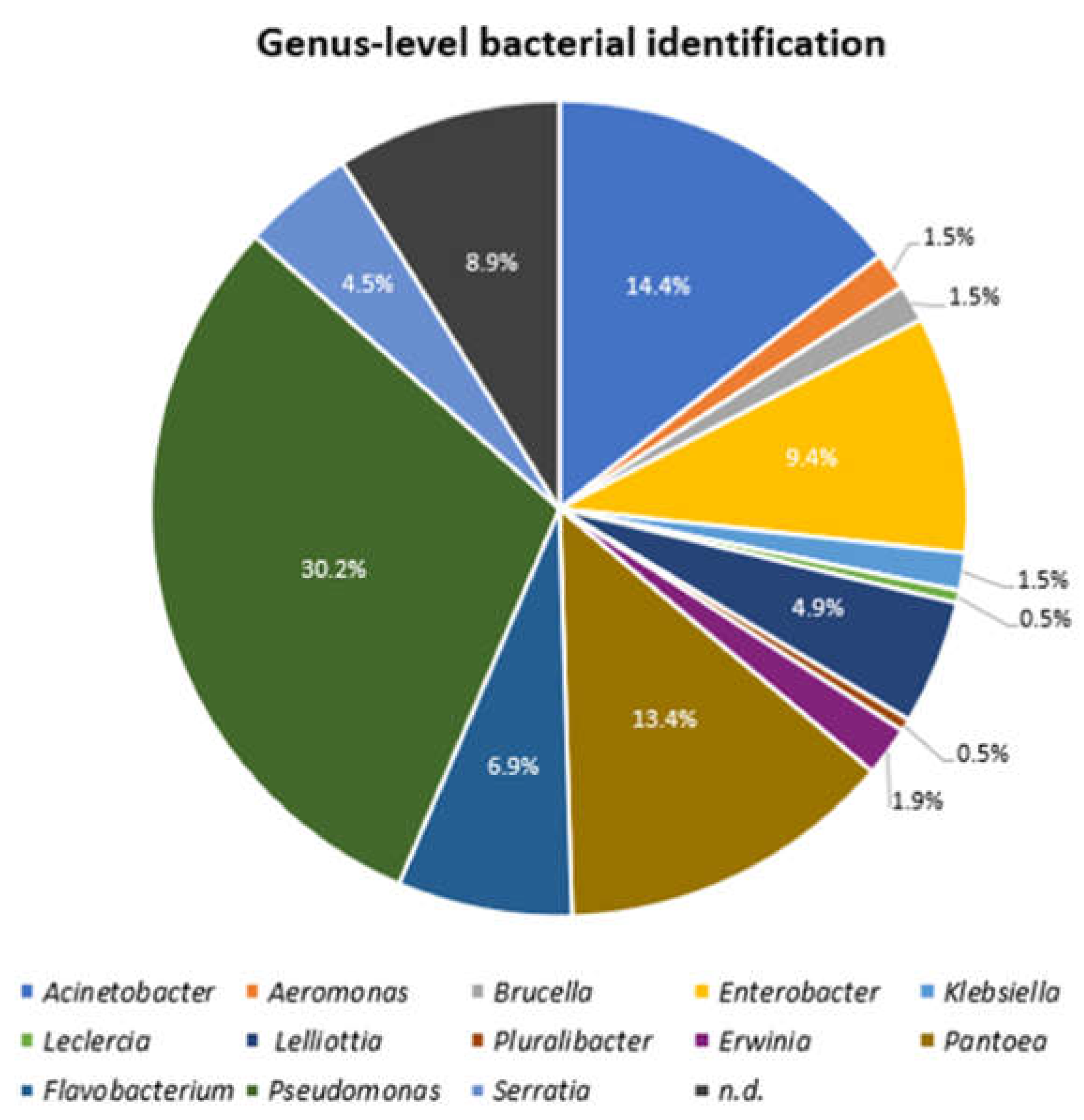
3.3. Strain Identification

In this study, 202 strains from the 28 lettuce samples cultivated in South Korea and the surrounding soils were selected as distinct strains. The principal method of 16S rRNA gene sequencing used in this study was Nanopore Flongle sequencing. However, as 66 strains could not be sequenced either because of contamination of sequencing inhibitors or lack of DNA, those strains were sequenced by Sanger sequencing. From these 202 strains, 184 could be assigned either to the genera Pseudomonas (n = 61, 30.2%), Acinetobacter (n = 29, 14.4%), Pantoea (n = 27, 13.4%), Enterobacter (n = 19, 9.4%), Flavobacterium (n = 14, 6.9%), Lelliottia (n = 10, 4.9%), Serratia (n = 9, 4.5%), Erwinia (n = 4, 1.9%), Aeromonas (n = 3, 1.5%), Brucella (n = 3, 1.5%), Klebsiella (n = 3, 1.5%), Leclercia (n = 1, 0.5%), and Pluralibacter (n = 1, 0.5%) based on 16S rRNA gene sequencing using the EzTaxon database for identification [39] (Figure 1). However, eighteen isolates (8.9%) could not be unequivocally identified to the genus level by the 16S rRNA gene sequencing method as the sequencing result identified two or more related genera (Figure 1).

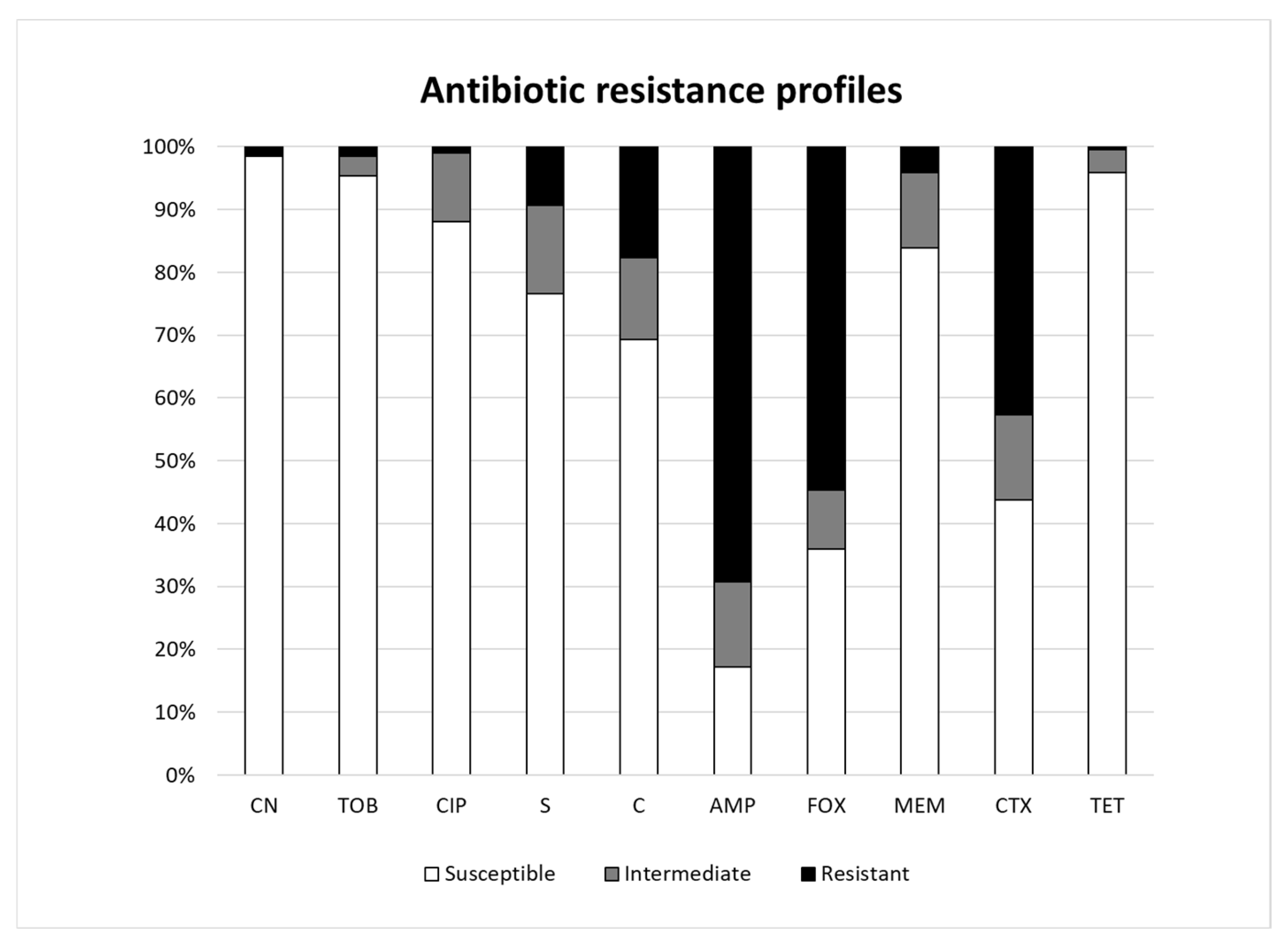
3.4. Antibiotic Resistance Profiles

All 202 strains were screened for antibiotic resistance using the disc diffusion method according to the CLSI guidelines. Nine Pseudomonas spp. and one Acinetobacter spp. out of the two hundred and two strains did not grow on MH agar plates and could therefore not be used for antibiotic susceptibility testing. The results classified strains as being either susceptible, intermediate, or resistant based on the diameter of the inhibition zone size. In this study, 133 (69.3%) and 105 (54.7%) strains showed a resistance phenotype to ampicillin and cefoxitin, respectively (Figure 2). Of the 192 strains tested in this study, 23 (11.9%) strains were fully susceptible to the antibiotics, while 30 (15.6%) strains showed an intermediate phenotype and were resistant to at least one antibiotic. A total of 84 strains were classified as susceptible to cefotaxime, while 82 (42.7%) strains and 26 (13.5%) strains showed resistant and intermediate phenotypes, respectively. For each antibiotic, strains belonging to the Pseudomonadaceae showed a generally high level of resistance at the family level. The resistance phenotype to gentamicin, tobramycin, ciprofloxacin, and tetracycline occurred only at low incidences of 1.6%, 1.6%, 1.0%, and 0.5%, respectively. However, a few isolates exhibited an intermediate phenotype to ciprofloxacin (n = 21, 10.9%), tobramycin (n = 6, 3.1%), and tetracycline (n = 7, 3.6%) (Figure 2). Only eight strains (six Pseudomonas spp., one Klebsiella spp., and one Acinetobacter spp.) were resistant to meropenem, which is a last-resort antibiotic for multidrug-resistant bacterial infections.

3.5. Whole Genome Sequencing

The genomes of fifteen selected isolates were sequenced and de novo assembled to generate draft genome sequences. The total raw sequence data obtained for genome coverage of these strains ranged from 25 (strain V98_8) to 147 (strain V89_4). The contig numbers of the fifteen isolates after sequencing ranged between 21 and 113, and the genome sizes ranged between 3.27 and 6.51 Mbp (Table 2). The highest G+C content (mol%) was 62.3% for strain V104_10 and the lowest was 38.64% for strain V89_4. The N50 values ranged from 115019 (V98_8) to 2533332 (V114_1) and the number of CDS were between 3116 and 6043. Six of these strains (V98_8, V88_4, V104_6, V104_10, V89_7, and V90_4) did not possess any sequences related to acquired antibiotic resistance, whereas nine strains contained more than one sequence related to potentially acquired antibiotic resistance from the ResFinder database. Of all the isolates which were fully genome-sequenced in this study, only the isolate V90_4 was found to contain an IncFII sequence related to a Gram-negative plasmid incompatibility type, by using the PlasmidFinder database (Table 2). Plasmids belonging to this incompatibility type are known to often be transferable and to carry antibiotic resistance [40]. In addition, the ResFinder database detected acquired antibiotic resistance genes in the other genome-sequenced strains, including a gene encoding a fosfomycin resistance protein fosA and fosA2, genes for the antibiotic efflux pump OqxA and QqxB, (fluoro)quinolone-resistance gene qnrE1, and β-lactamase genes cphA1, ampH, blaMIR-6, blaMOX-4, blaACT-12, blaOXA-304, blaADC-25, blaSST1, and blaOKP-A-11. Furthermore, genes for aminoglycoside resistance (aac(6′)-Ic) and for tetracycline resistance (tet(41)) could also be detected among the sequenced strains.
All of the genome-sequenced isolates were precisely identified using the average nucleotide identity (ANI) calculations and digital DNA–DNA hybridization values (dDDH) following the minimum guideline for species identification by these methods proposed in a previous study (Table 2) [41]. Except for the V89_13 strain, all strains that had generated draft genome sequences in this study were clearly identified by ANI and DDH analysis, along with closely related type strains above the species delineation cutoff values, and the results of the identification are shown in Table 2. Interestingly, the isolate V 89_13 showed the highest relationship to the Lelliottia jeotgali PFL01T type strain, with an ANI value of 91.12% similarity. Additionally, the dDDH value between V89_13 and the closest type strain Lelliottia jeotgali PFL01T was 43.6% (Table 2). This showed that both the ANI and in silico DDH analysis values for strain V89_13 and the closest type strain Lelliottia jeotgali PFL01T were below the cutoff value used for species delineation (Table 2), indicating that the strain probably represented a novel Lelliottia species, which will be further taxonomically investigated in follow-up studies.
Table 2. Summary of whole genome sequencing of 15 selected isolates. n.d., not detected; AMP, ampicillin; C, chloramphenicol; S, streptomycin; CIP, ciprofloxacin; MEM, meropenem; CTX, cefotaxime; FOX, cefoxitin.
V98_8V88_4V104_6V104_10V89_4V89_7V106_11V108_6V87_3
No. of contigs1134858663034375835
N50115,019382,325138,943195,255250,587215,337320,651163,040211,224
GC content (mol%)59.9960.5561.7762.338.6443.0353.3353.3755.70
Total length (bp)6,514,0746,264,1704,687,3765,620,5824,009,5863,276,0904,899,7094,944,3624,706,154
Genome coveragex 25x 103x 46x 32x 147x 68x 41x 63x 104
No. of CDSs604357254326523937713116495650324443
No. of tRNAs495362564565574047
No. of rRNAs356533554
Acquired resistance gene(s)n.d.n.d.n.d.n.d.blaOXA-304, blaADC-25n.d.OqxBOqxBfosA, blaMIR-6
Plasmid sequence(s)n.d.n.d.n.d.n.d.n.d.n.d.n.d.n.d.n.d.
Antibiotic resistancen.d.C, AMP, FOX, CTXn.d.AMP, FOX, CTXC, AMP, FOX, CTXAMP, MEM, CTXn.d.AMPAMP, FOX
OrthoANI identification (% similarity of top-hit)Pseudomonas umsongensis DSM 16611T (96.75%)Pseudomonas glycinae MS586T (96.48%)Pseudomonas fulva DSM 17717T (99.48%)Pseudomonas monteilii DSM 14164T (98.0%)Acineotbacter oleivorans JCM 16667T (96.91%)Acinetobacter soli KCTC 22184T (98.53%)Pantoea ananatis LMG 2665T (99.07%)Pantoea ananatis LMG 2665T (99.16%)Enterobacter cancerogenus ATCC 33241T (98.55%)
in silico DDH identification (% similarity of top-hit)Pseudomonas umsongensis DSM 16611T (71.9%)Pseudomonas glycinae MS586T (71.1%)Pseudomonas fulva DSM 17717T (96.1%)Pseudomonas monteilii DSM 14164T (83.4%)Acineotbacter oleivorans JCM 16667T (72.9%)Acinetobacter soli KCTC 22184T (88.2%)Pantoea ananatis LMG 2665T (92.5%)Pantoea ananatis LMG 2665T (93.2%)Enterobacter cancerogenus ATCC 33241T (87.4%)
Accession no.JASCAE000000000JASCAF000000000JASCAG000000000JASCAH000000000JASCAI000000000JASCAJ000000000JASCAK000000000JASCAL000000000JASCAM000000000
V87_3V89_11V114_1V89_13V90_4V115_8V90_14
No. of contigs35582146795249
N50211,224201,2952,533,332173,726134,120194,518197,099
GC content (mol%)55.7054.6059.8955.8456.4158.2161.53
Total length (bp)4,706,1544,700,0254,932,6114,846,0695,149,4125,253,8244,749,641
Genome coveragex 104x 115x 41x 85x 103x 40x 127
No. of CDSs4443450947204561497650844422
No. of tRNAs47367648526556
No. of rRNAs4445665
Acquired resistance gene(s)fosA, blaMIR-6blaACT-12, OqxA, OqxB, fosA2aac(6′)-Ic, blaSST-1, OqxB, tet(41)fosA2, OqxA, OqxBn.d.OqxA, OqxB, fosA, blaOKP-A-11 ampH, blaMOX-4, cphA1
Plasmid sequence(s)n.d.n.d.n.d.n.d.IncFII(Yp)n.d.n.d.
Antibiotic resistanceAMP, FOXAMP, FOXAMPn.d.AMPCIP, AMP, MEM, CTXAMP
OrthoANI identification (% similarity of top-hit)Enterobacter cancerogenus ATCC 33241T (98.55%)Enterobacter ludwigii DSM 16688T (98.88%)Serratia marcescens ATCC 13880T (98.64%)Lelliottia jeotgali PFL01T (91.19%), Lelliottia amnigena LMG2784T (85.24%), Lelliottia nimipressuralis CIP 104980T (84.15%)Erwinia aphidicola JCM 21238T (99.02%)Klebsiella quasipneumoniae subsp. quasipneumoniae 01A030T (99.19%)Aeromonas hydrophila ATCC 7966T (96.91%)
in silico DDH identification (% similarity of top-hit)Enterobacter cancerogenus ATCC 33241T (87.4%)Enterobacter ludwigii DSM 16688T (91.4%)Serratia marcescens ATCC 13880T (89.1%)Lelliottia jeotgali PFL01T (43.6%),
Lelliottia nimipressuralis CCUG 25894T (27.8%), Lelliottia amnigena LMG2784T (29.1%)
Erwinia aphidicola JCM 21238T (91.9%)Klebsiella quasipneumoniae subsp. quasipneumoniae 01A030T (93.9%)Aeromonas hydrophila ATCC 7966T (73.3%)
Accession no.JASCAM000000000JASCAN000000000JASCAO000000000JASCAP000000000JASCAQ000000000JASCAR000000000JASCAS000000000

4. Discussion

In this study, Gram-negative bacteria isolated from lettuce and the surrounding soil in Korea were identified and characterized to obtain a preliminary understanding of the safety of these types of produce. The microbial ecology of agricultural environments was investigated using a culture-based method, focusing on a particular microorganism, i.e., Enterobacterales, by using selective media for these bacteria. RAPD-PCR was successfully used to discriminate distinct strains among multiple isolates of the same strain. In addition to the RAPD fingerprint comparison, 16S rRNA sequencing was applied to identify all isolates using the EZTaxon database. After both RAPD comparison and 16S-sequence-based identification, 202 strains of out 248 isolates were selected and the predominating families of the 202 strains were Pseudomonadaceae (30.2%), Enterobacteriaceae (16.8%), Erwiniaceae (15.3%), and Moraxellaceae (14.4%). At the genus level, Pseudomonas (30.2%) was the most prevalent genus, followed by Acinetobacter (14.4%), Pantoea (13.4%), and Enterobacter (9.4%). A previous study showed that Pseudomonas was the predominant Gram-negative bacteria present in the processing of endive lettuce and the low-temperature storage of fresh-cut lettuce [2]. In another study that investigated lettuce samples in Korea using culture-independent 16S rRNA gene amplicon metagenome analysis, Gram-positive Bacillus and Exiguobacterium, and Gram-negative Pseudomonas were the dominant genera observed in lettuce [5]. However, it has been reported that the microflora compositions in lettuce can differ depending on the site and time of sample collection [5].
Pseudomonas aeruginosa, Acinetobacter baumannii, and Enterobacter cloacae are specific species within these genera Pseudomonas, Acinetobacter, and Enterobacter, respectively, and are considered typical opportunistic pathogens that can cause serious infections in infants, the elderly, and immunocompromised hosts. Furthermore, though detected in lower numbers (4.5%), Serratia is important since Serratia marcescens, which amounted to 55.6% of the total Serratia strains found, has recently emerged as an important cause of nosocomial infections [42]. Therefore, it is necessary to regularly monitor the prevalence of potential clinical pathogens belonging to Serratia, along with those included in the genera Pseudomonas, Acinetobacter, and Enterobacter.
In a previous metagenomic study, Acinetobacter, Pantoea, and Enterobacter, which were the dominant isolates from both lettuce and cultivating environments in our study, did not predominate in the Chinese cabbage or the cabbage farming environments in Korea [43]. Such a discrepancy might arise from differences in the microbiota between different products such as cabbage and lettuce, as well as differences in analysis methods between culture-independent metagenome analysis and culturing Gram-negative bacteria using VRBD agar plates. It can be advantageous to use the culture-independent method because it provides information about the total microbial community. However, in our study, culturing allowed for the detection of the subset of antibiotic-resistant, potentially pathogenic or pathogenic Gram-negative bacteria which may carry transferable antibiotic resistance genes. This, coupled with whole genome sequence (WGS) analysis of representative species applied together with several bioinformatic pipelines, was able to support rapid potential pathogen identification and the detection of acquired or intrinsic antibiotic resistance genes [44].
This study used Nanopore sequencing as a method for 16S rRNA gene sequencing. Applying this method for bacterial species identification is a novel approach, while being low cost and robust. By not only supporting the almost complete 16S rRNA gene but also controlling sequencing quality using the NanoCLUST [30] program, which generates a consensus sequence from long-read sequencing results of individual 16S rRNA PCR products of strains, it is possible to correct the error rate of the long-read sequencing method and to verify the purity of the 16S rRNA gene products. If PCR products are contaminated, different clusters are drawn on the plot based on calculations made by the NanoCLUST, which helps to confirm if the extracted DNA is pure or not, i.e., whether it stemmed from a pure culture or not [29].
Antibiotic resistance tested by the disc diffusion method revealed that the isolates from lettuce and the surrounding soil in the Korean agricultural environment tested were highly resistant to ampicillin (69.7%), with Pseudomonas (P.) being the dominant genus. The previous review study of Rahman et al. [16] reported that six of forty studies frequently isolated ampicillin-resistant Pseudomonas spp. as well as ESBL-producing Pseudomonas spp. from fresh produce samples. This ampicillin-resistant profile in the genus Pseudomonas is consistent with the study conducted with fresh produce, which found that Pseudomonas spp. Recovered from lettuce samples frequently exhibited resistance to ampicillin [45]. Additionally, culturable bacteria isolated from environmental water samples in Korea were found to be highly resistant to the ampicillin antibiotic [46]. In addition, the resistance to cefoxitin (54.7%), a second-generation cephalosporin antibiotic, was the second most frequently occurring resistance profile. This reflects the wide use of cefoxitin in human and veterinary medicines. A notable result was that 84 (42.7%) out of 192 isolates in this study also showed resistance towards cefotaxime. Of these strains, Pseudomonadaceae (54.8%) and Moraxellaceae (32.1%) were primarily identified. A previous study on P. aeruginosa by Pang et al. [47] showed the outer membrane permeability and efflux system of this strain caused a high level of intrinsic resistance to antibiotics. In this study, the Pseudomonas glycinae V88_4 isolate, for which non-acquired antibiotic resistance genes were detected in the ResFinder analysis, was resistant to ampicillin, cefoxitin, cefotaxime, and chloramphenicol. In addition, one strain identified as Acinetobacter soli V89_7 was also found to contain non-acquired antibiotic resistance genes by ResFinder, but showed resistance to ampicillin, meropenem, and cefotaxime. The high resistance to cefotaxime observed in these two strains could indicate that intrinsic resistance factors, such as point mutations in target genes, may be attributed to resistance.
Cefotaxime is a third-generation cephalosporin that is important for treating infections caused by members of the Enterobacteriaceae, i.e., Enterobacter spp., Klebsiella spp., and Escherichia coli. However, many clinical case studies reported isolates of these bacteria from infected patients to show resistance to this antibiotic [48]. Furthermore, the clinical Enterobacteriaceae isolates that were resistant to cefotaxime showed multidrug resistance properties. The primary reason for cefotaxime resistance in the study [48] was attributed to CTX-M-type beta lactamase genes, which were present in 76% of the Enterobacteriaceae, i.e., E. coli and Klebsiella spp. This implies that if cefotaxime-resistant enterobacterial isolates in fresh produce would possess ESBLs and that these would potentially be transferred to humans via horizontal gene transfer, this could result in a serious public health threat. In this study, it is possible that 42.7% of cefotaxime-resistant isolates are capable of producing one or more CTX-M-type beta lactamases. Therefore, further investigation is necessary to determine the presence of ESBL genes in these isolates using both physiological tests, i.e., MAST disc diffusion test, and molecular methods such as whole genome sequencing.
The incidence of chloramphenicol resistance among the bacteria in this study was 17.7%. Although chloramphenicol is not prescribed for humans and food-producing animals in the European Union (EU), it is still important to monitor the presence of chloramphenicol resistance genes in bacteria that are isolated from soil, plants, vegetables, and agricultural environments to ensure food safety for consumers [49]. Since chloramphenicol-resistant bacteria often display resistance to other antibiotics such as tetracycline and kanamycin, and harbor mobile genetic elements containing resistance plasmids and/or transposons [19].
Additionally, there was a low level of resistance towards aminoglycosides, which include gentamicin (1.6%), tobramycin (1.6%), and streptomycin (9.4%). According to the report published by the National Institute of Food and Drug Safety Evaluation and Ministry of Agriculture, Food and Rural Affairs, sales of streptomycin to livestock and fisheries in Korea were about 3.3 times higher than gentamicin, which might explain higher resistances observed in our experiments (www.mfds.go.kr (accessed on 22 August 2022)). A previous study of Schwaiger et al. [50] reported antibiotic-resistant Pseudomonas spp. isolated from vegetables at the marketing stage and showed similar resistance patterns as observed also in our study with regard to resistance incidences to gentamicin and tobramycin (<4%) and streptomycin (11%).
It is notable that there was low resistance to ciprofloxacin (1.0%) and to tetracycline (0.5%) in this study. Tetracyclines have in the past been used as therapeutic antibiotics in animal husbandry and for the control of plant diseases in Korea. The amounts of tetracyclines used in poultry flocks as therapy against diseases are still relatively high in several countries, which results in the accumulation of antibiotic substrates in the environment. This in turn, may influence the development of tetracycline resistance in foodborne bacteria, and the spread of these bacteria to the communities and hospitals via foods.
Carbapenem is another critical antibiotic that is used as a last resort to treat infections. From our experiment, 4.1% of bacteria were resistant to meropenem, which belongs to the class of carbapenem antibiotics. Primarily, Pseudomonas spp. (6), Acinetobacter spp. (1), and an Enterobacteriaceae (1) were found to be resistant to this antibiotic. Since genes related to resistance to carbapenem give further resistance to other antibiotics and often coexist with ESBL genes, it is highly likely that these resistant strains are potential ESBL-producing strains [7,16]. Furthermore, carbapenem-resistant, Gram-negative microorganisms have recently become critical issues in nosocomial infections. Among them, carbapenem-resistant Acinetobacter baumannii, Pseudomonas aeruginosa, Escherichia coli, and Klebsiella pneumoniae are WHO-designated ‘priority pathogens’ posing serious threats to human health [15]. As shown by clinical sample-based studies, genes including blaCTX-M, blaSHV, blaTEM, blaOXA, and blaNDM have been frequently found in carbapenem-resistant strains.
The genome sequences of 15 isolates were used to precisely identify the isolates as well as to determine the presence of potentially acquired antibiotic resistance genes, as well as plasmid-related sequences. ResFinder analysis showed a Serratia spp. isolate (V114_1) possessed resistance genes to antibiotics belonging to four different classes. Two strains (Pseudomonas glyciane V88_4 and Acinetobacter soli V89_7) which did not possess any acquired antibiotic resistance genes in the ResFinder analysis, appeared to be cefotaxime-resistant, because these strains were able to grow and showed only a small diameter inhibition zone (<18 mm) in resistance tests with this antibiotic. These discrepancies between phenotypic and genotypic resistance profiles may be due to the possible presence of new AMR gene variations which have not previously been identified, or on the other resistance mechanisms, such as efflux pumps and point mutations. This indicates that the determination of antimicrobial resistance should not be demonstrated only by searching for a specific gene that confers resistance to the antibiotic, but this should also be correlated with phenotypic resistance tests.
Genomic characterization of the fifteen Gram-negative bacteria from Korean soil and lettuce samples showed the low level (one out of fifteen strains) presence of potentially transferable antibiotic resistance genes on the genomes, indicating that the transfer of antibiotic resistances by potential pathogenic enterobacteria via fresh produce in Korea is a low but likely possibility. Fresh produce should therefore be continuously monitored for the occurrence and sources of entry of bacteria carrying transferable antibiotic resistances in order to find ways to lower the presence of these bacteria in the fresh produce food chain.

Supplementary Materials

The following supporting information can be downloaded at: https://www.mdpi.com/article/10.3390/microorganisms11051241/s1. Figure S1: Dendrogram obtained by UPGMA clustering using the Pearson correlation coefficient of RAPD-PCR fingerprints of 248 Gram-negative isolates from lettuces and different fertilized soil samples. Selected strains for 16S rRNA gene sequencing were marked with an under bar. *; selected isolates for whole genome sequencing.

Author Contributions

Conceptualization, G.-S.C., C.M.A.P.F. and K.-K.O.; methodology, I.K., S.J., B.-E.K. and M.-I.J.; investigations, S.J., I.K. and G.-S.C.; data analysis, S.J., I.K. and G.-S.C.; resources, B.-E.K., M.-I.J. and K.-K.O.; writing—original draft preparation S.J., I.K., G.-S.C. and C.M.A.P.F.; writing—review and editing, G.-S.C. and C.M.A.P.F.; funding acquisition, K.-K.O., G.-S.C. and C.M.A.P.F. All authors have read and agreed to the published version of the manuscript.

Funding

This work was supported by the Rural Development Administration (RDA) Cooperative Research Program (Project No. PJ016306), Republic of Korea.

Data Availability Statement

The draft genome sequences were deposited in DDB/ENA/GeneBank under the accession numbers listed in Table 2, PJ016303.

Acknowledgments

We thank J. Obermeyer and K. Magnussen for their help in the management of all bacterial laboratory work.

Conflicts of Interest

The authors declare no conflict of interest.

References

  1. Leff, J.W.; Fierer, N. Bacterial communities associated with the surfaces of fresh fruits and vegetables. PLoS ONE 2013, 8, e59310. [Google Scholar] [CrossRef]
  2. Sun, Y.; Zhao, X.; Ma, Y.; Ma, Z.; He, Z.; Zhao, W.; Wang, P.; Zhao, S.; Wang, D. Investigation on the Microbial Diversity of Fresh-Cut Lettuce during Processing and Storage Using High Throughput Sequencing and Their Relationship with Quality. Foods 2022, 11, 1683. [Google Scholar] [CrossRef] [PubMed]
  3. Berger, C.N.; Sodha, S.V.; Shaw, R.K.; Griffin, P.M.; Pink, D.; Hand, P.; Frankel, G. Fresh fruit and vegetables as vehicles for the transmission of human pathogens. Environ. Microbiol. 2010, 12, 2385–2397. [Google Scholar] [CrossRef] [PubMed]
  4. Castro-Ibáñez, I.; Gil, M.I.; Allende, A. Ready-to-eat vegetables: Current problems and potential solutions to reduce microbial risk in the production chain. Food Sci. Technol. 2017, 85, 284–292. [Google Scholar] [CrossRef]
  5. Yu, Y.C.; Yum, S.J.; Jeon, D.Y.; Jeong, H.G. Analysis of the Microbiota on Lettuce (Lactuca sativa L.) Cultivated in South Korea to Identify Foodborne Pathogens. J. Microbiol. Biotechnol. 2018, 28, 1318–1331. [Google Scholar] [CrossRef]
  6. Abdelfattah, A.; Freilich, S.; Bartuv, R.; Zhimo, V.Y.; Kumar, A.; Biasi, A.; Salim, S.; Feygenberg, O.; Burchard, E.; Dardick, C.; et al. Global analysis of the apple fruit microbiome: Are all apples the same? Environ. Microbiol. 2021, 23, 6038–6055. [Google Scholar] [CrossRef]
  7. Food and Agriculture Organization of the United Nations. Effects of Fruit and Vegetable Intakes on Direct and Indirect Health Outcomes: Background Paper for the FAO/WHO International Workshop on Fruits and Vegetables 2020; FAO: Rome, Italy, 2021. [Google Scholar]
  8. Aiyedun, S.O.; Onarinde, B.A.; Swainson, M.; Dixon, R.A. Foodborne outbreaks of microbial infection from fresh produce in Europe and North America: A systematic review of data from this millennium. Int. J. Food Sci. Technol. 2021, 56, 2215–2223. [Google Scholar] [CrossRef]
  9. FAO/WHO. Summary Report of the Joint FAO/WHO Expert Meeting on Microbiological Risk Assessment on the Prevention and Control of Microbiological Hazards in Fresh Fruits and Vegetables (Part 1: Administrative Procedures, Meeting Scope/Objectives, Data Collection; Part 2 General Principle and Fresh Fruits and Vegetables); FAO: Rome, Italy, 2021. [Google Scholar]
  10. Fiedler, G.; Kabisch, J.; Bohnlein, C.; Huch, M.; Becker, B.; Cho, G.S.; Franz, C. Presence of Human Pathogens in Produce from Retail Markets in Northern Germany. Foodborne Pathog. Dis. 2017, 14, 502–509. [Google Scholar] [CrossRef]
  11. Carstens, C.K.; Salazar, J.K.; Darkoh, C. Multistate Outbreaks of Foodborne Illness in the United States Associated with Fresh Produce From 2010 to 2017. Front. Microbiol. 2019, 10, 2667. [Google Scholar] [CrossRef]
  12. Authority, E.F.S. Shiga toxin-producing E. coli (STEC) O104:H4 2011 outbreaks in Europe: Taking Stock. EFSA J. 2011, 9, 2390. [Google Scholar] [CrossRef]
  13. Mellmann, A.; Harmsen, D.; Cummings, C.A.; Zentz, E.B.; Leopold, S.R.; Rico, A.; Prior, K.; Szczepanowski, R.; Ji, Y.; Zhang, W.; et al. Prospective genomic characterization of the German enterohemorrhagic Escherichia coli O104:H4 outbreak by rapid next generation sequencing technology. PLoS ONE 2011, 6, e22751. [Google Scholar] [CrossRef]
  14. Österblad, M.; Pensala, O.; Peterzéns, M.; Heleniusc, H.; Huovinen, P. Antimicrobial susceptibility of Enterobacteriaceae isolated from vegetables. J. Antimicrob. Chemother. 1999, 43, 503–509. [Google Scholar] [CrossRef] [PubMed]
  15. Al-Kharousi, Z.S.; Guizani, N.; Al-Sadi, A.M.; Al-Bulushi, I.M.; Shaharoona, B. Hiding in Fresh Fruits and Vegetables: Opportunistic Pathogens May Cross Geographical Barriers. Int. J. Microbiol. 2016, 2016, 4292417. [Google Scholar] [CrossRef]
  16. Rahman, M.; Alam, M.-U.; Luies, S.K.; Kamal, A.; Ferdous, S.; Lin, A.; Sharior, F.; Khan, R.; Rahman, Z.; Parvez, S.M.; et al. Contamination of Fresh Produce with Antibiotic-Resistant Bacteria and Associated Risks to Human Health: A Scoping Review. Int. J. Environ. Res. Public Health 2022, 19, 360. [Google Scholar] [CrossRef]
  17. Bezanson, G.S.; MacInnis, R.; Potter, G.; Hughes, T. Presence and potential for horizontal transfer of antibiotic resistance in oxidase-positive bacteria populating raw salad vegetables. Int. J. Food Microbiol. 2008, 127, 37–42. [Google Scholar] [CrossRef]
  18. Pleva, P.; Janalíková, M.; Pavlíčková, S.; Lecomte, M.; Godillon, T.; Holko, I. Characterization of Escherichia coli strains isolated from raw vegetables. Potravin. Slovak J. Food Sci. 2018, 12, 304–312. [Google Scholar] [CrossRef]
  19. Liu, S.; Kilonzo-Nthenge, A. Prevalence of Multidrug-Resistant Bacteria from U.S.-Grown and Imported Fresh Produce Retailed in Chain Supermarkets and Ethnic Stores of Davidson County, Tennessee. J. Food Prot. 2017, 80, 506–514. [Google Scholar] [CrossRef] [PubMed]
  20. Abatcha, M.G.; Effarizah, M.E.; Rusul, G. Prevalence, antimicrobial resistance, resistance genes and class 1 integrons of Salmonella serovars in leafy vegetables, chicken carcasses and related processing environments in Malaysian fresh food markets. Food Control 2018, 91, 170–180. [Google Scholar] [CrossRef]
  21. Freitag, C.; Michael, G.B.; Li, J.; Kadlec, K.; Wang, Y.; Hassel, M.; Schwarz, S. Occurrence and characterisation of ESBL-encoding plasmids among Escherichia coli isolates from fresh vegetables. Vet. Microbiol. 2018, 219, 63–69. [Google Scholar] [CrossRef]
  22. Li, Y.; Cao, W.; Liang, S.; Yamasaki, S.; Chen, X.; Shi, L.; Ye, L. Metagenomic characterization of bacterial community and antibiotic resistance genes in representative ready-to-eat food in southern China. Sci. Rep. 2020, 10, 15175. [Google Scholar] [CrossRef]
  23. Song, J.; Oh, S.S.; Kim, J.; Shin, J. Extended-spectrum β-lactamase-producing Escherichia coli isolated from raw vegetables in South Korea. Sci. Rep. 2020, 10, 19721. [Google Scholar] [CrossRef] [PubMed]
  24. Kim, H.S.; Chon, J.W.; Kim, Y.J.; Kim, D.H.; Kim, M.S.; Seo, K.H. Prevalence and characterization of extended-spectrum-β-lactamase-producing Escherichia coli and Klebsiella pneumoniae in ready-to-eat vegetables. Int. J. Food Microbiol. 2015, 207, 83–86. [Google Scholar] [CrossRef]
  25. Buck, J.D. Nonstaining (KOH) method for determination of gram reactions of marine bacteria. Appl. Environ. Microbiol. 1982, 44, 992–993. [Google Scholar] [CrossRef] [PubMed]
  26. Zhang, G.W.; Kotiw, M.; Daggard, G. A RAPD-PCR genotyping assay which correlates with serotypes of group B streptococci. Lett. Appl. Microbiol. 2002, 35, 247–250. [Google Scholar] [CrossRef]
  27. Araújo, S.; Silva, I.A.; Tacão, M.; Patinha, C.; Alves, A.; Henriques, I. Characterization of antibiotic resistant and pathogenic Escherichia coli in irrigation water and vegetables in household farms. Int. J. Food Microbiol. 2017, 257, 192–200. [Google Scholar] [CrossRef]
  28. CLSI. Performance Standards for Antimicrobial Susbeptibility Testing. 30th ed. CLSI supplement M100. Clinical and Laboratory Standards Institute. Available online: https://clsi.org/media/3481/m100ed30_sample.pdf (accessed on 15 October 2020).
  29. Borges, A.S.G.; Basu, M.; Brinks, E.; Bang, C.; Cho, G.-S.; Baines, J.F.; Franke, A.; Franz, C.M.A.P. Fast Identification Method for Screening Bacteria from Faecal Samples Using Oxford Nanopore Technologies MinION Sequencing. Curr. Microbiol. 2023, 80, 101. [Google Scholar] [CrossRef] [PubMed]
  30. Rodriguez-Perez, H.; Ciuffreda, L.; Flores, C. NanoCLUST: A species-level analysis of 16S rRNA nanopore sequencing data. Bioinformatics 2021, 37, 1600–1601. [Google Scholar] [CrossRef]
  31. Bolger, A.M.; Lohse, M.; Usadel, B. Trimmomatic: A flexible trimmer for Illumina sequence data. Bioinformatics 2014, 30, 2114–2120. [Google Scholar] [CrossRef]
  32. Bankevich, A.; Nurk, S.; Antipov, D.; Gurevich, A.A.; Dvorkin, M.; Kulikov, A.S.; Lesin, V.M.; Nikolenko, S.I.; Pham, S.; Prjibelski, A.D.; et al. SPAdes: A new genome assembly algorithm and its applications to single-cell sequencing. J. Comput. Biol. 2012, 19, 455–477. [Google Scholar] [CrossRef]
  33. Seemann, T. Prokka: Rapid prokaryotic genome annotation. Bioinformatics 2014, 30, 2068–2069. [Google Scholar] [CrossRef]
  34. Bharat, A.; Petkau, A.; Avery, B.P.; Chen, J.C.; Folster, J.P.; Carson, C.A.; Kearney, A.; Nadon, C.; Mabon, P.; Thiessen, J.; et al. Correlation between Phenotypic and In Silico Detection of Antimicrobial Resistance in Salmonella enterica in Canada Using Staramr. Microorganisms 2022, 10, 292. [Google Scholar] [CrossRef] [PubMed]
  35. Carattoli, A.; Zankari, E.; Garcia-Fernandez, A.; Voldby Larsen, M.; Lund, O.; Villa, L.; Moller Aarestrup, F.; Hasman, H. In silico detection and typing of plasmids using PlasmidFinder and plasmid multilocus sequence typing. Antimicrob. Agents Chemother. 2014, 58, 3895–3903. [Google Scholar] [CrossRef] [PubMed]
  36. Zankari, E.; Hasman, H.; Cosentino, S.; Vestergaard, M.; Rasmussen, S.; Lund, O.; Aarestrup, F.M.; Larsen, M.V. Identification of acquired antimicrobial resistance genes. J. Antimicrob. Chemother. 2012, 67, 2640–2644. [Google Scholar] [CrossRef] [PubMed]
  37. Lee, I.; Ouk Kim, Y.; Park, S.C.; Chun, J. OrthoANI: An improved algorithm and software for calculating average nucleotide identity. Int. J. Syst. Evol. Microbiol. 2016, 66, 1100–1103. [Google Scholar] [CrossRef] [PubMed]
  38. Meier-Kolthoff, J.P.; Auch, A.F.; Klenk, H.P.; Goker, M. Genome sequence-based species delimitation with confidence intervals and improved distance functions. BMC Bioinform. 2013, 14, 60. [Google Scholar] [CrossRef] [PubMed]
  39. Kim, O.S.; Cho, Y.J.; Lee, K.; Yoon, S.H.; Kim, M.; Na, H.; Park, S.C.; Jeon, Y.S.; Lee, J.H.; Yi, H.; et al. Introducing EzTaxon-e: A prokaryotic 16S rRNA gene sequence database with phylotypes that represent uncultured species. Int. J. Syst. Evol. Microbiol. 2012, 62, 716–721. [Google Scholar] [CrossRef] [PubMed]
  40. Blau, K.; Bettermann, A.; Jechalke, S.; Fornefeld, E.; Vanrobaeys, Y.; Stalder, T.; Top, E.M.; Smalla, K. The Transferable Resistome of Produce. mBio 2018, 9, e01300-18. [Google Scholar] [CrossRef]
  41. Chun, J.; Oren, A.; Ventosa, A.; Christensen, H.; Arahal, D.R.; da Costa, M.S.; Rooney, A.P.; Yi, H.; Xu, X.-W.; De Meyer, S.; et al. Proposed minimal standards for the use of genome data for the taxonomy of prokaryotes. Int. J. Syst. Evol. Microbiol. 2018, 68, 461–466. [Google Scholar] [CrossRef]
  42. Ghaith, D.M.; Zafer, M.M.; Ismail, D.K.; Al-Agamy, M.H.; Bohol, M.F.F.; Al-Qahtani, A.; Al-Ahdal, M.N.; Elnagdy, S.M.; Mostafa, I.Y. First reported nosocomial outbreak of Serratia marcescens harboring bla (IMP-4) and bla (VIM-2) in a neonatal intensive care unit in Cairo, Egypt. Infect. Drug Resist. 2018, 11, 2211–2217. [Google Scholar] [CrossRef]
  43. Kim, D.; Hong, S.; Kim, Y.T.; Ryu, S.; Kim, H.B.; Lee, J.H. Metagenomic Approach to Identifying Foodborne Pathogens on Chinese Cabbage. J. Microbiol. Biotechnol. 2018, 28, 227–235. [Google Scholar] [CrossRef]
  44. Vasala, A.; Hytönen, V.P.; Laitinen, O.H. Modern Tools for Rapid Diagnostics of Antimicrobial Resistance. Front. Cell. Infect. Microbiol. 2020, 10, 308. [Google Scholar] [CrossRef] [PubMed]
  45. Allydice-Francis, K.; Brown, P.D. Diversity of Antimicrobial Resistance and Virulence Determinants in Pseudomonas aeruginosa Associated with Fresh Vegetables. Int. J. Microbiol. 2012, 2012, 426241. [Google Scholar] [CrossRef] [PubMed]
  46. Go Eun, B.; Chung, I.-Y.; Kim, H.; Seok, K.-S.; Kim, B.; Yoo, Y.-J.; Jang, Y.; Chae, J.-C. Diversity of ampicillin resistant bacteria in domestic streams. Korean J. Microbiol. 2015, 51, 440–443. [Google Scholar] [CrossRef]
  47. Pang, Z.; Raudonis, R.; Glick, B.R.; Lin, T.-J.; Cheng, Z. Antibiotic resistance in Pseudomonas aeruginosa: Mechanisms and alternative therapeutic strategies. Biotechnol. Adv. 2019, 37, 177–192. [Google Scholar] [CrossRef]
  48. Noguchi, T.; Matsumura, Y.; Yamamoto, M.; Nagao, M.; Takakura, S.; Ichiyama, S. Clinical and microbiologic characteristics of cefotaxime-non-susceptible Enterobacteriaceae bacteremia: A case control study. BMC Infect. Dis 2017, 17, 44. [Google Scholar] [CrossRef]
  49. EFSA. Scientific Opinion on Chloramphenicol in food and feed. EFSA J. 2014, 12, 3907. [Google Scholar] [CrossRef]
  50. Schwaiger, K.; Helmke, K.; Hölzel, C.; Bauer, J. Antibiotic resistance in bacteria isolated from vegetables with regards to the marketing stage (farm vs. supermarket). Int. J. Food Microbiol. 2011, 148, 191–196. [Google Scholar] [CrossRef]
Figure 1. Bacterial identification of 202 Gram-negative strains using 16S rRNA gene sequencing based on the EzTaxon databases [39]. The percentages of each genera are indicated. n.d.; the sequencing result identified two or more closely related genera.
Microorganisms 11 01241 g001
Figure 2. Antibiotic resistance profiles of Gram-negative bacteria isolated from lettuce and surrounding soils in Korea. Determination of antibiotic resistance based on inhibition zone diameters (mm) as resistant, susceptible, and intermediate according to the CLSI guidelines. CN, gentamicin; TOB, tobramycin; CIP, ciprofloxacin; S, streptomycin; C, chloramphenicol; AMP, ampicillin; FOX, cefoxitin; MEM, meropenem (MEM, 10 µg); CTX, cefotaxime; TET, tetracycline.
Microorganisms 11 01241 g002
Table 1. Number of isolates obtained from the different types of surrounding soils and lettuces leaves.
Fertilizer SourceSurrounding SoilsLettuce Leaves
Non-treated soil2044
Cow manure526
Pig manure325
Poultry manure318
Chemical fertilizer1025
Chemical fertilizer + cow and poultry manure1223
Chemical fertilizer + cow + poultry + pig manure1321
Total number of isolates66182
Disclaimer/Publisher’s Note: The statements, opinions and data contained in all publications are solely those of the individual author(s) and contributor(s) and not of MDPI and/or the editor(s). MDPI and/or the editor(s) disclaim responsibility for any injury to people or property resulting from any ideas, methods, instructions or products referred to in the content.

