Next Article in Journal
Identification of Emerging Industrial Biotechnology Chassis Vibrio natriegens as a Novel High Salt-Tolerant and Feedstock Flexibility Electroactive Microorganism for Microbial Fuel Cell
Next Article in Special Issue
GroEL Secreted from Bacillus subtilis Natto Exerted a Crucial Role for Anti-Inflammatory IL-10 Induction in THP-1 Cells
Previous Article in Journal
Gut Microbiome and Neurodevelopmental Disorders: A Link Yet to Be Disclosed
Previous Article in Special Issue
Characterization and Genetic Diversity of Bacillus cereus Strains Isolated from Baby Wipes
 
 
Correction published on 30 June 2023, see Microorganisms 2023, 11(7), 1716.
Article

Analysis of Industrial Bacillus Species as Potential Probiotics for Dietary Supplements

by
Beata Łubkowska
1,*,
Joanna Jeżewska-Frąckowiak
2,
Michał Sroczyński
2,
Magdalena Dzitkowska-Zabielska
1,
Aleksandra Bojarczuk
1,
Piotr M. Skowron
2 and
Paweł Cięszczyk
1
1
Faculty of Physical Education, Gdansk University of Physical Education and Sport, K. Gorskiego 1, 80-336 Gdansk, Poland
2
Department of Molecular Biotechnology, Faculty of Chemistry, University of Gdansk, Wita Stwosza 63, 80-308 Gdansk, Poland
*
Author to whom correspondence should be addressed.
Microorganisms 2023, 11(2), 488; https://doi.org/10.3390/microorganisms11020488
Submission received: 23 December 2022 / Revised: 3 February 2023 / Accepted: 8 February 2023 / Published: 16 February 2023 / Corrected: 30 June 2023
(This article belongs to the Special Issue An Update on Bacillus)

Abstract

:
So far, Bacillus species bacteria are being used as bacteria concentrates, supplementing cleaning preparations in order to reduce odor and expel pathogenic bacteria. Here, we discuss the potential of Bacillus species as ‘natural’ probiotics and evaluate their microbiological characteristics. An industrially used microbiological concentrates and their components of mixed Bacillus species cultures were tested, which may be a promising bacteria source for food probiotic preparation for supplementary diet. In this study, antagonistic activities and probiotic potential of Bacillus species, derived from an industrial microbiological concentrate, were demonstrated. The cell free supernatants (CFS) from Bacillus licheniformis mostly inhibited the growth of foodborne pathogenic bacteria, such as Escherichia coli O157:H7 ATCC 35150, Salmonella Enteritidis KCCM 12021, and Staphylococcus aureus KCCM 11335, while some of Bacillus strains showed synergistic effect with foodborne pathogenic bacteria. Moreover, Bacillus strains identified by the MALDI TOF-MS method were found sensitive to chloramphenicol, kanamycin, and rifampicin. B. licheniformis and B. cereus displayed the least sensitivity to the other tested antibiotics, such as ampicillin, ampicillin and sulfbactam, streptomycin, and oxacillin and bacitracin. Furthermore, some of the bacterial species detected extended their growth range from the mesophilic to moderately thermophilic range, up to 54 °C. Thus, their potential sensitivity to thermophilic TP-84 bacteriophage, infecting thermophilic Bacilli, was tested for the purpose of isolation a new bacterial host for engineered bionanoparticles construction. We reason that the natural environmental microflora of non-pathogenic Bacillus species, especially B. licheniformis, can become a present probiotic remedy for many contemporary issues related to gastrointestinal tract health, especially for individuals under metabolic strain or for the increasingly growing group of lactose-intolerant people.

1. Introduction

A considerable group of bacterial probiotics is based on the Bacillus genus, with commonly mentioned B. subtilis, B. coagulans, B. pumilus, B. licheniformis, and B. clausii [1]. These species are a field of rising scientific interest and they offer a potential for industrial applications, being commonly found in the Asian and West African Bacillus fermented foods (BFFs), based on soybeans and locust beans [2]. Bacillus species are also promising and particularly important for an fast growing group of lactose-intolerant individuals. The species are typically found in non-dairy fermented products, for example in such traditional fermented foods as Japanese natto (fermented soybeans), Korean kimchi (fermented vegetables, mainly cabbage), and Vietnamese fish sauce as well as in drinks, juices, and on raw and unprocessed fruits and vegetables [3,4]. Bacillus species are an alternative to sustain the everyday microbiological balance in human organisms deprived of lactic acid bacteria (LAB) strain sources.
The knowledge of the physiology and biochemistry of bacteria of the genus Bacillus as well as the availability of many genetic engineering tools that allow to “manage” their metabolism, facilitates the use of these microorganisms in the food industry [5]. Especially, a special role play α-amylases, which currently account for 25% of all enzyme preparations on the world market. The performed analyzes allow to conclude that the amylases synthesized by B. licheniformis and B. subtilis show activity in a wide pH range (3.6–10.0) as well as at high temperatures (up to 100 °C) [6].
The Bacillus species share sporulation ability, forming one oval endospore per cell. This is a crucial feature for Bacillus species to survive environmental stress and harsh conditions of growth, preservation, storage, and distribution. Spore formers show vast tolerance and survivability in extreme temperatures, pH (even bile fluids), salt, dehydration, or poor nutrition [7,8]. Moreover, the Bacillus probiotic spore formers are perfect model microorganisms able to survive stabilization methods used in powder product generation such as freeze-drying (lyophilization) or drying, which both involve cell dehydration [9,10]. The present trends in probiotic delivery involve a whole palette of microencapsulation methods, which can significantly increase cell viability during freezing or drying processes [11]. An example of a stabilizing solution for Bacillus, but also Lactobacillus and Bifidobacteria, is vegetable oil (sunflower seed, olive, maize, soya, line seed, sesame, rice) or animal (fish) oil with the addition of polysaccharides, such as maltodextrin or inulin [12].
In this paper, we examinate a microorganisms species devised for industrial and household use, as a potential prototype preparation, possibly subjected certain modifications, for dietary supplementation. The examination includes morphological and quantitative assessment of Bacillus species as the second (next to Lactobacilli) meaningful group of bacterial probiotics. This potential probiotic product comprises mainly of bimodal bacteria belonging to the Bacillus genus, such as B. subtilis, B. atrophaeus, B. cereus, B. licheniformis, B. pumilus, and B. amyloliquefaciens. In this study, we assessed the antagonistic activity of the Bacilli strains against foodborne pathogenic bacteria such as Escherichia coli O157:H7 ATCC 35150, Salmonella Enteritidis KCCM 12021, and Staphylococcus aureus KCCM 11335, as well as their probiotic potential. Furthermore, we evaluated the Bacillus species for its sensitivity to antibiotics and tested its potential sensitivity to thermophilic TP-84 bacteriophage, infecting thermophilic Bacilli, as we are currently carry out a programme for engineering TP-84 bacteriophage bionanoparticles [13,14].

2. Materials and Methods

2.1. Bacteria, Reagents and Equipment

The microbiological samples were from Piotr Skowron’s private collection. Before analysis, the long-stored samples were thoroughly mixed prior to analysis. The TYM nutrition was prepared from Pepton K [20 g/L] and yeast extract at [4 g/L], which were purchased from BTL Species z o.o. company (Lodz, Poland). At the end, CaCl2 [5 mM/L], MgCl2 [10 mM/L] from WarChem Sp z o.o. (Warsaw, Poland) and D-fructose [0.5%/L] from BTL Species z o.o. company (Lodz, Poland) were added. For agar TYM plates, 20% agar from BD™ Difco™ company was added. The pH of the microbial mediums was 6.7 (±0.2). All other reagents were purchased from Sigma-Aldrich (St Louis, MO, USA).

2.2. Bacterial Isolation and Cultivation

Bacterial biomass from probiotic product was isolated using Sigma 1–14 K microcentrifuge (SciQuip, Wem, UK) and preparative Sigma 3–18 K centrifuge (SciOuip, UK). Bacterial cultivation was conducted in sterilized media, both solid and liquid, in stabilized-temperature incubators (Binder, Tuttlingen, Germany) and Excela E25 incubator-shaker (New-Brunswick Scientific, Edison, NJ, USA). Bacterial biomass was aliquoted into the liquid TYM nutrition in a ratio of 1:9, using serial dilutions from 10−1 to 10−7. Bacterial cultures on solid agar medium were conducted in the temperature profile ranging for: 37, 40, 45, 48, 51, 54, and 57 °C. Finally, 100 µL of a given dilution were spread on agar surface and incubated for 17 h.

2.3. Microscopic and Quantitative Morphological Analysis of Bacteria

Bacterial colonies of Bacillus species grown in solid agar culture were counted using a CH-20 colony counter (ChemLand, Starogard, Poland). Bacterial observation was conducted using an MBL 800T light microscope (Olympus, Tokyo, Japan) under 1000× magnification. Bacillus strains were identified according to assays previously described: Gram-method, Wirtz staining, and negative Maneval’s method [15,16,17,18].

2.4. MALDI-TOF Bacterial Species Identification

Potential probiotic strains’ pure cultures were streaked on TYM agar plates to obtain single colonies and subsequently isolated after 17 h incubation at 37 and 40 °C. The MALDI-TOF method was used for the bacterial species identification [19]. MALDI-TOF mass spectrometry analysis was performed using MALDI Bio-typer (Bruker Daltonics, Billerica, MA, USA) at Medical Laboratories Bruss (Gdynia, Poland). The resulting spectra were compared with intracellular protein profile databases for microbiological species (not shown). Quantification and documentation were conducted using a UV custom Canon EOS documentation system.

2.5. Antagonistic Activities

Escherichia coli O157:H7 ATCC 35150, Salmonella Enteritidis KCCM 12021, and Staphylococcus aureus KCCM 11335 were obtained from the University of Gdansk (Gdansk, Poland). All Bacillus species strains were grown in TYM medium at 45 °C and pathogenic bacteria were grown in nutrient broth at 37 °C, both 200 rpm. Cell free supernatants (CFS) were collected after the Bacillus species strains were cultured in TYM medium for 24 h at 45 °C, then were filtrated used sterile syringe filter (PES 0.45 μm 33 mm). E. coli O157:H7 ATCC 35150, S. Enteritidis KCCM 12021, and S. aureus KCCM 11335 were incubated in the same broth for 24 h and diluted to 0.06 at 600 nm, which is equivalent to the McFarland standard 0,5. Then, 3 mL of each test strain was mixed with 3 mL volume of CFS from the bacterial culture Bacillus species and incubated for 37 °C, 200 rpm for 24 h. The growth of foodborne pathogenic bacteria in the presence or absence of CFS was determined at 600 nm.

2.6. Antibiotics Susceptibility

Antibiotic susceptibility test of the Bacillus species strains was performed using standardized Single-Disc Method, as described previously [20]. All antibiotics, except for bacitracin B10, streptomycin S10, and kanamycin K30, which were purchased from BioMaxima S.A. (Lublin, Polska), used in this study were obtained from Emapol Species z o.o. (Gdansk, Poland). Sources of antibiotics were as follows: kanamycin K30 ug, ampicillin AM10, ampicillin & sulfbactam SAM20, streptomycin S10, rifampicin RA5, oxacillin OX5, bacitracin B10, and chloramphenicol C30. The overnight bacterial cultures were rejuvenated and adjusted to OD = 0.3 (at 600 nm), then were subsequently rubbed onto the surface of TYM agar plates. On 60 mL of the solid medium, 250 µL of the culture of the appropriate probiotic bacteria Bacillus species was rubbed. A commercial dispenser was used to apply antibiotic discs. All antibiotic discs were the standard high-concentration discs from 5 to 30 µg/µL. The incubation time was from 20 to 24 h at 37 °C. Zone diameters were measured against a black background illuminated with a high-intensity lamp to provide transmitted light. Inhibition of approximately 80% or more of the growth around a disc was considered to be a zone of inhibition. Some strains exhibited an inner area of light growth immediately around the antibiotic disc with an obvious area of inhibition outside this hazy growth. In these instances, the outer zone of inhibition was measured. The most obvious zone of inhibition was measured in those instances when several concentric zones were observed around an antibiotic disc. Zone diameters of 7 mm did not correspond to any measurable zone of inhibition since the antibiotic discs were between 6 and 7 mm in diameter.

2.7. TP-84 Bacteriophage Sensitivity Tests

We tested a potential sensitivity to thermophilic TP-84 bacteriophage, infecting thermophilic Bacilli, for the purpose of isolation a new bacterial host for engineered bionanoparticles construction. We tested all 6 species of bacteria that were detected in the tested probiotic by using the MALDI-TOF method. In each case, bacterial lawns were prepared from freshly grown bacterial cultures at OD=0.3 (at 600 nm). 30 µL of the appropriate culture were rubbed onto the surface of solid agar medium and incubated for 1 h. Subsequently, spot tests were performed on these plates with dilutions of bacteriophage TP-84 from 0 to −8. Next, 3 μL of the appropriate dilution was applied to the bacterial lawns. The plates were incubated at the same temperatures as in 2.3, i.e., 37, 40, 45, 48, 51, 54, and 57 °C for 20 h. After this time, observations were made to look for bacteriophage plaques.

2.8. Statistical Analysis

All experiments were carried out in triplicate. Data were presented as the mean ± standard deviation (SD) for the indicated number of independently performed experiments. The level p < 0.05 was considered statistically significant using one-way analysis of variance (ANOVAs).

3. Results

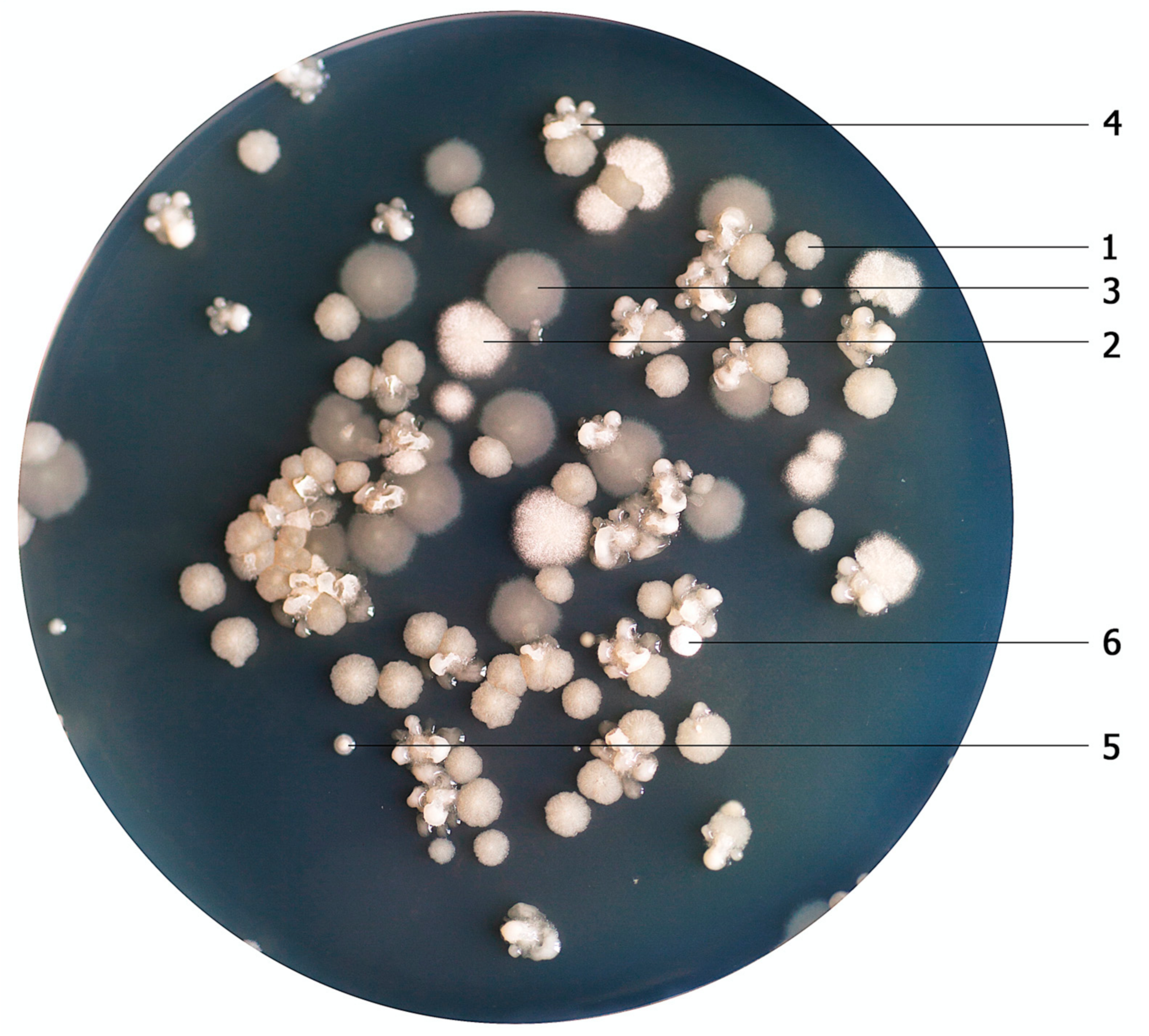
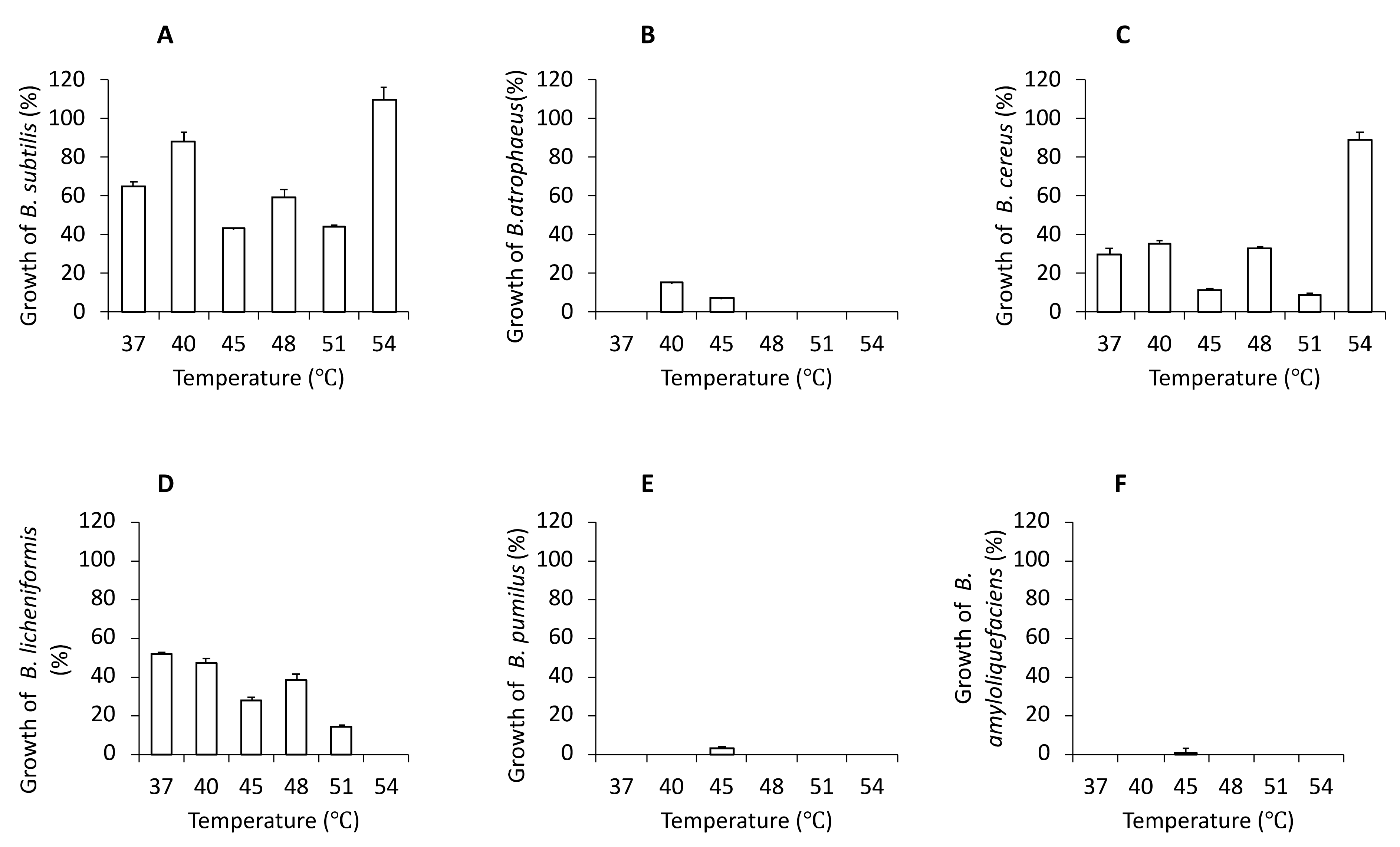
3.1. Selection and Quantification of Bacterial Cultures

We have previously analyzed a few of commercial ‘industrial probiotics-like’ preparations intended for the use as ‘friendly bacteria’ supplements in various cleaning preparations [19]. The use of bacteria cocktails in industrial and household products inevitably leads to their contact with humans. For sheer safety reasons, they are very likely to penetrate the human gastrointestinal tract, their producers had to test them and thus, unintentionally, they provided them as human probiotics. In this study, we have found that some of those species are also common in a numerous other applications, which may be a basis for the development of probiotic food preparation. Six different species of the Bacillus group (B. subtilis, B. atrophaeus, B. cereus, B. licheniformis, B. pumilus, B. amyloliquefaciens) were determined, as shown in Figure 1. For the tested product, the temperature 45 °C was used as the most adequate. Environmental Bacillus strains were counted quantitatively at a range of temperatures (Supplementary Table S1), while a quantitative analysis of their growth depending on temperature is presented in Figure 2A–F. At a temperature of 40 °C, Bacillus strains were less numerous, and fever species were identified. Here, B. subtilis dominated, followed by B. licheniformis and B. cereus. Interestingly, at a temperature of 54 °C, B. subtilis and B. cereus were the most abundant, which suggests their facultatively thermophilic character despite their fuzzy shape and smaller size of their colonies. None of the Bacillus strains grew at 57 °C. Furthermore, the optimal cultivation time for colonies features evaluation, in the temperatures from 37 °C to 57 °C, turned out to be 17 h. A longer incubation time (24 h) resulted in bigger colony size.
Graph A in Figure 2, presents the growth of B. subtilis across the entire temperature spectrum from 37 °C to 54 °C, with the highest abundance observed at 54 °C. B. atrophaeus grows only at two temperatures: 40 °C and 45 °C (Figure 2B). It is worth noticing that B. cereus (Figure 2C) grows to comparable amounts at two temperatures: 37 °C and 48 °C. On the other hand, though B. cereus grows most abundantly at 54 °C, its colonies have a ‘blurred’ shape and spread bacterial background. We also determined that B. licheniformis grows in the temperature range from 37 °C to 51 °C, as shown in Figure 2D. However, these data could indicate that the number of colonies of B. licheniformis decreased when temperature increased. Furthermore, we found out that B. pumilus presented in Figure 2E and B. amyloliquefaciens presented in Figure 2F only grow as single colonies and only at the temperature of 45 °C. We suggest that perhaps these bacteria may be just a trace contaminant present in the tested Bacillus preparations.

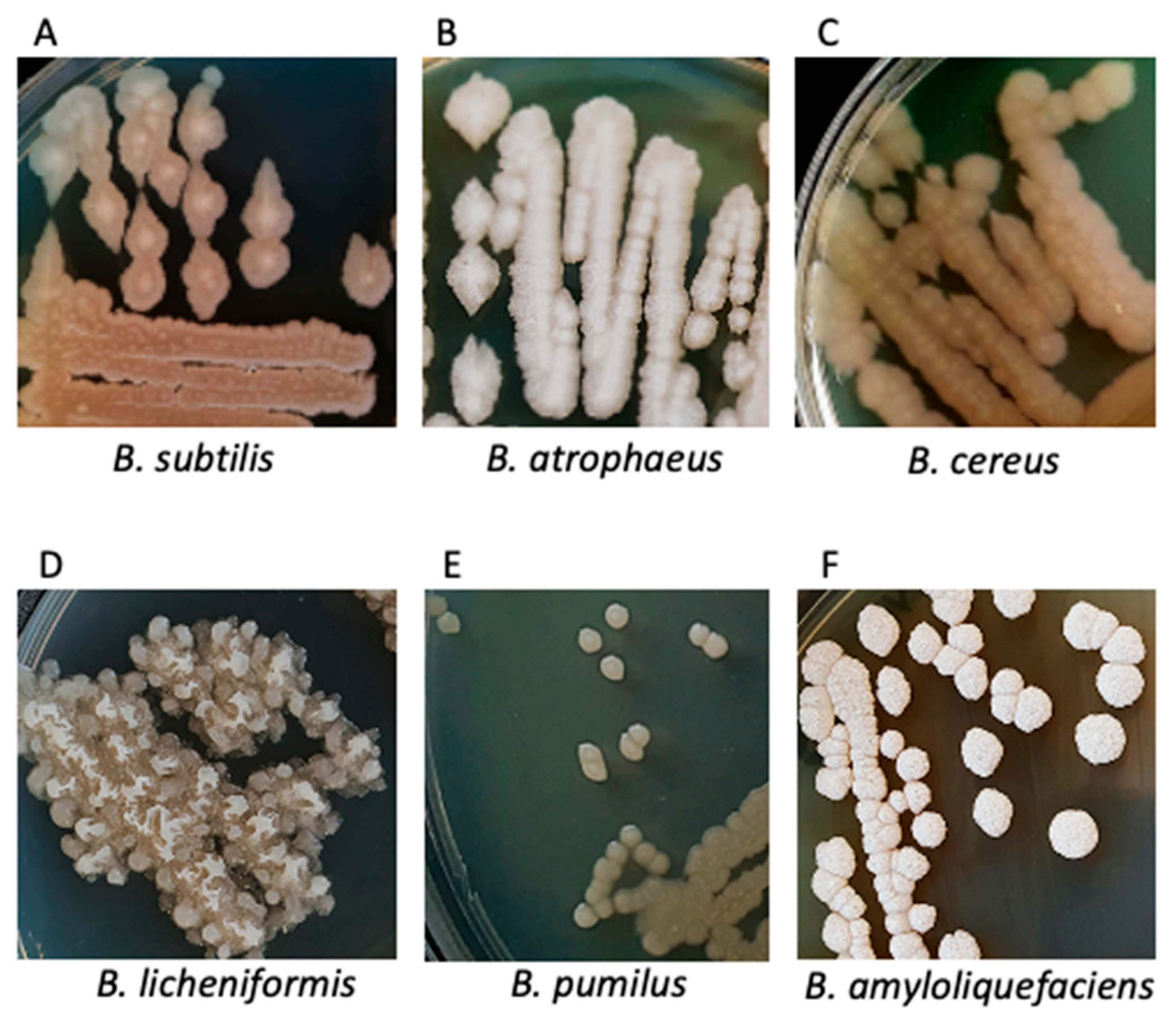
3.2. Characteristics of Bacterial Cultures, Colonies and Bacterial Cells

We also determined the morphology of bacterial cultures, shown in Figure 3A–F. On an agar plate with the well separated colonies of Bacillus species, factors such as shape, color, size, and production of extracellular enzymes were examined. As we previously described, the growth of one species of Bacillus species in different temperatures was sometimes varied more than between two different species grown in the same conditions. What’s more, as observed under the microscope, this phenomenon occurred regarding the shape and sizes of single cells.
We compared B. subtilis colonies (Figure 3A) present to other species of the Bacillus group. While B. subtilis were round and flat, other Bacillus species/strains had more of an irregular shape. Moreover, B. subtilis were average in size and had a cream-like color with a lighter dot in the center. In addition, when we incubated at a temperature of 54 °C, the colonies were more transparent and had a white ring (Supplementary Figure S1). This appearance can be explained by the motility characteristics of B. subtilis, which seems to increase with higher temperatures, where mucoid or slimy colonies appear [21]. Microscopic study clearly showed that B. subtilis are Gram(+)blue-violet rods (Supplementary Figure S2, panel A). Interestingly, heat-resistant endospores of B. subtilis were observed in around half of bacteria in the Wirtz staining (Supplementary Figure S2, panel B). The conditions of nutrient limitation drive the population of B. subtilis cells to form a mixed population, where half of the population activates the genetic regulator of sporulation and the second half omitted this path, which was demonstrated in previous reports [22]. Furthermore, B. subtilis has medium-sized, enveloped mucoid rods, which has been well documented using the Maneval’s tests (Supplementary Figure S2, panel C).
Subsequently, B. atrophaeus was examined on nutrition agar (Figure 3B). The results showed large individual colonies (2–5 mm) that had flat and uneven surface covered with a delicate white, non-hemolytic coating with irregular edges. In addition, B. atrophaeus were the brightest colored of all tested Bacillus species found in this potential probiotic dietary product. Further, our study demonstrated in Gram’s tests that B. atrophaeus are Gram(+) purple rods with a pink center (Supplementary Figure S2, panel A). Interestingly, in the Wirtz test, B. atrophaeus stained blue but did not show spore-forming. The reason for this is unknown, but we speculated that it is apparently related to the atypical biochemical composition of the spores (Supplementary Figure S2, panel B). Moreover, our Maneval’s study showed that, depending on the growth phase, B. atrophaeus formed longer and shorter envelopes with round endospores at the ends (Supplementary Figure S2, panel C).
Previous report by Lim et al. [23] demonstrated that the form of the B. cereus colonies varies, depending on the strain. Our study showed that, in the tested Bacillus mixtures, B. cereus were flat, growing as large single colonies or forming clusters. They were colored gray and/or brown with a lighter center and a wavy edge (Figure 3C). Furthermore, our analyses revealed that there was a wide zone of hemolysis around the colony of B. cereus, characteristic of a β-hemolytic bacterium, capable of erythrocytes hemolysis (Supplementary Figure S3). Gram’s staining showed purple Gram(+) rods surrounded by a pink border (Supplementary Figure S2, panel A). While other Bacillus rods were short or medium length, B. cereus were the longest. Furthermore, after Maneval’s envelopes test, long, thin rods with envelopes were also observed (Supplementary Figure S2, panel C). Wirtz staining showed many endospores of B. cereus (Supplementary Figure S2, panel B).
We examined B. licheniformis on an nutrition agar (Figure 3D). In this study, numerous clusters of B. licheniformis grew, colored white and beige in flower-like in shapes. Despite their medium sizes (3–4 mm), the colonies of B. licheniformis built up high to reach to the lid of the plate. Furthermore, cultivated on solid plates with other bacteria (Bacillus species mixture diluted 10−7), they were attached to other Bacillus strains colonies. Further, B. licheniformis demonstrated Gram(+) middle size and violet rods morphology (Supplementary Figure S2, panel A). Cultivated in difficult living conditions (temperature above 51 °C) they tended to form spores, while in temperature 37 °C, a vegetative state in Wirtz staining was observed (Supplementary Figure S2, panel B). In addition, in Maneval’s test, B. licheniformis exhibited production of envelopes (Supplementary Figure S2, panel C).
Isolated B. pumilus colonies were small (1–2 mm), pearly, with concentric growth rings. In addition, B. pumilus colonies we distinguishable from outnumbering them other Bacillus species only at the temperature of 45 °C upon higher dilution of the bacteria concentrate. After cooling to a temperature of 6 °C, the bacteria turned bright yellow. Hence, we suspect that apparently this treatment induced genes coding for dye production. Interestingly, in the Gram staining analyses, B. pumilus appeared in the form of pink rods, and/or pink rods with purple dots inside the bacteria (Supplementary Figure S2, panel A). Later, in the Wirtz test, a large amount of spore-forming were seen (Supplementary Figure S2, panel B). Moreover, in comparison to other strains, oval-shaped colonies of B. pumilus were formed with thin envelopes (Supplementary Figure S2, panel C).
In the same way, also on nutrition agar we examined B. amyloliquefaciens (Figure 3F). The results showed distinct white-cream colored colonies that built above the surface of the agar plates up to about 1–3 mm. In addition, the colonies had the roughest surfaces and waviest edges of all isolated Bacillus. Similarly to B. pumilus, B. amyloliquefaciens was isolated from the potential probiotic grown at 45 °C as a single colony. Cultivated separately on solid agar medium, it grew in large numbers and released yellow biofilm into the medium (Supplementary Figure S4). Previous reports suggest that B. amyloliquefaciens produces γ-polyglutamic acid (γ-PGA) [24]. Our microscope observation indicated that B. amyloliquefaciens were long, purple Gram(+) rods with a blue line (Supplementary Figure S2, panel A). Furthermore, the image of green dots indicating spores and blurred shapes was obtained by staining with the Wirtz method (Supplementary Figure S2, panel B). The Maneval test demonstrated that formed clusters, with the envelopes fused together to form a white, irregularly shaped surface (Supplementary Figure S2, panel C).

3.3. MALDI-TOF Mass Spectrometry Analysis of Bacteria

In this study, the microbiological identity of bacterial species/strains present was investigated by microbial protein spectrum (proteome) mass spectroscopy analysis in MALDI-TOF assays as described [25,26]. These data allowed the distinguishing of closely related Bacillus species. B. subtilis, B. atrophaeus, B. cereus, B. licheniformis, B. pumilus, B. amyloliquefaciens were determined in the samples of the analyzed product. Reliable identification to the genus and species level using the Bruker MALDI Biotyper mass spectrometer was confirmed. These results indicated, that the value of the strain identification index was respectively: for B. subtilis—2.27, B. atrophaeus—1.89, B. cereus—1.96, B. licheniformis—2.22, B. pumilus—2.10, B. amyloliquefaciens—1.96. Therefore, due to the fact that for three species—B. atrophaeus, B. cereus, and B. amyloliquefaciens—the confidence factor was slightly below the value of 2.0, the presence of these species in the tested product, although highly probably, leaves a margin of lower confidence.

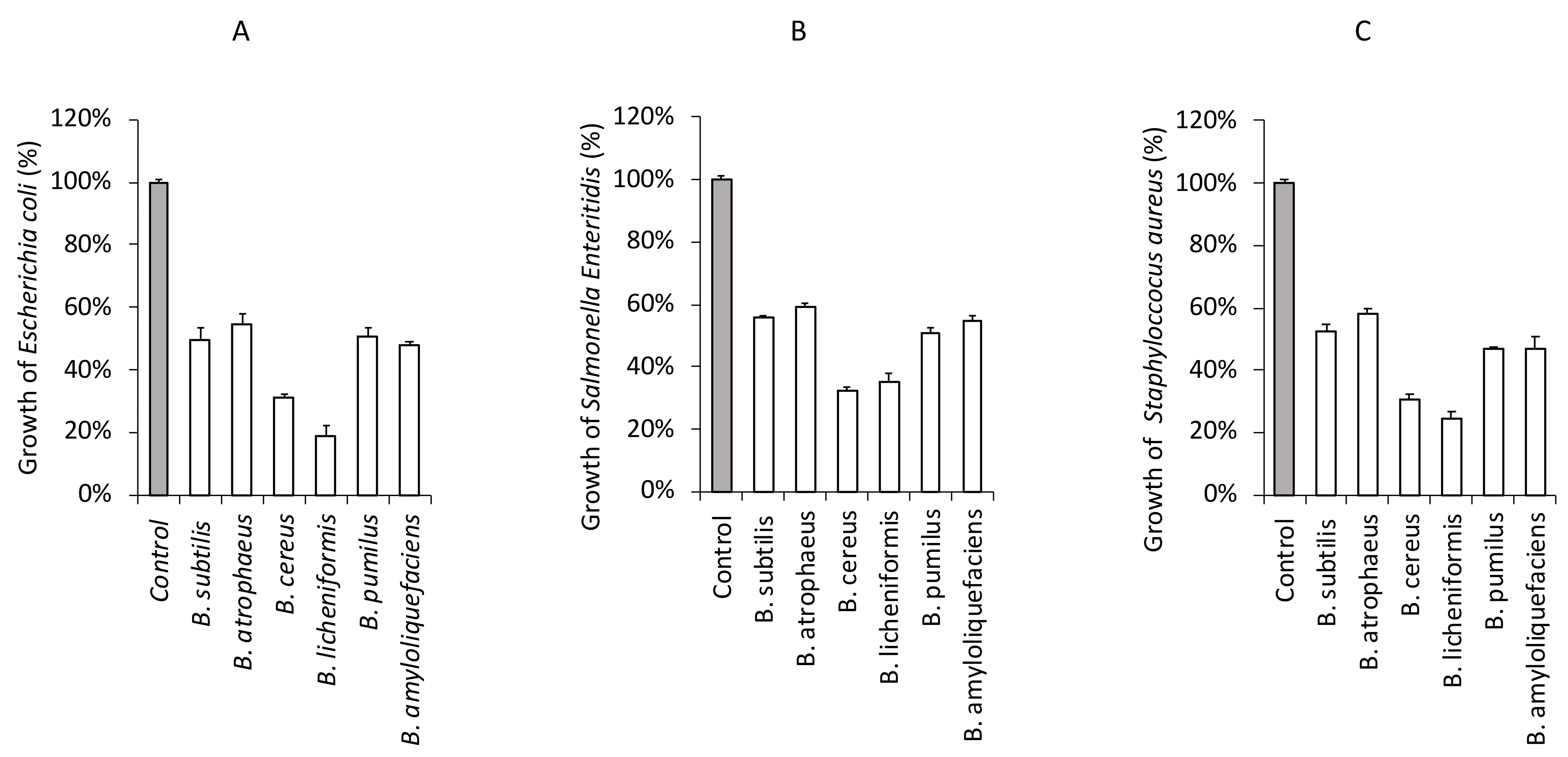
3.4. Antagonistic Effect of Foodborne Pathogenic Bacteria in the Presence of CFS from Bacillus Strains

As a result of this study, antagonistic activity of CFS from the Bacillus strains against foodborne pathogenic bacteria was determined as shown in Figure 4A–C and Supplementary Table S2. All the CFS partially inhibited the growth of E. coli O157:H7 ATCC 35150 (Figure 4A), Salmonella Enteritidis KCCM 12021 (Figure 4B), and S. aureus KCCM 11335 (Figure 4C).
Isolated B. subtilis showed antagonistic activity against in about 50% of both gram-positive (S. aureus) and gram-negative bacteria (E. coli, S. enteritidis). This study also revealed a weak antagonistic activity of B. atrophaeus. It inhibited the activity of food-borne pathogenic bacteria only in 40%, which indicates that this bacterium is of low functionality as a probiotic bacterium. B. cereus exhibits a wide range of zone of inhibition and was bacteriostatic for three of the tested pathogens in 70%. The above result indicates that the isolate might produce some substances that have an inhibitory effect on a wide range of bacteria. The results revealed that B. licheniformis exhibited various degrees of inhibitory activities against foodborne pathogenic bacteria, which indicates a broad spectrum of inhibition patterns of this organism. Maximum inhibitory activity was observed against E. coli (85%), followed by S. aureus (77%) and S. enteritidis (68%). B. pumilus showed antagonistic activity against in about 50% of foodborne pathogenic bacteria, similarly to S. subtilis. On the other hand, B. amyoliquefaciens showed antagonistic activity against E. coli and S. aureus in over 50%, like previous reports indicated [27]. A minor inhibitory effect for S. enteritidis (45%) was observed.
To further confirm these results, an agar well diffusion assay was performed using the neutralized CFS (adjusted to pH 3.5 by adding 1 M HCl). Since Bacillus produce several antimicrobial compounds including organic acids, bacteriocins, enzymes, and antibiotics, we tested whatever neutralized CFS would be able to inhibit the growth of pathogenic bacteria. However, these results suggest that the neutralized CFS were unable to inhibit the growth of E. coli O157:H7 ATCC 35150, Salmonella Enteritidis KCCM 12021, and S. aureus KCCM 11335 (data not shown). These data could indicate that the inhibition of foodborne pathogenic bacterial growth was due to organic acids or non-bacteriocin compounds from the Bacillus strains.

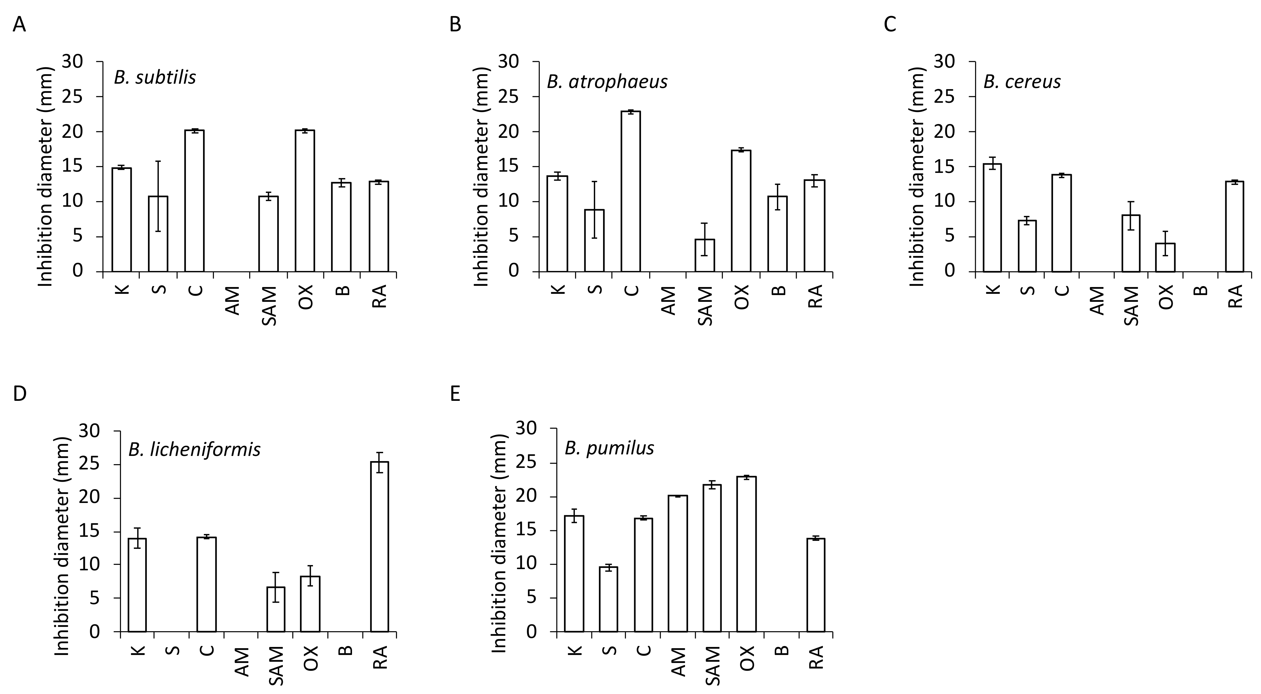
3.5. Antibiotic Resistance

In this study, we determined whether organisms are resistant (R) or susceptible (S) to the antibiotics, as shown in Table 1. The data for triplicate incubation plates with antibiotics are presented on diagrams (Figure 5A–E). In addition, the zones of growth inhibition of Bacillus are presented in Supplementary Figure S5A–E and using the interpretative zones diameters given in Supplementary Table S3. All the isolated Bacillus species revealed resistance to ampicillin, with one exception—B. pumilus.
As displayed in Figure 5A and Supplementary Table S3, B. subtilis showed sensitivity to kanamycin, chloramphenicol, oxacillin, bacitracin, rifampicin, and complex of ampicillin and sulfbactam. In addition, for chloramphenicol and oxacillin, we observed particularly large inhibition zones. Another antibiotic, streptomycin-showed ambiguous outcomes because it appeared in a few colonies growing next to the disc of streptomycin, which possibly comprise spontaneous R mutants.
B. atrophaeus (Figure 5B, Supplementary Table S3) showed sensitivity to chloramphenicol with the biggest zone of growth inhibition; followed by oxacillin, kanamycin, and rifampicin. On the contrary, bacitracin, streptomycin, and the complex of ampicillin and sulfbactam revealed its resistance or regrowth. The results indicated the appearance of a few colonies of resistant B. atrophaeus, or its regrowth, following the decomposition of the antimicrobial agent.
Furthermore, the study showed that chloramphenicol and rifampicin caused very clear zones of growth inhibition of B. cereus (Figure 5C, Supplementary Table S3), while for complex of ampicillin and sulfbactam, streptomycin, and oxacillin, clear zones of inhibition were not recorded. Similar ambiguous results were obtained with kanamycin which, despite causing the largest inhibition zone, left it with small amount of bacteria.
B. licheniformis (Figure 5D, Supplementary Table S3) exhibited sensitivity to chloramphenicol only. Although big zones of growth inhibition of B. licheniformis to rifampicin and kanamycin were observed, again regrowth of a few single colonies was evident. This was similar for oxacillin and complex ampicillin and sulfbactam, where the zones of inhibition were very small. B. licheniformis was only one bacterium isolated from Bacillus species, which proved resistant to three antibiotics: bacitracin, streptomycin and ampicillin. Moreover, the additional brightening zones around the antibiotic discs displayed a pattern very characteristic of B. licheniformis, resembling sunflowers.
B. pumilus (Figure 5E, Supplementary Table S3) showed sensitivity to all antibiotics, except bacitracin, similarly to B. cereus and B. licheniformis. B. pumilus as the only one from isolated Bacillus species gave clear results, as very big zones of growth inhibition without regrowth were observed. Moreover, additional very wide brightening zones around the antibiotic discs, especially for ampicillin and sulfbactam, oxacillin, kanamycin, and ampicillin, were observed.
No zones of inhibited growth for B. amyloliquefaciens have been determined due to the very low content of this bacterium in the potential Bacillus probiotic source.

3.6. TP-84 Bacteriophage Sensitivity Tests

We evaluated the Bacillus species identified by MALDI-TOF for potential sensitivity to thermophilic TP-84 bacteriophage, infecting thermophilic Bacilli, as we are currently carry out a programme for engineering TP-84 bacteriophage bionanoparticles [13,14]. At 54 °C, only two bacterial species (B. subtilis, B. cereus) multiplied, while at 51 °C, three of the bacteria detected (B. subtilis, B. cereus, B. licheniformis) multiplied, as shown in Figure 1. Thus they were subjected to TP-84 infections. However, all of them exhibited resistance to TP-84 bacteriophage.

4. Discussion

Commercially available probiotics originate from the gastrointestinal tract of humans and animals as well as from dairy fermented foods. Probiotic Bacillus strains, when applied in the form of health foods and dietary supplements or functional feeds and feed supplements, have numerous documented beneficial effects on humans and animals [1]. Although a number of Bacillus are involved in the process of fermentation, only B. subtilis has been extensively studied as a probiotic agent for purposes such as adhesive characteristics to human intestinal epithelial cells, as well as antioxidant and anti-microbial activities [28]. Thus, in this study, we characterized the antagonistic activities and probiotic potential of Bacillus species isolated from a commercial concentrate, and showed their sensitivity/resistance to antibiotics. We confirmed their probiotic usage potential-isolated B. subtilis showed antagonistic activity against both gram-positive (S. aureus) and gram-negative bacteria (E. coli, S. enteritidis) (Figure 4). Further analysis provided that B. licheniformis exhibited the best degrees of inhibitory activities against the foodborne pathogenic bacteria, with a maximum inhibition degree for E. coli (Figure 4).
Each potential probiotic strain has its own specific properties regarding antibiotic resistance. The knowledge of susceptibility and resistance of Bacillus strains to antibiotics is an important prerequisite for probiotics formulations designers. Although antibiotic resistance of Bacillus strains might be considered a major concern for probiotic application, the determination of antibiotic resistance among Bacillus strains is confounded by testing methods [29]. Our study showed that most of the Bacillus strains tested were sensitive to chloramphenicol and rifampicin (Table 1, Figure 5 and Supplementary Figure S5). Furthermore, isolated Bacillus derived from probiotic were resistant to ampicillin, except one—B. pumilus. Still, intrinsic resistance to antibiotics is not considered as a risk to animal and human health [30]. In some cases, such ‘human neutral’ antibiotic resistance may be beneficial during prolonged antibiotic therapies, where existing human microbiome is damaged. In such a case, Bacillus species probiotics may temporarily fill in this niche.
Bacillus biofilm formation supports the host organism against the gastrointestinal tract (GIT), urogenital tract (UGT), and urinary tract (UT) infections, while modulating immune system activity [2]. Our further analyses revealed that B. amyloliquefaciens also form biofilms (Supplementary Figure S4). The balancing effect and favorable colonization by Bacillus probiotics are sustained, even if an administered preparation contains spores [31] or if the sporulation occurs in upper parts of the GIT in the stomach or due to bile activity. The Bacillus species probiotic biofilm introduces biochemical effects such as antimicrobial and enzymatic activity, thus contributing to protection from dysbiosis as well as GIT and other infections. Moreover, in vitro models of the human stomach and human studies with probiotic Bacillus species reveal the mechanisms of its life cycle and sporulation [1,32]. Our microscope observation indicated the present of endospores for all isolated Bacillus species (Supplementary Figure S2).
Despite identification of six Bacillus species on an agar medium plates (Figure 3) has been demonstrated, spectroscopic analysis identified with high degree of confidence only three out of six initially detected Bacillus. Previous reports confirmed that the detected B. subtilis, B. licheniformis, and B. pumilus play the most important role in probiotic bacteria preparations. These bacteria are currently used in the production of food for both humans and animals [33,34,35]. Previous reports demonstrated that one of example of potentially pathogenic Bacillus species are B. cereus and B. anthracis, as some of its strains produce toxins with different levels of toxicity, posing a human or animal-health risk [28,36]. Thus these bacteria should not be included in the development of bimodal probiotic from Bacillus species. However, in the analyzed conglomerate only a small number of B. cereus were recorded as growing on a solid medium at temperatures of 40 °C and 45 °C (Figure 2B), and thus they may be an unintended contamination during large scale cultivation of mixed Bacillus species population. In addition, B. cereus colonies are typically larger, thus this species might actually be a closely related B. cereus-like species. Furthermore, it the MALDI-TOF factor of 1.99 is low-to-medium as shown, this isolate may represent a variant of a ‘human-friendly’ species B. atrophaeus, containing some more common proteins with B. anthracis. Within such a closely related ‘Bacillus group’, horizontal gene transfer undoubtedly is a common phenomenon. The above analysis points to the possibility of exploring Bacillus preparations used for other than human use purposes for valuable species, which eventually could be used for new formulations as human probiotics upon further extensive testing.
Despite the beneficial activity of Bacillus strains belonging to the safety group 1, a number of strains can pose a substantial health risk, carrying genes for various toxins or antibiotic resistance [37]. Furthermore, a promising future application of the probiotic Bacillus species might be microflora biocontrol in the human body and the closest human environment. Although the environmental probiotic Bacillus species display the potential to support human microflora, the controversies regarding the safety of certain strains is a key factor in their still-limited application [37].

Supplementary Materials

The following supporting information can be downloaded at: https://www.mdpi.com/2076-2607/11/2/488/s1, Figure S1: Growth of B. subtilis on agar plate in 54 °C; Figure S2: Microscopic analyses of Bacillus species cells by Gram staining (panel A), Wirtz staining (panel B) and Maneval tests (panel C); Figure S3: The colony of B. cereus with a wide zone of hemolysis after incubated in temperature 45 °C; Figure S4: Colonies of B. amyloliquefaciens with a yellow biofilm secreted into the agar medium; Figure S5: Antibiotic sensitivity test of isolated probiotic Bacillus derived from the probiotic preparation; Table S1: Quantification of bacterial species in the temperature range tested; Table S2: The antagonistic activity of CFS from the Bacillus strains against foodborne pathogenic bacteria; Table S3: Zone of inhibition growth of the Bacillus sp. derived from probiotic.

Author Contributions

Conceptualization, B.Ł. and J.J.-F.; Methodology, A.B. and P.M.S.; Formal analysis, M.S. and M.D.-Z.; Investigation, B.Ł.; Resources, M.S., P.M.S. and P.C.; Writing—original draft, B.Ł. and J.J.-F.; Supervision, B.Ł. and P.M.S.; Funding acquisition, P.M.S. All authors have read and agreed to the published version of the manuscript.

Funding

The work was supported by TECHMATSTRATEG2/410747/11/NCBR/2019 grant to University of Gdansk, Faculty of Chemistry, Molecular Biotechnology Department.

Data Availability Statement

The authors confirm that the data supporting the findings of this study are available within the article and its supplementary materials.

Conflicts of Interest

The authors declare that the research was conducted in the absence of any commercial or financial relationships that could be construed as a potential conflict of interest.

References

  1. Jeżewska-Frąckowiak, J.; Seroczyńska, K.; Banaszczyk, J.; Jedrzejczak, G.; Żylicz-Stachula, A.; Skowron, P.M. The promises and risks of probiotic Bacillus species. Acta Biochim. Pol. 2018, 65, 509–519. [Google Scholar] [CrossRef] [PubMed]
  2. Gopikrishna, T.; Suresh Kumar, H.K.; Perumal, K.; Elangovan, E. Impact of Bacillus in fermented soybean foods on human health. Ann. Microbiol. 2021, 71, 30. [Google Scholar] [CrossRef]
  3. Cutting, S.M. Bacillus probiotics. Food Microbiol. 2011, 28, 214–220. [Google Scholar] [CrossRef] [PubMed]
  4. Sornplang, P.; Piyadeatsoontorn, S. Probiotic isolates from unconventional sources: A review. J. Anim. Sci. Technol. 2016, 58, 26. [Google Scholar] [CrossRef] [PubMed]
  5. Schallmey, M.; Singh, A.; Ward, O.P. Developments in the use of Bacillus species for industrial production. Can. J. Microbiol. 2004, 50, 1–17. [Google Scholar] [CrossRef]
  6. Raul, D.; Biswas, T.; Mukhopadhyay, S.; Kumar Das, S.; Gupta, S. Production and Partial Purification of Alpha Amylase from Bacillus subtilis (MTCC 121) Using Solid State Fermentation. Biochem. Res. Int. 2014, 2014, 568141. [Google Scholar] [CrossRef]
  7. Jezewska-Frackowiak, J.; Seroczynska, K.; Banaszczyk, J.; Wozniak, D.; Skowron, M.; Ozog, A.; Zylicz-Stachula, A.; Ossowski, T.; Skowron, P.M. Detection of endospore producing Bacillus species from commercial probiotics and their preliminary microbiological characterization. J. Environ. Biol. 2017, 38, 1435–1440. [Google Scholar] [CrossRef]
  8. Yao, M.; Xie, J.; Du, H.; McClements, D.J.; Xiao, H.; Li, L. Progress in microencapsulation of probiotics: A review. Compr. Rev. Food Sci. Food Saf. 2020, 19, 857–874. [Google Scholar] [CrossRef]
  9. Goderska, K. Different methods of probiotics stabilization. In Probiotics; Rigobelo, E., Ed.; InTech: Rijeka, Croatia, 2012. [Google Scholar] [CrossRef]
  10. Kiepś, J.; Dembczyński, R. Current Trends in the Production of Probiotic Formulations. Foods 2022, 11, 2330. [Google Scholar] [CrossRef]
  11. Šipailienė, A.; Petraitytė, S. Encapsulation of Probiotics: Proper Selection of the Probiotic Strain and the Influence of Encapsulation Technology and Materials on the Viability of Encapsulated Microorganisms. Probiotics Antimicrob. Proteins 2018, 10, 1–10. [Google Scholar] [CrossRef]
  12. Mantzouridou, F.; Spanou, A.; Kiosseoglou, V. An inulin-based dressing emulsion as a potential probiotic food carrier. Food Res. Int. 2012, 46, 260–269. [Google Scholar] [CrossRef]
  13. Skowron, P.M.; Kropinski, A.M.; Zebrowska, J.; Janus, L.; Szemiako, K.; Czajkowska, E.; Maciejewska, N.; Skowron, M.; Łoś, J.; Łoś, M.; et al. Correction: Sequence, genome organization, annotation and proteomics of the thermophilic, 47.7-kb Geobacillus stearothermophilus bacteriophage TP-84 and its classification in the new Tp84virus genus. PLoS ONE 2018, 13, e0196798, Erratum in PLoS ONE 2018, 13, e0195449. [Google Scholar] [CrossRef]
  14. Skowron, P.M.; Krawczun, N.; Żebrowska, J.; Krefft, D.; Żołnierkiewicz, O.; Bielawa, M.; Jeżewska-Frąckowiak, J.; Janus, Ł.; Witkowska, M.; Palczewska, M.; et al. A vector-enzymatic DNA fragment amplification-expression technology for construction of artificial, concatemeric DNA, RNA and proteins for novel biomaterials, biomedical and industrial applications. Mater. Sci. Eng. C 2020, 108, 110426. [Google Scholar] [CrossRef]
  15. Available online: https://science.umd.edu/CBMG/faculty/asmith/BSCI223/lab3.pdf (accessed on 2 November 2022).
  16. Schaeffer, A.B.; Fulton, M.D. A Simplified Method of Staining Endospores. Science 1933, 77, 194. [Google Scholar] [CrossRef]
  17. Maneval, W.E. Staining bacteria and yeast with acid dyes. Stain Technol. 1941, 16, 13–19. [Google Scholar] [CrossRef]
  18. Kunicki–Goldfinger, W. The Life of Bacteria; PWN Scientific Publishing House: Warsaw, Poland, 1998; ISBN 978-83-01-14378-7. [Google Scholar]
  19. Jeżewska-Frąckowiak, J.; Żebrowska, J.; Czajkowska, E.; Jasińska, J.; Pęksa, M.; Jędrzejczak, G.; Skowron, P.M. Identification of bacterial species in probiotic consortiums in selected commercial cleaning preparations. Acta Biochim. Pol. 2019, 66, 215–222. [Google Scholar] [CrossRef]
  20. Wikins, T.D.; Holdeman, L.V.; Abramson, I.J.; Moore, W.E. Standardized single-disc method for antibiotic susceptibility testing of anaerobic bacteria. Antimicrob. Agents Chemother. 1972, 1, 451–459. [Google Scholar] [CrossRef]
  21. Berkeley, R.M.; Heyndrickx, N.L.; De Vos, P. Applications and Systematics of Bacillus and Relatives; Wiley-Blackwell: Oxford, UK, 2008. [Google Scholar]
  22. Kearns, D.B.; Losick, R. Cell population heterogeneity during growth of Bacillus subtilis. Genes 2005, 19, 3083–3094. [Google Scholar] [CrossRef]
  23. Lim, E.S.; Baek, S.Y.; Oh, T.; Koo, M.; Lee, J.Y.; Kim, H.J.; Kim, J.S. Strain variation in Bacillus cereus biofilms and their susceptibility to extracellular matrix-degrading enzymes. PLoS ONE 2021, 16, e0245708. [Google Scholar] [CrossRef]
  24. Liu, J.; He, D.; Li, X.Z.; Gao, S.; Wu, H.; Liu, W.; Gao, X.; Zhou, T. Gamma-polyglutamic acid (gamma-PGA) produced by Bacillus amyloliquefaciens C06 promoting its colonization on fruit surface. Int. J. Food Microbiol. 2010, 142, 190–197. [Google Scholar] [CrossRef]
  25. Azarko, J.; Wendt, U. Identyfikacja drobnoustrojów—Porównanie metody biochemicznej i spektrometrii masowej. Diagn. Lab. 2011, 47, 409–417. [Google Scholar]
  26. Kosikowska, P.; Pikula, M.; Langa, P.; Trzonkowski, P.; Obuchowski, M.; Lesner, A. Synthesis and Evaluation of Biological Activity of Antimicrobial--Pro-Proliferative Peptide Conjugates. PLoS ONE 2015, 10, e0140377. [Google Scholar] [CrossRef] [PubMed]
  27. Afrin, S.; Bhuiyan, M.N.I. Antagonistic activity of Bacillus amyloliquefaciens subspecies Amyloliquefaciens against multidrug resistant Serratia rubidaea. BioRxiv 2019. [CrossRef]
  28. Elshaghabee, F.M.F.; Rokana, N.; Gulhane, R.D.; Sharma, C.; Panwar, H. Bacillus as Potential Probiotics: Status, Concerns, and Future Perspectives. Front. Microbiol. 2017, 8, 1490. [Google Scholar] [CrossRef]
  29. Huys, G.; D’Haene, K.; Swings, J. Influence of the culture medium on antibiotic susceptibility testing of food-associated lactic acid bacteria with the agar overlay disc diffusion method. Lett. Appl. Microbiol. 2002, 34, 402–406. [Google Scholar] [CrossRef]
  30. Al Kassaa, I.; Hober, D.; Hamze, M.; Chihib, N.E.; Drider, D. Antiviral potential of lactic acid bacteria and their bacteriocins. Probiotics Antimicrob. Proteins 2014, 6, 177–185. [Google Scholar] [CrossRef]
  31. Coppi, F.; Ruoppolo, M.; Mandressi, A.; Bellorofonte, C.; Gonnella, G.; Trinchieri, A. Results of treatment with Bacillus subtilis spores (Enterogermina) after antibiotic therapy in 95 patients with infection calculosis. Chemioterapia 1985, 4, 467–470. [Google Scholar]
  32. Bernardeau, M.; Lehtinen, M.J.; Forssten, S.D.; Nurminen, P. Importance of the gastrointestinal life cycle of Bacillus for probiotic functionality. J. Food Sci. Technol. 2017, 54, 2570–2584. [Google Scholar] [CrossRef]
  33. UK Standards for Microbiology Investigations. Identification of Bacillus species. In Bacteriology—Identification 9; Standards Unit, PHE, Microbiology Services; Public Health England: London, UK, 2015; pp. 1–27. [Google Scholar]
  34. Romo-Barrera, C.M.; Castrillón-Rivera, L.E.; Palma-Ramos, A.; Castañeda-Sánchez, J.I.; Luna-Herrera, J. Bacillus licheniformis and Bacillus subtilis, Probiotics That Induce the Formation of Macrophage Extracellular Traps. Microorganisms 2021, 9, 2027. [Google Scholar] [CrossRef]
  35. Hill, J.E.; Baiano, J.C.F.; Barnes, A.C. Isolation of a novel strain of “B. pumilus” from penaeid shrimp that is inhibitory against marine pathogens. J. Fish Dis. 2009, 32, 1007–1016. [Google Scholar] [CrossRef]
  36. Zakowska, D.; Bartoszcze, M.; Niemcewicz, M.; Bielawska-Drózd, A.; Kocik, J. New aspects of the infection mechanisms of Bacillus anthracis. Ann. Agric. Environ. Med. 2012, 19, 613–618. [Google Scholar]
  37. Pals, K.L.; Chang, R.T.; Ryan, A.J.; Gisolfi, C.V. Effect of running intensity on intestinal permeability. J. Appl. Physiol. 1985, 82, 571–576. [Google Scholar] [CrossRef]
Figure 1. Identification of probiotic bacteria colonies detected in the course of commercial preparation analysis in the growth temperature of 45 °C. Dilution of 10−7. Cultures marked: (1) B. subtilis, (2) B. atrophaeus, (3) B. cereus, (4) B. licheniformis, (5) B. pumilus, (6) B. amyloliquefaciens.
Microorganisms 11 00488 g001
Figure 2. Changes of growth of Bacillus strains dependents of temperature range in triplicate. (A) B. subtilis, (B) B. atrophaeus, (C) B. cereus, (D) B. licheniformis, (E) B. pumilus, (F) B. amyloliquefaciens.
Microorganisms 11 00488 g002
Figure 3. The morphology of bacterial cultures of Bacillus species on agar plate.
Microorganisms 11 00488 g003
Figure 4. Inhibition of foodborne pathogenic bacterial growth in the presence of CFS from Bacillus strains isolated from potential probiotic source. Escherichia coli O157:H7 ATCC 35150 (A), Salmonella Enteritidis KCCM 12021 (B), or Staphylococcus aureus KCCM 11335 (C) were incubated in the presence of CFS from the Bacillus strains (B. subtilis, B. atrophaeus, B. cereus, B. licheniformis, B. pumilus, B. amyloliquefaciens) at 37 °C for 24 h. The bacterial growth was determined at OD600. The growth rate of foodborne pathogenic bacteria without CFS was assigned to 100% (Control).
Microorganisms 11 00488 g004
Figure 5. Antibiotic resistance and susceptibility of the Bacillus derived from a potential probiotic source. Data for triplicate.
Microorganisms 11 00488 g005
Table 1. Antibiotic resistance of the Bacillus species derived from a potential probiotic source.
Bacillus SpeciesAntibiotics
KSCAMSAMOXBRA
B. subtilisSNDSRSSSS
B. atrophaeusS *ND *S * R *ND *S *ND *S *
B. cereusNDNDSRNDNDRS
B. licheniformisND *R *S *R *ND *ND *R *ND *
B. pumilusS *S *S *S *S *S *R *S *
B. amyloliquefaciensNDNDNDNDNDNDNDND
Results are shown antibiotics susceptibility. R—means resistant to the antibiotics, no antimicrobial effect; S—sensitivity to the antibiotics, true zone of growth inhibition; ND— ambiguous outcome or potential resistance or regrowth; an asterisk (*) indicates that additional clear zones were visible. Antibiotics discs were as follows: K—kanamycin (30 μg), AM—ampicillin (10 μg), SAM—ampicillin & sulfbactam (20 μg), S—streptomycin (10 μg), RA—rifampicin (5 μg), OX—oxacillin (5 μg), B—bacitracin (10 μg), and C—chloramphenicol (30 μg).
Disclaimer/Publisher’s Note: The statements, opinions and data contained in all publications are solely those of the individual author(s) and contributor(s) and not of MDPI and/or the editor(s). MDPI and/or the editor(s) disclaim responsibility for any injury to people or property resulting from any ideas, methods, instructions or products referred to in the content.

