Article

Comparative Genomic Analysis Reveals the Emergence of ST-231 and ST-395 Klebsiella pneumoniae Strains Associated with the High Transmissibility of blaKPC Plasmids

1
Medical Laboratory, Diwan Health Centre, Muscat 130, Oman
2
Department of Microbiology and Immunology, College of Medicine and Health Sciences, Sultan Qaboos University, Muscat 123, Oman
3
Microbiology and Immunology Diagnostic Laboratory, Department of Microbiology and Immunology, Sultan Qaboos University Hospital, Muscat 123, Oman
*
Author to whom correspondence should be addressed.
Microorganisms 2023, 11(10), 2411; https://doi.org/10.3390/microorganisms11102411
Submission received: 3 September 2023 / Revised: 21 September 2023 / Accepted: 23 September 2023 / Published: 27 September 2023
(This article belongs to the Special Issue Mobile Genetic Elements in Pathogens)

Abstract

:
Conjugative transposons in Gram-negative bacteria have a significant role in the dissemination of antibiotic-resistance-conferring genes between bacteria. This study aims to genomically characterize plasmids and conjugative transposons carrying integrons in clinical isolates of Klebsiella pneumoniae. The genetic composition of conjugative transposons and phenotypic assessment of 50 multidrug-resistant K. pneumoniae isolates from a tertiary-care hospital (SQUH), Muscat, Oman, were investigated. Horizontal transferability was investigated by filter mating conjugation experiments. Whole-genome sequencing (WGS) was performed to determine the sequence type (ST), acquired resistome, and plasmidome of integron-carrying strains. Class 1 integrons were detected in 96% of isolates and, among integron-positive isolates, 18 stains contained variable regions. Horizontal transferability by conjugation confirmed the successful transfer of integrons between cells and WGS confirmed their presence in conjugative plasmids. Dihydrofolate reductase (dfrA14) was the most prevalent (34.8%) gene cassette in class 1 integrons. MLST analysis detected predominantly ST-231 and ST-395. BlaOXA-232 and blaCTX-M-15 were the most frequently detected carbapenemases and beta-lactamases in the sequenced isolates. This study highlighted the high transmissibility of MDR-conferring conjugative plasmids in clinical isolates of K. pneumoniae. Therefore, the wise use of antibiotics and the adherence to effective infection control measures are necessary to limit the further dissemination of multidrug-resistant bacteria.

1. Introduction

Klebsiella pneumoniae is an opportunistic pathogen that is associated with different serious nosocomial infections, including pneumonia, septicemia, meningitis, and urinary tract infections (UTIs) [1,2]. To treat the infections caused by extended-spectrum beta-lactamase (ESBL)-producing K. pneumoniae, carbapenem antibiotics are the drugs of choice; however, they are considered as last-resort antibiotics [3]. Moreover, the number of K. pneumoniae carbapenemase (KPC) enzyme producers has been increasing [4,5,6,7]. The spread of resistance determinants has been facilitated by horizontal gene transfer mechanisms mainly via mobile genetic elements (MGEs). Integrons, for example, are semi-mobile platforms that recognize and capture mobile gene cassettes and transform them to usable genes by ensuring their appropriate expression [8,9,10]. They are located on different MGEs, such as plasmids, transposons, and pathogenicity islands (PAIs), which enable their movement between different bacteria [11,12]. A number of studies showed that most K. pneumoniae clinical isolates carried class 1 integrons, whereas class 2 integrons were only present in 1–2% of the isolates and rarely harbored class 3 integron [13]. A significant association between integron-positive isolates and antibiotic resistance for some drugs was observed, including piperacillin-tazobactam, ciprofloxacin, cefotaxime, and ceftazidime [12,14,15,16,17]. Both class 1 and class 2 integrons have gene cassettes encoding resistance to trimethoprim (dfr) as a predominant gene, which may be due to the long-term usage of this antibiotic [18,19,20].
Class 1 integron is associated with transposons derived from Tn402, which can bind in a larger transposon such as Tn21. Over 80 different antibiotic-resistance-encoding gene cassettes are associated with class 1 integrons, but most integrons have the gene aadA, which encodes streptomycin–spectinomycin resistance [8]. Moreover, class 1 integrons have a 3′-conserved segment that contains a sulI gene-encoding resistance to sulfonamides and a qacEΔ1 gene-encoding resistance to quaternary ammonium compounds [21]. In Oman, there has been an increasing concern regarding the spread of MDR K. pneumoniae and KPC producers in clinical settings [22,23,24]. However, molecular characterization is still lacking. Therefore, we aim, in this study, to genomically analyze MDR K. pneumoniae carrying class 1 integrons, in terms of the structure, sequence types (STs), and antimicrobial phenotype and genotype. In addition, we investigate the association between antimicrobial susceptibility and the presence of integrons in clinical isolates of K. pneumoniae. Finally, we conduct conjugation experiments to evaluate the horizontal-transfer capability, which is key to predict the future dissemination and implementation of intervention strategies.

2. Materials and Methods

2.1. Bacterial Strains

A total of fifty (n = 50: ESBL (n = 27), XDR (n = 21), and PDR (n = 2) strains = K. pneumoniae isolates were collected in this study from the Diagnostic Microbiology and Immunology Laboratory in Sultan Qaboos University Hospital (SQUH), Muscat, Sultanate of Oman between July 2019 and October 2019. The isolates were mostly from urine (n = 25), respiratory (n = 10), wound (n = 9), bloodstream (n = 4), body fluid (n = 1), and biopsy (n = 1) samples. All isolates were processed as per the Clinical and Laboratory Standards Institute guidelines [25]. The isolates were cultured in a selective medium (Cefuroxime Cysteine Lactose Electrolyte-Deficient agar, Oxoid, Basingstoke Hampshire, UK) prior to preservation in sterile CryoBeads (Mast Diagnostics, Derby, UK) at −80 °C for further analysis (Table 1).

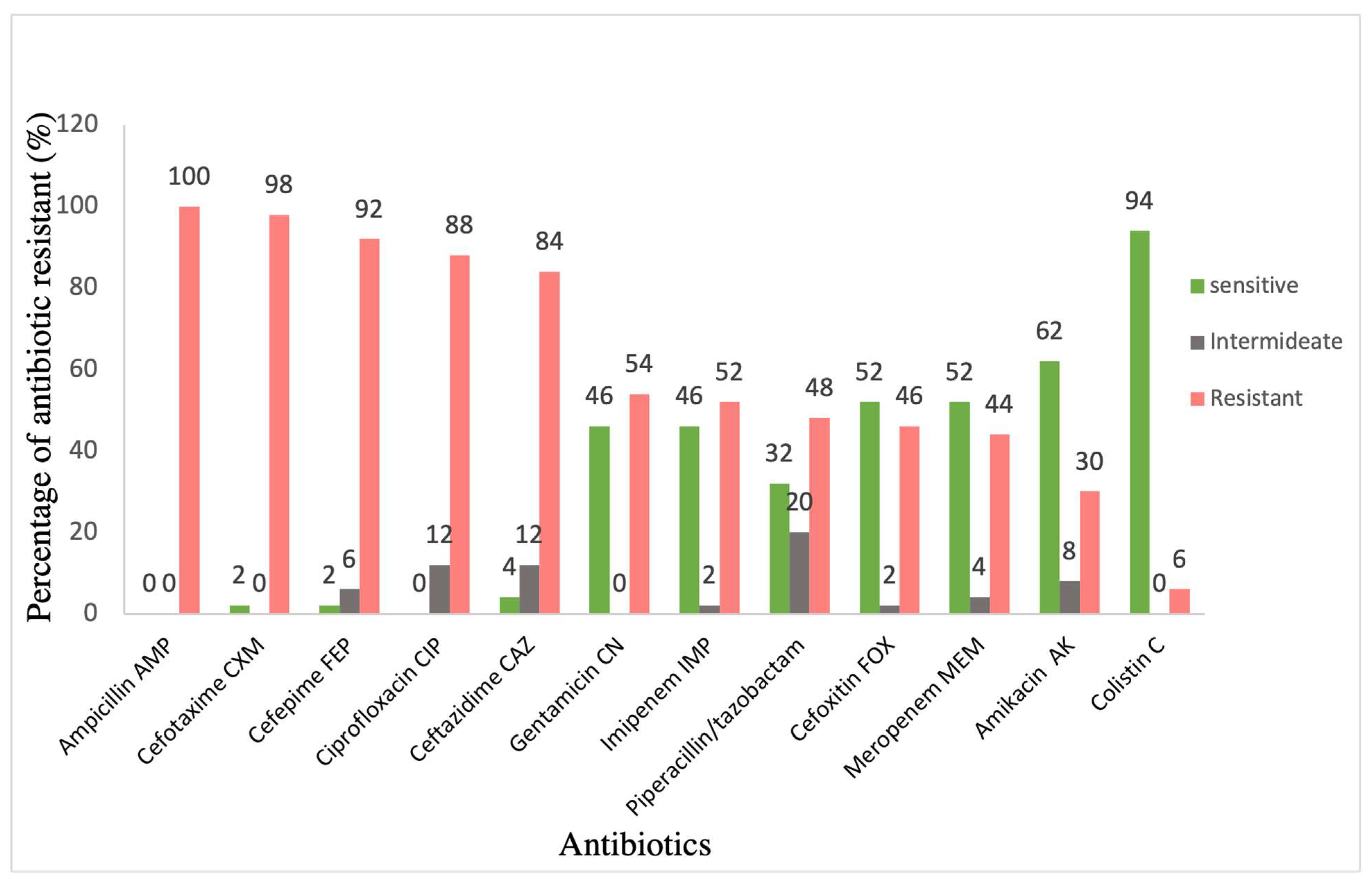
2.2. Antimicrobial Susceptibility

The antimicrobial susceptibility profiles of the K. pneumoniae isolates were assessed using two methods: disk diffusion method and BD PhoenixTM automated system (Becton Dickinson Diagnostic Systems, Sparks, MD, USA). For both methods, three to five bacterial colonies from overnight pure cultures were suspended in normal saline (Fisher Chemical, Cramlington, UK) and adjusted to a 0.5 McFarland standard (approximately 1–2 × 10⁸ CFU/mL) using a CrystalSpec nephelometer (BD Diagnostics, Sparks Glencoe, MD, USA), according to the manufacturer’s recommendations. After 15 min, the suspension was spread onto a Mueller–Hinton agar (MHA) surface (Oxoid, Hampshire, UK) and left for 1–2 min at room temperature to be absorbed. After 15 min, the selected antibiotic disks (BioMérieux and Liofilchem, Germany) were placed on the inoculated MHA plates using sterile forceps. The plates were incubated at 37 °C for 18–24 h. The antibiotics were selected according to the CLSI standard as follows: ampicillin (AMP 10 µg), piperacillin/tazobactam (TZP 110 µg), cefepime (FEP 30 µg), cefotaxime (CTX 30 µg), cefoxitin (FOX 30 µg), ceftazidime (CAZ 30 µg), imipenem (IPM 10 µg), meropenem (MEM 10 µg), amikacin (AK 30 µg), gentamicin (CN 10 µg), and ciprofloxacin (CIP 5 µg). E. coli (ATCC 25922) and P. aeruginosa (ATCC 27853) were used as the susceptible control strains. The interpretive categories and zone diameter breakpoints, nearest to the whole mm for each antibiotic, are listed in Table 2. The BD PhoenixTM automated system was used to test the susceptibility of colistin and tigecycline, and to confirm the antimicrobial susceptibility of other antibiotics tested by the disk diffusion method. For colistin, >1 mg/L was considered resistant according to CLSI breakpoints [25]. The European Committee on Antimicrobial Susceptibility Testing (EUCAST) interpretive criteria was used for tigecycline, with ≤0.5 mg/L considered as sensitive [26]. The ESBL production was further confirmed using the disk diffusion method. Both cefotaxime and ceftazidime alone and in combination with clavulanic acid were used. An increase in diameter of ≥5 mm with the clavulanic acid compared to the antibiotic alone was interpreted as positive for ESBL production. The XDR and PDR isolates were characterized genotypically to the carbapenemase genotype level using Xpert Carba-R (Cepheid, Sunnyvale, CA, USA).

2.3. Genomic DNA Extraction and Purification

The DNA was extracted using Qiagen kit (QIAamp® genomic DNA kit, Hilden, Germany) as described in the manufacturer’s instructions with slight modifications. One to four colonies were suspended in 10 mL Mueller–Hinton broth (Oxoid, Mampshire, UK) and left overnight on a shaking incubator set to 250 rpm at 37 °C. Then, the bacterial suspension was centrifuged for 15 min at 4000× g. The amount of 20–40 mg of pelleted bacterial cells was re-suspended in a pre-lysis buffer (100 μL TE buffer and 0.1 μL RNase A 100 mg/mL) (Thermo Fisher Scientific, Winsford, UK). The cells were well resuspended by pipetting up and down several times. After that, the samples were incubated again at 37 °C at a 400 rpm shaking incubator for 30–60 min (Innova 4000, New Brunswick Scientific, Hertfordshire, UK). A volume of 1μL Proteinase K (stock concentration = 20 mg/mL) + 99 μL TE were added to the sample to obtain a final volume of 200 μL in 1.5 mL microcentrifuge tube. Then, 200 μL of the sample was mixed with 400 μL of lysis solution and incubated at 65 °C for 15 min using a heat block (Eppendorf ThermoStat plus, Hamburg, Germany). As per the manufacturer’s instruction, the samples were washed twice using the columns. The DNA pellet was eluted in a final volume of 50–100 μL of nuclease-free H2O (QIAamp® genomic DNA kit, Hilden, Germany). The extracted DNA was aliquoted into 2 vials, which were stored at 4 °C and −80 °C for future use. The boiling method was used to extract the DNA from trans-conjugant colonies in the conjugation experiment [28]. Three to five colonies were removed from the plate and resuspended in 30 μL of nuclease-free water. Then, the samples were heated at 100 °C for 10 min in a heat block. Cells were then pelleted by centrifuging the samples at 8000× g for 1 min. The supernatant was used as a template for PCR.

2.4. Polymerase Chain Reaction (PCR)

2.4.1. Detection and Characterization of Classes 1, 2, and 3 Integrons

PCR assays were performed using Go-Taq DNA polymerase (Promega Ltd., Madison, WI, USA). The cycle conditions were adjusted depending on the gene size and the primer’s melting temperature. The detection of classes 1, 2, and 3 integrons was investigated by the amplification of integrase genes intl1, intl2, and intl3, respectively. The PCR reaction mixture consisted of 5 μL of 5X PCR green buffer; 0.5 mL of dNTPs (10 μM); 1 μL of both forward and reverse primers (10 μM); 0.1 μL of Go-Tag DNA polymerase (5 μ/mL); and 1.5 μL of template DNA. Then, nuclease-free water was added until the total volume reached 25 μL. A reaction mixture without a DNA template was used as a negative control. The same PCR reaction mixture was used to amplify variable-region genes. For the DNA that was extracted by the boiling method, dimethyl sulphoxide (DMSO) was added to encourage the annealing of the primers to the template and further enhance the amplification. A thermocycler (Eppendorf master cycler®, Merk, Darmstadt, Germany) was used for PCR amplification and the cycling conditions for Go-Tag enzyme are mentioned in Table 3. Annealing temperatures, which are different for each gene depending on the primer’s melting temperature, are listed in Table 4. The specific primers for detecting integrase genes and integron-specific variable regions were used as previously described [18]. Gel electrophoresis results can be found in the Supplementary Materials.

2.4.2. PCR Purification

PCR products were purified using QIAquick PCR and Gel Cleanup Kit (Qiagen, Hilden, Germany). Five volumes of buffer PB were added to 1 volume of the PCR sample and then mixed. To bind DNA, the mixture was applied to the QIAquick column and centrifuged for 30–60 s at 17,900× g at room temperature. After centrifugation, the column was placed in a clean 2 mL collection tube and the tube containing the filtrate was discarded. Then, 750 μL of buffer PE was added to the QIAquick column and centrifuged again for 30–60 s at the same previous conditions. Again, the column was placed in a clean 2 mL collection tube and the tube containing the filtrate was discarded. Then, the QIAquick column was centrifuged once more for 1 min to remove the residual wash buffer. Finally, the QIAquick column was placed in a clean 1.5 mL microcentrifuge tube to which 30 mL of elution buffer (10 mM Tris·HCl, pH 8.5) was added. The tube was incubated at room temperature for 1 min and then centrifuged for 30–60 s at 17,900× g. The purified DNA was stored at −80 °C for future use.

2.5. Gel Electrophoresis

All PCR products were visualized by using 2% agarose gel electrophoresis containing 1× TBE (ThermoFisher Scientific, Waltham, MA, USA) and MIDORI Green Direct (NIPPON-genetics, Europe). The bands were visualized by using G:Box Chemi-XR5 device (Syngene, India). DNA fragments sizes were estimated by using the GeneRuler 1 kb DNA Ladder (ThermoFisher Scientific, USA).

2.6. Whole-Genome DNA Sequencing

Whole-genome sequencing (WGS) in this study was performed at microbesNG by Illumina next-generation sequencing at a minimum coverage of 30× (https://microbesng.co.uk/, Birmingham, accessed on 20 July 2022). The genomic DNA was prepared and then was sent for sequencing following the protocol provided by the sequencing facility. The assembled sequences were then retrieved from the websites and analyzed accordingly.

2.7. Bioinformatics Analyses

WGS data of K. pneumoniae isolates files were analyzed using the center for genomic epidemiology server (CGE) (http://cge.cbs.dtu.dk/services, accessed on 2 July 2022). At CGE, the CSI phylogeny tool [29] was used to investigate the relatedness between the strains based on single nucleotide polymorphism (SNP) identification. The assembled genome data for all samples were returned to the server as input. The reference genome was K. pneumoniae subsp. pneumoniae HS11286 (GeneBank: CP003200). The SNPs were localized and filtered based on the default settings, which include a minimum distance of ten bases between the SNPs, a sequencing depth of ten bases, and a minimum SNPs quality of 30. In addition, the Z-score for each SNP must be above 1.96. Then, the data file was visualized and managed by using an online tool, Interactive Tree of Life (iTOL) [30]. The ResFinder tool from the GCE server was used in this study to detect the acquired antimicrobial resistance genes and their specific location on the sequence [31]. MLST and PlasmidFinder were used to detect Multilocus sequence typing (MLST) and plasmids, respectively [32,33]. Along with ResFinder tool in CGE, the Comprehensive Antibiotic Resistance Database (CARD) (https://card.mcmaster.ca/home, accessed on 20 July 2020) was used to detect the presence of putative antibiotic-resistance genes by using the Resistance gene identifier (RGI) tool. This tool, in addition, can detect known point mutations within the resistance-conferring genes. Artemis software version (18.2.0) from the Sanger Institute was used for the visualization of WGS features. Moreover, GenBank from the National Center for Biotechnology Information (NCBI) (https://www.ncbi.nlm.nih.gov/genbank/, accessed on 3 August 2020) was used to search in the DNA database for similar DNA sequences and the Basic Local Alignment Search Tool (BLAST) was used to search for similarities between DNA sequences (https://blast.ncbi.nlm.nih.gov/Blast.cgi, accessed on 3 August 2020).

2.8. Conjugation Experiment

Conjugation experiments were performed for K. pneumoniae strains with variable regions as donors, and E. coli HB101 as a recipient by using the filter mating method [34]. To obtain the standard concentration of the required antibiotic (Amoxicillin and streptomycin (Sigma-Aldrich, St. Louis, MO, USA)) solution, the powder was weighed on a calibrated analytical balance and dissolved in an appropriate solvent (as per the manufacturer’s instructions). K. pneumoniae isolates were cultured in a Luria agar (LA) (Lab M®, Manchester, UK) plate with 100 mg/mL of amoxicillin. The E. coli HB101 strain was cultured in the Luria agar (LA) plate with 50 mg/mL of streptomycin. Both strains were incubated overnight at 37 °C. On the next day, a single colony from previous plates was inoculated in 5 mL of Luria broth (LB) (Lab M®, Manchester, UK) with the same antibiotic selection and incubated overnight at 37 °C on a shaking incubator. Then, a 1:100 dilution of both cultures was inoculated in 5 mL fresh LB and incubated again on the shaking incubator at 37 °C until the bacteria grew to their exponential phase (OD600 0.2), which was measured using Bio-Photometer (Eppendorf, Germany). After that, 1 mL of each culture was centrifuged at 8000× g for 5 min to harvest the cells. The pellets were washed twice with 1X Phosphate-Buffered Saline (PBS) (prepared from 10X PBS, pH 7.4, ThermoFisher Scientific, MA, USA) to remove the remaining antibiotics. Then, the pellets were re-suspended in 200 µL of PBS. The recipient, donor, and mixed bacterial suspension were spotted onto the center of the 0.45 mm pore size membrane (Whatman Ltd. Sigma-Aldrich, St. Louis, MO, USA) and placed on LA without antibiotics. The spots were allowed to dry and then incubated overnight at 37 °C. Subsequently, filter papers were washed in 1 mL 1X PBS to detach the bacterial cells. Serial dilutions up to 10–8 were performed for the donor, recipient, and mixed bacteria by using PBS as a diluent. The trans-conjugants are selected on an LA plate supplemented with both amoxicillin and streptomycin. For colony-forming unit (CFU) counts, dilutions of the donor were plated on LA with 100 µg/mL of amoxicillin and the recipient in 50 µg/mL of streptomycin. All plates were incubated at 37 °C overnight. The efficiency of trans-conjugation is calculated using the following formulas:
% Efficiency of transconjugation = nTransconjugants/nRecipient Recovered (Cfu/mL) × 100

3. Results

Class 1 integrons were detected in 92% of isolates (46/50) using IntI1F and IntI1R primers (Table 4). The screening of classes 2 and 3 integrons by detecting the presence of IntI2 and IntI3 genes, respectively, showed that these were not present in the collection. The analysis of the antimicrobial susceptibility patterns of the 50 K. pneumoniae isolates was performed (Figure 1). All clinical isolates were resistant to ampicillin (100%); most of the isolates were resistant to cefotaxime (98%), cefepime (92%) and ceftazidime (84%); and almost half of them showed resistance to piperacillin/tazobactam (48%). Moreover, 88% of the isolates showed resistance to ciprofloxacin. Furthermore, these isolates showed intermediate resistance to gentamicin (54%), imipenem (52%), and, to a lesser extent, to amikacin and colistin (30% and 6%).
According to the results obtained by the PhoenixBD semi-automated system, 54% of K. pneumoniae isolates (n = 27) were identified as ESBLs, whereas 42% (n = 21) of isolates were identified as XDR and two isolates (4%) as PDR. Almost all XDRs were susceptible to colistin, except three isolates (Kp 2, Kp 22, and Kp 50) (Table 5). In addition, 11.8% and 5.6% of the XDRs were susceptible to amikacin and gentamicin, respectively. Some of the XDR isolates were susceptible to amikacin, gentamicin, imipenem, and meropenem along with colistin (Table 5).
The conjugation was performed between variable region-positive isolates as donors and E. coli HB101 strain as a recipient. Seven representative strains were selected for testing (Kp 5, Kp 21, Kp 27, Kp 37, Kp 42, Kp 49, and Kp 50). The trans-conjugant PCR products are shown in (Supplementary File). All isolates were positive for the IntI1 gene, which confirmed the successful occurrence of conjugation via horizontal transfer. In the tested strains, the constituents of gene cassettes, plasmids, and sequencing types were analyzed by sequencing. The WGS data show that, in our tested strains, plasmids were detected in all E. coli trans-conjugants, which confirms that these conjugative plasmids are highly transmissible in the E. coli conjugation experiments. The efficiency of trans-conjugation for each strain was calculated and ranged between 0.07% and 17.3% (Figure 2). This exemplifies the role of these plasmids as the main vehicle for the transmission of integrons, and subsequently the transport of various types of gene cassettes from the donor to the recipient strains.
Out of the total 50 isolates of K. pneumoniae, 24 isolates were analyzed by WGS, including the 7 isolates which were successful in conjugation. Sixteen of these isolates were positive for the variable regions (Kp 5, 7, 10, 11, 15, 16, 21, 28, 37, 40, 42, 43, 44, 45, 46, and 49) and two isolates showed bands with an unexpected size for the intI3 gene (Kp 4 and Kp 22). According to previous studies, these bands could be variants of integrase genes [35]. Among the 24 isolates, 3 were integron-negative isolates by the PCR method (Kp 4, Kp 27, and Kp 30). However, WGS showed that two of these isolates were positive for class 1 integron (Kp 27 and Kp 30), whereas Kp 4 was still negative. Therefore, Kp 4 isolate was excluded from the subsequent molecular analysis.
The MLST of all sequenced isolates was determined by the online MLST database from the Center of Genomics Epidemiology (CGE) [32]. Nine of the isolates belong to sequence type (ST-231) (Kp 5, 6, 7, 10, 11, 15, 28, 30, and 45), six isolates are ST-395 (Kp16, 22, 41, 43, 44, and 50), and two isolates are ST-405 (Kp 42 and Kp 46). Only one isolate belongs to each of the following miscellaneous sequence types: ST-37 (Kp 40), ST-45 (Kp 21), ST-147 (Kp 49), ST-280 (Kp 27), ST-1710 (Kp37), and ST-1741(Kp 25).
The WGS data of 23 integron-positive isolates were used to construct a whole-genome phylogenetic single-nucleotide polymorphism (SNP) study (Figure 3), and the output from the analysis shows three main clusters of strains. The first cluster is composed of nine isolates belonging to ST-231. The second cluster of strains is in the ST-395 group (n = 6). The clinical data of the above-mentioned two STs indicate the frequent moving of patients from the Intensive Care Unit (ICU) to the male medical ward or vice versa. However, the third cluster consisted of two strains only belonging to ST-405. The remaining strains belong to miscellaneous sequence types (ST-37, ST-147, ST-280, ST-1741, ST-45, ST-13, ST-17, and ST-1710).
A close-up analysis of the two predominant STs (ST-231 and ST-395) was conducted to predict if there are any possible recent outbreaks through clustering (Figure 4). The SNP tree for ST-231 and ST-395 was created individually and it was observed that the PDR isolates (Kp6 and Kp11) branched from the same clade, suggesting a possible vertical transmission among patients. Similarly, among the other isolates, ST-231 and ST-395 stains are very closely related.
The demographic data for thirteen strains that belong to ST-231 (n = 9) and ST-395 (n = 4), which account for most strains, are listed in Table 6. Ninety-two percent of the patients were males aged over 50 years (69.2%). The length of stay varied between patients, ranging from 1 to 134 days. Most of the patients were admitted to the ICU (61.5%) and the rest of the patients were admitted to intermediate care wards (male medical wards). Highly critical ICU patients usually require broad-spectrum antibiotics, which exert tremendous selection pressure, thus driving antimicrobial-resistance bacteria to thrive further in these settings.
At the level of ST, more than two-thirds (77.8%) of ST-231 and all ST-395 strains were isolated from patients admitted either to the ICU or male medical wards or moved between the two places interchangeably. Almost all patients have various risk factors contributing to their likelihood of acquiring highly resistant K. pneumoniae strains, including ICU admission, ventilation, urinary catheters, central venous catheters, and hospitalization, for more than 7 days [36]. Unfortunately, the mortality rate among these patients was very high (84.6%).
All sequenced isolates carried a wide range of acquired antimicrobial-resistance-conferring genes. Fourteen isolates harbored the blaOXA-232 gene, all of which were XDR and PDR isolates except KP 7, which was the only XDR isolate that did not carry the blaOXA-232 gene (Figure 3). bla-NDM5 was detected in only one isolate (Kp 49). Based on the WGS data and Carba-R, 87% of XDR and PDR isolates (20/23) have blaOXA-48-like, 8.7% (2/23) have blaNDM, and one isolate (4.3%) has both blaNDM and blaOXA48-like genes. There was a high level of agreement between the WGS and GeneXpert results for the tested isolates (8/9). The blaOXA-1 allele was detected in eight isolates, most of which belong to ST-395 (n = 6). The ESBL gene blaCTX-M-15 was detected in all isolates, while blaTEM-1B was seen in 15 isolates. Moreover, 10 isolates were positive for blaSHV-1, 9 isolates for blaSHV-11, 2 for blaSHV-76, and 2 for blaSHV-27.
A number of various genes conferring resistance against aminoglycosides were detected, including aac(6′)-Ib-cr, aac(6′)-Ib Hangzhou, aadA1,2, aac(3)-lId, aph(3′)-Ia, strA, strB, armA, and rmtB. A fosfomycin-resistance-conferring gene (fosA6) was detected in all isolates except Kp 49, which harbored fosA5. In addition, six different genes encoding quinolone resistance were detected in the isolates, which are aac(6′)-Ib-cr, oqxA, oqxB, qnrS1, B1, and B66. Moreover, rifampicin- (arr-2) and trimethoprim-resistance-encoding genes (dfr) were found in 10 and 11 isolates, respectively.
The tetracycline-resistance gene, tetA, was detected in four ESBL isolates, whereas tetD in two XDR isolates. According to the Phoenix automated system, all of these were tigecycline-resistant, except one ESBL isolate that carried the tetA gene (Kp 46). The gene of the acrAB efflux pumps’ regulator (marA), which contributes to resistance against tigecycline and other antibiotic classes, was detected in all sequenced isolates. In addition, K. pneumoniae efflux pumps (Kpn E, F, G, and H) that confer resistance to different antibiotic classes, such as macrolide, aminoglycoside, cephalosporin, tetracycline, rifamycin, and colistin, were detected in all isolates.
The four colistin-resistance isolates belonging to ST-231 and ST-395 (Kp 6, Kp 11, Kp 22, and Kp 50) were negative for plasmid-mediated mcr genes; therefore, a further analysis of the SNPs in PhoPQ operons was performed. No chromosomal mutations were detected in the regulatory two-component systems (TCSs) PmrAB and crrAB, and in the mgrB gene, a negative regulator gene of TCSs. However, all isolates harbored the regulatory TCSs (PhoPQ). Our analysis of the PhoPQ operons in the colistin-resistant isolates identified four novel, undescribed SNPs in the PhoP genes with the amino acid substitutions as follows: (Val130Glu), (Gln147His), (Gln131Glu), and (Pro129Leu) (Table 7). The significance of these SNPs is yet to be determined, as none of these substitutions have been described.
Most of the tested isolates showed an agreement between their resistance phenotypic patterns to different antibiotic families and the presence of resistance genes, as shown in Table 8. Carbapenems, cephalosporin, and quinolone antibiotics showed a complete level of agreement between phenotype and genotype. However, aminoglycoside antibiotics (AK, CN) showed a low level of agreement, with 21 isolates carrying resistance genes and only 9 and 15 isolates with phenotypic-resistance patterns to amikacin and gentamicin, respectively. This observation could be because these resistance-conferring genes might not be expressed. The expression of these genes needs to be further investigated, which was not within the scope of this study.
Using WGS data, the variable region of 23 isolates, including the trans-conjugant strains (Kp 5, Kp 21, Kp 27, Kp 37, Kp 42, Kp 49, and Kp 50), were analyzed to detect gene cassettes (Table 9 and Figure 3). Ten different gene cassettes were identified in 16 strains with a variable region, including those encoding resistance to aminoglycoside (aadA, aacA4, aac(6′)-Ib, APH(3″)-Ia, and ant1) trimethoprim (dfrA5, dfrA12, and dfrA14), rifampin (arr2 and arr3), chloramphenicol (catB3, catB8, and cmlA1), macrolide (ereA2 and erm), and quaternary ammonium compound (emrE). The most frequent gene cassette was composed of the dfrA14 gene alone, which was present in eight isolates (34.8%). However, three isolates showed empty integrons (In0) without any gene cassette insertions (Kp 16, Kp 22, and Kp 25). One of them had a variable region (Kp 16), while the other two did not (Kp 22 and Kp 25).
The comparative analysis of integrons was performed by locating the gene cassettes within the variable region. Mapping was conducted based on the genes between the primers of the variable region that varied among strains (Table 9). It was observed that there was a contig break in the middle of the integron cassettes in some strains and the downstream sequence (3′-end) was found in a separate contig. Therefore, the genetic maps of the integrons were drawn after the manual reassembly of the various components (Figure 5).
Among the detected plasmids, eight of them harbored antibiotic-resistance genes (Figure 6 and Table 10). The most prevalent plasmid is pKPQIL-IT, which is present in 15 strains that belong to ST-395 and ST-231. It is a 115,300 bp in size, IncFIB(QIL) replicon carrying genes associated with resistance to β-lactams (blaTEM-1 and blaKPC-3).
Plasmid pKP3-A is a 7605 bp linear ColKP3 replicon, carrying the blaOXA-181 gene. ST-231 and ST-405 share the uniqueness of pKP3-A that the integron is interrupted by insertion sequences (Figure 5). These isolates exhibit highly similar genotypes. All produced OXA-181, and the majority also have plasmid mediated-blaTEM-1, blaKPC-3 genes. Unlike Xpert Carba-R, the plasmid finder tool detected the kpc gene from WGS data in all ST-231 and ST-405 isolates. This could suggest that the copy numbers of the plasmid might be low, or the gene might have not been expressed and hence could not be detected by the GeneXpert system.
Isolates Kp 41 and Kp 50 are the only two strains carrying IncFIB (pNDM-Mar) and IncHI1B (pNDM-Mar) plasmids. In these isolates, blaNDM-1 was within the pNDM-Mar plasmid. Isolate Kp 49 (ST-147) had blaNDM-1 as part of the (ble-blaNMD-1) operon, where it was flanked by bleomycin-resistance (ble) gene and N-(5′-phosphoribosyl) anthranilate isomerase (trpF) gene. Moreover, the plasmid composition of Kp 49 had IncR, IncFII, and pKP3-A. The plasmid pAMA1167-NDM-5 is present only in seven of the ST-231 isolates. It is a 11,310 bp sized, IncFII(pAMA1167-NDM-5) replicon with genes encoding resistance against aminoglycosides (aph(3″)-Ib, aph(6)-Id, aadA2, aadA5, aac(3)-IIa, and aac(6)-Ib-cr5), β-lactams (blaNDM-5, blaOXA-1, blaCTX-M-15-1, and blaTEM-1), chloramphenicol (cat), sulfonamides (Sul1 and Sul2), trimethoprim (dfrA12 and dfrA17), tetracycline (tet(b) and tet(c)), and macrolides (emrE and mp(A)). The sequence analysis of the pKPN-IT plasmid revealed that it is a 208,191-bp IncFIB(K) replicon carrying (aadA2, cat, Mph(A), Sul1, and dfrA12) genes. It is found in the KP 10, 25, 27, 37, 42, and 46 isolates. However, the plasmid pBK30683 is a 139,941-bp FIA replicon present only in one isolate (KP 37).

4. Discussion

The present study focused on the molecular characterizations of integrons as a common class of mobile elements and their significance in the dissemination of multi-drug resistance genes among K. pneumoniae. Whole-genome sequence (WGS) data were analyzed to determine the sequence type as well as characterization of integron-carrying plasmids. In addition, WGS was used for studying the correlation between antimicrobial-resistance genotype and phenotype, and the level of agreement was analyzed. Moreover, the phylogenetic relatedness of the isolates was associated with the patient’s demographic data to explore the possible spread of mobile elements. Most of the studies in the region investigate the epidemiology of AMR, with a few studies concerning the prevalence of mobile genetic elements, such as genomic islands and their molecular characteristics, in Gram-negative bacteria (GNB), such as A. baumannii and K. pneumoniae [22,23,37,38,39,40,41]. Moreover, another study that was conducted in Gram-negative bacteria isolates in Palestinian hospitals focused on integrons and their role in AMR dissemination [42]. To our knowledge, this is the first study in Oman that assesses the transmissibility of plasmids by examining the efficiency of conjugation in vitro, thus emphasizing the importance of this mechanism in the spontaneous transfer of integron-carried plasmids intra-specially.
In the current study, 96% (48/50) of our K. pneumoniae isolates carried class 1 integrons, whereas none of the isolates contained either classes 2 or class 3 integrons. This finding is similar to the results of previous studies that were conducted in Iran [43,44]. In addition, a previous study showed that 100% of the MDR isolates were found to be positive for class 1 integrons, 36% for class 2, and none for class 3 integrons [12]. However, other studies detected class 2 integrons at low prevalence levels of 1.7% and 8.3% [42,45].
A high prevalence of class 1 integrons among our MDR isolates was observed and therefore it was the focus of this study. The presence of class 1 integrons in most of the isolates suggests that these genetic elements confer a significant advantage to their hosts, where exposure to antibiotic overuse creates a selective pressure in hospital environments [12,46]. The absence of both classes 2 and class 3 integrons from the isolates in this study is expected since these classes are relatively uncommon [8,47,48,49]. Moreover, this might be the case because our selection is limited to representative MDR isolates, which are unusually difficult to treat over a short period of time, which might not reflect the actual prevalence. Therefore, a larger sample size, including screening specimens, is more likely to reveal the other classes of integrons.
Our findings show that 37.5% (18/48) of integron-positive isolates were carrying variable regions. By the analysis of WGS data, a total of 10 different gene cassettes were detected in these isolates. The latter isolates encode for aminoglycoside, trimethoprim, rifampin, chloramphenicol, macrolide, and quaternary ammonium compounds. Trimethoprim-resistant gene cassettes (dfrA14), which encodes for dihydrofolate reductase enzymes, were found to be predominant in the isolates in this study (34.7%). Several studies showed that dfr genes were the most frequent gene cassettes present in integrons [18,42,50,51]. The stability of this gene cassette in class 1 integrons might indicate the overuse of the trimethoprim for a long period of time, specially to treat urinary tract infections [18]. Moreover, despite the initial results of the variable region, which was detected by employing conventional PCR using primers that bind in the 5′- and 3′-conserved regions, further WGS analysis showed that 13% of isolates with positive variable regions carried empty gene cassettes. Moreover, previous studies reported the presence of sul1 at the 3′CS region of the integrons [52,53,54]. However, it was only observed in (3/20) isolates, despite the amplification of the 5′CS–3′CS region by PCR. This has been described in Salmonella enterica [55,56]. The non-classical structure of integrons, where intI1 and/or 5′ end of integrons is truncated, were also observed in two strains (Kp 16 and Kp 25), which might suggest that amelioration of IntI1 confers a selective advantage for the host bacteria [35].
Simple PCR assays tend to overestimate the presence of integron cassettes and cannot provide further information on the subsequent changes in the integron cassettes, such as deletions and insertions. In fact, gene cassettes undergo continuous changes in their compositions, which means that these cassettes can integrate antimicrobial-resistant genes once conditions are favorable. Truncations and inversions within the integron structures are not uncommon and could not be further detected by simple PCR. Therefore, WGS data were key in bridging the gap on the various components within these integrons.
Plasmids are considered as main carriers for antibiotic-resistant genes through HGT mechanisms. Spontaneous plasmid conjugation is possible when the plasmid has a compatible mechanism of transfer. A plasmid is classified as conjugative when it helps other conjugative elements, such as integrons, to move between different strains [57]. In the current study, the trans-conjugant colonies were positive for intI1 gene in all the representative strains, with trans-conjugation efficiency ranging between 0.05% and 17.3%. This confirmed the successful transfer of integrons between cells via HGT. WGS data for the tested isolates supports our hypothesis, as plasmids were identified in all the tested isolates, which is similar to the result of a previous study [18]. Being carried on plasmids, these integrons might have a significant fitness cost since more copies of plasmids can be present compared to chromosomally located integrons. This observation has been reported in previous studies on K. pneumoniae as well as other Gram-negative organisms [58,59].
Most of our sequenced isolates showed a hierarchical relationship between different types of MGEs. The detected gene cassettes were embedded inside integrons, which were in turn inserted inside transposons, which are flanked by insertion sequences (IS) that can confer mobility to transposons. Some of these composite transposons were incorporated into conjugative plasmids that offer transfer to other cells [60]. Most of the isolates in this study (Kp 5, 21, 27, 28, 37, 40, 41, 42, 44, 46, 49, and Kp 50) have integrons inserted in transposons that are flanked by IS (Figure 4). All above-mentioned strains had a similar IS, which is IS6 and IS6100 (Kp 42 and Kp 46). IS6100 belongs to the IS6 family of transposable elements forming co-integrates as an endpoint of transposition, which was originally isolated from Mycobacterium fortuitum [61]. In general, different previous studies showed the same relationship, where the integrons were in transposons that were imbedded in a conjugative plasmid [62,63]. However, MGEs, including Tns and ISs, disrupt the integron structure.
The presence of IS6 on both sides of the Dcm-methylation operon, which is a type II restriction–modification (RM) system and next to the integron denotes that RM systems, are mobile and located in plasmids. These systems are involved in genome rearrangements and enhance virulence and resistance plasmid dissemination by carriage on other MGEs, as in our study on integrons [64,65,66]. The existence of these IS6 in the isolates in this study entails their importance in disseminating antibiotic-resistance genes among various genera and species of bacteria [67,68]. In this study, the specific capture of resistance cassettes by the integrons signifies apparent bias driven by the selective pressure of antibiotic therapy regimes. Other studies have also shown that integrons could carry catabolic genes, which proves that integrons play a broader role in bacterial evolution [10,69].
ST-231, the most abundant STs found in this study, is widely distributed in South-West Asia with clonal dissemination in Singapore, Brunei, and Darussalam between 2013 and 2015 [70,71]. In addition, in India, ST-231 strains were reported as a predominant ST in 22 isolates (45%) [72]. In Europe, the first occurrence of the MDR K. pneumoniae ST-231 clone was confirmed in Switzerland [73]. This dissemination may represent a global public threat toward a new epidemic clone. ST-395 has been reported as the most common MDR K. pneumoniae clone (69%) in North-Eastern France as well as an outbreak in an ICU in Italy [74,75].
In a previous study conducted in isolates from the Arabian Peninsula, different ST types were detected in MDR K. pneumoniae in Gulf countries, including Oman; however, ST-231 was not among these STs [76]. Similarly, previous studies were conducted in Oman and Saudi Arabia that have concluded that no ST-231 strain was found [77,78]. The absence of ST-231 possibly indicates a recent emergence of ST-231 in Oman. However, ST-147 was one of the commonly detected STs from K. pneumoniae isolates in the two previous studies [76,77], whereas in the present study, only one strain belongs to ST-147.
Both of these STs, ST-395 carrying strains and 77.8% of ST-231, were clustered in the same wards (mainly medical and ICU wards) with a significant mortality rate (84.6%). This observation highly suggests that there is a horizontal transfer of resistance-conferring genes, which is alarming. The heavy use of broad-spectrum antibiotics for critically ill patients in high-dependency areas and ICU patients selects for highly resistant strains and enhances the spread of resistance determinants. Furthermore, this may indicate the inefficient infection control practices that allowed the dissemination of plasmid-mediated resistance in the hospital. This finding corresponds to a study that was conducted in carbapenem-resistant GNB in SQUH, where 87% of infections were healthcare-associated with a 62% mortality rate [23].
Over the last 15 to 20 years, carbapenem-hydrolyzing β-lactamases, including OXA-48-like and NDM-type carbapenemases, disseminated and emerged in Enterobacteriaceae all over the world [79,80]. Carbapenemase OXA-48-like differs from the classical OXA-48 by one to five amino acids, in which they hydrolyze carbapenems and penicillins but do not affect extended-spectrum cephalosporin [80].
OXA-232-producing K. pneumoniae (OXA232Kp) was identified for the first time on a 6.1-kb ColE-type non-conjugative plasmid in France in 2013 from a patient who returned from India and, since then, it has spread worldwide [79,80,81]. OXA-232 is considered as a point-mutation derivative from OXA-181 with one amino acid difference [82]. In this study, WGS and Xpert Carba-R data detected the presence of the blaOXA-48-like gene in 87% (20/23) of our XDR and PDR isolates. These genes were identified by WGS as blaOXA-232 in 93.3% (14/15) of our XDR- and PDR-sequenced isolates. All of them belonged to ST-231 and ST-395. The level of agreement between WGS- and GeneXpert-tested isolates was highly significant (n = 8/9), with only one isolate that was negative for the OXA gene at GeneXpert and positive in WGS (Kp 43). Illumina WGS has good coverage for sequencing the whole genome, whereas GeneXpert is based on real-time PCR, and even with 97% specificity, it might miss some allelic variants. The plasmid analysis of our strains showed that the OXA-181 gene (or OXA-232) was carried on pKP3-A (7605 bp) replicon-type ColKP3. It was expressed only in the XDR- and PDR OXA-232-positive strains. This is similar to a previous study, where 33% and 100% of OXA-232 isolates were carried on ColKP3 plasmid in the USA and China, respectively [82,83].
On the other hand, NDM was first detected in 2008 in a Swedish patient of Indian origin and then spread all over the world [84]. In this study, Xpert Carba-R results show that only 4% (2/50) of the isolates have the NDM gene, whereas only one isolate (4.3%) that belongs to ST-147 harbors both OXA-48-like (blaOXA-232) and NDM genes (blaNDM-5) according to the WGS data. A study was conducted in the UAE to characterize carbapenem-resistant Enterobacteriaceae in the Arabian Peninsula, showing that NDM and OXA-48-like genes are the most-detected genes with rates of 46.5% and 32.5%, respectively. In the isolates collected from Oman, 46% and 44.4% of the isolates were carrying NDM and OXA-48-like genes (blaOXA-181), respectively, whereas only 1.6% carried both genes [76].
Along with the carbapenem genes, ESBL genes, including bla(CTX-M), bla(TEM), and bla(SHV), were detected, and reported in different studies [71,72]. All bla(CTX-M) genes were identified, including blaCTX-M-15, and it was the dominant ESBL amongst our strains since it was produced by all sequenced isolates. One main factor that contributes to the CTX-M-type ESBL distribution in K. pneumoniae is conjugative plasmids, particularly those belonging to IncF with specific insertion sequences [85]. Moreover, all bla(TEM) genes belonged to blaTEM-1 and were detected in 69.6%, whereas SHV types were characterized as blaSHV-1, blaSHV-11, blaSHV-27, and blaSHV-76. All ST-231 strains carried blaSHV-1, while ST-395 strains carried blaSHV-11. Moreover, OXA-1 was positive in all ST-395 strains only with an association with other ESBL genes (blaSHV-11, blaCTX-M-15, blaTEM-1). The association between OXA-1 and CTX-M-15 genes renders isolates resistant to β-lactam–β-lactamase inhibitor combinations. Noteworthily, a wide variety of GNB carried the OXA-1 gene in plasmid and integron locations [86]. Plasmid analysis in this study determined that the blaOXA-1 gene along with blaCTX-M-15 was located in IncF plasmids IncFII and IncFII (pAMA1167-NDM-5).
Resistance to colistin, a drug that is used as the last line drug in the treatment of extensively resistant pathogens, is increasingly reported in Enterobacteriaceae, particularly K. pneumoniae [87]. Several mechanisms are involved in colistin resistance, mostly the emergence of mobilized colistin-resistance (mcr) genes via plasmid and the mutations in the chromosomal gene (mgrB) and operons (PmrAB and PhoPQ), which are associated with the biosynthesis and modification of lipopolysaccharide (LPS). The PhoPQ regulatory system is activated at low concentrations of Mg2+ or Ca2+ and acidic PH [88]. The PhoQ (sensor kinase) activates PhoP (regulator protein) by phosphorylation, which in turn activates pmrFHIJKLM operons. These operons lead to LPS modification by adding 4-amino-4-deoxy-L-arabinose (L-Ara4N) and phosphoethanolamine (PETN) to lipid A. This modification neutralizes the negative charge of LPS leading to the low affinity of LPS to positively charged colistin [88,89,90].
Four sequenced isolates were resistant to colistin via BMD; however, all of these strains were negative for the plasmid-mediated colistin-resistance gene (mcr) and harbored a truncated mgrB gene. Bioinformatics analysis revealed the presence of four SNPs in the PhoP genes with the following amino acid substitutions: (Val77Glu), (Gln147His), (Gln131Glu), and (Pro129Thr). A further analysis of the promoter region of PhoPQ operon did not reveal any SNPs. A previous study reported different SNPs in PhoP (Val3Phe and Ser86Leu) and PhoQ (Leu26Pro) that were found to have a role in colistin resistance when tested by real-time PCR [91]. However, the detected SNPs in this study have not been described before, and therefore, a further expression analysis by real-time PCR is needed to show their role along with the truncated mgrB gene in colistin resistance. Moreover, colistin resistance might occur due to other resistance mechanisms, such as capsule overproduction, which causes a reduction in the interaction between colistin and its target site at K. pneumoniae (LPS) [88,92]. Furthermore, all isolates in this study carried K. pneumoniae efflux pumps (Kpn E, F, G, H), which might contribute to colistin resistance.
Tigecycline has broad-spectrum activity against Gram-positive and Gram-negative bacteria and demonstrates efficacy and safety as salvage therapy for MDR/XDR bacteria [37,40,41,93,94]. However, tigecycline resistance is becoming more common in ESBL-producing, MDR, XDR, and carbapenem-resistant isolates [95,96]. In this study, most of the sequenced isolates (17/19) were tigecycline-resistant, while the remaining four isolates were not tested. The WGS data show that the efflux pumps’ regulator gene (marA) is present in all isolates (n = 23), including the two sensitive ones (Kp37 and Kp46). In this context, a study reported that the marA gene, along with other regulatory pathways, could mediate the resistance to tigecycline through upregulating the acrAB efflux pump, even in the absence of ram A [97]. In another study, the development of tigecycline resistance was attributed to the tetA gene [98]. In our study, the tetA gene was detected in four ESBL isolates, from which only two were resistant. Thus, it is more likely that the marA regulator is engaged in tigecycline resistance.
Several studies reported that the armA gene was in conjugative plasmids of the IncL/M or IncFIA plasmid, while rmtB in IncFI or IncFIA [99,100]. Interestingly, these two genes were only identified in Kp 49, which belongs to ST-147, and it is an OXA-232- and NDM-5-producing isolate. In addition, IncFIA(HI1) and IncFII plasmids were uniquely present in this isolate only, and a further search of rmtB and armA genes by BLAST using WGS data confirmed the localization of these two genes in the (IncFII) plasmid.
Among carbapenems, cephalosporin, and quinolones, all isolates possessed expressed genes with a complete agreement between AMR genotypic findings and phenotypic expression. These results are close to what has been detected in a previous study [101]. In contrast, aminoglycoside antibiotics (amikacin and gentamicin) had high non-expressed genes (57.1% and 28.6%, respectively). Since there are other mechanisms behind AMR, the presence or absence of certain genes might not be a sufficient indicator of the isolate’s resistance profile. Furthermore, genotypic analysis has to be correlated with the phenotypic findings of various mechanisms involved.
The versatility and ubiquity of integrons in bacterial genomes indicate the key role of these mobile elements in bacterial adaptation. The abundance of repeat sequences of ISs and Tns within the integron structures in our WGS data pertains to the tremendous selective pressure of the antibiotic regimens used in the hospital microbial environment. These integrons are very potent capture systems with a limitless capacity to exchange antibiotic gene cassettes as well as other genes that increase their fitness. Integrons with combinations of various antibiotic cassettes have been termed multi-resistance integrons (MRIs) in Gram-negative bacteria [102].
Genomic plasticity was apparent in a few cases where there was a deletion of non-essential genes or empty integrons, which confirms that these integron systems are performing adaptive rather than housekeeping functions. In support with this hypothesis, two of the isolates we examined had integrons with no antibiotic gene cassettes and the intI1 gene was partially truncated (Kp 16 and Kp25). The different attachment sites and the variety of open reading frames of hypothetical or unknown functions within these integron systems are compelling. Further detailed analysis is required to unravel the importance of integrons among other MGEs towards the adaptation of bacterial evolution.

5. Conclusions

In summary, this study demonstrated a high prevalence of class 1 integrons (96%) in MDR K. pneumoniae in SQUH and the absence of classes 2 and class 3 integrons. The data of WGS confirmed the presence of diverse integron-carried gene cassettes, with dfrA as the predominant cassette. In addition, two main STs (ST-231 and ST-395) were detected with the dominance of OXA-232 carbapenemase, while the NDM-5 type was identified in one isolate. The sequence typing and genotypic characterization of the isolates by WGS revealed a possible break in the infection control in the male medical ward (R3) and ICU causing the intra-hospital transmission and spread of carbapenem resistance. The dissemination of integrons by the horizontal transfer of the conjugative plasmids play a vital role in the spread and exchange of resistance genes between bacteria of the same or different species [85,103]. This phenomenon imposes a tremendous threat to the currently available antibiotic regimens and necessitates the adoption of strict antimicrobial stewardship programs to prevent the further dissemination of AMROs as well as the improvement of infection control measures.

Supplementary Materials

The following supporting information can be downloaded at: https://www.mdpi.com/article/10.3390/microorganisms11102411/s1. Supplementary Figure S1: Agarose gel electrophoresis of class 1 integrons (IntI1 gene). (A–D) lane (1) 1 Kb DNA ladder, lane (2–19) K. pneumoniae isolates in (A), (19–30) in (B), (31–40) in (C) isolates 12, 13, 14 and 15 are repeated, and (41–50) in (D). (−ve) negative control for integron gene. Supplementary Figure S2: Agarose gel electrophoresis of variable regions genes in 50 K. pneumoniae isolates. (A,B), lane (1) 1 Kb DNA ladder, lane (2–29) K. pneumoniae isolates in (A), and (2–19) in (B). (−ve) negative control for the variable gene. Supplementary Figure S3: Agarose gel electrophoresis of class 1 integron gene in trans-conjugant colonies from the conjugation experiment (n = 7). lane (1) 1 Kb DNA ladder, lane (2–8) int1I as conjugation experiment products (+ve) positive control, and (−ve) negative control.

Author Contributions

Conceptualization, Z.A.-J.; methodology, M.A.-M. and Z.A.-J.; software, M.A.-M. and Z.A.-J.; validation, M.R., Z.A.-M. and Z.A.-J.; formal analysis, M.A.-M. and M.A.-Q.; investigation, M.A.-M.; resources, Z.A.-M.; data curation, Z.A.-J. and M.R.; writing—original draft preparation, M.A.-M.; writing—review and editing, Z.A.-J.; visualization, M.R.; supervision, Z.A.-J. and M.R.; project administration, Z.A.-J. and Z.A.-M.; funding acquisition, Z.A.-J. and M.R. All authors have read and agreed to the published version of the manuscript.

Funding

This research was funded by Sultan Qaboos University Internal Grant, Muscat, Oman; grant number: IG/MED/MICR/21/01.

Institutional Review Board Statement

This study was approved by the Medical Research Ethics Committee (MREC), College of Medicine and Health Sciences, Sultan Qaboos University, Oman. The MREC approval number 1896.

Data Availability Statement

All supporting data can be found in the Supplementary files. All whole-genome sequencing data were deposited in GenBank at accession number PRJNA999478.

Acknowledgments

We would like to express our sincere thanks to the sequencing company provided by MicrobesNG (Birmingham, UK) (https://microbesng.com, accessed on 4 July 2022) for performing WGS and bioinformatics analysis. We would like to thank the supervising staff of the College of Agriculture, Department of Food Microbiology, for providing instrumental support to conduct some experiments for cultivating the bacterial isolates.

Conflicts of Interest

The authors declare no conflict of interest.

References

  1. Podschun, R.; Ullmann, U. Klebsiella Spp. as Nosocomial Pathogens: Epidemiology, Taxonomy, Typing Methods, and Pathogenicity Factors. Clin. Microbiol. Rev. 1998, 11, 589–603. [Google Scholar] [CrossRef] [PubMed]
  2. Ashurst, J.V.; Dawson, A. Klebsiella Pneumonia; StatPearls Publishing: Treasure Island, FL, USA, 2018. [Google Scholar]
  3. Aurilio, C.; Sansone, P.; Barbarisi, M.; Pota, V.; Giaccari, L.G.; Coppolino, F.; Barbarisi, A.; Passavanti, M.B.; Pace, M.C. Mechanisms of Action of Carbapenem Resistance. Antibiotics 2022, 11, 421. [Google Scholar] [CrossRef] [PubMed]
  4. Metwally, L.; Gomaa, N.; Attallah, M.; Kamel, N. High Prevalence of Klebsiella Pneumoniae Carbapenemase-Mediated Resistance in K. Pneumoniae Isolates from Egypt. East. Mediterr. Health J. 2013, 19, 947–952. [Google Scholar] [CrossRef]
  5. Ainoda, Y.; Aoki, K.; Ishii, Y.; Okuda, K.; Furukawa, H.; Manabe, R.; Sahara, T.; Nakamura-Uchiyama, F.; Kurosu, H.; Ando, Y.; et al. Klebsiella Pneumoniae Carbapenemase (KPC)-Producing Klebsiella Pneumoniae ST258 Isolated from a Japanese Patient without a History of Foreign Travel—A New Public Health Concern in Japan: A Case Report 21 History and Archaeology 2103 Historical Studies. BMC Infect. Dis. 2019, 19, 20. [Google Scholar] [CrossRef]
  6. Aires-De-Sousa, M.; De La Rosa, J.M.O.; Gonçalves, M.L.; Pereira, A.L.; Nordmann, P.; Poirel, L. Epidemiology of Carbapenemase-Producing Klebsiella Pneumoniae in a Hospital, Portugal. Emerg. Infect. Dis. 2019, 25, 1632–1638. [Google Scholar] [CrossRef]
  7. Arnold, R.S.; Thom, K.A.; Sharma, S.; Phillips, M.; Kristie Johnson, J.; Morgan, D.J. Emergence of Klebsiella Pneumoniae Carbapenemase-Producing Bacteria. South. Med. J. 2011, 104, 40–45. [Google Scholar] [CrossRef] [PubMed]
  8. Mazel, D. Integrons: Agents of Bacterial Evolution. Nat. Rev. Microbiol. 2006, 4, 608–620. [Google Scholar] [CrossRef]
  9. Fluit, A.C.; Schmitz, F.J. Resistance Integrons and Super-Integrons. Clin. Microbiol. Infect. 2004, 10, 272–288. [Google Scholar] [CrossRef]
  10. Gillings, M.R. Integrons: Past, Present, and Future. Microbiol. Mol. Biol. Rev. 2014, 78, 257–277. [Google Scholar] [CrossRef]
  11. Tseng, C.H.; Huang, Y.T.; Mao, Y.C.; Lai, C.H.; Yeh, T.K.; Ho, C.M.; Liu, P.Y. Insight into the Mechanisms of Carbapenem Resistance in Klebsiella Pneumoniae: A Study on IS26 Integrons, Beta-Lactamases, Porin Modifications, and Plasmidome Analysis. Antibiotics 2023, 12, 749. [Google Scholar] [CrossRef]
  12. Firoozeh, F.; Mahluji, Z.; Khorshidi, A.; Zibaei, M. Molecular Characterization of Class 1, 2 and 3 Integrons in Clinical Multi-Drug Resistant Klebsiella Pneumoniae Isolates. Antimicrob. Resist. Infect. Control 2019, 8, 59. [Google Scholar] [CrossRef] [PubMed]
  13. Shukla, S.; Joshi, P.; Trivedi, P.; Akinwotu, O.; Gajjar, D. Genomic Islands in Klebsiella Pneumoniae. In Microbial Genomic Islands in Adaptation and Pathogenicity; Springer Nature Singapore: Singapore, 2023. [Google Scholar] [CrossRef]
  14. Di Pilato, V.; Principe, L.; Andriani, L.; Aiezza, N.; Coppi, M.; Ricci, S.; Giani, T.; Luzzaro, F.; Rossolini, G.M. Deciphering Variable Resistance to Novel Carbapenem-Based β-Lactamase Inhibitor Combinations in a Multi-Clonal Outbreak Caused by Klebsiella Pneumoniae Carbapenemase (KPC)-Producing Klebsiella Pneumoniae Resistant to Ceftazidime/Avibactam. Clin. Microbiol. Infect. 2023, 29, 537.e1–537.e8. [Google Scholar] [CrossRef] [PubMed]
  15. Castillo-Polo, J.A.; Hernández-García, M.; Morosini, M.I.; Pérez-Viso, B.; Soriano, C.; De Pablo, R.; Cantón, R.; Ruiz-Garbajosa, P. Outbreak by KPC-62-Producing ST307 Klebsiella Pneumoniae Isolates Resistant to Ceftazidime/Avibactam and Cefiderocol in a University Hospital in Madrid, Spain. J. Antimicrob. Chemother. 2023, 78, 1259–1264. [Google Scholar] [CrossRef]
  16. Jahanbin, F.; Marashifard, M.; Jamshidi, S.; Zamanzadeh, M.; Dehshiri, M.; Hosseini, S.A.A.M.; Khoramrooz, S.S. Investigation of Integron-Associated Resistance Gene Cassettes in Urinary Isolates of Klebsiella Pneumoniae in Yasuj, Southwestern Iran during 2015-16. Avicenna J. Med. Biotechnol. 2020, 12, 124–131. [Google Scholar]
  17. Derakhshan, S.; Peerayeh, S.N.; Bakhshi, B. Association between Presence of Virulence Genes and Antibiotic Resistance in Clinical Klebsiella Pneumoniae Isolates. Lab. Med. 2016, 47, 306–311. [Google Scholar] [CrossRef] [PubMed]
  18. Xu, X.; Li, X.; Luo, M.; Liu, P.; Su, K.; Qing, Y.; Chen, S.; Qiu, J.; Li, Y. Molecular Characterisations of Integrons in Clinical Isolates of Klebsiella Pneumoniae in a Chinese Tertiary Hospital. Microb. Pathog. 2017, 104, 164–170. [Google Scholar] [CrossRef]
  19. Solberg, O.D.; Ajiboye, R.M.; Riley, L.W. Origin of Class 1 and 2 Integrons and Gene Cassettes in a Population-Based Sample of Uropathogenic Escherichia Coli. J. Clin. Microbiol. 2006, 44, 1347–1351. [Google Scholar] [CrossRef]
  20. Deng, Y.; Bao, X.; Ji, L.; Chen, L.; Liu, J.; Miao, J.; Chen, D.; Bian, H.; Li, Y.; Yu, G. Resistance Integrons: Class 1, 2 and 3 Integrons. Ann. Clin. Microbiol. Antimicrob. 2015, 14, 45. [Google Scholar] [CrossRef]
  21. Drouin, F.; Mélançon, J.; Roy, P.H. The IntI-like Tyrosine Recombinase of Shewanella Oneidensis is Active as an Integron Integrase. J. Bacteriol. 2002, 184, 1811–1815. [Google Scholar] [CrossRef]
  22. Al Balushi, M.; Kumar, R.; Al-Rashdi, A.; Ratna, A.; Al-Jabri, A.; Al-Shekaili, N.; Rani, R.; Al Sumri, S.; Al-Ghabshi, L.; Al-Abri, S.; et al. Genomic Analysis of the Emerging Carbapenem-Resistant Klebsiella Pneumoniae Sequence Type 11 Harbouring Klebsiella Pneumoniae Carbapenemase (KPC) in Oman. J. Infect. Public Health 2022, 15, 1089–1096. [Google Scholar] [CrossRef]
  23. Balkhair, A.; Al-Muharrmi, Z.; Al’Adawi, B.; Al Busaidi, I.; Taher, H.B.; Al-Siyabi, T.; Al Amin, M.; Hassan, K.S. Prevalence and 30-Day All-Cause Mortality of Carbapenem-and Colistin-Resistant Bacteraemia Caused by Acinetobacter Baumannii, Pseudomonas Aeruginosa, and Klebsiella Pneumoniae: Description of a Decade-Long Trend. Int. J. Infect. Dis. 2019, 85, 10–15. [Google Scholar] [CrossRef] [PubMed]
  24. Balkhair, A.; Al-Farsi, Y.M.; Al-Muharrmi, Z.; Al-Rashdi, R.; Al-Jabri, M.; Neilson, F.; Al-Adawi, S.S.; El-Beeli, M.; Al-Adawi, S. Epidemiology of Multi-Drug Resistant Organisms in a Teaching Hospital in Oman: A One-Year Hospital-Based Study. Sci. World J. 2014, 2014, 157102. [Google Scholar] [CrossRef] [PubMed]
  25. CLSI. M100 Performance Standards for Antimicrobial Susceptibility Testing, 32nd ed.; Clinical Laboratory Standard Institute: Wayne, NY, USA, 2021. [Google Scholar]
  26. The European Committee on Antimicrobial Susceptibility Testing. Breakpoint Tables for Interpretation of MICs and Zone Diameters; Version 10.0; The European Committee on Antimicrobial Susceptibility Testing: Brussels, Belgium, 2020; Available online: http://www.eucast.org (accessed on 23 December 2020).
  27. CLSI. Performance Standards for Antimicrobial Susceptibility Testing A CLSI Supplement for Global Application; Clinical and Laboratory Standards Institute: Wayne, NY, USA, 2020. [Google Scholar]
  28. Queipo-Ortuño, M.I.; De Dios Colmenero, J.; Macias, M.; Bravo, M.J.; Morata, P. Preparation of Bacterial DNA Template by Boiling and Effect of Immunoglobulin g as an Inhibitor in Real-Time PCR for Serum Samples from Patients with Brucellosis. Clin. Vaccine Immunol. 2008, 15, 293–296. [Google Scholar] [CrossRef]
  29. Kaas, R.S.; Leekitcharoenphon, P.; Aarestrup, F.M.; Lund, O. Solving the Problem of Comparing Whole Bacterial Genomes across Different Sequencing Platforms. PLoS ONE 2014, 9, e104984. [Google Scholar] [CrossRef] [PubMed]
  30. Letunic, I.; Bork, P. Interactive Tree of Life (ITOL) v5: An Online Tool for Phylogenetic Tree Display and Annotation. Nucleic Acids Res. 2021, 49, W293–W296. [Google Scholar] [CrossRef]
  31. Bortolaia, V.; Kaas, R.S.; Ruppe, E.; Roberts, M.C.; Schwarz, S.; Cattoir, V.; Philippon, A.; Allesoe, R.L.; Rebelo, A.R.; Florensa, A.F.; et al. ResFinder 4.0 for Predictions of Phenotypes from Genotypes. J. Antimicrob. Chemother. 2020, 75, 3491–3500. [Google Scholar] [CrossRef]
  32. Larsen, J.; Enright, M.C.; Godoy, D.; Spratt, B.G.; Larsen, A.R.; Skov, R.L. Multilocus Sequence Typing Scheme for Staphylococcus Aureus: Revision of the Gmk Locus. J. Clin. Microbiol. 2012, 50, 2538–2539. [Google Scholar] [CrossRef]
  33. Carattoli, A.; Zankari, E.; Garciá-Fernández, A.; Larsen, M.V.; Lund, O.; Villa, L.; Aarestrup, F.M.; Hasman, H. PlasmidFinder and PMLST: In Silico Detection and Typing of Plasmid. Antimicrob. Agents Chemother. 2014, 58, 3895–3903. [Google Scholar] [CrossRef]
  34. Velhner, M.; Todorović, D.; Novović, K.; Jovčić, B.; Lazić, G.; Kojić, M.; Kehrenberg, C. Characterization of Antibiotic Resistance in Escherichia Coli Isolates from Black-Headed Gulls (Larus Ridibundus) Present in the City of Novi Sad, Serbia. Vet. Res. Commun. 2021, 45, 199–209. [Google Scholar] [CrossRef]
  35. Filipić, B.; Malešević, M.; Vasiljević, Z.; Novović, K.; Kojić, M.; Jovčić, B. Comparative Genomics of Trimethoprim-Sulfamethoxazole-Resistant Achromobacter Xylosoxidans Clinical Isolates from Serbia Reveals Shortened Variant of Class 1 Integron Integrase Gene. Folia Microbiol. 2023, 68, 431–440. [Google Scholar] [CrossRef]
  36. Malek, M.M.; Amer, F.A.; Allam, A.A.; El-Sokkary, R.H.; Gheith, T.; Arafa, M.A. Occurrence of Classes I and II Integrons in Enterobacteriaceae Collected from Zagazig University Hospitals, Egypt. Front. Microbiol. 2015, 6, 601. [Google Scholar] [CrossRef] [PubMed]
  37. Al-Jabri, Z.; Zamudio, R.; Horvath-Papp, E.; Ralph, J.D.; Al-Muharrami, Z.; Rajakumar, K.; Oggioni, M.R. Integrase-Controlled Excision of Metal-Resistance Genomic Islands in Acinetobacter Baumannii. Genes 2018, 9, 366. [Google Scholar] [CrossRef] [PubMed]
  38. Al-Jabri, Z.J.; Busaidi, B.A.; Muzahmi, M.A.; Shabibi, Z.A.; Rizvi, M.; Rashdi, A.A.; Al-Jardani, A.; Farzand, R. Diversity of the Sequence Type Determines the Unique Genetic Arrangement of K-Loci in Hypervirulent Klebsiella Pneumoniae Clinical Strains in Review. Available online: www.frontiersin.org (accessed on 2 August 2023).
  39. Dandachi, I.; Chaddad, A.; Hanna, J.; Matta, J.; Daoud, Z. Understanding the Epidemiology of Multi-Drug Resistant Gram-Negative Bacilli in the Middle East Using a One Health Approach. Front. Microbiol. 2019, 10, 1941. [Google Scholar] [CrossRef]
  40. AL Quraini, M.; AL Jabri, Z.; Sami, H.; Mahindroo, J.; Taneja, N.; AL Muharrmi, Z.; AL Busaidi, I.; Rizvi, M. Exploring Synergistic Combinations in Extended and Pan-Drug Resistant (XDR and PDR) Whole Genome Sequenced Acinetobacter Baumannii. Microorganisms 2023, 11, 1409. [Google Scholar] [CrossRef] [PubMed]
  41. Al-Quraini, M.; Rizvi, M.; Al-Jabri, Z.; Sami, H.; Al-Muzahmi, M.; Al-Muharrmi, Z.; Taneja, N.; Al-Busaidi, I.; Soman, R. Assessment of In-Vitro Synergy of Fosfomycin with Meropenem, Amikacin and Tigecycline in Whole Genome Sequenced Extended and Pan Drug Resistant Klebsiella Pneumoniae: Exploring A Colistin Sparing Protocol. Antibiotics 2022, 11, 153. [Google Scholar] [CrossRef] [PubMed]
  42. Hussein, A.I.A.; Ahmed, A.M.; Sato, M.; Shimamoto, T. Characterization of Integrons and Antimicrobial Resistance Genes in Clinical Isolates of Gram-Negative Bacteria from Palestinian Hospitals. Microbiol. Immunol. 2009, 53, 595–602. [Google Scholar] [CrossRef]
  43. Derakhshan, S.; Peerayeh, S.N.; Fallah, F.; Bakhshi, B.; Rahbar, M.; Ashrafi, A. Detection of Class 1, 2, and 3 Integrons among Klebsiella Pneumoniae Isolated from Children in Tehran Hospitals. Arch. Pediatr. Infect. Dis. 2014, 1, 164–168. [Google Scholar] [CrossRef]
  44. Mohammadi, R.; Haddadi, A.; Harzandi, N. Prevalence of Integrons as the Carrier of Multidrug Resistance Genes among Clinical Isolates of Klebsiella. J. Med. Bacteriol. 2019, 8, 23–30. [Google Scholar]
  45. Xu, H.; Davies, J.; Miao, V. Molecular Characterization of Class 3 Integrons from Delftia spp. J. Bacteriol. 2007, 189, 6276–6283. [Google Scholar] [CrossRef]
  46. Hobson, C.A.; Pierrat, G.; Tenaillon, O.; Bonacorsi, S.; Bercot, B.; Jaouen, E.; Jacquier, H.; Birgy, A. Klebsiella Pneumoniae Carbapenemase Variants Resistant to Ceftazidime-Avibactam: An Evolutionary Overview. Antimicrob. Agents Chemother. 2022, 66, e0044722. [Google Scholar] [CrossRef]
  47. Yang, L.; Zhang, G.; Zhao, Q.; Guo, L.; Yang, J. Molecular Characteristics of Clinical IMP-Producing Klebsiella Pneumoniae Isolates: Novel IMP-90 and Integron In2147. Ann. Clin. Microbiol. Antimicrob. 2023, 22, 38. [Google Scholar] [CrossRef] [PubMed]
  48. Omar, F.H.; Ibrahim, A.H. The Prevalence of Integron Class I and II Among Multi-Drug Resistance Producing Klebsiella Pneumonia. Iraqi J. Agric. Sci. 2023, 54, 619–629. [Google Scholar] [CrossRef]
  49. Collis, C.M.; Kim, M.J.; Partridge, S.R.; Stokes, H.W.; Hall, R.M. Characterization of the Class 3 Integron and the Site-Specific Recombination System It Determines. J. Bacteriol. 2002, 184, 3017–3026. [Google Scholar] [CrossRef]
  50. Li, B.; Hu, Y.; Wang, Q.; Yi, Y.; Woo, P.C.Y.; Jing, H.; Zhu, B.; Liu, C.H. Structural Diversity of Class 1 Integrons and Their Associated Gene Cassettes in Klebsiella Pneumoniae Isolates from a Hospital in China. PLoS ONE 2013, 8, e75805. [Google Scholar] [CrossRef] [PubMed]
  51. da Lima, A.M.S.; de Melo, M.E.S.; Alves, L.C.; Brayner, F.A.; Lopes, A.C.S. Investigation of Class 1 Integrons in Klebsiella Pneumoniae Clinical and Microbiota Isolates Belonging to Different Phylogenetic Groups in Recife, State of Pernambuco. Rev. Soc. Bras. Med. Trop. 2014, 47, 165–169. [Google Scholar] [CrossRef] [PubMed]
  52. Levesque, C.; Piche, L.; Larose, C.; Roy, P.H. PCR Mapping of Integrons Reveals Several Novel Combinations of Resistance Genes. Antimicrob. Agents Chemother. 1995, 39, 185–191. [Google Scholar] [CrossRef] [PubMed]
  53. Antunes, P.; Machado, J.; Sousa, J.C.; Peixe, L. Dissemination of Sulfonamide Resistance Genes (Sul1, Sul2, and Sul3) in Portuguese Salmonella Enterica Strains and Relation with Integrons. Antimicrob. Agents Chemother. 2005, 49, 836–839. [Google Scholar] [CrossRef]
  54. Domínguez, M.; Miranda, C.D.; Fuentes, O.; De La Fuente, M.; Godoy, F.A.; Bello-Toledo, H.; González-Rocha, G. Occurrence of Transferable Integrons and Suland Dfrgenes among Sulfonamide-and/or Trimethoprim-Resistant Bacteria Isolated from Chilean Salmonid Farms. Front. Microbiol. 2019, 10, 748. [Google Scholar] [CrossRef]
  55. Chuanchuen, R.; Koowatananukul, C.; Khemtong, S. Characterization of Class 1 Integrons with Unusual 3′ Conserved Region from Salmonella Enterica Isolates. Southeast Asian J. Trop. Med. Public Health 2008, 39, 419–424. [Google Scholar]
  56. Evershed, N.J.; Levings, R.S.; Wilson, N.L.; Djordjevic, S.P.; Hall, R.M. Unusual Class 1 Integron-Associated Gene Cassette Configuration Found in IncA/C Plasmids from Salmonella Enterica. Antimicrob. Agents Chemother. 2009, 53, 2640–2642. [Google Scholar] [CrossRef]
  57. Domingues, S.; da Silva, G.J.; Nielsen, K.M. Integrons: Vehicles and Pathways for Horizontal Dissemination in Bacteria. Mob. Genet. Elements 2012, 2, 211–223. [Google Scholar] [CrossRef] [PubMed]
  58. Huang, J.; Lan, F.; Lu, Y.; Li, B. Characterization of Integrons and Antimicrobial Resistance in Escherichia Coli Sequence Type 131 Isolates. Can. J. Infect. Dis. Med. Microbiol. 2020, 2020, 3826186. [Google Scholar] [CrossRef] [PubMed]
  59. Ploy, M.C.; Chainier, D.; Thi, N.H.T.; Poilane, I.; Cruaud, P.; Denis, F.; Collignon, A.; Lambert, T. Integron-Associated Antibiotic Resistance in Salmonella Enterica Serovar Typhi from Asia. Antimicrob. Agents Chemother. 2003, 47, 1427–1429. [Google Scholar] [CrossRef] [PubMed]
  60. Norman, A.; Hansen, L.H.; Sørensen, S.J. Conjugative Plasmids: Vessels of the Communal Gene Pool. Philos. Trans. R. Soc. B Biol. Sci. 2009, 364, 2275–2289. [Google Scholar] [CrossRef]
  61. Herron, P.R.; Evans, M.C.; Dyson, P.J. Low Target Site Specificity of an IS6100-Based Mini-Transposon, Tn1792, Developed for Transposon Mutagenesis of Antibiotic-Producing Streptomyces. FEMS Microbiol. Lett. 1999, 171, 215–221. [Google Scholar] [CrossRef]
  62. Wang, D.; Hou, W.; Chen, J.; Yang, L.; Liu, Z.; Yin, Z.; Feng, J.; Zhou, D. Characterization of Novel Integrons, In1085 and In1086, and the Surrounding Genes in Plasmids from Enterobacteriaceae, and the Role for AttCaadA16 Structural Features during Atti1 × Attc Integration. Front. Microbiol. 2017, 8, 1003. [Google Scholar] [CrossRef]
  63. Martínez-García, S.; Rodríguez-Martínez, S.; Cancino-Diaz, M.E.; Cancino-Diaz, J.C. Extracellular Proteases of Staphylococcus Epidermidis: Roles as Virulence Factors and Their Participation in Biofilm. APMIS 2018, 126, 177–185. [Google Scholar] [CrossRef]
  64. Oliveira, P.H.; Touchon, M.; Rocha, E.P.C. The Interplay of Restriction-Modification Systems with Mobile Genetic Elements and Their Prokaryotic Hosts. Nucleic Acids Res. 2014, 42, 10618–10631. [Google Scholar] [CrossRef]
  65. Greene, P.J.; Gupta, M.; Boyer, H.W.; Brown, W.E.; Rosenberg, J.M. Sequence Analysis of the DNA Encoding the Eco RI Endonuclease and Methylase. J. Biol. Chem. 1981, 256, 2143–2153. [Google Scholar] [CrossRef]
  66. Shetty, V.P.; Akshay, S.D.; Rai, P.; Deekshit, V.K. Integrons as the Potential Targets for Combating Multidrug Resistance in Enterobacteriaceae Using CRISPR- Cas9 Technique. J. Appl. Microbiol. 2023, 134, lxad137. [Google Scholar] [CrossRef]
  67. Wang, L.; Zhu, M.; Yan, C.; Zhang, Y.; He, X.; Wu, L.; Xu, J.; Lu, J.; Bao, Q.; Hu, Y.; et al. Class 1 Integrons and Multiple Mobile Genetic Elements in Clinical Isolates of the Klebsiella Pneumoniae Complex from a Tertiary Hospital in Eastern China. Front. Microbiol. 2023, 14, 985102. [Google Scholar] [CrossRef] [PubMed]
  68. Gai, Z.; Wang, X.; Liu, X.; Tai, C.; Tang, H.; He, X.; Wu, G.; Deng, Z.; Xu, P. The Genes Coding for the Conversion of Carbazole to Catechol Are Flanked by IS6100 Elements in Sphingomonas sp. Strain XLDN2-5. PLoS ONE 2010, 5, e10018. [Google Scholar] [CrossRef] [PubMed]
  69. An, R.; Qi, Y.; Zhang, X.X.; Ma, L. Xenogenetic Evolutionary of Integrons Promotes the Environmental Pollution of Antibiotic Resistance Genes—Challenges, Progress and Prospects. Water Res. 2023, 231, 119629. [Google Scholar] [CrossRef]
  70. Abdul Momin, M.H.F.; Liakopoulos, A.; Phee, L.M.; Wareham, D.W. Emergence and Nosocomial Spread of Carbapenem-Resistant OXA-232-Producing Klebsiella Pneumoniae in Brunei Darussalam. J. Glob. Antimicrob. Resist. 2017, 9, 96–99. [Google Scholar] [CrossRef] [PubMed]
  71. Teo, J.W.P.; Kurup, A.; Lin, R.T.P.; Hsien, K.T. Emergence of Clinical Klebsiella Pneumoniae Producing OXA-232 Carbapenemase in Singapore. New Microbes New Infect. 2013, 1, 13–15. [Google Scholar] [CrossRef]
  72. Shankar, C.; Mathur, P.; Venkatesan, M.; Pragasam, A.K.; Anandan, S.; Khurana, S.; Veeraraghavan, B. Rapidly Disseminating Bla OXA-232 Carrying Klebsiella Pneumoniae Belonging to ST231 in India: Multiple and Varied Mobile Genetic Elements. BMC Microbiol. 2019, 19, 137. [Google Scholar] [CrossRef]
  73. Mancini, S.; Poirel, L.; Tritten, M.L.; Lienhard, R.; Bassi, C.; Nordmann, P. Emergence of an MDR Klebsiella Pneumoniae ST231 Producing OXA-232 and RmtF in Switzerland. J. Antimicrob. Chemother. 2018, 73, 821–823. [Google Scholar] [CrossRef]
  74. Muggeo, A.; Guillard, T.; Klein, F.; Reffuveille, F.; François, C.; Babosan, A.; Bajolet, O.; Bertrand, X.; de Champs, C. Spread of Klebsiella Pneumoniae ST395 Non-Susceptible to Carbapenems and Resistant to Fluoroquinolones in North-Eastern France. J. Glob. Antimicrob. Resist. 2018, 13, 98–103. [Google Scholar] [CrossRef]
  75. Maida, C.M.; Bonura, C.; Geraci, D.M.; Graziano, G.; Carattoli, A.; Rizzo, A.; Torregrossa, M.V.; Vecchio, D.; Giuffrè, M. Outbreak of ST395 KPC-Producing Klebsiella Pneumoniae in a Neonatal Intensive Care Unit in Palermo, Italy. Infect. Control Hosp. Epidemiol. 2018, 39, 496–498. [Google Scholar] [CrossRef]
  76. Sonnevend, Á.; Ghazawi, A.A.; Hashmey, R.; Jamal, W.; Rotimi, V.O.; Shibl, A.M.; Al-Jardani, A.; Al-Abri, S.S.; Tariq, W.U.Z.; Weber, S.; et al. Characterization of Carbapenem-Resistant Enterobacteriaceae with High Rate of Autochthonous Transmission in the Arabian Peninsula. PLoS ONE 2015, 10, e0131372. [Google Scholar] [CrossRef]
  77. Dortet, L.; Poirel, L.; Al Yaqoubi, F.; Nordmann, P. NDM-1, OXA-48 and OXA-181 Carbapenemase-Producing Enterobacteriaceae in Sultanate of Oman. Clin. Microbiol. Infect. 2012, 18, E144–E148. [Google Scholar] [CrossRef] [PubMed]
  78. uz Zaman, T.; Alrodayyan, M.; AlBladi, M.; AlDrees, M.; Siddique, M.I.; Aljohani, S.; Balkhy, H.H. Clonal Diversity and Genetic Profiling of Antibiotic Resistance among Multidrug/Carbapenem-Resistant Klebsiella Pneumoniae Isolates from a Tertiary Care Hospital in Saudi Arabia. BMC Infect. Dis. 2018, 18, 205. [Google Scholar] [CrossRef] [PubMed]
  79. Mataseje, L.F.; Boyd, D.A.; Fuller, J.; Haldane, D.; Hoang, L.; Lefebvre, B.; Melano, R.G.; Poutanen, S.; Van Caeseele, P.; Mulvey, M.R. Characterization of OXA-48-like Carbapenemase Producers in Canada, 2011–2014. J. Antimicrob. Chemother. 2018, 73, 626–633. [Google Scholar] [CrossRef] [PubMed]
  80. Findlay, J.; Hopkins, K.L.; Loy, R.; Doumith, M.; Meunier, D.; Hill, R.; Pike, R.; Mustafa, N.; Livermore, D.M.; Woodford, N. OXA-48-like Carbapenemases in the UK: An Analysis of Isolates and Cases from 2007 to 2014. J. Antimicrob. Chemother. 2017, 72, 1340–1349. [Google Scholar] [CrossRef]
  81. Poirel, L.; Potron, A.; Nordmann, P. OXA-48-like Carbapenemases: The Phantom Menace. J. Antimicrob. Chemother. 2012, 67, 1597–1606. [Google Scholar] [CrossRef]
  82. Li, X.; Ma, W.; Qin, Q.; Liu, S.; Ye, L.; Yang, J.; Li, B. Nosocomial Spread of OXA-232-Producing Klebsiella Pneumoniae ST15 in a Teaching Hospital, Shanghai, China. BMC Microbiol. 2019, 19, 235. [Google Scholar] [CrossRef]
  83. Lutgring, J.D.; Zhu, W.; De Man, T.J.B.; Avillan, J.J.; Anderson, K.F.; Lonsway, D.R.; Rowe, L.A.; Batra, D.; Rasheed, J.K.; Limbago, B.M. Phenotypic and Genotypic Characterization of Enterobacteriaceae Producing Oxacillinase-48-like Carbapenemases, United States. Emerg. Infect. Dis. 2018, 24, 700–709. [Google Scholar] [CrossRef]
  84. Zarfel, G.; Hoenigl, M.; Leitner, E.; Salzer, H.J.F.; Feierl, G.; Masoud, L.; Valentin, T.; Krause, R.; Grisold, A.J. Emergence of New Delhi Metallo-β-Lactamase, Austria. Emerg. Infect. Dis. 2011, 17, 129–130. [Google Scholar] [CrossRef]
  85. Mathers, A.J.; Peirano, G.; Pitout, J.D.D. The Role of Epidemic Resistance Plasmids and International High-Risk Clones in the Spread of Multidrug-Resistant Enterobacteriaceae. Clin. Microbiol. Rev. 2015, 28, 565–591. [Google Scholar] [CrossRef]
  86. Poirel, L.; Naas, T.; Nordmann, P. Class D Ss-Lactamases: Diversity, Epidemiology and Genetics. Antimicrob. Agents Chemother. 2009, 54, 24–38. [Google Scholar] [CrossRef]
  87. Poirel, L.; Jayol, A.; Nordmann, P. Polymyxins: Antibacterial Activity, Susceptibility Testing, and Resistance Mechanisms Encoded by Plasmids or Chromosomes. Clin. Microbiol. Rev. 2017, 30, 557–596. [Google Scholar] [CrossRef]
  88. Aghapour, Z.; Gholizadeh, P.; Ganbarov, K.; Bialvaei, A.Z.; Mahmood, S.S.; Tanomand, A.; Yousefi, M.; Asgharzadeh, M.; Yousefi, B.; Kafil, H.S. Molecular Mechanisms Related to Colistin Resistance in Enterobacteriaceae. Infect. Drug Resist. 2019, 12, 965–975. [Google Scholar] [CrossRef] [PubMed]
  89. Jayol, A.; Nordmann, P.; Brink, A.; Poirel, L. Heteroresistance to Colistin in Klebsiella Pneumoniae Associated with Alterations in the PhoPQ Regulatory System. Antimicrob. Agents Chemother. 2015, 59, 2780–2784. [Google Scholar] [CrossRef] [PubMed]
  90. Wright, M.S.; Suzuki, Y.; Jones, M.B.; Marshall, S.H.; Rudin, S.D.; Van Duin, D.; Kaye, K.; Jacobs, M.R.; Bonomo, R.A.; Adamsa, M.D. Genomic and Transcriptomic Analyses of Colistin-Resistant Clinical Isolates of Klebsiella Pneumoniae Reveal Multiple Pathways of Resistance. Antimicrob. Agents Chemother. 2015, 59, 536–543. [Google Scholar] [CrossRef] [PubMed]
  91. Cheng, Y.H.; Lin, T.L.; Pan, Y.J.; Wang, Y.P.; Lin, Y.T.; Wang, J.T. Colistin Resistance Mechanisms in Klebsiella Pneumoniae Strains from Taiwan. Antimicrob. Agents Chemother. 2015, 59, 2909–2913. [Google Scholar] [CrossRef]
  92. Kuhn, J.M.; Di, Y.P. Determination of Mutational Timing of Colistin-Resistance Genes through Klebsiella Pneumoniae Evolution. Pharmaceutics 2023, 15, 270. [Google Scholar] [CrossRef]
  93. Song, Y.; Hu, L.; Shu, Q.; Ye, J.; Liang, J.; Chen, X.; Tan, L. Tigecycline Salvage Therapy for Critically Ill Children with Multidrug-Resistant/Extensively Drug-Resistant Infections after Surgery. Int. J. Infect. Dis. 2018, 75, 82–88. [Google Scholar] [CrossRef]
  94. Al-Jabri, Z.; Al-Shabibi, Z.; Al-Bimani, A.; Al-Hinai, A.; Al-Shabibi, A.; Rizvi, M. Whole Genome Sequencing of Methicillin-Resistant Staphylococcus Epidermidis Clinical Isolates Reveals Variable Composite SCCmec ACME among Different STs in a Tertiary Care Hospital in Oman. Microorganisms 2021, 9, 1824. [Google Scholar] [CrossRef]
  95. Pournaras, S.; Koumaki, V.; Spanakis, N.; Gennimata, V.; Tsakris, A. Current Perspectives on Tigecycline Resistance in Enterobacteriaceae: Susceptibility Testing Issues and Mechanisms of Resistance. Int. J. Antimicrob. Agents 2016, 48, 11–18. [Google Scholar] [CrossRef]
  96. Wang, X.; Xie, Y.; Li, G.; Liu, J.; Li, X.; Tian, L.; Sun, J.; Ou, H.Y.; Qu, H. Whole-Genome-Sequencing Characterization of Bloodstream Infection-Causing Hypervirulent Klebsiella Pneumoniae of Capsular Serotype K2 and ST374. Virulence 2018, 9, 510–521. [Google Scholar] [CrossRef]
  97. Veleba, M.; Schneiders, T. Tigecycline Resistance Can Occur Independently of the RamA Gene in Klebsiella Pneumoniae. Antimicrob. Agents Chemother. 2012, 56, 4466–4467. [Google Scholar] [CrossRef] [PubMed]
  98. Du, X.; He, F.; Shi, Q.; Zhao, F.; Xu, J.; Fu, Y.; Yu, Y. The Rapid Emergence of Tigecycline Resistance in BlaKPC-2 Harboring Klebsiella Pneumoniae, as Mediated in Vivo by Mutation in TetA during Tigecycline Treatment. Front. Microbiol. 2018, 9, 648. [Google Scholar] [CrossRef] [PubMed]
  99. Bogaerts, P.; Galimand, M.; Bauraing, C.; Deplano, A.; Vanhoof, R.; De Mendonca, R.; Rodriguez-Villalobos, H.; Struelens, M.; Glupczynski, Y. Emergence of ArmA and RmtB Aminoglycoside Resistance 16S RRNA Methylases in Belgium. J. Antimicrob. Chemother. 2007, 59, 459–464. [Google Scholar] [CrossRef]
  100. Ayad, A.; Drissi, M.; de Curraize, C.; Dupont, C.; Hartmann, A.; Solanas, S.; Siebor, E.; Amoureux, L.; Neuwirth, C. Occurence of ArmA and RmtB Aminoglycoside Resistance 16S RRNA Methylases in Extended-Spectrum β-Lactamases Producing Escherichia Coli in Algerian Hospitals. Front. Microbiol. 2016, 7, 1409. [Google Scholar] [CrossRef] [PubMed]
  101. Ragupathi, N.K.D.; Bakthavatchalam, Y.D.; Mathur, P.; Pragasam, A.K.; Walia, K.; Ohri, V.C.; Veeraraghavan, B. Plasmid Profiles among Some ESKAPE Pathogens in a Tertiary Care Centre in South India. Indian J. Med. Res. 2019, 149, 222–231. [Google Scholar] [CrossRef]
  102. Rowe-Magnus, D.A.; Guerout, A.M.; Ploncard, P.; Dychinco, B.; Davies, J.; Mazel, D. The Evolutionary History of Chromosomal Super-Integrons Provides an Ancestry for Multiresistant Integrons. Proc. Natl. Acad. Sci. USA 2001, 98, 652–657. [Google Scholar] [CrossRef]
  103. Lerminiaux, N.A.; Cameron, A.D.S. Horizontal Transfer of Antibiotic Resistance Genes in Clinical Environments. Can. J. Microbiol. 2019, 65, 34–44. [Google Scholar] [CrossRef]
Figure 1. Bar chart representing the percentage of antimicrobial susceptibility of K. pneumoniae isolates against different antibiotics.
Microorganisms 11 02411 g001
Figure 2. Bar chart representing the percentage of trans-conjugation efficiency of K. pneumoniae isolates as donors and E. coli HB101 as a recipient.
Microorganisms 11 02411 g002
Figure 3. Maximum likelihood phylogenetic tree constructed from WGS data for 23 K. pneumoniae isolates. The phylogenetic tree is annotated with the isolate’s number and sequence type (ST) as follows: Green: ST-231, purple: ST-405, and red: ST-395. Black boxes to the right of each strain number illustrate the distribution of antibiotic-resistance genes; absent genes are shown in white boxes. K. pneumoniae HS11286 was used as a reference strain in this phylogenetic tree (accession number CP003200).
Microorganisms 11 02411 g003
Figure 4. SNPs trees of K. pneumoniae strains (ST-231 and ST-395). A phylogeny tree showing the relatedness of the K. pneumoniae strains that belong to (A) ST-231 (n = 9) and (B) ST-395 (n = 6). Isolates are labeled according to their numbers and sequence type (ST). Green: ST-231 and red: ST-395. K. pneumoniae HS11286 was used as a reference isolate (accession number CP003200). An online tool (iTOL) was used to draw and edit the tree.
Microorganisms 11 02411 g004
Figure 5. Schematic map of the composition of the integrons in K. pneumoniae strains. Genes and ORFs are denoted by arrowheads indicating the direction of transcription and colored based on the gene function classifications as shown in the key in the lower part of the figure. Shaded areas denote regions of homology (>95% nucleotide sequence identity). Grouping of strains is based on the sequence similarity as follows: ST-231 strains (Kp 41, Kp 43, Kp 44, and Kp 50), (Kp 27, Kp 37, Kp 42, and Kp 46), (Kp 40 and Kp 21), (Kp 5 and Kp 15) and (Kp 49, Kp 28, and Kp10).
Microorganisms 11 02411 g005
Figure 6. Bar chart representing the distribution of plasmid replicon types identified among K. pneumoniae (n = 23). Plasmid types were detected based on WGS data by using the ResFinder online tool.
Microorganisms 11 02411 g006
Table 1. Clinical information of the K. pneumoniae isolates (n = 50).
IsolateMonth of IsolationType of ResistanceSpecimen TypeHospital Unit
Kp1JulyXDRTracheal aspirateEmergency
Kp 2JulyXDRUrineEmergency
Kp 3JulyESBLUrineOncology
Kp 4JulyESBLUrinePediatrics
Kp 5JulyXDRUrineMale Medical
Kp 6JulyPANTracheal aspirateICU
Kp 7JulyXDRWoundMale Medical
Kp 8JulyESBLUrineDay Care
Kp 9JulyESBLBiopsySurgery
Kp 10JulyESBLUrineOncology
Kp 11AugustPANTracheal aspirateMale Medical
Kp 12AugustESBLPusSurgery
Kp 13AugustXDRBlood cultureEmergency
Kp 14AugustESBLWoundMale Medical
Kp 15AugustXDRCatheter urineMale Medical
Kp 16AugustXDRSputumICU
Kp 17AugustESBLCatheter urinePediatrics
Kp 18AugustESBLUrineEmergency
Kp 19AugustESBLUrinePediatrics
Kp 20AugustESBLUrinePediatrics
Kp 21AugustESBLurineEmergency
Kp 22SeptemberXDRWoundICU
Kp 23SeptemberESBLBlood cultureEmergency
Kp 24SeptemberXDRSkinEmergency
Kp 25SeptemberESBLPeritoneal fluidMale Medical
Kp 26SeptemberESBLwoundSurgery
Kp 27SeptemberESBLUrineEmergency
Kp 28SeptemberXDRUrineMale Medical
Kp 29SeptemberXDRUrineDay Care
Kp 30SeptemberXDRWoundMale Medical
Kp 31SeptemberXDRSputumICU
Kp 32SeptemberXDRbronchial washEmergency
Kp 33SeptemberESBLwoundFemale Medical
Kp 34SeptemberESBLUrinePediatrics
Kp 35SeptemberESBLBlood cultureFemale Medical
Kp 36SeptemberESBLUrineEmergency
Kp 37SeptemberESBLUrineUrology
Kp 38OctoberESBLUrineEmergency
Kp 39OctoberESBLUrineEmergency
Kp 40OctoberESBLTracheal aspirateNeonatal unit
Kp 41OctoberXDRWoundMale Medical
Kp 42OctoberESBLBlood cultureNeonatal unit
Kp 43OctoberXDRTracheal aspirateMale Medical
Kp 44OctoberXDRUrineMale Medical
Kp 45OctoberXDRTracheal aspirateMale Medical
Kp 46OctoberESBLTracheal aspirateNeonatal unit
Kp 47OctoberESBLUrineFemale Medical
Kp 48OctoberXDRUrineSurgery
Kp 49OctoberXDRUrineMale Medical
Kp 50OctoberXDRUrineICU
Table 2. Interpretive categories and zone diameter breakpoints, nearest to the whole mm [27].
Zone Diameter Breakpoints (mm)
AntibioticDisk ContentSusceptibleIntermediateResistant
AmpicillinAMP 10 µg≥1714–16≤13
Piperacillin–tazobactamTZP 110 µg≥2118–20≤17
CefepimeFEP 30 µg≥2519–24≤18
CefotaximeCTX 30 µg≥2623–25≤22
CefoxitinFOX 30 µg≥1815–17≤14
CeftazidimeCAZ 30 µg≥2118–20≤17
ImipenemIMP 10 µg≥2320–22≤19
MeropenemMEM 10 µg≥2320–22≤18
GentamicinCN 30 µg≥1513–14≤12
AmikacinAK 10 µg≥1715–16≤14
CiprofloxacinCIP 5 µg≥3121–30≤20
Table 3. PCR cycling conditions for Go-Taq polymerases.
StepTemperatureTime
Initial denaturation95 °C2 min
Denaturation95 °C30 s
Annealing 30 s
Extension72 °C1 min/kb
Final extension72 °C10 min
Hold15 °C
Table 4. List of the specific primers used in this study. All primers were obtained from [18].
Gene Annealing TemperatureNucleotide Sequence (5′-3′)Expected Size
Class 1 integrase geneIntl156 °CIntI1F (ACGAGCGCAAGGTTTCGGT)
IntI1R (GAAAGGTCTGGTCATACATG)
565
Class 2 integrase geneIntl252 °CIntI2F (GTGCAACGCATTTTGCAGG)
IntI2R (CAACGGAGTCATGCAGATG)
403
Class 3 integrase geneIntl357 °CIntI3F (CATTTGTGTTGTGGACGGC)
IntI3R (GACAGATACGTGTTTGGCAA)
717
Variable regions 52 °C5′-CS (GGCATCCAAGCAGCAAG)
3′-CS (AAGCAGACTTGACCTGAT)
Uncertain
Table 5. Antimicrobial susceptibility profile of the XDR (n = 21) and PDR (n = 2) isolates of K. pneumoniae.
IsolateAMPCTXFEPCIPCAZTZPFOXIPMMEMCNAKCL
Kp 1RRRRRRRRRRIS
Kp 2RRRRRRRRRSIR
Kp 5RRRRRRRRRRRS
Kp 6 PDRRRRRRRRRRRRR
Kp 7RRRRRIRSSRRS
Kp 11 PDRRRRRRRRRRRRR
Kp 13RRRRRRRIRRRS
Kp 15RRRRRRRRRRRS
Kp 16RRRRRRRRRSSS
Kp 22RRRRRSRRRSSR
Kp 24RRRRRRRRRSSS
Kp 28RRRRRRRRRRRS
Kp 29RRRRRRRRRRRS
Kp 30RRRRRRRRRRRS
Kp 31RRRRRRRRRRRS
Kp 32RRRRIRRRRRRS
Kp 41RRRRRRRRRRSS
Kp 43RRRRRRRRRRSS
Kp 44RRRRRRRRRRIS
Kp 45RRRRRRRRRRRS
Kp 48RRRRRRRRRRRS
Kp 49RRRRRRRRRRRS
Kp 50RRRRRRRRRRSR
Abbreviations: Kp = Klebsiella pneumoniae, AMP = ampicillin, CTX = ceftriaxone, FEP = cefepime, CIP = ciprofloxacin, CAZ = ceftazidime, TZP = piperacillin–tazobactam, FOX = cefoxitin, IPM = imipenem, MEM = meropenem, CN = gentamicin, AK = amikacin, CL = colistin.
Table 6. Demographic features and clinical characteristics of the patients.
Demographic and Clinical CharacteristicsCase Patients, n = 13
Male, n (%)12 (92%)
Age
Adults ≤ 24 years, n (%)1 (7.7%)
From 25–50 years, n (%)3 (23.1%)
More than 50 years, n (%)9 (69.2%)
Age at first positive culture
Mean age of adults, years (range)59.7 (20–86)
Length of stay
Median length of stay after a first positive culture, days (range)48 (1–134)
Hospital location
Intensive Care Unit (ICU), n (%)8 (61.5%)
Intermediate care ward, n (%)5 (83.5%)
Table 7. The detected amino acid changes in PhoP and PhoQ in colistin-resistant strains (n = 4).
Isolate (Kp)PhoPPhoQ
Kp 6 and Kp 113 SNPs−Ve
(Gln147His)
(Gln131Glu)
(Pro129Leu)
Kp 22 and Kp 503 SNPs−Ve
(Val130Glu) (Gln147His)
(Gln131Glu)
Table 8. Level of agreement of antimicrobial-resistance genes and phenotypic resistance for K. pneumoniae isolates.
K. pneumoniae (n = 23)Phenotypic ResistancePositive GenesLevel of Agreement Genotype with Phenotypic Expression %
AminoglycosidesAmikacin92142.90%
Gentamicin152171.40%
Quinolones2323100%
Carbapenems1414100%
Cephalosporins2323100%
Table 9. The detected gene cassettes in the K. pneumoniae isolates (n = 23).
IsolateST TypeGene Cassette
Kp 4037dfrA12, ant1
Kp 49147Arr3, ereA2, aadA, cmlA1
Kp 2145dfra12, ant1
Kp 41395dfrA14,
Kp 43395dfrA12, APH(3″)-Ia
Kp 44395dfrA14
Kp 50395dfrA14
Kp 16395In0
Kp 22395In0
Kp 5231aac(6′)-Ib, arr2
Kp 6231dfrA12, emrE, ant1
Kp 7231dfrA12, emrE, ant1
Kp 10231dfrA5
Kp 11231aacA4, cat1, ant1
Kp 15231aacA4, emrE, ant1
Kp 28231Ant1, erm, cat1
Kp 30231aacA4
Kp 45231dfrA14
Kp 42405dfrA14
Kp 46405dfrA14
Kp 27280dfrA14
Kp 251741In0
Kp 371710dfrA14
Table 10. Plasmids existent in K. pneumoniae isolates and their features.
Isolate (KP)PlasmidSize (bp)RepliconResistance Genes
5, 6, 7, 10, 11, 15, 16, 22, 28, 30, 41, 43, 44, 45, 50pKPQIL-IT115,300IncFIB (QIL)blaTEM-1, blaKPC-3
5, 6, 11, 15, 16, 22, 28, 30, 41, 43, 44, 45, 49, 50pKP3-A7605ColKP3BlaOXA-181
5, 6, 7, 15, 28, 30, 45pAMA1167-NDM-511,310IncFII (pAMA1167-NDM-5)aadA5, aadA2, aac(3)-IId, aph(6)-Id, aph(3″)-Ib, aac(6′)-Ib-cr5, blaNDM-5, blaOXA-1, blaCTX-M-15-1, blaTEM-1
dfrA17, dfrA12, Mph(A), Sul1, Sul2, emrE, tet(b), tet(C), cat
10, 25, 27, 37, 42, 46pKPN-IT208,191IncFIB (K)aadA2, cat, Mph(A), Sul1, dfrA12
21, 40pCAV1099-14113,992IncFIB(K) (pCAV1099-14)dfrA19, APH(3″)-Ib, APH(3′)-Ia, QnrB52
41, 50pNDM-MAR267,242IncFIB (pNDM-Mar)
IncHI1B (pNDM-Mar)
aac(6′)-Ib, blaOXA-1, blaNDM-1, cat, QnrB1
27, 49pK24598,264IncRaacC2, strA, strB, dfrA14, catA2, Qnrs, blaSH2A, blaTEM
49pC15-1a92,353IncFIIaac(6′)-Ib, aac(3)-II, blaTEM-1, blaOXA-1, blaCTX-M-15-1, tet(A)
37pBK30683139,941FIA (pBK30683)dfrA14, StrA, StrB, blaTEM-1, blaOXA-9, blaKPC-3 Sul2,ant(3″)-Ia
Disclaimer/Publisher’s Note: The statements, opinions and data contained in all publications are solely those of the individual author(s) and contributor(s) and not of MDPI and/or the editor(s). MDPI and/or the editor(s) disclaim responsibility for any injury to people or property resulting from any ideas, methods, instructions or products referred to in the content.

