Article

iChip-Inspired Isolation, Bioactivities and Dereplication of Actinomycetota from Portuguese Beach Sediments

by
José Diogo Neves dos Santos
1,2,*,
Susana Afonso João
1,2,
Jesús Martín
3,
Francisca Vicente
3,
Fernando Reyes
3 and
Olga Maria Lage
1,2
1
Department of Biology, Faculty of Sciences, University of Porto, Rua do Campo Alegre S/N, 4169-007 Porto, Portugal
2
Interdisciplinary Centre of Marine and Environmental Research, University of Porto, Terminal de Cruzeiros do Porto de Leixões, Avenida General Norton de Matos, S/N, 4450-208 Matosinhos, Portugal
3
Fundación MEDINA, Centro de Excelencia en Investigación de Medicamentos Innovadores en Andalucía, Avenida del Conocimiento, 34 Parque Tecnológico de Ciencias de la Salud, 18016 Granada, Spain
*
Author to whom correspondence should be addressed.
Microorganisms 2022, 10(7), 1471; https://doi.org/10.3390/microorganisms10071471
Submission received: 1 July 2022 / Revised: 13 July 2022 / Accepted: 19 July 2022 / Published: 20 July 2022

Abstract

:
Oceans hold a stunning number of unique microorganisms, which remain unstudied by culture-dependent methods due to failures in establishing the right conditions for these organisms to grow. In this work, an isolation effort inspired by the iChip was performed using marine sediments from Memoria beach, Portugal. The isolates obtained were identified by 16S rRNA gene analysis, fingerprinted using BOX-PCR and ERIC-PCR, searched for the putative presence of secondary metabolism genes associated with polyketide synthase I (PKS-I) and non-ribosomal peptide synthetases (NRPS), screened for antimicrobial activity against Escherichia coli ATCC 25922 and Staphylococcus aureus ATCC 29213, and had bioactive extracts dereplicated by LC/HRMS. Of the 158 isolated strains, 96 were affiliated with the phylum Actinomycetota, PKS-I and NRPS genes were detected in 53 actinomycetotal strains, and 11 proved to be bioactive (10 against E. coli, 1 against S. aureus and 1 against both pathogens). Further bioactivities were explored using an “one strain many compounds” approach, with six strains showing continued bioactivity and one showing a novel one. Extract dereplication showed the presence of several known bioactive molecules and potential novel ones in the bioactive extracts. These results indicate the use of the bacteria isolated here as sources of new bioactive natural products.

1. Introduction

Oceans cover over approximately 70% of the Earth’s surface and are involved in every aspect of all biogeochemical cycles [1]. Moreover, it is estimated that the oceans contain around 6 gigatons of carbon [2] and are home to an incredible diversity of organisms. Yet, it is estimated that humans have only explored about 20% of the ocean [3]. Many of the biogeochemical processes that take place in the oceans are carried out by the present microbiological community. Bacteria, which are estimated to account for 106 cells per millilitre of sea water [4], represent approximately 1.3 gigatons of carbon [2] and are thought to have a higher diversity than all the organisms existing in tropical rainforests [5]. As bacteria play major roles in maintaining the cycles of elements, bacteria have the potential to cause a revolution in all biotechnological fields, such as in the fight against global warming and even in the field of medicine [6].
Nature has long been the inspiration behind the search for novel medicines. Poultices, plant mixtures and tonics have been used since antiquity for treating multiple conditions. Evidence found on Nubian mummies points to the use of a mixture containing tetracycline in the possible treatment of fractures [7] and, most notably, extracts of willow bark (genus Salix) containing salicylic acid, a drug that was first isolated in 1828, were used as far back as the ancient Sumerians [8].
Marine bacterial natural products hold particular interest in fields such as antimicrobial and anticancer research. This can be ascribed to the fact that marine bacteria, namely members of Actinomycetota, Pseudomonadota, Bacillota and Cyanobacteria, are great reservoirs of bioactive molecules [9]. These include novel carbon skeletons, such as taromycin B [10] and janthinopolyenemycins [11], and molecules in clinical phase trials such as bryostatins [12] and salinisporamide A [13].
Many of these bioactive molecules are biosynthesised by two groups of enzymes, the polyketide synthases (PKS) and the non-ribosomal peptide synthetases (NRPS). PKSs are a family of enzyme complexes that produce polyketides similar to molecular LEGO, which are classified into three classes, types I, II and III [14]. On the other hand, NRPSs are a cluster of modular enzymes that catalyse peptides in a similar way as in ribosomal protein synthesis. Non-ribosomal peptide synthetases produce a peptide chain of two to forty-eight residues in length and are able to use a variety of standard and non-proteinogenic amino acids [15]. The presence of these genes in newly discovered organisms can indicate the possibility of discovering novel natural products.
This phylum is composed of Gram-positive, aerobic, nonmotile bacteria with a high G + C content in their DNA. Actinomycetota show a large range of morphologies, which can vary from spore-forming aerial mycelium to asporogenous rods and cocci. Actinomycetota can be found in most natural habitats, and the class Actinomycetia, and in particular, the Actinomycetales, are responsible for the production of over two-thirds of naturally derived antibiotics [16] and novel bioactive molecules isolated in the past 20 years [6,9]. In fact, salinisporamide A and taromycin B were isolated from bacteria belonging to Actinomycetota. Furthermore, Actinomycetota are a source of many enzymes responsible for a range of industrial and bioremediation processes [17]. These facts make this phylum predominant in biotechnological studies.
Metagenomic data have revealed that marine Actinomycetota display substantial diversity, including new subclasses and orders [18]. Initial theories postulated that marine Actinomycetota were, in fact, simply the run-off of spores and cells from terrestrial and freshwater sources [19]. However, the discovery of obligate marine taxa, such as Salinispora [20] and Marinispora [21], came to disprove this theory. Furthermore, many novel taxonomic strains are being isolated from diverse habitats that vary from mangroves [22] and kelp forests [23] to deep-sea sediments [24,25]. Moreover, genomic approaches have proven that deep-sea sediments may contain over a thousand different actinobacterial taxa, a significant percentage of which are believed to be novel taxonomic groups [26].
Conventional techniques for isolating Actinomycetota include physical and chemical treatments and nutritional formulations of the isolating media. Physical treatments can vary from the use of high temperatures (heat) or dryness, to selectively killing vegetative cells and stimulating the germination of spores, while chemical treatments with usual antibiotics are used to control the number of fast-growing bacteria [27]. Moreover, antifungal compounds such as cycloheximide are also used. Medium nutritional formulations are adjusted to include complex sugars and polymers, of which Actinomycetota are known biological degraders [28]. However, the domestication of new marine actinobacterial taxa can be challenging. Marine Actinomycetota have needed to adapt to the unique and extreme temperatures, pressure and nutritional conditions present in the ocean. This latter condition is particularly difficult to simulate under laboratory conditions, since micronutrients can differ significantly over time in nature as the microbiological community interacts with each other, meaning that many taxa remain to be brought into axenic culture [26,29]. Thus, new, smarter isolation techniques need to be applied to the isolation of novel bacteria. A promising technique is the isolation chip (iChip) [30]. The iChip allows for the in situ enrichment of cells, thus facilitating the domestication of new strains [31]. Briefly, this device comprises a plate with multiple wells, which are inoculated with a gelled suspension of environmental bacterial cells, covered by porous membranes and sealed, creating miniature diffusion chambers [30]. The cells are then incubated under their natural conditions, with particular regard to nutritional availability.
In this study, in situ conditions were simulated for an isolation effort inspired by the iChip technology. The isolation was performed using marine beach sediments from Memoria beach, on the northern coast of Portugal. The obtained isolates were identified based on 16S rRNA gene analysis. Furthermore, secondary metabolism genes associated with the enzymes PKS-I and NRPS were searched for and antimicrobial activity against Escherichia coli ATCC 25,922 and Staphylococcus aureus ATCC 29,213 tested in strains with relevant secondary metabolism genes.

2. Materials and Methods

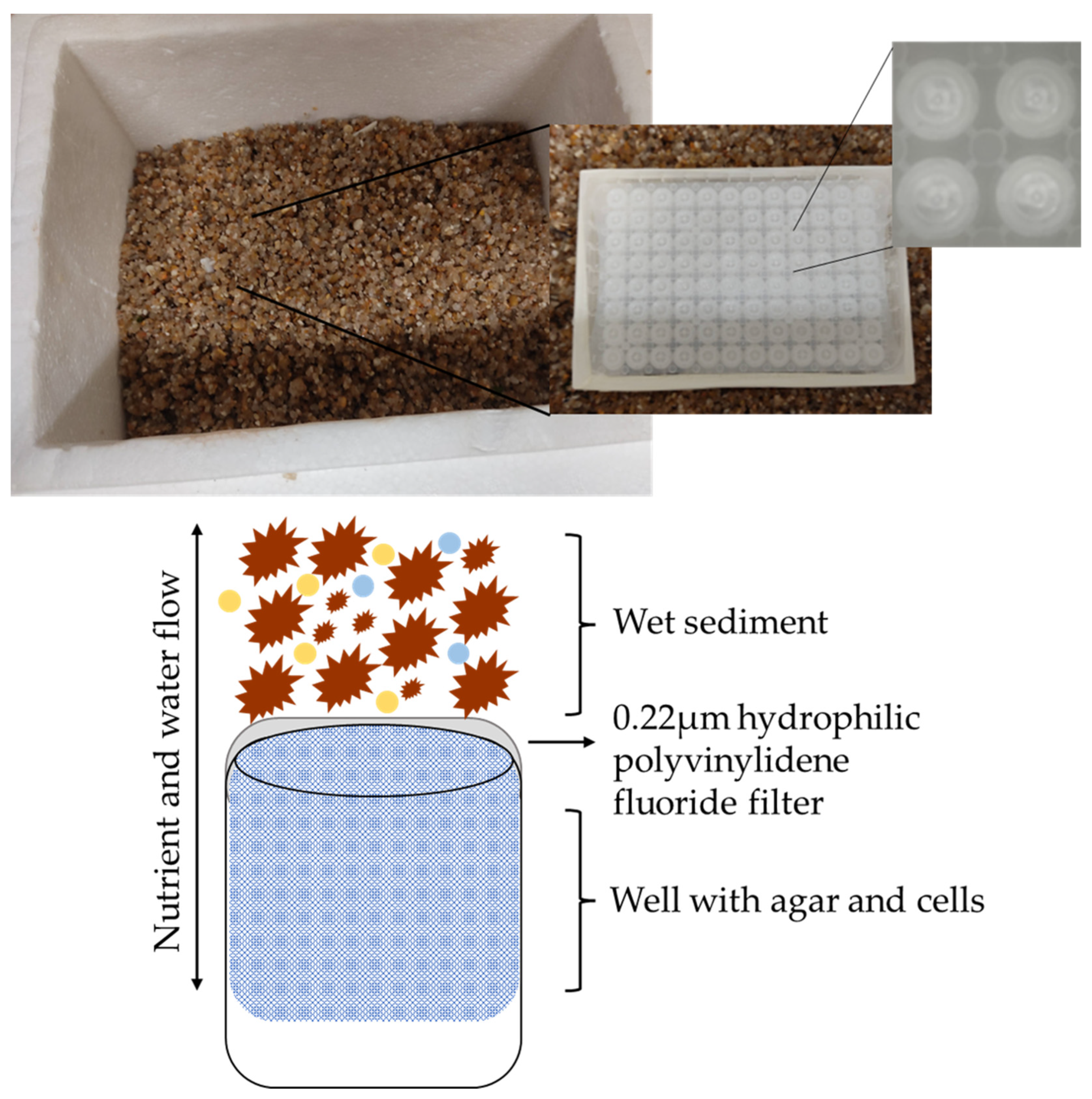
2.1. Sampling and Isolation

Wet sediment samples were retrieved from Memoria beach in Perafita, Matosinhos (41°13′50.35″ N 8°43′16.61″ W), Portugal in March 2020. Approximately 25 g of sediments was collected in a sterile falcon tube and a recipient of 21 × 15 × 14 cm was about half filled with wet sediments from the same beach (Figure 1). The sediment samples were brought to the laboratory refrigerated. To simulate the miniature diffusion chambers present in the iChip culturing system [30], a MultiScreen® 96-Well Filtration Plate was used. The MultiScreen® 96-Well Filtration Plate has a 0.22 µm hydrophilic polyvinylidene fluoride filter on the bottom side.
Around 10 mL of sediment was added to a falcon tube and 10 mL of sterile seawater added to create a cell suspension. Cells were counted using a Thoma counting chamber, and a gelled suspension with 10 cells per 100 μL was prepared. This suspension was a mixture of sterile natural seawater with agar at 0.8% (w/v). One hundred microliters of this suspension were then seeded in each well of the 96-Well Filtration Plate. The plate’s upper lid was closed and sealed with Parafilm® and the whole plate placed in a box, with the filter end of the plate covered with wet sediment from the beach. The box was kept in the dark, at room temperature. After incubation for 60 days, the bacterial growth in the gelled wells was inoculated into medium M600 (0.1% w/v peptone, 0.1% w/v yeast extract, 5 mM Tris-HCl pH 7.5, 0.1% w/v glucose, 0.1% v/v of vitamin solution (0.1 μg mL−1 cyanocobalamin, 2.0 μg mL−1 biotin, 5.0 μg mL−1 thiamine-HCl, 5.0 μg mL−1 Ca-pantothenate, 2.0 μg mL−1 folic acid, 5.0 μg mL−1 riboflavin, and 5.0 μg mL−1 nicotinamide) and 0.2% v/v of Hutner’s solution (99 mg/L FeSO4.7H2O, 12.67 mg/L NaMoO4.2H2O, 3.34 g/L CaCl2.2H2O, 29.70 g/L MgSO4.7H2O, 50 mL/L “44” metals solution, and 10.0 g/L nitrilotriacetic acid; for 100 mL of “44” metals: 250 mg ethylenediaminetetraacetic acid, 1095 mg ZnSO4.7H2O, 500 mg FeSO4.7H2O, 154 mg MnSO4.H2O, 39.2 mg CuSO4.5H2O, 24.8 mg Co(NO3)2.6H2O, and 17.7 mg Na2B4O7.10H2O) [32] and marine agar (MA) (0.5% w/v peptone, 0.1% w/v yeast extract, and 1L aged natural seawater) plates and incubated at 25 °C until colonies were observed and picked. When pure cultures were obtained, they were kept in the isolation medium (M600 or MA) and cryopreserved in M600 or marine broth supplemented with 20% (v/v) glycerol, at −80 °C.

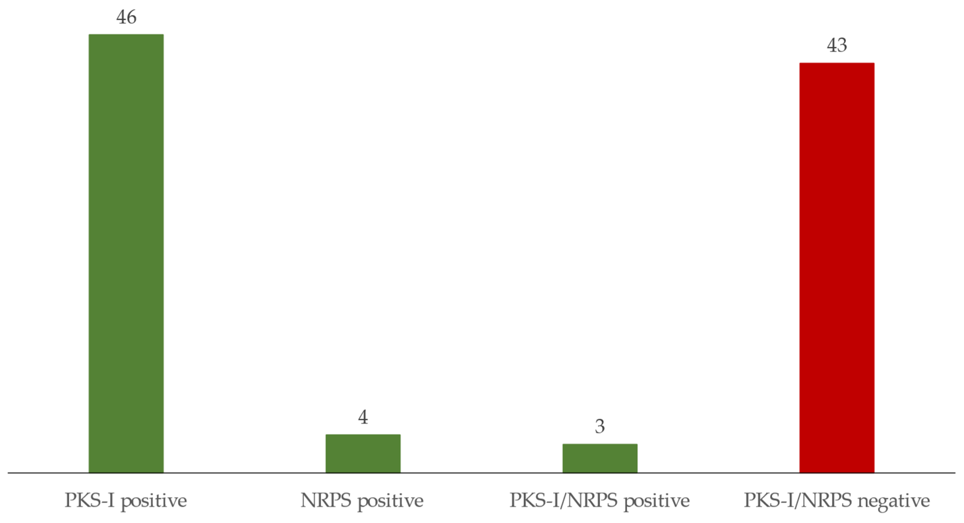
2.2. Identification of the Strain’s Phylogeny and Detection of Secondary-Metabolite-Associated Genes

Pure cultures of the isolated strains were used for genomic DNA extraction. DNA was extracted with the E.Z.N.A.® Bacterial DNA Isolation Kit (Omega Bio-Tek, Norcross, TN, USA) and strains were identified by analysing their 16S rRNA gene. This gene was amplified by polymerase chain reaction (PCR) with the primers 27F and 1492R, following the protocol described by Bondoso et al. [33]. PCR products were purified using the Illustra™ GFX™ PCR DNA and Gel Band Purification Kit and sequenced by Sanger sequencing at Eurofins Genomics. Sequences were analysed using Geneious R11 and the consensus 16S rRNA gene sequences obtained were deposited in the National Center for Biotechnology Information Search (NCBI) database. The phylogeny was inferred using the 16S-based ID tool in the EzBioCloud platform [34]. To further assist in the differentiation of actinobacterial strains, BOX-PCR and ERIC-PCR fingerprinting were performed on strains with the same 16S rRNA gene affiliation, using the primers BOX-A1R and ERIC1R and ERIC2, respectively. PCR protocols were performed as described in Lage et al. [35]. PCR products were separated by electrophoresis in a 1.5% agarose gel Tris–acetate–EDTA buffer, and phylogenetic dendrograms were constructed using PyElph with an unweighted pair group method with arithmetic mean for clustering the distance matrix [36].
For the detection of biosynthetic gene clusters, PCR amplification was performed with the degenerate primers MDPQQRf (5′-RTRGAYCCNCAGCAICG-3′) and HGTGTr (5′-VGTNCCNGTGCCRTG-3′) aimed at the α-keto synthase of PKS-I motif [37] and MTRF2 (5′-GCNGG(C/T)GG(C/T)GCNTA(C/T)GTNCC-3′) and DKF (5′-GTGCCGGTNCCRTGNGYYTC-3′) for core motif-V of NRPS [38]. PCR protocol was performed as described by Graça et al. [39]. The expected amplicon size of PKS-I and NRPS genes was 750 and 1000 bp, respectively.

2.3. Strain Fermentation and Extraction

The isolated Actinomycetota with amplified PKS-I, NRPS or both genes were chosen for antimicrobial screening. Cultures were fermented in plates containing 25 mL of modified M13 medium (M607) [32] for 15 days, at 25 °C, in the dark. The cultures were collected and steeped in 100 mL of ethyl-acetate overnight for bioactive molecule extraction. The suspension in ethyl-acetate was collected and the agar was washed twice with 10 mL ethyl-acetate, which was added to the extraction suspension. The ethyl-acetate was dried to completeness and the extract was dissolved in 500 µL of 20% (v/v) DMSO. Additionally, an unfermented medium contained in a Petri dish was extracted with the same protocol to serve as a medium control. To further explore the antimicrobial activity, bioactive strains were refermented in different media. These included modified M607 and M600, MA, CGY [40] and a 1:10 diluted version of M607 medium, and the cultures were extracted as described above.

2.4. Antimicrobial Screening

Antimicrobial screening of the extracts was performed against E. coli ATCC 25,922 and S. aureus ATCC 29,213 as described previously by Santos et al. [41]. Briefly, single colonies of each target microorganism were incubated in nutrient broth (NB) overnight, at 37 °C and 220 rpm. Cultures were then diluted to obtain an inoculum with 5.0 × 105 cells/mL. Then, 90 µL/well of the corresponding diluted inoculum was mixed with 10 µL of extract in triplicate. Streptomycin and ampicillin at 10 mg/mL were used as positive controls for E. coli ATCC 25,922 and S. aureus ATCC 29213, respectively. Negative internal controls were included. These controls comprise solvent (DMSO) and bacterial growth controls and, additionally, medium controls. Absorbance (at 600 nm) was measured in a Thermo Scientific™ Multiskan™ GO. The percentage of growth inhibition was calculated using the following equation:
% inhibition = 100 100 × ( T FE T 0 E ) ( T FB T 0 B ) ( T FG T 0 G ) ( T FB T 0 B )
where T0 is the absorbance at 0 h, TF is the absorbance at 24 h, E is the extract well, B is blank wells and G is the control growth wells.
Each extract was tested three times on different days with new inocula (n = 3). Extracts were considered to have an inhibitory effect if the target growth was reduced by more than 50% in at least two assays and the average was also above the 50% threshold.

2.5. Dereplication of Extracts

Extract dereplication was performed by liquid chromatography/high-resolution mass spectroscopy (LC/HRMS) with an Agilent 1200 Rapid Resolution HPLC interfaced with a Bruker maXis mass spectrometer. The column used was a Zorbax SB-C8 column (2.1 mm × 30 mm, 3.5 mm particle size), with two solvents used for the mobile phase. Both solvents were composed of water and acetonitrile, in a 90:10 ratio for solvent A, and in a 10:90 ratio for B, and 13 mM ammonium formate and 0.01% trifluoracetic acid were added to both. The mass spectrometer was operated in positive ESI mode. To obtain putative component identification, the retention time and exact mass of the components were compared against Fundación MEDINA’s high-resolution mass spectrometry database. For the components with no matches in the MEDINA database, the predicted molecular formula and exact mass were searched for in the Chapman and Hall Dictionary of Natural Products (DNP) database. If a plausible match was found, considering the exact mass/molecular formula, the producing microorganism, and the target assay, the molecule was reported as a suggested component of the fraction [42].

3. Results

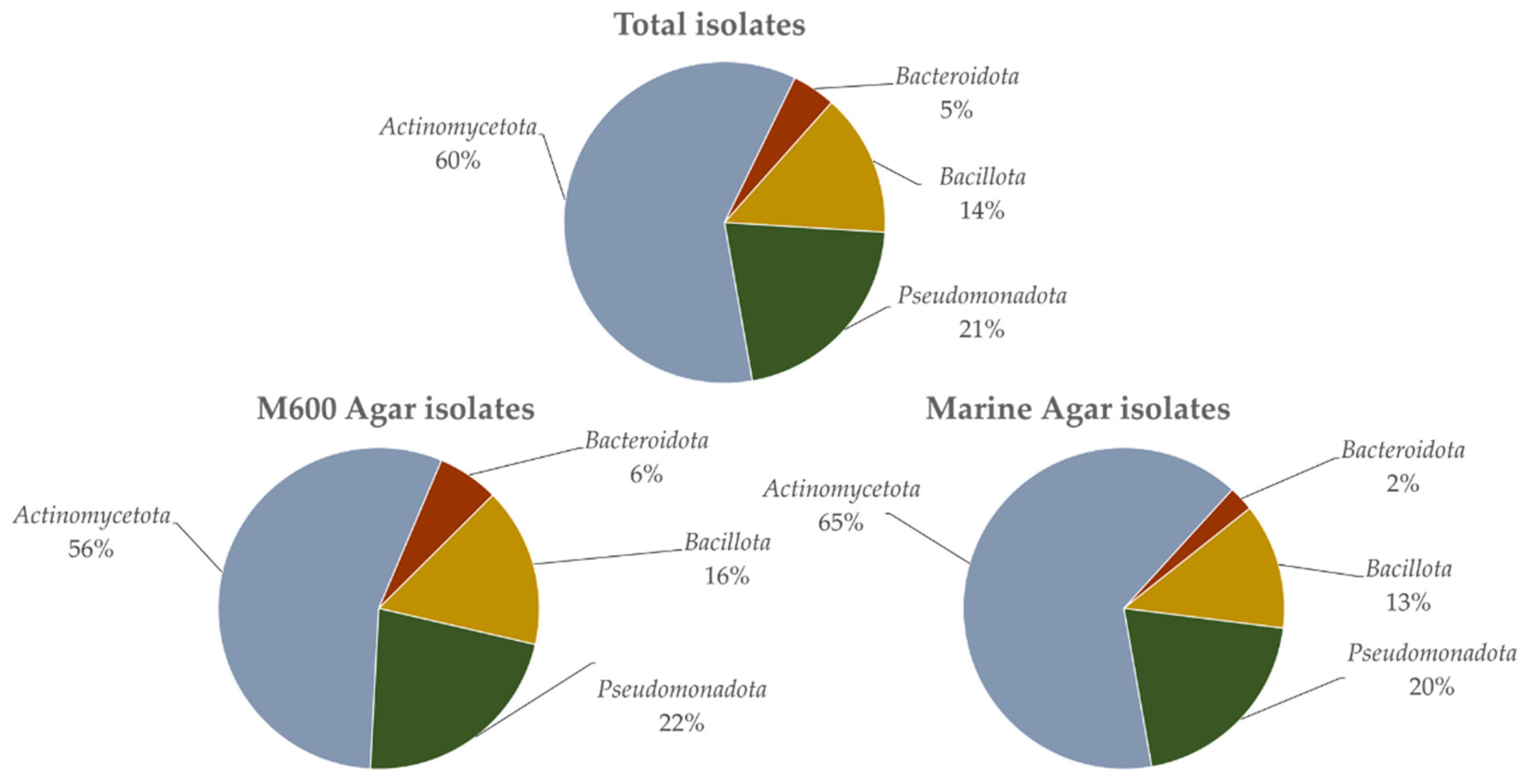
3.1. Isolation and Identification of Strains

Overall, 192 isolates were obtained, but only 158 were identified due to the lack of viability of several isolates. The 158 strains were identified through 16S rRNA gene-based analysis (Table S1). In total, 79 strains were obtained in both M600 and MA. In strains isolated in M600, 45 belonged to Actinomycetota, 17 to Pseudomonadota, 12 to Bacillota and 4 to Bacteroidota. In the strains isolated in marine agar, 51 belonged to Actinomycetota, 16 to Pseudomonadota, 10 to Bacillota and 4 to Bacteroidota. All strains of 16S rRNA gene sequences were deposited in NCBI’s GenBank database with accession numbers MW739985 to MW740142.
In general, Actinomycetota made up the majority of the isolated strains (Figure 2 and Table S1), in a total of 96 isolates corresponding to 60.8% of the sequenced isolates. Furthermore, 33 strains (20.9%) of the isolates belonged to Pseudomonadota, 22 strains (13.9%) to Bacillota, and 7 strains (4.4%) to the Bacteroidota (Figure 2 and Table S2). At the genus level (Table S2), the majority belonged to the genus Streptomyces (48 isolates, 30%), followed by Nocardiopsis (29 isolates, 18.1%), Bacillus (19 isolates, 12.0%) and Pseudoalteromonas (9 isolates, 5.7%). Moreover, at least two or more strains were isolated from genera such as Psychrobacter (6 isolates, 3.75%), Rhodococcus (5 isolates, 3.125%), Arthrobacter (4 isolates, 2.5%), Microbacterium, Aquimarina, Cobetia, Limimaricola, Sulfitobacter, Tritonibacter (each with 3 isolates, 1.9%), and Dietzia, Arenibacter, Alkalihalobacillus and Marinobacter (each with 2 isolates, 1.3%). From the genera Corynebacterium, Kocuria, Micromonospora, Nocardia, Plantibacter, Catalinimonas, Tenacibaculum, Fictibacillus, Henriciella, Phaeobacter, Providencia and Vibrio, only one isolate (0.63% each) was obtained (Figure S1 and Table S2). Only one strain, PMIC_1C1B, showed a similar percentage below the species threshold of 98.70% [43], indicating that it possibly constitutes a novel taxon. When considering the media used for the isolation, 81 viable strains were obtained in medium M600, while in medium MA, 79 strains were retrieved (Figure S1 and Table S2). However, while strains belonging to the four phyla were isolated in both media, in M600, isolates belonged to 21 different genera, while in MA, isolates belonged to 18 genera (Figure S1 and Table S2). Moreover, M600 isolates showed higher diversity at the phylum level: Actinomycetota (10 in M600, 5 in MA) and Bacteroidota (three in M600, two in MA). On the other hand, MA isolates showed greater diversity in the phyla Bacillota (one in M600, three in MA) and Pseudomonadota (seven in M600, eight in MA) (Figure S1 and Table S2). Furthermore, while genera such as Streptomyces were equally isolated in both media, the isolates of Corynebacterium, Dietzia, Kocuria, Microbacterium, Micromonospora, Nocardia, Aquimarina, Limimaricola, Marinobacter and Phaeobacter were only retrieved in medium M600, evidencing the greater diversity obtained in this medium (Figure S1). Yet, genera such as Nocardiopsis appeared to prefer MA (23 isolates compared to the 6 in M600).
The genetic differentiation of closely related strains was assessed using BOX-PCR and ERIC-PCR and a 20% difference threshold for separation between genotypes [35] was applied. The results obtained show a clear grouping of strains into the different species of bacteria (Figures S2–S11). Examples of bacterial strains with similar genotypes include Arthrobacter gandavensis PMIC_1E12B, PMIC_1F10B.1, PMIC_1F10C.1 and PMIC_2F9 (Figure S2) and strains with different genotypes such as Streptomyces ardesiacus PMIC_2D8A, PMIC_2D8B, PMIC_1C8A, PMIC_2C8A and PMIC_2C8B (three identified genotypes) (Figure S8). While in some cases there was a perfect match between the BOX-PCR and the ERIC-PCR fingerprinting, that was not the case in others, such as the strains of Nocardiopsis prasina (BOX-PCR identified two genotypes while ERIC-PCR identified three different genotypes) (Figure S4), Nocardiopsis alba (seven genotypes in the BOX-PCR and two in the ERIC-PCR) (Figure S5), Streptomyces hydrogenans (BOX-PCR did not identify any different genotypes, while three were identified in ERIC-PCR) (Figure S9) and Streptomyces xiamenensis (four identified in BOX-PCR, and two in ERIC-PCR) (Figure S11).

3.2. Antimicrobial Screening

All the strains of Actinomycetota were analysed for the putative presence of genes of NRPS and PKS-I enzyme complexes. Out of the 96 strains screened, a total of 53 strains with either one or both NRPS and PKS-I genes were detected (Table 1 and Figure 3). However, only three were positive for both genes (S. ardesiacus strain PMIC_2C8B, R. erythropolis strain PMIC_1E9B and Rhodococcus coprophilus strain PMIC_2E10) and only four presented the NRPS gene (Table 1 and Figure 3) (Kocuria polaris strain PMIC_1H7A, Streptomyces albidoflavus PMIC_1C12A, S. xiamenensis PMIC_2C2B and Rhodococcus qingshengii strain PMIC_2E9C). The PKS-I gene was the most widespread among the isolated strains, with 23 Nocardiopsis, 18 Streptomyces, 2 Arthrobacter and 1 Nocardia, Plantibacter and Rhodococcus strains, in a total of 46 strains (Table 1 and Figure 3). The 43 strains that did not amplify any of the genes (Table 1 and Figure 3) belonged to the genera Streptomyces (27), Nocardiopsis (6), Microbacterium (3, including the possible novel taxon, PMIC_1C1B), Arthrobacter (2), Dietzia (2), Corynebacterium (1), Micromonospora (1) and Rhodococcus (1).
As it has been shown that oligotrophy induces greater bioactivity in Actinomycetota [44], antimicrobial screening with the strains that putatively amplified at least one PKS-I or one NRPS gene was conducted with the extracts of these strains in medium M607. This medium is comparatively more oligotrophic than the isolation media MA or M600. As the bioactive compounds are products of secondary metabolism, the strains were allowed to grow for 15 days. This guarantees that all attained the stationary growth phase.
Of the 53 tested strains, only 12 strains were bioactive, with values above 50% growth inhibition (Table 1). Ten strains showed activity only against E. coli ATCC 25922, and one was bioactive only against S. aureus ATCC 29,213 (Table 1). Streptomyces flavoviridis strain PMIC_1A8B was bioactive against both targets, and all the bioactive strains putatively possessed genes associated with the PKS-I cluster (Table 1).
The one strain many compounds (OSMAC) [45] approach was performed with the 12 bioactive strains in media of varying levels of oligotrophy, 1:10 M607, M607, M600, MA and CGY (Table 2). Regarding E. coli ATCC 25922, some extracts did not show bioactivity, namely A. gandavensis PMIC_1E12B, N. alba PMIC_1A11B.2 and PMIC_1F6A.3, R. coprophilus PMIC_1E10C and S. albidoflavus PMIC_2C12. For strains N. alba PMIC_2A11B.1, S. flavoviridis PMIC_1A8B, S. griseoflavus PMIC_1D9B and S. hydrogenans PMIC_1I1A, a decrease in the degree of bioactivity compared to the first screening was observed, but the growth inhibition values were near the bioactivity threshold. Increased growth inhibition was verified for N. nova PMIC_1A10B, especially in the extracts from media 1:10 M607 (100%) and CGY (87%), and S. setonii in extracts from media 1:10 M607 (93.1%) and M607 (70.4%). N. alba PMIC_2H2C.2, which in the first screening showed growth inhibition values below the threshold (34.4%), showed an increase in bioactivity inhibition, especially in medium 1:10 M607 (91.7% inhibition). In relation to S. aureus ATCC 29213, 1:10 M607, M607 and M600 extracts from S. flavoviridis PMIC_1A8B showed very high inhibition values (100.0%, 77.2%, and 100.0%, respectively) (Table 2). Furthermore, S. griseoflavus PMIC_1D9B only became bioactive in CGY medium (91.1% inhibition), S. hydrogenans PMIC_1I1A became bioactive in 1:10 M607 medium (73.2% inhibition), and N. alba PMIC_1F6A.3 lost bioactivity.
All of the bioactive extracts were dereplicated (Table S3), and the results from the initial and the OSMAC screenings are presented in Table 3. Extract dereplication is a critical step in natural product discovery, as it putatively detects and identifies known molecules in this early stage of the screening process. Certain compounds were often putatively found in the dereplications. This was the case of the diketopiperazines cyclo(L-Leu-L-Pro) (almost always present) and cyclo(Pro-Tyr), and the derivative of tyramine, N-acetyltyramine, with anti-Vibrio anguillarum [46], antifungal [47] and antimalaria activity [48], respectively. Germicidin A, a pyranone autoregulator of sporulation [49], was putatively present in the extracts from N. alba (PMIC_1A11B.2 and PMIC_2A11B.1). The antibiotic X-14952B, a 20-membered macrolide lactone with broad-spectrum antibacterial properties [50,51], was found in the extracts of N. alba PMIC_2H2C.2 and PMIC_2A11B.1. Also present in the extract of N. alba PMIC_1F6A.3 was cyclo(Tyr-Leu), a diketopiperazine that exhibits antibacterial [52] and antifungal [53] activities. Germicidin G and surugamide A were putatively present in S. albidoflavus PMIC_2C12 and S. hydrogenans PMIC_ I1A. Just like germicidin A, germicidin G also regulates spore germination [54,55], and surugamide A is a cyclic octapeptide with anticancer properties [56]. Ansalactam A, putatively present in S. albidoflavus PMIC_2C12 and S. griseoflavus PMIC_1D9B extracts, is an ansa macrolide with possible antibacterial activity [57] that was first isolated from a marine-sediment-derived bacterium of the genus Streptomyces [58]. 3-acetylamino-N-2-thienylpropanamide, with reported cytotoxic activity [59], was consistently found in S. griseoflavus PMIC_1D9B extracts. The antibiotic MKN-003B was present in all of the extracts from S. hydrogenans PMIC_1I1A. This strain also putatively produces surugamide E, blastomycin, antimycin A13 and antimycin A11. MKN-003B is a lactone with reported antifungal activity [60], although Lacret et al. [61] demonstrated no antifungal activities against Candida albicans MY1055 and Aspergillus fumigatus ATCC46645 and no antibacterial activity against methicillin-resistant S. aureus MB5393 and E. coli MB2884. Surugamide E, like surugamide A, is a cyclic octapeptide with cytotoxic activity, [56] and blastomycin is a polyene with fungicide properties [62]. Antimycins A11 and A13 are macrodiolides with potent and mild antifungal activity, respectively [63]. The extracts of S. setonii strain PMIC_1F12B showed the presence of corynecin I and chloramphenicol. Chloramphenicol is a dichloro-substituted acetamide with broad-spectrum antibacterial activity [64]. Corynecin I is a chloramphenicol-like acyl-nitrophenylpropylamine that also has broad-spectrum antibacterial activity [65,66].
In the various dereplications of the bioactive extracts, several non-identified molecules were found, for which the chemical formulae are provided in Table 3. Some formulae did not match any known compound in the DNP, namely C9H10N2O, C12H25NO3, C14H29NO3, C16H24O3, C20H31NO4S, C22H44O12, C23H13ClO4S2, C24H48O13, C27H53N5O10 and C27H54N10O10. Others matched more than one compound, although they might also constitute new natural products. This is the case of the following formulae: (1) C20H13N3O6 was found to match two compounds, the antibiotics A 33,853 and U 60394, both active against Gram-positive bacteria. (2) C13H22O3 matched with 10 coincidences in the DNP. Of these, 5-(6-hydroxy-7-methyloctyl)-2(5h)-furanone, trihomononactic acid lactone, 7-ethyl-10-hydroxy-7-undecene-3,6-dione, 7-ethyl-9-hydroxy-7-undecene-3,6-dione and 7-ethyl-4-hydroxy-7-undecene-3,6-dione showed no known antimicrobial activity. Acaterin, 3-(1-hydroxyoctyl)-5-methyl-2(5H)-furanone is an inhibitor of acyl-cholesterol acyltransferase [67]. MKN 003C, a butenolide containing a branched side chain very similar to MKN-003B, showed antifouling activity [60]. 5-(6-hydroxy-6-methyloctyl)-2(5H)-furanone showed anti-adenoviral activity, while 5-(7-hydroxy-6-methyloctyl)-2(5H)-furanone and 5-(6-methyloctyl)-2(5H)-furanone, (5S,6′S)-form, 7′S-hydroxy, showed cytotoxic activity [68]. (3) C15H24O3 matched with nine molecules in the DNP (Table 3). 10,15-dihydroxy-4-cadinen-3-one is a sesquiterpene [69], nocapyrone R is an α-pyrone [70], nocardiopyrone A is also an α-pyrone [71], presulficidin C is a triketide pyrone [72], and streptoone C is a ketonic acid [73], all with no known relevant biological importance. Kandenol A is a sesquiterpene with weak antimicrobial activities against Bacillus subtilis and Mycobacterium vaccae [74], nocapyrone H is an α-pyrone with anti-inflammatory effects [75], marinactinone A is a γ-pyrone with cytotoxic activity [76] and photopyrone A is also an α-pyrone that plays a role in quorum-sensing signalling for Photorhabdus luminescens [77]. (4) C15H24O4 matched with 10 different molecules in the DNP. Of these, 1,3,5-bisabolatriene-1,7,11,15-tetrol and 2,6-farnesadiene-1,12-dioic acid, presulficidin D [72], and nocapyrones A, I, J, N and O [78,79] have no known biological importance. Kandenol B and D are sesquiterpenes with weak antimicrobial activities against B. subtilis and M. vaccae [74].

4. Discussion

The beach sand microbiota (micropsammon, the microorganisms inhabiting supratidal and intertidal sand [80]) has been poorly studied [81]. This environment seems to be dominated by Pseudomonadota and Bacteroidota [82,83,84], as also shown in a study of coastal sediments in three sites in Quanzhou Bay, Fujian Province, China, where Huang et al. [85] isolated 1036 bacterial strains, most of which were Pseudomonadota (75.77%), and only 13.32% were Actinomycetota. However, in our study with the modified iChip methodology and without any especial selective treatment, a much higher number of Actinomycetota (60.8%) compared to the lower percentage of Pseudomonadota (20.9%) and Bacteroidota (4.4%) was obtained. This methodology also allowed the obtainment of a great diversity at genus level (29 different genera). About the same number of isolates were obtained in the two media used (Figure 3 and Table S2), and a higher diversity at genus level was obtained in M600 compared with medium MA. These results may indicate that the nutritional formulation of M600 might be more adequate to isolate the diverse microbiological community present in marine sediments. In fact, the composition of M600 includes Hutner’s basal salts [86] and a vitamin solution [87] as supplements that are not available in MA medium. Vitamins are essential for many microorganisms that are unable to synthetize them. Furthermore, some micronutrients, such as Fe2+, which can be used as alternative energy sources [88], limit the growth of microorganisms if they are deficient in the culture medium.
With the iChip in the present study, and considering only the Actinomycetota, 50% belong to the genus Streptomyces and 30.2% to Nocardiopsis. The iChip approach allowed for the isolation of a great quantity of Streptomyces and Nocardiopsis, while only one Micromonospora-related isolate and no Salinispora strains were obtained. Other studies were also focused exclusively on the isolation of Actinomycetota. Ribeiro et al. [89], whose aim was to specifically obtain sporogenous Actinomycetota from marine sediments in Cepães beach, Esposende (north of Memoria beach), used three different media, two of which were selective for Actinomycetota. With these media, out of a total of 52 actinobacterial isolates, 67% belonged to the genus Micromonospora, 17% to Streptomyces and 8% to Arthrobacter. In a study of the coast of Diu Island, Gujarat, India, inter-tidal sediments were dried in a laminar flow hood for 16 h and isolation media were supplemented with cycloheximide and nalidixic acid. In the study, the authors isolated 148 Actinomycetota strains, of which Streptomyces spp. accounted for 61.5%, Micromonospora spp. 15.4%, Nocardiopsis spp. 10.2%, Saccharomonospora spp. 5.1%, Actinomadura spp. 2.6%, Glycomyces spp. 2.6% and Nocardia spp. 2.6% [90]. In another isolation experiment focused also only on Actinomycetota, Prieto-Davo et al. [91] isolated a total of 400 actinobacterial strains from near-shore and off-shore sediments in the Madeira archipelago, the majority belonging to Streptomyces (44.5%), Micromonospora (22.5%), Salinispora (8.5%) and Nocardiopsis (7%). Salinispora spp. are believed to inhabit only tropical and subtropical regions (to which the Madeira archipelago belongs) of the oceans, where thousands of strains have been isolated [92,93,94], while Memoria beach is in a temperate zone and bathed by cold Atlantic waters. Thus, this may be the reason for the absence of isolates from this genus in the present study. Unlike any of the previous isolation attempts, no particular pre-treatments (use of heat, dryness of sediments or antibiotics) were necessary to isolate Actinomycetota in the iChip isolation attempt. Additionally, the agar medium plates did not require any treatment with antibiotics to minimize the development of fast-growing bacteria, as the suspension placed in the wells usually contained only one or two organisms (Table S1). This technique greatly reduced and simplified the isolation process and allowed the obtainment of a high diversity of bacterial genera and species.
The detection of the presence of secondary-metabolite-associated genes, such as PKS-I or NRPS genes, is an important first step to assess the biotechnological potential of the strains under study [6,41]. This molecular analysis showed that about 55% of the tested strains putatively possess one or both genes and allowed our antimicrobial screening to focus on these strains. In the comparatively more oligotrophic M607 medium, 12 strains (five Streptomyces, four Nocardiopsis, one Arthrobacter, one Nocardia and one Rhodococcus) were bioactive.
The literature reveals how relevant these genera are in the discovery of unique and biotechnologically useful organic molecules [6]. Subsequently, the OSMAC screening shows that some extracts no longer showed bioactivity. While the loss of bioactivity is difficult to justify, a factor that may be determinant is the composition of the natural seawater used in media formulations, the composition of which varies over time. Moreover, peptone and yeast extracts may also play a role, as the exact chemical compositions of peptone and yeast extracts are not fully known and may have specific signalling molecules interfering with bioactive compound production [95,96].
Data on the bioactivity of strains from the genus Arthrobacter are still limited. While these strains are present and abundant in many environments, soils and sediments appear to be their preferred habitat. The genus is an industrially relevant bioactive group and a source of glutamic acid, α-ketoglutaric acid and riboflavin [97]. Furthermore, Rojas et al. [40] showed that two strains of Arthrobacter agilis isolated from biological mats from the Antarctic region produced bioactive cyclic thiazolyl peptides that proved to be effective against Gram-positive bacteria, in particular, S. aureus ATCC 29213. Our results show that Arthrobacter gandavensis PMIC_1E12B produced an extract with bioactive properties against E. coli ATCC 25922, which could be due to the presence of two diketopiperazines (Table 2), but not against S. aureus ATCC 29213.
The bacteria from the genus Nocardia are a well-known source of bioactive natural products, and in the review by Dhakal et al. [98], 47 major bioactive molecules isolated from Nocardia spp. were described, showing a vast chemical diversity from which 26 molecules showed antimicrobial activity. Curiously, regarding antibacterial or other antimicrobial activities, none appeared to be associated with strains affiliated with N. nova, a genus that was bioactive in our study. The anti-E. coli ATCC 25,922 bioactivity observed by N. nova PMIC_1A10B may be due to the putative presence of cyclo(L-Leu-L-Pro), cyclo(Pro-Tyr) or six unidentified molecules. The results herein show that this strain putatively possesses a great capacity for producing molecules with antimicrobial properties and may point to the presence of novel natural products.
Regarding the genus Nocardiopsis, reports on the production of natural products with antimicrobial properties are abundant [99,100]. Nocardiopsis-affiliated strains have been shown to produce various compounds, such as the phenazines 1,6-dihydroxyphenazine and 1,6-dihydroxyphenazine 5,10-dioxide, which were isolated from the marine strain OPC-15, affiliated with Nocardiopsis dassonvillei [101]. These phenazines showed antimicrobial activity against Proteus mirabilis and B. subtilis. From a marine-derived N. alba, the thiopeptide TP-1161 was isolated [102]. TP-1161 displayed broad-spectrum antibacterial activity against Staphylococcus haemolyticus, Staphylococcus epidermidis, S. aureus, Streptococcus pneumoniae, Enterococcus faecalis and Enterococcus faecium. The production by N. alba of several diketopiperazines that possess antibacterial properties has been reported [103,104]. For example, albonoursin and methoxylated albonoursin, isolated from N. alba strain ATCC BAA-2165, have been shown to inhibit the growth of the bee pathogen Paenibacillus larvae [105]. Moreover, a novel derivative of 3-acetyl-dimethyl sterol, which was isolated from a marine actinomycetes N. alba MCCB 110, showed promising antibacterial activity against the shrimp aquaculture pathogen Vibrio harveyi [106]. In fact, the dereplication of the extracts of the four strains of N. alba of the present study consistently showed the presence of diketopiperazines (Table 3). Furthermore, the presence of N-acetyltyramine and the antibiotic X-14952B may explain the results obtained for extracts from the CGY and M600 media. Curiously, 1:10 M607 extract showed only the presence of cyclo(L-Leu-L-Pro), yet it proved to be the most bioactive. Several non-identified molecules, such as C15H24O4, C16H24O3 and C27H52N10O10, may also explain the bioactivity due to N. alba strains. The results presented here show the capability of the genus Nocardiopsis to produce relevant bioactive compounds, pointing to the presence of novel natural products.
Although strains PMIC_1A11B.2, PMIC_2A11B.1, PMIC_1F6A.3 and PMIC_2H2C.2 are all 100% phylogenetically affiliated with N. alba DSM 43377, ERIC-PCR and BOX-PCR fingerprinting showed that they belong to two different genotypes. With the ERIC-PCR, PMIC_1A11B.2, PMIC_2A11B.1 and PMIC_1F6A.3 were grouped in one genotype and PMIC_2H2C.2 in another, while in the BOX-PCR, PMIC_1A11B.2 and PMIC_2A11B.1 were grouped in one genotype and PMIC_1F6A.3 and PMIC_2H2C.2 in a second. The bioactive profile of the first extract also showed differences regarding the strains, with strains PMIC_1A11B.2, PMIC_2A11B.1 and PMIC_1F6A.3 showing high inhibition of E. coli ATCC 25922, and strain PMIC_2H2C.2 showing mild anti-S. aureus ATCC 29,213 activity, which, interestingly, coincides with the ERIC-PCR fingerprinting cluster results.
Data regarding the genus Rhodococcus show its biotechnological potential. Rhodococcus-affiliated strains have been shown to produce lariatins, cyclic peptides with selected bioactivity against Mycobacterium tuberculosis [107], and rhodostreptomycins, aminoglycosides with bioactivity against E. coli, S. aureus and B. subtillis [108]. However, no data are available on the bioactive potential of Rhodococcus coprophilus. The strain tested here, R. coprophilus PMIC_1E10C, showed very strong anti-E. coli activity (Table 2), which may be due to the putative presence of the bioactive diketopiperazine cyclo(l-leu-l-pro) and an unidentifiable molecule with the formula C14H29NO3. Further testing with this strain may prove useful in determining additional bioactivities and maybe even novel natural products, as there is a clear gap in the knowledge within this genus, which has prime biotechnological potential.
The genus Streptomyces is the gold standard of natural products and drug discovery research, with more than 6000 different compounds being described with an origin in this genus [109]. Moreover, strains closely associated with the ones obtained with the iChip approach have already been shown to be reservoirs of biotechnologically interesting molecules. For example, S. albidoflavus strain I07A-01824 was shown to produce Antimycin A18, an effective antifungal agent [110]. For S. flavoviridis ATCC 21892, abundant data show its ability to produce natural products with antimicrobial properties [111]. An example is zorbamycin, a glycopeptide antibiotic first reported in 1971 that can rapidly induce the degradation of deoxyribonucleic and ribonucleic acids in B. subtilis and E. coli [112]. S. griseoflavus is also a well-known producer of natural products, such as bicozamycin, an antibacterial agent that inhibits the Rho protein of E. coli [113], and colabomycins A-C, which have anti-Gram-positive bacteria and cytotoxic activities [114]. S. hydrogenans strain KMFA-1 was shown to produce extracts with antifungal properties [115]. Moreover, Lewis et al. [116] recently showed that strains closely related to S. hydrogenans and S. albidoflavus produce the polyene antifungal candicidin. S. setonii strains also have antifungal properties, due to the compound FR109615, an aminocyclopentane isolated from S. setonii strain no. 7562 [117,118].
The bioactivity found in the Streptomyces strains detected in this work may be justified by the action of molecules such as diketopiperazines, N-acetyltyramine, antibiotic MKN-003B, germicidin G, surugamide A and E, blastmycin, antimycins A11 and A13, ansalactam A, corynecin I and chloramphenicol, as well as non-identified molecules such as C13H22O3, C20H13N3O6, C22H44O12, C27H53N5O10 and C23H13ClO4S2. This last molecule seems to be of special interest, as the presence of a halogen (chlorine) and two sulphur atoms may indicate potential bioactivity [119,120]. Overall, these results suggest the relevance of the genus Streptomyces for the discovery of natural products and drug discovery research, as the extracts of the strains studied here showed antibacterial activity and putatively contain bioactive compounds and possible novel natural products.

5. Conclusions

The in situ conditions simulated by the use of an iChip-inspired methodology allowed the recovery of a large number of diverse bacteria from marine sediments, which is demonstrated by the high number of overall domesticated species and the number of genotypes that were identified using PCR fingerprinting techniques. Moreover, a putative novel taxon was isolated (98.48% 16S rRNA gene similarity to the closest known strain). Regarding the bioactive potential of the isolated actinobacterial strains, a high number possessed genes associated with bioactive potential, and 12 strains had at least one inhibitory effect on E. coli ATCC 25,922 and/or S. aureus ATCC 29213. The dereplication of their extracts showed the putative presence of several known bioactive molecules, and of some with unknown identity that might constitute new natural antibiotics. In conclusion, the highly diverse Actinomycetota isolated in this study have great biosynthetic potential and, thus, may prove to be useful biotechnological tools.

Supplementary Materials

The following supporting information can be downloaded at: https://www.mdpi.com/article/10.3390/microorganisms10071471/s1, Figure S1: Distribution of the genera of the obtained strains per medium used. Figure S2: (A) ERIC-PCR fingerprinting performed on strains belonging to Arthorbacter gandavensis, Dietzia maris, Rhodococcus coprophilus and Rhodococcus erythropolis. (B) BOX-PCR fingerprinting performed on strains belonging to A. gandavensis, D. maris, R. coprophilus and R. erythropolis; Figure S3: (A) ERIC-PCR fingerprinting performed on strains belonging to Streptomyces griseoflavus, Streptomyces ambofaciens and Nocardiopsis umidischolae. (B) BOX-PCR fingerprinting performed on strains belonging to S. griseoflavus, S. ambofaciens and N. umidischolae; Figure S4: (A) ERIC-PCR fingerprinting performed on strains belonging to Nocardiopsis prasina. (B) BOX-PCR fingerprinting performed on strains belonging to N. prasina; Figure S5: (A) ERIC-PCR fingerprinting performed on strains belonging to Nocardiopsis alba. (B) BOX-PCR fingerprinting performed on strains belonging to N. alba; Figure S6: (A) ERIC-PCR fingerprinting performed on strains belonging to Streptomyces albidoflavus. (B) BOX-PCR fingerprinting performed on strains belonging to S. albidoflavus; Figure S7: (A) ERIC-PCR fingerprinting performed on strains belonging to Streptomyces albogriseolus. (B) BOX-PCR fingerprinting performed on strains belonging to S. albogriseolus; Figure S8: (A) ERIC-PCR fingerprinting performed on strains belonging to Streptomyces ardesiacus. (B) BOX-PCR fingerprinting performed on strains belonging to S. ardesiacus; Figure S9: (A) ERIC-PCR fingerprinting performed on strains belonging to Streptomyces hydrogenans. (B) BOX-PCR fingerprinting performed on strains belonging to S. hydrogenans; Figure S10: (A) ERIC-PCR fingerprinting performed on strains belonging to Streptomyces setonii. (B) BOX-PCR fingerprinting performed on strains belonging to S. setonii; Figure S11: (A) ERIC-PCR fingerprinting performed on strains belonging to Streptomyces xiamenensis. (B) BOX-PCR fingerprinting performed on strains belonging to S. xiamenensis. Table S1: Isolated strains’ ID, affiliation, phyla and 16S rRNA gene GenBank accession number; Table S2: Total and per medium distribution of isolated strains by genera; Table S3: All putatively identified and non-identified molecules present in the extracts.

Author Contributions

Experiment design was developed by J.D.N.d.S., F.V., F.R. and O.M.L. Isolation and identification work was performed by J.D.N.d.S. Bioactivity screening work was performed by J.D.N.d.S. and S.A.J. Dereplication work was performed by J.M. and F.R. Manuscript writing was performed by J.D.N.d.S. and O.M.L. and manuscript revision by J.D.N.d.S., S.A.J., J.M., F.V., F.R. and O.M.L. All authors have read and agreed to the published version of the manuscript.

Funding

This research was partially supported by the FCT—Foundation for Science and Technology through the projects UIDB/04423/2020 and UIDP/04423/2020. J.D.N. Santos is supported by an FCT doctoral grant (SFRH/BD/145576/2019). F. Vicente, J. Martín and F. Reyes are supported by Fundación MEDINA.

Institutional Review Board Statement

Not applicable.

Informed Consent Statement

Not applicable.

Data Availability Statement

Not applicable.

Acknowledgments

The authors thank Inês Vitorino for the help in collecting the samples.

Conflicts of Interest

The authors declare no conflict of interest.

References

  1. NOAA (Ed.) Ocean Literacy: The Essential Principles and Fundamental Concepts of Ocean Sciences for Learners of All Ages; NOAA: Washington, DC, USA, 2020.
  2. Bar-On, Y.M.; Phillips, R.; Milo, R. The biomass distribution on Earth. Proc. Natl. Acad Sci. USA 2018, 115, 6506–6511. [Google Scholar] [CrossRef] [Green Version]
  3. NOAA. How Much of the Ocean Have We Explored? Available online: https://oceanservice.noaa.gov/facts/exploration.html (accessed on 7 March 2021).
  4. Hunter-Cevera, J.; Karl, D.; Buckley, M. Marine Microbial Diversity: The Key to Earth’s Habitability. In Proceedings of the American Academy of Microbiology, San Francisco, CA, USA, 8–10 April 2005. [Google Scholar]
  5. Haefner, B. Drugs from the deep: Marine natural products as drug candidates. Drug Discov. Today 2003, 8, 536–544. [Google Scholar] [CrossRef]
  6. Santos, J.D.; Vitorino, I.; Reyes, F.; Vicente, F.; Lage, O.M. From ocean to medicine: Pharmaceutical applications of metabolites from marine bacteria. Antibiotics 2020, 9, 455. [Google Scholar] [CrossRef] [PubMed]
  7. Armelagos, G.J.; Kolbacher, K.; Collins, K.; Cook, J.; Krafeld-Daugherty, M. Tetracycline consumption in prehistory. In Tetracyclines in Biology, Chemistry and Medicine; Nelson, M., Hillen, W., Greenwald, R.A., Eds.; Birkhäuser Basel: Basel, Switzerland, 2001; pp. 219–236. [Google Scholar] [CrossRef]
  8. Montinari, M.R.; Minelli, S.; De Caterina, R. The first 3500 years of aspirin history from its roots—A concise summary. Vasc. Pharm. 2019, 113, 1–8. [Google Scholar] [CrossRef]
  9. Carroll, A.R.; Copp, B.R.; Davis, R.A.; Keyzers, R.A.; Prinsep, M.R. Marine natural products. Nat. Prod. Rep. 2020, 37, 175–223. [Google Scholar] [CrossRef]
  10. Reynolds, K.A.; Luhavaya, H.; Li, J.; Dahesh, S.; Nizet, V.; Yamanaka, K.; Moore, B.S. Isolation and structure elucidation of lipopeptide antibiotic taromycin B from the activated taromycin biosynthetic gene cluster. J. Antibiot. 2018, 71, 333–338. [Google Scholar] [CrossRef]
  11. Anjum, K.; Sadiq, I.; Chen, L.; Kaleem, S.; Li, X.-C.; Zhang, Z.; Lian, X.-Y. Novel antifungal janthinopolyenemycins A and B from a co-culture of marine-associated Janthinobacterium spp. ZZ145 and ZZ148. Tetrahedron Lett. 2018, 59, 3490–3494. [Google Scholar] [CrossRef]
  12. Sudek, S.; Lopanik, N.B.; Waggoner, L.E.; Hildebrand, M.; Anderson, C.; Liu, H.; Patel, A.; Sherman, D.H.; Haygood, M.G. Identification of the putative bryostatin polyketide synthase gene cluster from “Candidatus Endobugula sertula”, the uncultivated microbial symbiont of the marine bryozoan. Bugula Neritina J. Nat. Prod. 2007, 70, 67–74. [Google Scholar] [CrossRef]
  13. Dyshlovoy, S.A.; Honecker, F. Marine compounds and cancer: The first two decades of XXI century. Mar. Drugs 2019, 18, 20. [Google Scholar] [CrossRef] [Green Version]
  14. Dutta, S.; Whicher, J.R.; Hansen, D.A.; Hale, W.A.; Chemler, J.A.; Congdon, G.R.; Narayan, A.R.; Hakansson, K.; Sherman, D.H.; Smith, J.L.; et al. Structure of a modular polyketide synthase. Nature 2014, 510, 512–517. [Google Scholar] [CrossRef] [Green Version]
  15. Strieker, M.; Tanovic, A.; Marahiel, M.A. Nonribosomal peptide synthetases: Structures and dynamics. Curr. Opin. Struct. Biol. 2010, 20, 234–240. [Google Scholar] [CrossRef] [PubMed]
  16. Bentley, S.D.; Chater, K.F.; Cerdeño-Tárraga, A.M.; Challis, G.L.; Thomson, N.R.; James, K.D.; Harris, D.E.; Quail, M.A.; Kieser, H.; Harper, D.; et al. Complete genome sequence of the model actinomycete Streptomyces coelicolor A3(2). Nature 2002, 417, 141–147. [Google Scholar] [CrossRef] [PubMed]
  17. Lacombe-Harvey, M.-È.; Brzezinski, R.; Beaulieu, C. Chitinolytic functions in actinobacteria: Ecology, enzymes, and evolution. Appl. Microbiol. Biotechnol. 2018, 102, 7219–7230. [Google Scholar] [CrossRef] [PubMed] [Green Version]
  18. Jensen, P.R.; Lauro, F.M. An assessment of actinobacterial diversity in the marine environment. Antonie Van Leeuwenhoek 2008, 94, 51–62. [Google Scholar] [CrossRef] [Green Version]
  19. Goodfellow, M.; Williams, S.T. Ecology of actinomycetes. Annu. Rev. Microbiol. 1983, 37, 189–216. [Google Scholar] [CrossRef]
  20. Maldonado, L.A.; Fenical, W.; Jensen, P.R.; Kauffman, C.A.; Mincer, T.J.; Ward, A.C.; Bull, A.T.; Goodfellow, M. Salinispora arenicola gen. nov., sp. nov. and Salinispora tropica sp. nov., obligate marine actinomycetes belonging to the family Micromonosporaceae. Int. J. Syst. Evol. Microbiol. 2005, 55, 1759–1766. [Google Scholar] [CrossRef] [Green Version]
  21. Kwon, H.C.; Kauffman, C.A.; Jensen, P.R.; Fenical, W. Marinomycins A-D, antitumor-antibiotics of a new structure class from a marine actinomycete of the recently discovered genus “Marinispora”. J. Am. Chem. Soc. 2006, 128, 1622–1632. [Google Scholar] [CrossRef]
  22. Azman, A.S.; Othman, I.; Velu, S.S.; Chan, K.G.; Lee, L.H. Mangrove rare actinobacteria: Taxonomy, natural compound, and discovery of bioactivity. Front. Microbiol. 2015, 6, 856. [Google Scholar] [CrossRef]
  23. Girão, M.; Ribeiro, I.; Ribeiro, T.; Azevedo, I.C.; Pereira, F.; Urbatzka, R.; Leão, P.N.; Carvalho, M.F. Actinobacteria isolated from Laminaria ochroleuca: A source of new bioactive compounds. Front. Microbiol. 2019, 10, 683. [Google Scholar] [CrossRef] [Green Version]
  24. Kamjam, M.; Sivalingam, P.; Deng, Z.; Hong, K. Deep sea actinomycetes and their secondary metabolites. Front. Microbiol. 2017, 8, 760. [Google Scholar] [CrossRef] [Green Version]
  25. Chen, P.; Zhang, L.; Guo, X.; Dai, X.; Liu, L.; Xi, L.; Wang, J.; Song, L.; Wang, Y.; Zhu, Y.; et al. Diversity, biogeography, and biodegradation potential of Actinobacteria in the deep-sea sediments along the southwest Indian ridge. Front. Microbiol. 2016, 7, 1340. [Google Scholar] [CrossRef] [PubMed] [Green Version]
  26. Stach, E.M.; Bull, A.T. Estimating and comparing the diversity of marine Actinobacteria. Antonie Van Leeuwenhoek 2005, 87, 3–9. [Google Scholar] [CrossRef] [PubMed]
  27. Mincer, T.J.; Fenical, W.; Jensen, P.R. Culture-dependent and culture-independent diversity within the obligate marine actinomycete genus Salinispora. Appl. Environ. Microbiol. 2005, 71, 7019–7028. [Google Scholar] [CrossRef] [PubMed] [Green Version]
  28. Manivasagan, P.; Venkatesan, J.; Kim, S.-K. Introduction to marine Actinobacteria. In Marine Microbiology; Kim, S.-K., Ed.; Wiley-VCH Verlag GmbH & Co. KGaA: Weinheim, Germany, 2013; pp. 1–19. [Google Scholar] [CrossRef]
  29. Rinke, C.; Schwientek, P.; Sczyrba, A.; Ivanova, N.N.; Anderson, I.J.; Cheng, J.F.; Darling, A.; Malfatti, S.; Swan, B.K.; Gies, E.A.; et al. Insights into the phylogeny and coding potential of microbial dark matter. Nature 2013, 499, 431–437. [Google Scholar] [CrossRef] [Green Version]
  30. Nichols, D.; Cahoon, N.; Trakhtenberg, E.M.; Pham, L.; Mehta, A.; Belanger, A.; Kanigan, T.; Lewis, K.; Epstein, S.S. Use of ichip for high-throughput in situ cultivation of “uncultivable” microbial species. Appl. Environ. Microbiol. 2010, 76, 2445–2450. [Google Scholar] [CrossRef] [Green Version]
  31. Berdy, B.; Spoering, A.L.; Ling, L.L.; Epstein, S.S. In situ cultivation of previously uncultivable microorganisms using the ichip. Nat. Protoc. 2017, 12, 2232–2242. [Google Scholar] [CrossRef]
  32. Lage, O.M.; Bondoso, J. Planctomycetes diversity associated with macroalgae. FEMS Microbiol. Ecol. 2011, 78, 366–375. [Google Scholar] [CrossRef] [Green Version]
  33. Bondoso, J.; Albuquerque, L.; Nobre, M.F.; Lobo-da-Cunha, A.; da Costa, M.S.; Lage, O.M. Aquisphaera giovannonii gen. nov., sp. nov., a planctomycete isolated from a freshwater aquarium. Int. J. Syst. Evol. Microbiol. 2011, 61, 2844–2850. [Google Scholar] [CrossRef]
  34. Yoon, S.H.; Ha, S.M.; Kwon, S.; Lim, J.; Kim, Y.; Seo, H.; Chun, J. Introducing EzBioCloud: A taxonomically united database of 16S rRNA gene sequences and whole-genome assemblies. Int. J. Syst. Evol. Microbiol. 2017, 67, 1613–1617. [Google Scholar] [CrossRef]
  35. Lage, O.M.; Bondoso, J.; Viana, F. Isolation and characterization of Planctomycetes from the sediments of a fish farm wastewater treatment tank. Arch. Microbiol. 2012, 194, 879–885. [Google Scholar] [CrossRef]
  36. Pavel, A.B.; Vasile, C.I. PyElph—A software tool for gel images analysis and phylogenetics. BMC Bioinform. 2012, 13, 9. [Google Scholar] [CrossRef] [PubMed] [Green Version]
  37. Kim, T.K.; Garson, M.J.; Fuerst, J.A. Marine actinomycetes related to the ‘Salinospora’ group from the Great Barrier Reef sponge Pseudoceratina clavata. Environ. Microbiol. 2005, 7, 509–518. [Google Scholar] [CrossRef]
  38. Neilan, B.A.; Dittmann, E.; Rouhiainen, L.; Bass, R.A.; Schaub, V.; Sivonen, K.; Börner, T. Nonribosomal peptide synthesis and toxigenicity of Cyanobacteria. J. Bacteriol. 1999, 181, 4089. [Google Scholar] [CrossRef] [PubMed] [Green Version]
  39. Graça, A.P.; Calisto, R.; Lage, O.M. Planctomycetes as novel source of bioactive molecules. Front. Microbiol. 2016, 7, 1241. [Google Scholar] [CrossRef] [PubMed] [Green Version]
  40. Rojas, J.L.; Martín, J.; Tormo, J.R.; Vicente, F.; Brunati, M.; Ciciliato, I.; Losi, D.; Van Trappen, S.; Mergaert, J.; Swings, J.; et al. Bacterial diversity from benthic mats of Antarctic lakes as a source of new bioactive metabolites. Mar. Genom. 2009, 2, 33–41. [Google Scholar] [CrossRef]
  41. Santos, J.D.; Vitorino, I.; De la Cruz, M.; Díaz, C.; Cautain, B.; Annang, F.; Pérez-Moreno, G.; Gonzalez Martinez, I.; Tormo, J.R.; Martín, J.M.; et al. Bioactivities and extract dereplication of actinomycetales isolated from marine sponges. Front. Microbiol. 2019, 10, 727. [Google Scholar] [CrossRef]
  42. Perez-Victoria, I.; Martin, J.; Reyes, F. Combined LC/UV/MS and NMR strategies for the dereplication of marine natural products. Planta Med. 2016, 82, 857–871. [Google Scholar] [CrossRef] [Green Version]
  43. Yarza, P.; Yilmaz, P.; Pruesse, E.; Glockner, F.O.; Ludwig, W.; Schleifer, K.H.; Whitman, W.B.; Euzeby, J.; Amann, R.; Rossello-Mora, R. Uniting the classification of cultured and uncultured bacteria and archaea using 16S rRNA gene sequences. Nat. Rev. Microbiol. 2014, 12, 635–645. [Google Scholar] [CrossRef]
  44. Wang, D.S.; Xue, Q.H.; Ma, Y.Y.; Wei, X.L.; Chen, J.; He, F. Oligotrophy is helpful for the isolation of bioactive actinomycetes. Indian J. Microbiol. 2014, 54, 178–184. [Google Scholar] [CrossRef] [Green Version]
  45. Romano, S.; Jackson, S.A.; Patry, S.; Dobson, A.D.W. Extending the “One Strain Many Compounds” (OSMAC) principle to marine microorganisms. Mar. Drugs 2018, 16, 244. [Google Scholar] [CrossRef] [Green Version]
  46. Fdhila, F.; Vazquez, V.; Sanchez, J.L.; Riguera, R. dd-diketopiperazines: Antibiotics active against Vibrio anguillarum isolated from marine bacteria associated with cultures of Pecten maximus. J. Nat. Prod. 2003, 66, 1299–1301. [Google Scholar] [CrossRef] [PubMed]
  47. Cimmino, A.; Puopolo, G.; Perazzolli, M.; Andolfi, A.; Melck, D.; Pertot, I.; Evidente, A. Cyclo(L-PRO-L-TYR), The fungicide isolated from Lysobacter capsici AZ78: A structure–activity relationship study. Chem. Heterocycl. Compd. 2014, 50, 290–295. [Google Scholar] [CrossRef]
  48. Plouffe, D.; Brinker, A.; McNamara, C.; Henson, K.; Kato, N.; Kuhen, K.; Nagle, A.; Adrian, F.; Matzen, J.T.; Anderson, P.; et al. In silico activity profiling reveals the mechanism of action of antimalarials discovered in a high-throughput screen. Proc. Natl. Acad Sci. USA 2008, 105, 9059–9064. [Google Scholar] [CrossRef] [PubMed] [Green Version]
  49. Petersen, F.; Zahner, H.; Metzger, J.W.; Freund, S.; Hummel, R.P. Germicidin, an autoregulative germination inhibitor of Streptomyces viridochromogenes NRRL B-1551. J. Antibiot 1993, 46, 1126–1138. [Google Scholar] [CrossRef] [Green Version]
  50. Omura, S.; Nakagawa, A.; Imamura, N.; Kushida, K.; Liu, C.M.; Sello, L.H.; Westley, J.W. Structure of a new macrolide antibiotic, X-14952B. J. Antibiot 1985, 38, 674–676. [Google Scholar] [CrossRef] [PubMed]
  51. Li, N.; Chen, S.; Yan, Z.; Han, J.; Ta, Y.; Pu, T.; Wang, Y. Antimicrobial activity and identification of the biosynthetic gene cluster of X-14952B from Streptomyces sp. 135. Front. Microbiol. 2021, 12, 703093. [Google Scholar] [CrossRef]
  52. Scopel, M.; Abraham, W.R.; Henriques, A.T.; Macedo, A.J. Dipeptide cis-cyclo(Leucyl-Tyrosyl) produced by sponge associated Penicillium sp. F37 inhibits biofilm formation of the pathogenic Staphylococcus epidermidis. Bioorg. Med. Chem. Lett. 2013, 23, 624–626. [Google Scholar] [CrossRef]
  53. Kumar, N.; Gorantla, J.N.; Mohandas, C.; Nambisan, B.; Lankalapalli, R.S. Isolation and antifungal properties of cyclo(D-Tyr-L-Leu) diketopiperazine isolated from Bacillus sp. associated with rhabditid entomopathogenic nematode. Nat. Prod. Res. 2013, 27, 2168–2172. [Google Scholar] [CrossRef]
  54. Zhao, T.; Chang, Y.; Zhu, T.; Li, J.; Gu, Q.; Li, D.; Che, Q.; Zhang, G. alpha-Pyrone derivatives with cyto-protective activity from two Takla Makan desert soil derived actinomycete Nocardiopsis strains recovered in seawater based medium. Nat. Prod. Res. 2019, 33, 2498–2506. [Google Scholar] [CrossRef]
  55. Xu, Z.; Ding, L.; Hertweck, C. A branched extender unit shared between two orthogonal polyketide pathways in an endophyte. Angew. Chem. Int. Ed. Engl. 2011, 50, 4667–4670. [Google Scholar] [CrossRef]
  56. Takada, K.; Ninomiya, A.; Naruse, M.; Sun, Y.; Miyazaki, M.; Nogi, Y.; Okada, S.; Matsunaga, S. Surugamides A-E, cyclic octapeptides with four D-amino acid residues, from a marine Streptomyces sp.: LC-MS-aided inspection of partial hydrolysates for the distinction of D- and L-amino acid residues in the sequence. J. Org. Chem. 2013, 78, 6746–6750. [Google Scholar] [CrossRef] [PubMed]
  57. Le, T.C.; Yang, I.; Yoon, Y.J.; Nam, S.J.; Fenical, W. Ansalactams B-D illustrate further biosynthetic plasticity within the ansamycin pathway. Org. Lett. 2016, 18, 2256–2259. [Google Scholar] [CrossRef] [PubMed]
  58. Wilson, M.C.; Nam, S.J.; Gulder, T.A.; Kauffman, C.A.; Jensen, P.R.; Fenical, W.; Moore, B.S. Structure and biosynthesis of the marine streptomycete ansamycin ansalactam A and its distinctive branched chain polyketide extender unit. J. Am. Chem. Soc. 2011, 133, 1971–1977. [Google Scholar] [CrossRef] [PubMed] [Green Version]
  59. Ye, X.; Chai, W.; Lian, X.Y.; Zhang, Z. Novel propanamide analogue and antiproliferative diketopiperazines from mangrove Streptomyces sp. Q24. Nat. Prod. Res. 2017, 31, 1390–1396. [Google Scholar] [CrossRef]
  60. Cho, K.W.; Lee, H.S.; Rho, J.R.; Kim, T.S.; Mo, S.J.; Shin, J. New lactone-containing metabolites from a marine-derived bacterium of the genus Streptomyces. J. Nat. Prod. 2001, 64, 664–667. [Google Scholar] [CrossRef] [PubMed]
  61. Lacret, R.; Oves-Costales, D.; Gomez, C.; Diaz, C.; de la Cruz, M.; Perez-Victoria, I.; Vicente, F.; Genilloud, O.; Reyes, F. New ikarugamycin derivatives with antifungal and antibacterial properties from Streptomyces zhaozhouensis. Mar. Drugs 2014, 13, 128–140. [Google Scholar] [CrossRef] [Green Version]
  62. Watanabe, K.; Tanaka, T.; Fukuhara, K.; Miyairi, N.; Yonehara, H.; Umezawa, H. Blastmycin, a new antibiotic from Streptomyces sp. J. Antibiot 1957, 10, 39–45. [Google Scholar]
  63. Hosotani, N.; Kumagai, K.; Nakagawa, H.; Shimatani, T.; Saji, I. Antimycins A10 approximately A16, seven new antimycin antibiotics produced by Streptomyces spp. SPA-10191 and SPA-8893. J. Antibiot. 2005, 58, 460–467. [Google Scholar] [CrossRef]
  64. Ambrose, P.J. Clinical pharmacokinetics of chloramphenicol and chloramphenicol succinate. Clin. Pharm. 1984, 9, 222–238. [Google Scholar] [CrossRef]
  65. Nakano, H.; Tomita, F.; Yamaguchi, K.; Nagashima, M.; Suzuki, T. Corynecin (chloramphenicol analogs) fermentation studies: Selective production of Corynecin I by Corynebacterium hydrocarboclastus grown on acetate. Biotechnol. Bioeng. 1977, 19, 1009–1018. [Google Scholar] [CrossRef]
  66. Undabarrena, A.; Valencia, R.; Cumsille, A.; Zamora-Leiva, L.; Castro-Nallar, E.; Barona-Gomez, F.; Camara, B. Rhodococcus comparative genomics reveals a phylogenomic-dependent non-ribosomal peptide synthetase distribution: Insights into biosynthetic gene cluster connection to an orphan metabolite. Microb. Genom. 2021, 7, 000621. [Google Scholar] [CrossRef]
  67. Naganuma, S.; Sakai, K.; Hasumi, K.; Endo, A. Acaterin, a novel inhibitor of acyl-CoA: Cholesterol acyltransferase produced by Pseudomonas sp. A92. J. Antibiot 1992, 45, 1216–1221. [Google Scholar] [CrossRef] [Green Version]
  68. Strand, M.; Carlsson, M.; Uvell, H.; Islam, K.; Edlund, K.; Cullman, I.; Altermark, B.; Mei, Y.F.; Elofsson, M.; Willassen, N.P.; et al. Isolation and characterization of anti-adenoviral secondary metabolites from marine actinobacteria. Mar. Drugs 2014, 12, 799–821. [Google Scholar] [CrossRef] [PubMed] [Green Version]
  69. Ding, L.; Pfoh, R.; Rühl, S.; Qin, S.; Laatsch, H. T-Muurolol sesquiterpenes from the marine Streptomyces sp. M491 and revision of the configuration of previously reported amorphanes. J. Nat. Prod. 2009, 72, 99–101. [Google Scholar] [CrossRef] [PubMed]
  70. Kim, Y.; Ogura, H.; Akasaka, K.; Oikawa, T.; Matsuura, N.; Imada, C.; Yasuda, H.; Igarashi, Y. Nocapyrones: Alpha- and gamma-pyrones from a marine-derived Nocardiopsis sp. Mar. Drugs 2014, 12, 4110–4125. [Google Scholar] [CrossRef] [PubMed] [Green Version]
  71. Raju, R.; Piggott, A.M.; Quezada, M.; Capon, R.J. Nocardiopsins C and D and nocardiopyrone A: New polyketides from an Australian marine-derived Nocardiopsis sp. Tetrahedron 2013, 69, 692–698. [Google Scholar] [CrossRef]
  72. Tang, X.; Eitel, K.; Kaysser, L.; Kulik, A.; Grond, S.; Gust, B. A two-step sulfation in antibiotic biosynthesis requires a type III polyketide synthase. Nat. Chem. Biol. 2013, 9, 610–615. [Google Scholar] [CrossRef]
  73. Tian, H.; Shafi, J.; Ji, M.; Bi, Y.; Yu, Z. Antimicrobial metabolites from Streptomyces sp. SN0280. J. Nat. Prod. 2017, 80, 1015–1019. [Google Scholar] [CrossRef]
  74. Ding, L.; Maier, A.; Fiebig, H.-H.; Lin, W.-H.; Peschel, G.; Hertweck, C. Kandenols A–E, eudesmenes from an endophytic Streptomyces sp. of the mangrove tree Kandelia candel. J. Nat. Prod. 2012, 75, 2223–2227. [Google Scholar] [CrossRef]
  75. Kim, M.C.; Kwon, O.-W.; Park, J.-S.; Kim, S.Y.; Kwon, H.C. Nocapyrones H–J, 3,6-disubstituted α-pyrones from the marine actinomycete Nocardiopsis sp. KMF-001. Chem. Pharm. Bull. 2013, 61, 511–515. [Google Scholar] [CrossRef] [Green Version]
  76. Wang, F.; Tian, X.; Huang, C.; Li, Q.; Zhang, S. Marinactinones A–C, new γ-pyrones from marine actinomycete Marinactinospora thermotolerans SCSIO 00606. J. Antibiot. 2011, 64, 189–192. [Google Scholar] [CrossRef] [PubMed] [Green Version]
  77. Brachmann, A.O.; Brameyer, S.; Kresovic, D.; Hitkova, I.; Kopp, Y.; Manske, C.; Schubert, K.; Bode, H.B.; Heermann, R. Pyrones as bacterial signaling molecules. Nat. Chem. Biol. 2013, 9, 573–578. [Google Scholar] [CrossRef] [PubMed]
  78. Lin, Z.; Torres, J.P.; Ammon, M.A.; Marett, L.; Teichert, R.W.; Reilly, C.A.; Kwan, J.C.; Hughen, R.W.; Flores, M.; Tianero, M.D.; et al. A bacterial source for mollusk pyrone polyketides. Chem. Biol. 2013, 20, 73–81. [Google Scholar] [CrossRef] [PubMed] [Green Version]
  79. Schneemann, I.; Ohlendorf, B.; Zinecker, H.; Nagel, K.; Wiese, J.; Imhoff, J.F. Nocapyrones A−D, γ-pyrones from a Nocardiopsis strain isolated from the marine sponge Halichondria panicea. J. Nat. Prod. 2010, 73, 1444–1447. [Google Scholar] [CrossRef] [Green Version]
  80. Neel, J.K. A Limnological investigation of the psammon in douglas lake, michigan, with especial reference to shoal and shoreline dynamics. Trans. Am. Microsc. Soc. 1948, 67, 1–53. [Google Scholar] [CrossRef]
  81. Whitman, R.; Harwood, V.J.; Edge, T.A.; Nevers, M.; Byappanahalli, M.; Vijayavel, K.; Brandao, J.; Sadowsky, M.J.; Alm, E.W.; Crowe, A.; et al. Microbes in beach sands: Integrating environment, ecology and public health. Rev. Environ. Sci. Biotechnol. 2014, 13, 329–368. [Google Scholar] [CrossRef] [Green Version]
  82. Probandt, D.; Eickhorst, T.; Ellrott, A.; Amann, R.; Knittel, K. Microbial life on a sand grain: From bulk sediment to single grains. ISME J. 2018, 12, 623–633. [Google Scholar] [CrossRef] [Green Version]
  83. Gobet, A.; Böer, S.I.; Huse, S.M.; van Beusekom, J.E.E.; Quince, C.; Sogin, M.L.; Boetius, A.; Ramette, A. Diversity and dynamics of rare and of resident bacterial populations in coastal sands. ISME J. 2012, 6, 542–553. [Google Scholar] [CrossRef] [Green Version]
  84. Musat, N.; Werner, U.; Knittel, K.; Kolb, S.; Dodenhof, T.; van Beusekom, J.E.; de Beer, D.; Dubilier, N.; Amann, R. Microbial community structure of sandy intertidal sediments in the North Sea, Sylt-Romo Basin, Wadden Sea. Syst. Appl. Microbiol. 2006, 29, 333–348. [Google Scholar] [CrossRef]
  85. Huang, Z.; Mo, S.; Yan, L.; Wei, X.; Huang, Y.; Zhang, L.; Zhang, S.; Liu, J.; Xiao, Q.; Lin, H.; et al. A simple culture method enhances the recovery of culturable actinobacteria from coastal sediments. Front. Microbiol. 2021, 12, 1451. [Google Scholar] [CrossRef]
  86. Cohen-Bazire, G.; Sistrom, W.R.; Stanier, R.Y. Kinetic studies of pigment synthesis by non-sulfur purple bacteria. J. Cell. Comp. Physiol. 1957, 49, 25–68. [Google Scholar] [CrossRef]
  87. Schlesner, H. Pirella marina sp. nov., a budding, peptidoglycan-less bacterium from brackish water. Syst. Appl. Microbiol. 1986, 8, 177–180. [Google Scholar] [CrossRef]
  88. Field, E.K.; Kato, S.; Findlay, A.J.; MacDonald, D.J.; Chiu, B.K.; Luther III, G.W.; Chan, C.S. Planktonic marine iron oxidizers drive iron mineralization under low-oxygen conditions. Geobiology 2016, 14, 499–508. [Google Scholar] [CrossRef]
  89. Ribeiro, I.; Girão, M.; Alexandrino, D.A.M.; Ribeiro, T.; Santos, C.; Pereira, F.; Mucha, A.P.; Urbatzka, R.; Leão, P.N.; Carvalho, M.F. Diversity and bioactive potential of actinobacteria isolated from a coastal marine sediment in northern Portugal. Microorganisms 2020, 8, 1691. [Google Scholar] [CrossRef] [PubMed]
  90. Jose, P.A.; Jha, B. Intertidal marine sediment harbours Actinobacteria with promising bioactive and biosynthetic potential. Sci. Rep. 2017, 7, 10041. [Google Scholar] [CrossRef] [PubMed]
  91. Prieto-Davo, A.; Dias, T.; Gomes, S.E.; Rodrigues, S.; Parera-Valadez, Y.; Borralho, P.M.; Pereira, F.; Rodrigues, C.M.; Santos-Sanches, I.; Gaudencio, S.P. The Madeira archipelago as a significant source of marine-derived actinomycete diversity with anticancer and antimicrobial potential. Front. Microbiol. 2016, 7, 1594. [Google Scholar] [CrossRef]
  92. Jensen, P.R.; Mafnas, C. Biogeography of the marine actinomycete Salinispora. Environ. Microbiol. 2006, 8, 1881–1888. [Google Scholar] [CrossRef]
  93. Jensen, P.R.; Gontang, E.; Mafnas, C.; Mincer, T.J.; Fenical, W. Culturable marine actinomycete diversity from tropical Pacific Ocean sediments. Environ. Microbiol. 2005, 7, 1039–1048. [Google Scholar] [CrossRef] [Green Version]
  94. Jensen, P.R.; Mincer, T.J.; Williams, P.G.; Fenical, W. Marine actinomycete diversity and natural product discovery. Antonie Van Leeuwenhoek 2005, 87, 43–48. [Google Scholar] [CrossRef]
  95. Tamura, S.; Suzuki, A.; Aoki, Y.; Otake, N. Isolation of Several Diketopiperazines from Peptone. Agric. Biol. Chem. 1964, 28, 650–652. [Google Scholar] [CrossRef]
  96. Khare, E.; Tyagi, S.; Patil, K.S. Chapter 5—Language of plant-microbe-microbe interactions in rhizospheric ecosystems. In Molecular Aspects of Plant Beneficial Microbes in Agriculture; Sharma, V., Salwan, R., Al-Ani, L.K.T., Eds.; Academic Press: Cambridge, MA, USA, 2020; pp. 59–76. [Google Scholar] [CrossRef]
  97. Jones, D.; Keddie, R.M. The Genus Arthrobacter. In The Prokaryotes: Volume 3: Archaea. Bacteria: Firmicutes, Actinomycetes; Dworkin, M., Falkow, S., Rosenberg, E., Schleifer, K.-H., Stackebrandt, E., Eds.; Springer: New York, NY, USA, 2006; pp. 945–960. [Google Scholar] [CrossRef]
  98. Dhakal, D.; Rayamajhi, V.; Mishra, R.; Sohng, J.K. Bioactive molecules from Nocardia: Diversity, bioactivities and biosynthesis. J. Ind. Microbiol. Biotechnol. 2019, 46, 385–407. [Google Scholar] [CrossRef]
  99. Bennur, T.; Ravi Kumar, A.; Zinjarde, S.S.; Javdekar, V. Nocardiopsis species: A potential source of bioactive compounds. J. Appl. Microbiol. 2016, 120, 1–16. [Google Scholar] [CrossRef] [PubMed]
  100. Ibrahim, A.H.; Desoukey, S.Y.; Fouad, M.A.; Kamel, M.S.; Gulder, T.A.M.; Abdelmohsen, U.R. Natural product potential of the genus Nocardiopsis. Mar. Drugs 2018, 16, 147. [Google Scholar] [CrossRef] [PubMed]
  101. Tsujibo, H.; Sato, T.; Inui, M.; Yamamoto, H.; Inamori, Y. Intracellular accumulation of phenazine antibiotics produced by an alkalophilic actinomycete. I. Taxonomy, isolation and identification of the phenazine antibiotics. Agric. Biol. Chem. 1988, 52, 301–306. [Google Scholar] [CrossRef] [Green Version]
  102. Engelhardt, K.; Degnes, K.F.; Kemmler, M.; Bredholt, H.; Fjaervik, E.; Klinkenberg, G.; Sletta, H.; Ellingsen, T.E.; Zotchev, S.B. Production of a new thiopeptide antibiotic, TP-1161, by a marine Nocardiopsis species. Appl. Environ. Microbiol. 2010, 76, 4969–4976. [Google Scholar] [CrossRef] [Green Version]
  103. Sun, M.; Chen, X.; Li, W.; Lu, C.; Shen, Y. New diketopiperazine derivatives with cytotoxicity from Nocardiopsis sp. YIM M13066. J. Antibiot. 2017, 70, 795–797. [Google Scholar] [CrossRef] [PubMed]
  104. Chen, H.; Wan, C.; Zhang, L. A new diketopiperazine isolated from a Nocardiopsis strain TRM20105 guided by bioassay against Candida albicans. Nat. Prod. Res. 2019, 33, 3421–3425. [Google Scholar] [CrossRef]
  105. Li, Y.; Lai, Y.-M.; Lu, Y.; Yang, Y.-L.; Chen, S. Analysis of the biosynthesis of antibacterial cyclic dipeptides in Nocardiopsis alba. Arch. Microbiol. 2014, 196, 765–774. [Google Scholar] [CrossRef]
  106. Sunish, K.S.; Sreedharan, P.; Daniel, S.; Biji, M.; Rosamma, P.; Sukumaran, V.; Mohandas, A.; Singh, I.S.B. A novel substituted derivative of sterol from marine actinomycetes Nocardiopsis alba MCCB 110 antagonistic to the aquaculture pathogen Vibrio harveyi. Microb. Pathog. 2021, 157, 104967. [Google Scholar] [CrossRef]
  107. Iwatsuki, M.; Tomoda, H.; Uchida, R.; Gouda, H.; Hirono, S.; Ōmura, S. Lariatins, antimycobacterial peptides produced by Rhodococcus sp. K01−B0171, have a lasso structure. J. Am. Chem. Soc. 2006, 128, 7486–7491. [Google Scholar] [CrossRef]
  108. Kurosawa, K.; Ghiviriga, I.; Sambandan, T.G.; Lessard, P.A.; Barbara, J.E.; Rha, C.; Sinskey, A.J. Rhodostreptomycins, antibiotics biosynthesized following horizontal gene transfer from Streptomyces padanus to Rhodococcus fascians. J. Am. Chem. Soc. 2008, 130, 1126–1127. [Google Scholar] [CrossRef]
  109. Moumbock, A.F.A.; Gao, M.; Qaseem, A.; Li, J.; Kirchner, P.A.; Ndingkokhar, B.; Bekono, B.D.; Simoben, C.V.; Babiaka, S.B.; Malange, Y.I.; et al. StreptomeDB 3.0: An updated compendium of streptomycetes natural products. Nucleic Acids Res. 2021, 49, D600–D604. [Google Scholar] [CrossRef]
  110. Yan, L.-L.; Han, N.-N.; Zhang, Y.-Q.; Yu, L.-Y.; Chen, J.; Wei, Y.-Z.; Li, Q.-P.; Tao, L.; Zheng, G.-H.; Yang, S.-E.; et al. Antimycin A18 produced by an endophytic Streptomyces albidoflavus isolated from a mangrove plant. J. Antibiot. 2010, 63, 259–261. [Google Scholar] [CrossRef]
  111. Wang, L.; Yun, B.-S.; George, N.P.; Wendt-Pienkowski, E.; Galm, U.; Oh, T.-J.; Coughlin, J.M.; Zhang, G.; Tao, M.; Shen, B. Glycopeptide antitumor antibiotic zorbamycin from Streptomyces flavoviridis ATCC 21892:  Strain improvement and structure elucidation. J. Nat. Prod. 2007, 70, 402–406. [Google Scholar] [CrossRef] [Green Version]
  112. Reusser, F. Mode of Action of Zorbamycin. J. Bacteriol. 1971, 108, 30–37. [Google Scholar] [CrossRef] [Green Version]
  113. Ochi, K.; Tsurumi, Y.; Shigematsu, N.; Iwami, M.; Umehara, K.; Okuhara, M. Physiological analysis of bicozamycin high-producing Streptomyces griseoflavus used at industrial level. J. Antibiot 1988, 41, 1106–1115. [Google Scholar] [CrossRef]
  114. Grote, R.; Zeeck, A.; Drautz, H.; Zahner, H. Metabolic products of microorganisms. 244. Colabomycins, new antibiotics of the manumycin group from Streptomyces griseoflavus. I. Isolation, characterization and biological properties. J. Antibiot. 1988, 41, 1178–1185. [Google Scholar] [CrossRef] [Green Version]
  115. Palla, M.S.; Guntuku, G.S.; Muthyala, M.K.K.; Pingali, S.; Sahu, P.K. Isolation and molecular characterization of antifungal metabolite producing actinomycete from mangrove soil. Beni-Suef Univ. J. Basic Appl. Sci. 2018, 7, 250–256. [Google Scholar] [CrossRef]
  116. Lewis, R.A.; Devi, J.; Green, K.; Li, J.; Hopkins, A.; Hayles, J.; Nurse, P.; Errington, J.; Allenby, N.E.E. Screening and purification of natural products from actinomycetes that induce a “rounded” morphological phenotype in fission yeast. Nat. Prod. Bioprospect. 2021, 11, 431–445. [Google Scholar] [CrossRef]
  117. Kawabata, K.; Inamoto, Y.; Sakane, K.; Iwamoto, T.; Hashimoto, S. Synthesis and structure determination of FR109615, a new antifungal antibiotic. J. Antibiot 1990, 43, 513–518. [Google Scholar] [CrossRef]
  118. Iwamoto, T.; Tsujii, E.; Ezaki, M.; Fujie, A.; Hashimoto, S.; Okuhara, M.; Kohsaka, M.; Imanaka, H.; Kawabata, K.; Inamoto, Y.; et al. FR109615, a new antifungal antibiotic from Streptomyces setonii. Taxonomy, fermentation, isolation, physico-chemical properties and biological activity. J. Antibiot. 1990, 43, 1–7. [Google Scholar] [CrossRef]
  119. Neumann, C.S.; Fujimori, D.G.; Walsh, C.T. Halogenation strategies in natural product biosynthesis. Chem. Biol. 2008, 15, 99–109. [Google Scholar] [CrossRef] [Green Version]
  120. Hai, Y.; Wei, M.-Y.; Wang, C.-Y.; Gu, Y.-C.; Shao, C.-L. The intriguing chemistry and biology of sulfur-containing natural products from marine microorganisms (1987–2020). Mar. Life Sci. Technol. 2021, 3, 488–518. [Google Scholar] [CrossRef]
Figure 1. Scheme of the iChip methodology using the MultiScreen® 96-Well Filtration Plate (Nunc, Roskilde, Denmark) in a sediment box to simulate the natural environment.
Microorganisms 10 01471 g001
Figure 2. Distribution of isolated strains by phylum in total numbers and by isolation medium (M600 and MA).
Microorganisms 10 01471 g002
Figure 3. Number of isolated Actinomycetota strains putatively presenting PKS-I and NRPS genes.
Microorganisms 10 01471 g003
Table 1. Presence of secondary metabolism genes in Actinobacterial strains and mean inhibition of the extracts produced from the first extract.
Strain IDAffiliation NRPSPKS-I% Inhibition
%Taxonomic UnitE. coliS. aureus
PMIC_1E12B100.00Arthrobacter gandavensis R 5812N.AA64.839.6
PMIC_1F10C.1100.00Arthrobacter gandavensis R 5812N.AA1.72.9
PMIC_1H7A100.00Kocuria polaris CMS 76orAN.A1.9−16.3
PMIC_1A10B99.82Nocardia nova NBRC 15556N.AA50.6−12.7
PMIC_1A11B.2100.00Nocardiopsis alba DSM 43377N.AA76.317.6
PMIC_1F6A.399.76Nocardiopsis alba DSM 43377N.AA79.222.4
PMIC_2A11A.199.92Nocardiopsis alba DSM 43377N.AA39.619.2
PMIC_2A11B.1100.00Nocardiopsis alba DSM 43377N.AA77.7−8.3
PMIC_2A11B.2100.00Nocardiopsis alba DSM 43377N.AA12.94.2
PMIC_2A11B.3100.00Nocardiopsis alba DSM 43377N.AA37.422.6
PMIC_2F6A99.76Nocardiopsis alba DSM 43377N.AA45.230.8
PMIC_2F6B99.76Nocardiopsis alba DSM 43377N.AA15.71.0
PMIC_2F6C99.77Nocardiopsis alba DSM 43377N.AA19.418.6
PMIC_2H2A100.00Nocardiopsis alba DSM 43377N.AA26.726.8
PMIC_2H2C.1100.00Nocardiopsis alba DSM 43377N.AA38.748.5
PMIC_2H2C.2100.00Nocardiopsis alba DSM 43377N.AA33.452.0
PMIC_2B1A99.85Nocardiopsis prasina DSM 43845N.AA14.13.4
PMIC_2B1C99.82Nocardiopsis prasina DSM 43845N.AA12.817.0
PMIC_2B1D99.85Nocardiopsis prasina DSM 43845N.AA38.449.4
PMIC_2D10A99.84Nocardiopsis prasina DSM 43845N.AA20.014.9
PMIC_2D10B.199.75Nocardiopsis prasina DSM 43845N.AA35.622.2
PMIC_2D10B.299.84Nocardiopsis prasina DSM 43845N.AA19.87.5
PMIC_2D10C99.84Nocardiopsis prasina DSM 43845N.AA44.136.1
PMIC_2F12A99.82Nocardiopsis prasina DSM 43845N.AA−5.417.3
PMIC_2C3B.299.11Nocardiopsis umidischolae 66/93N.AA41.47.9
PMIC_2C3B.399.19Nocardiopsis umidischolae 66/93N.AA39.123.1
PMIC_2C3B.499.20Nocardiopsis umidischolae 66/93N.AA16.913.5
PMIC_2H699.81Plantibacter flavus VKM Ac-2504N.AA−3.4−9.8
PMIC_1E10C99.69Rhodococcus coprophilus NBRC 100603N.AA86.716.6
PMIC_2E1099.69Rhodococcus coprophilus NBRC 100603AA24−9.5
PMIC_1E9B100.00Rhodococcus erythropolis NBRC 15567AA8.9−0.9
PMIC_2E9C99.76Rhodococcus qingshengii JCM 15477AN.A3.2−11.5
PMIC_1C12A99.67Streptomyces albidoflavus DSM 40455AN.A1.9−18.2
PMIC_2C1299.69Streptomyces albidoflavus DSM 40455N.AA64.813.1
PMIC_1A8C99.66Streptomyces albogriseolus NRRL B-1305N.AA2.039.9
PMIC_2G8C99.68Streptomyces ambofaciens ATCC 23877N.AA30.912.0
PMIC_1C8A99.69Streptomyces ardesiacus NRRL B-1773N.AA34.012.3
PMIC_2C8A99.69Streptomyces ardesiacus NRRL B-1773N.AA33.131.9
PMIC_2C8B99.70Streptomyces ardesiacus NRRL B-1773AA10.7−2.0
PMIC_2D8A100.00Streptomyces ardesiacus NRRL B-1773N.AA20.810.4
PMIC_2D8B100.00Streptomyces ardesiacus NRRL B-1773N.AA27.628.4
PMIC_1A8B99.43Streptomyces flavoviridis NBRC 12772N.AA75.9100
PMIC_1D9A99.80Streptomyces griseoflavus LMG 19344N.AA30.14.8
PMIC_1D9B99.75Streptomyces griseoflavus LMG 19344N.AA64.69.8
PMIC_1I1A100.00Streptomyces hydrogenans NBRC 13475N.AA80.211.9
PMIC_1I1B100.00Streptomyces hydrogenans NBRC 13475N.AA30.126.6
PMIC_2C8C99.57Streptomyces hydrogenans NBRC 13475N.AA36.515.6
PMIC_2D11A.299.69Streptomyces hydrogenans NBRC 13475N.AA46.131.7
PMIC_2D11C99.70Streptomyces hydrogenans NBRC 13475N.AA21.426.1
PMIC_1F12B100.00Streptomyces setonii NRRL ISP-5322N.AA62.121.6
PMIC_2F12B100.00Streptomyces setonii NRRL ISP-5322N.AA28.540.8
PMIC_1B3A.199.52Streptomyces xiamenensis MCCC 1A01550N.AA29.415.5
PMIC_2C2B99.81Streptomyces xiamenensis MCCC 1A01550AN.A35.818.8
Culture medium extract 25.89.9
A = Amplified; N.A = Not Amplified.
Table 2. Bioactive actinomycetotal strains’ average inhibition of the extracts produced from all fermentations.
Strain IDAffiliation Inhibition (%) Inhibition (%)
E. coliS. aureus
%Taxonomic UnitM607OSMAC 1:10 M607OSMAC M607OSMAC M600OSMAC MAOSMAC CGYM607OSMAC 1:10 M607OSMAC M607OSMAC M600OSMAC MAOSMAC CGY
PMIC_1E12B100%Arthrobacter gandavensis R 581264.824.617.416.329.829.939.6−6.5−9.83.72.8−10.4
PMIC_1A10B99.82%Nocardia nova NBRC 1555650.6100.062.079.954.387.0−12.728.827.022.6−25.46.2
PMIC_1A11B.2100%Nocardiopsis alba DSM 4337776.323.617.416.329.829.917.6−10.1−9.83.72.8−10.4
PMIC_2A11B.1100%Nocardiopsis alba DSM 4337777.740.943.145.741.745.0−8.319.229.019.016.736.5
PMIC_1F6A.399.76%Nocardiopsis alba DSM 4337779.214.721.415.426.115.922.4−12.2−13.5−16.89.5−3.2
PMIC_2H2C.2100%Nocardiopsis alba DSM 4337733.491.742.954.744.168.152.026.530.940.331.741.1
PMIC_1E10C99.69%Rhodococcus coprophilus NBRC 10060386.731.244.948.042.733.116.623.924.87.619.321.0
PMIC_2C1299.69%Streptomyces albidoflavus DSM 4045564.822.035.431.032.20.013.119.8−14.6−9.2−3.7−15.3
PMIC_1A8B99.43%Streptomyces flavoviridis NBRC 1277275.939.141.840.542.446.7100.0100.077.2100.037.040.3
PMIC_1D9B99.75%Streptomyces griseoflavus LMG 1934464.657.254.354.243.298.39.827.834.327.35.091.1
PMIC_1I1A100%Streptomyces hydrogenans NBRC 1347580.253.744.049.744.952.011.973.214.814.319.117.0
PMIC_1F12B100%Streptomyces setonii NRRL ISP-532262.193.170.452.948.742.121.631.533.521.923.3−15.0
Culture medium extract25.89.011.715.49.3−0.29.914.712.010.25.55.9
Table 3. Putatively identified bioactive compounds and non-identified molecules present in the extracts.
Strain IDTaxonomic UnitMediumPutatively Detected Bioactive MoleculesNon-Identified Molecules
PMIC_1E12BArthrobacter gandavensisM607cyclo(Pro-Tyr), cyclo(L-Leu-L-Pro)
OSMAC 1:10 M607
OSMAC M607
OSMAC M600
OSMAC MA
OSMAC CGY
PMIC_1A10BNocardia novaM607N-acetyltyramine, cyclo(L-Leu-L-Pro)
OSMAC 1:10 M607N-acetyltyramine, cyclo(L-Leu-L-Pro)C9H10N2O; C15H24O3
OSMAC M607cyclo(Pro-Tyr), cyclo(L-Leu-L-Pro)C27H54N10O10
OSMAC M600cyclo(pro-tyr), N-acetyltyramine, cyclo(L-Leu-L-Pro)C22H44O12
OSMAC MAcyclo(pro-tyr), N-acetyltyramine, cyclo(L-Leu-L-Pro)
OSMAC CGYN-acetyltyramine, cyclo(L-Leu-L-Pro)C22H44O12; C24H48O13
PMIC_1A11B.2Nocardiopsis albaM607cyclo(pro-tyr), N-acetyltyramine, cyclo(L-Leu-L-Pro), germicidin AC12H25NO3, C14H29NO3
OSMAC 1:10 M607
OSMAC M607
OSMAC M600
OSMAC MA
OSMAC CGY
PMIC_2A11B.1Nocardiopsis albaM607cyclo(Pro-Trp), N-acetyltyramine, cyclo(L-Leu-L-Pro), germicidin A
OSMAC 1:10 M607
OSMAC M607
OSMAC M600N-acetyltyramine, cyclo(L-Leu-L-Pro), cyclo(Pro-Trp), germicidin A
OSMAC MA
OSMAC CGYN-acetyltyramine, cyclo(L-Leu-L-Pro), germicidin A, antibiotic X-14952B
PMIC_1F6A.3Nocardiopsis albaM607cyclo(Pro-Tyr), cyclo(Tyr-Leu), cyclo(L-Leu-L-Pro)C20H31NO4S
OSMAC 1:10 M607
OSMAC M607
OSMAC M600
OSMAC MA
OSMAC CGY
PMIC_2H2C.2Nocardiopsis albaM607cyclo(L-Leu-L-Pro)
OSMAC 1:10 M607cyclo(L-Leu-L-Pro)C27H52N10O10
OSMAC M607
OSMAC M600N-acetyltyramine, cyclo(L-Leu-L-Pro), antibiotic X-14952BC15H24O4
OSMAC MA
OSMAC CGYcyclo(Pro-Tyr), N-acetyltyramine, cyclo(L-Leu-L-Pro), antibiotic X-14952BC15H24O4, C16H24O3
PMIC_1E10CRhodococcus coprophilusM607cyclo(L-Leu-L-Pro)C14H29NO3
OSMAC 1:10 M607
OSMAC M607
OSMAC M600
OSMAC MA
OSMAC CGY
PMIC_2C12Streptomyces albidoflavusM607cyclo(Pro-Tyr), N-acetyltyramine, cyclo(L-Leu-L-Pro), germicidin G, surugamide A, ansalactam A
OSMAC 1:10 M607
OSMAC M607
OSMAC M600
OSMAC MA
OSMAC CGY
PMIC_1A8BStreptomyces flavoviridisM607N-acetyltyramine, cyclo(L-Leu-L-Pro),C20H13N3O6, C23H13ClO4S2
OSMAC 1:10 M607cyclo(L-Leu-L-Pro)C23H13ClO4S2
OSMAC M607cyclo(pro-tyr), N-acetyltyramine, cyclo(L-Leu-L-Pro)
OSMAC M600N-acetyltyramine, cyclo(L-Leu-L-Pro)C20H13N3O6, C23H13ClO4S2
OSMAC MA
OSMAC CGY
PMIC_1D9BStreptomyces griseoflavusM607N-acetyltyramine, cyclo(L-Leu-L-Pro), ansalactam A
OSMAC 1:10 M607N-acetyltyramine, ansalactam A
OSMAC M607cyclo(Pro-Tyr), N-acetyltyramine, cyclo(L-Leu-L-Pro), 3-acetylamino-N-2-thienylpropanamide, ansalactam A
OSMAC M600cyclo(Pro-Tyr), N-acetyltyramine, cyclo(L-Leu-L-Pro), 3-acetylamino-N-2-thienylpropanamide, ansalactam A
OSMAC MA
OSMAC CGYN-acetyltyramine, 3-acetylamino-N-2-thienylpropanamide, ansalactam A
PMIC_1I1AStreptomyces hydrogenansM607cyclo(Pro-Tyr), N-acetyltyramine, cyclo(L-Leu-L-Pro), antibiotic MKN-003B, germicidin G, surugamide AC13H22O3
OSMAC 1:10 M607cyclo(L-Leu-L-Pro), antibiotic MKN-003B, germicidin G, surugamide E, surugamide AC13H22O3
OSMAC M607
OSMAC M600
OSMAC MA
OSMAC CGYcyclo(Pro-Tyr), N-acetyltyramine, cyclo(L-Leu-L-Pro), antibiotic MKN-003B, germicidin G, surugamide E, surugamide A, blastmycin, antimycin A13, antimycin A11C13H22O3
PMIC_1F12BStreptomyces setoniiM607N-acetyltyramine, corynecin I, chloramphenicol
OSMAC 1:10 M607N-acetyltyramine, cyclo(L-Leu-L-Pro)C27H53N5O10
OSMAC M607
OSMAC M600N-acetyltyramine, cyclo(L-Leu-L-Pro)
OSMAC MA
OSMAC CGYN-acetyltyramine, cyclo(L-Leu-L-Pro)C22H44O12
−: Not dereplicated.
Publisher’s Note: MDPI stays neutral with regard to jurisdictional claims in published maps and institutional affiliations.

