
Detection of Tahyna Orthobunyavirus-Neutralizing Antibodies in Patients with Neuroinvasive Disease in Croatia

 ,
,  ,
,  , , ,
, , ,
Abstract
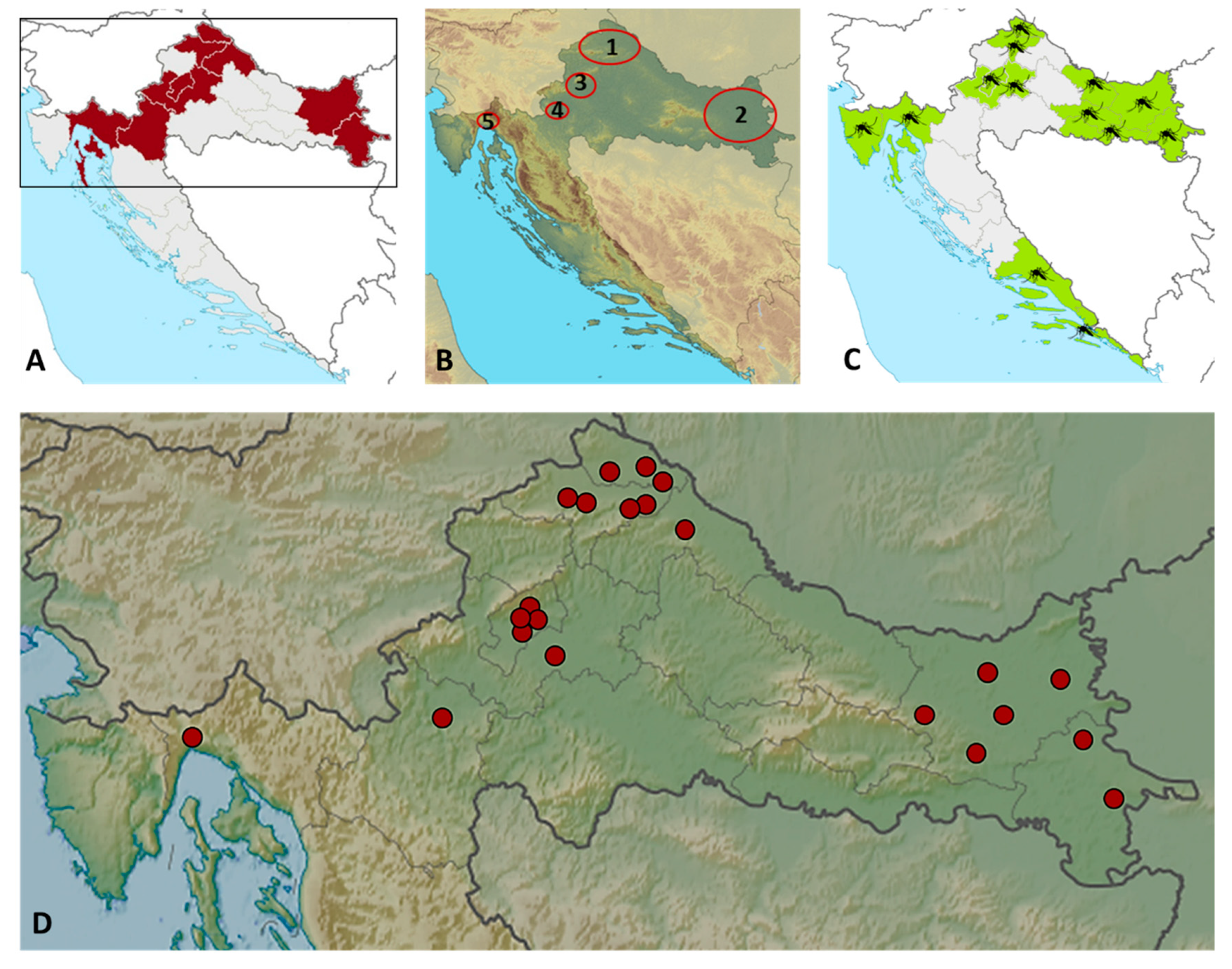
:1. Introduction
2. Materials and Methods
2.1. Patients
2.2. Methods
2.3. Statistical Analysis
3. Results
4. Discussion
5. Conclusions
Author Contributions
Funding
Institutional Review Board Statement
Informed Consent Statement
Data Availability Statement
Acknowledgments
Conflicts of Interest
References
- ICTV. Genus: Orthobunyavirus. Available online: https://talk.ictvonline.org/ictv-reports/ictv_online_report/negative-sense-rna-viruses/w/peribunyaviridae/1238/genus-orthobunyavirus (accessed on 18 May 2022).
- Bardos, V.; Danielova, V. The Tahyna virus—A virus isolated from mosquitoes in Czechoslovakia. J. Hyg. Epidemiol. Microbiol. Immunol. 1959, 3, 264–276. [Google Scholar] [PubMed]
- Camp, J.V.; Kniha, E.; Obwaller, A.G.; Walochnik, J.; Nowotny, N. The transmission ecology of Tahyna orthobunyavirus in Austria as revealed by longitudinal mosquito sampling and blood meal analysis in floodplain habitats. Parasites Vectors 2021, 14, 561. [Google Scholar] [CrossRef] [PubMed]
- Hubálek, Z. Mosquito-borne viruses in Europe. Parasitol. Res. 2008, 103 (Suppl. S1), S29–S43. [Google Scholar] [CrossRef] [PubMed]
- Hubalek, Z.; Zeman, P.; Halouzka, J.; Juricova, Z.; Bálková, H.; Sikutová, S.; Rudolf, I. Antibodies against mosquito-borne viruses in human population of an area of Central Bohemia affected by the flood of 2002. Epidemiol. Mikrobiol. Imunol. 2004, 53, 112–120. [Google Scholar] [PubMed]
- Hubalek, Z.; Sebesta, O.; Pesko, J.; Betasova, L.; Blazejova, H.; Venclikova, K.; Rudolf, I. Isolation of Tahyna Virus (California Encephalitis Group) From Anopheles hyrcanus (Diptera, Culicidae), a Mosquito Species New to, and Expanding in, Central Europe. J. Med. Entomol. 2014, 51, 1264–1267. [Google Scholar] [CrossRef]
- Camp, J.V.; Haider, R.; Porea, D.; Oslobanu, L.E.; Forgách, P.; Nowotny, N. Serological surveillance for Tahyna virus (California encephalitis orthobunyavirus, Peribunyaviridae) neutralizing antibodies in wild ungulates in Austria, Hungary and Romania. Zoonoses Public Health 2018, 65, 459–463. [Google Scholar] [CrossRef] [Green Version]
- Atkinson, B.; Hewson, R. Emerging arboviruses of clinical importance in Central Asia. J. Gen. Virol. 2018, 99, 1172–1184. [Google Scholar] [CrossRef]
- Bárdos, V.; Sixl, W.; Wisidagama, C.L.; Halouzka, J.; Stünzner, D.; Hubálek, Z.; Withalm, H. Prevalence of arbovirus antibodies in sera of animals in Sri Lanka. Bull. World Health Organ. 1983, 61, 987–990. [Google Scholar]
- Arunagiri, C.K.; Perera, L.P.; Abeykoon, S.B.; Peiris, J.S. A serologic study of California serogroup bunyaviruses in Sri Lanka. Am. J. Trop. Med. Hyg. 1991, 45, 377–382. [Google Scholar] [CrossRef]
- Lu, Z.; Lu, X.J.; Fu, S.H.; Zhang, S.; Li, Z.X.; Yao, X.H.; Feng, Y.P.; Lambert, A.J.; Ni da, X.; Wang, F.T.; et al. Tahyna virus and human infection, China. Emerg. Infect. Dis. 2009, 15, 306–309. [Google Scholar] [CrossRef]
- Li, W.; Cao, Y.; Fu, S.; Wang, J.; Li, M.; Jiang, S.; Wang, X.; Xing, S.; Feng, L.; Wang, Z.; et al. Tahyna virus infection, a neglected arboviral disease in the Qinghai-Tibet Plateau of China. Vector Borne Zoonotic Dis. 2014, 14, 353–357. [Google Scholar] [CrossRef] [PubMed] [Green Version]
- Bennett, R.S.; Gresko, A.K.; Murphy, B.R.; Whitehead, S.S. Tahyna virus genetics, infectivity, and immunogenicity in mice and monkeys. Virol. J. 2011, 8, 135. [Google Scholar] [CrossRef] [PubMed] [Green Version]
- Hubálek, Z.; Zeman, P.; Halouzka, J.; Juricová, Z.; Stovicková, E.; Bálková, H.; Sikutová, S.; Rudolf, I. Mosquitoborne viruses, Czech Republic, 2002. Emerg. Infect. Dis. 2005, 11, 116–118. [Google Scholar] [CrossRef] [PubMed]
- Xia, H.; Wang, Y.; Atoni, E.; Zhang, B.; Yuan, Z. Mosquito-Associated Viruses in China. Virol. Sin. 2018, 33, 5–20. [Google Scholar] [CrossRef] [Green Version]
- Hubálek, Z. History of Arbovirus Research in the Czech Republic. Viruses 2021, 13, 2334. [Google Scholar] [CrossRef]
- Vesenjak-Hirjan, J.; Galinović-Weisglass, M.; Urlić, V.; Bendiš, M.; Miović, P.; Vujošević, N.; Vuksanović, P. Occurrence of arboviruses in the Middle and the South Adriatic (Yugoslavia). In Arboviruses in the Mediterranean Countries; Vesenjak-Hirjan, J., Ed.; Gustav Fischer Verlag: Stuttgart, Germany; New York, NY, USA, 1980; pp. 303–310. [Google Scholar]
- Turković, B.; Brudnjak, Z. Arboviruses in Croatia. Acta Med. Croat. 1998, 52, 87–89. [Google Scholar]
- Ilic, M.; Barbic, L.; Bogdanic, M.; Tabain, I.; Savic, V.; Kosanovic Licina, M.L.; Kaic, B.; Jungic, A.; Vucelja, M.; Angelov, V.; et al. Tick-borne encephalitis outbreak following raw goat milk consumption in a new micro-location, Croatia, June 2019. Ticks Tick-Borne Dis. 2020, 11, 101513. [Google Scholar] [CrossRef]
- Vilibic-Cavlek, T.; Savic, V.; Sabadi, D.; Peric, L.; Barbic, L.; Klobucar, A.; Miklausic, B.; Tabain, I.; Santini, M.; Vucelja, M.; et al. Prevalence and molecular epidemiology of West Nile and Usutu virus infections in Croatia in the “One health” context, 2018. Transbound. Emerg. Dis. 2019, 66, 1946–1957. [Google Scholar] [CrossRef]
- Vilibic-Cavlek, T.; Zidovec-Lepej, S.; Ledina, D.; Knezevic, S.; Savic, V.; Tabain, I.; Ivic, I.; Slavuljica, I.; Bogdanic, M.; Grgic, I.; et al. Clinical, virological and immunological findings in patients with Toscana neuroinvasive disease in Croatia: Report of three cases. Trop. Med. Infect. Dis. 2020, 5, 144. [Google Scholar] [CrossRef]
- Vilibic-Cavlek, T.; Savic, V.; Klobucar, A.; Ferenc, T.; Ilic, M.; Bogdanic, M.; Tabain, I.; Stevanovic, V.; Santini, M.; Curman Posavec, M.; et al. Emerging Trends in the West Nile Virus Epidemiology in Croatia in the ‘One Health’ Context, 2011–2020. Trop. Med. Infect. Dis. 2021, 6, 140. [Google Scholar] [CrossRef]
- Vilibic-Cavlek, T.; Barbic, L.; Mrzljak, A.; Brnic, D.; Klobucar, A.; Ilic, M.; Janev-Holcer, N.; Bogdanic, M.; Jemersic, L.; Stevanovic, V.; et al. Emerging and Neglected Viruses of Zoonotic Importance in Croatia. Pathogens 2021, 10, 73. [Google Scholar] [CrossRef] [PubMed]
- Punda-Polić, V.; Jerončić, A.; Mohar, B.; Šiško Kraljević, K. Prevalence of Toscana virus antibodies in residents of Croatia. Clin. Microbiol. Infect. 2012, 18, E200–E203. [Google Scholar] [CrossRef] [PubMed] [Green Version]
- Vesenjak-Hirjan, J.; Galinović-Weisglass, M.; Brudnjak, Z.; Calisher, C.H.; Tovornik, D.; Lazuick, J.S.; Rendić, Z. Island of Brač—Focus of arbovirus infections. In Arboviruses in the Mediterranean Countries; Vesenjak-Hirjan, J., Ed.; Gustav Fischer Verlag: Stuttgart, Germany; New York, NY, USA, 1980; pp. 311–317. [Google Scholar]
- Vesenjak-Hirjan, J. Arboviruses in Yugoslavia. In Arboviruses in the Mediterranean Countries; Vesenjak-Hirjan, J., Ed.; Gustav Fischer Verlag: Stuttgart, Germany; New York, NY, USA, 1980; pp. 165–177. [Google Scholar]
- Borčić, B.; Punda, V. Sandfly fever epidemiology in Croatia. Acta Med. Iugosl. 1987, 41, 89–97. [Google Scholar] [PubMed]
- World Rivers. Rivers of Croatia. Available online: https://worldrivers.net/2018/03/07/rivers-of-croatia/ (accessed on 10 May 2022).
- Schwaiger, M.; Cassinotti, P. Development of a quantitative real-time RT-PCR assay with internal control for the laboratory detection of tick-borne encephalitis virus (TBEV) RNA. J. Clin. Virol. 2003, 27, 136–145. [Google Scholar] [CrossRef]
- Tang, Y.; Anne Hapip, C.; Liu, B.; Fang, C.T. Highly sensitive TaqMan RT-PCR assay for detection and quantification of both lineages of West Nile virus RNA. J. Clin. Virol. 2006, 36, 177–182. [Google Scholar] [CrossRef] [PubMed]
- Nikolay, B.; Weidmann, M.; Dupressoir, A.; Faye, O.; Boye, C.S.; Diallo, M.; Sall, A.A. Development of a Usutu virus specific real-time reverse transcription PCR assay based on sequenced strains from Africa and Europe. J. Virol. Methods 2014, 197, 51–54. [Google Scholar] [CrossRef] [Green Version]
- Weidmann, M.; Sanchez-Seco, M.P.; Sall, A.A.; Ly, P.O.; Thiongane, Y.; Lo, M.M.; Schley, H.; Hufert, F.T. Rapid detection of important human pathogenic Phleboviruses. J. Clin. Virol. 2008, 41, 138–142. [Google Scholar] [CrossRef]
- Li, H.; Cao, Y.X.; He, X.X.; Fu, S.H.; Lyu, Z.; He, Y.; Gao, X.Y.; Liang, G.D.; Wang, H.Y. Real-time RT-PCR Assay for the detection of Tahyna Virus. Biomed. Environ. Sci. 2015, 28, 374–377. [Google Scholar] [CrossRef]
- Matsuno, K.; Weisend, C.; Travassos da Rosa, A.P.; Anzick, S.L.; Dahlstrom, E.; Porcella, S.F.; Dorward, D.W.; Yu, X.J.; Tesh, R.B.; Ebihara, H. Characterization of the Bhanja serogroup viruses (Bunyaviridae): A novel species of the genus Phlebovirus and its relationship with other emerging tick-borne phleboviruses. J. Virol. 2013, 87, 3719–3728. [Google Scholar] [CrossRef] [Green Version]
- Bardos, V.; Sefcovicova, L. The presence of antibodies neutralizing Tahyna virus in the sera of inhabitants of some European, Asian, African and Australian countries. J. Hyg. Epidemiol. Microbiol. Immunol. 1961, 5, 501–504. [Google Scholar]
- Kolman, J.M.; Kopecký, K.; Rác, O. Serologic examination of human population in South Moravia (Czechoslovakia) on the presence of antibodies to arboviruses of the Alfavirus, Flavivirus, Turlock groups and Bunyamwera supergroup. Folia Parasitol. 1979, 26, 55–60. [Google Scholar]
- Heinz, F.; Asera, J. Presence of viruse-neutralizing antibodies of the Tahyna virus in the inhabitants of North Moravia. Folia Parasitol. 1972, 19, 315–320. [Google Scholar]
- Hubálek, Z.; Halouzka, J.; Juricová, Z.; Príkazský, Z.; Záková, J.; Sebesta, O. Surveillance of mosquito-borne viruses in Breclav after the flood of 1997. Epidemiol. Mikrobiol. Imunol. 1999, 48, 91–96. [Google Scholar] [PubMed]
- Avšič-Županc, T. Medically important araboviruses in Slovenia. Zdrav. Vestn. 1995, 64, 15–19. (In Slovenian) [Google Scholar]
- Kuniholm, M.H.; Wolfe, N.D.; Huang, C.Y.; Mpoudi-Ngole, E.; Tamoufe, U.; LeBreton, M.; Burke, D.S.; Gubler, D.J. Seroprevalence and distribution of Flaviviridae, Togaviridae, and Bunyaviridae arboviral infections in rural Cameroonian adults. Am. J. Trop. Med. Hyg. 2006, 74, 1078–1083. [Google Scholar] [CrossRef] [Green Version]
- Lv, Z.; Fu, S.H.; Wang, F.T.; Kosoy, O.L.; Nasci, R.S.; Liang, G.D. Investigation of Tahyna virus infection among unknown fever cases in Xinjiang, China. Bing Du Xue Bao 2011, 27, 71–74. [Google Scholar]
- Barakat, A.M.; Smura, T.; Kuivanen, S.; Huhtamo, E.; Kurkela, S.; Putkuri, N.; Hasony, H.J.; Al-Hello, H.; Vapalahti, O. The Presence and Seroprevalence of Arthropod-Borne Viruses in Nasiriyah Governorate, Southern Iraq: A Cross-Sectional Study. Am. J. Trop. Med. Hyg. 2016, 94, 794–799. [Google Scholar] [CrossRef] [Green Version]
- Arbovirus Surveillance Project. Institut Pasteur du Laos. Available online: https://www.pasteur.la/project-carried-on-in-the-lab-2016-2017-lao-fr1/research-and-development/ (accessed on 5 May 2022).
- Sonnleitner, S.T.; Lundström, J.; Baumgartner, R.; Simeoni, J.; Schennach, H.; Zelger, R.; Prader, A.; Schmutzhard, E.; Nowotny, N.; Walder, G. Investigations on California serogroup orthobunyaviruses in the Tyrols: First description of Tahyna virus in the Alps. Vector Borne Zoonotic Dis. 2014, 14, 272–277. [Google Scholar] [CrossRef]
- Glinskikh, N.P.; Fedotova, T.T.; Pereskokova, I.G.; Melnikov, V.G.; Volkova, L.I. The potentials for the comprehensive diagnosis of viral encephalitis in Sverdlovsk Province. Vopr. Virusol. 1994, 39, 190–191. [Google Scholar]
- Zelená, H.; Januska, J.; Raszka, J. Micromodification of virus-neutralisation assay with vital staining in 96-well plate and its use in diagnostics of Tahyna virus infections. Epidemiol. Mikrobiol. Imunol. 2008, 57, 106–110. [Google Scholar]
- Sudarić Bogojević, M.; Merdić, E.; Bogdanović, T. The flight distances of floodwater mosquitoes (Aedes vexans, Ochlerotatus sticticus and Ochlerotatus caspius) in Osijek, Eastern Croatia. Biologia 2011, 66, 678–683. [Google Scholar] [CrossRef]
- European Centre for Disease Prevention and Control and European Food Safety Authority. Mosquito Maps [Internet]. Stockholm: ECDC. 2021. Available online: https://ecdc.europa.eu/en/disease-vectors/surveillance-and-disease-data/mosquito-maps (accessed on 2 June 2022).
- Merdić, E.; Lovaković, T. Population dynamic of Aedes vexans and Ochlerotatus sticticus in flooded areas of the River Drava in Osijek, Croatia. J. Am. Mosq. Control Assoc. 2001, 17, 275–280. [Google Scholar] [PubMed]
- Evans, A.B.; Peterson, K.E. Cross reactivity of neutralizing antibodies to the encephalitic California Serogroup orthobunyaviruses varies by virus and genetic relatedness. Sci. Rep. 2021, 11, 16424. [Google Scholar] [CrossRef] [PubMed]
- Gu, H.X.; Spence, L.; Artsob, H.; Chia, W.K.; Th’ng, C.; Lampotang, V. Serological evidence of infection with California serogroup viruses (family Bunyaviridae) in residents of Long Hua, suburb of Shanghai, People’s Republic of China. Trans. R. Soc. Trop. Med. Hyg. 1984, 78, 780–781. [Google Scholar] [CrossRef]


| Virus | Serology | Manufacturer/Protocol | Sensitivity/Specificity a | RT-qPCR Protocol |
|---|---|---|---|---|
| TBEV | ELISA IgM/IgG, VNT | ELISA/IFA; Euroimmun, Lübeck, Germany VNT protocol; Ilic et al., 2020 [19] | IgG 100%/100% | Schwaiger and Cassinotti, 2003 [29] |
| IgG 100%/100% | ||||
| WNV | ELISA IgM/IgG, VNT | IgM 94.4%/99.8% | Tang at al., 2006 [30] | |
| IgG 99.5%/96.9% | ||||
| USUV | ELISA IgG, VNT | 100%/99% | Nikolay et al., 2014 [31] | |
| Phlebovirus Mosaic (TOSV, SFSV, SFNV, SFCV) | IFA IgM/IgG | IgM 100%/100% IgG 100%/92% | Weidmann et al., 2008 [32] | |
| TAHV | VNT | Li et al., 2015 [33] | ||
| BHAV | NT | Matsuno et al., 2013 [34] |
| Virus | RT-qPCR | IgG Positive N (%) | 95%CI |
|---|---|---|---|
| TBEV | Negative | 3 (1.4) | 0.3–3.9 |
| WNV | Negative | 6 (2.8) | 1.0–5.9 |
| USUV | Negative | 1 (0.5) | 0.0–2.5 |
| TOSV | Negative | 2 (0.9) | 0.1–3.3 |
| SFSV | NT | 2 (0.9) | 0.1–3.3 |
| TAHV | Negative | 22 (10.1) | 6.4–14.9 |
| BHAV | Negative | NT |
| Characteristic | Case 1 | Case 2 | Reference Values | |
|---|---|---|---|---|
| Demographic characteristics | Gender | Male | Female | |
| Age | 75 | 59 | ||
| Date of disease onset | 16 June 2019 | 18 October 2020 | ||
| Area of residence | Urban | Rural, close to a river | ||
| Clinical characteristics | Clinical presentation | Meningitis | Meningitis | |
| Clinical symptoms | Fever, headache, weakness | Fever, headache, weakness, nausea, photophobia | ||
| Underlying diseases | - | Hypertension | ||
| Outcome | Recovery | Recovery | ||
| CSF findings | Cells (mm3) | 412 | 218 | <5 |
| Mononuclear cells (%) | 78 | 70 | 100% | |
| Proteins (g/L) | 0.7 | 1.3 | 0.17–0.37 g/L | |
| Glucose (mmol/L) | 3.1 | 2.2 | 2.5–3.3 mmol/L | |
| TAHV RT-qPCR | CSF | Negative | Negative | |
| Urine | Negative | Negative | ||
| TAHV VNT (titer) | Serum | 320 | 640 | ≥10 positive |
| CSF | 5 | 10 | ≥5 positive |
| Characteristic | Tested | TAHV NT Antibodies N (%) | 95% CI | p | |
|---|---|---|---|---|---|
| N (%) | |||||
| Gender | Male | 144 (66.1) | 16 (11.1) | 6.5–17.4 | 0.485 |
| Female | 74 (33.9) | 6 (8.1) | 3.0–16.8 | ||
| Age group | <30 years | 57 (26.1) | 1 (1.8) | 0.0–9.4 | 0.001 |
| 30–49 years | 44 (20.2) | 2 (4.6) | 0.6–15.5 | ||
| 50–69 years | 76 (34.9) | 9 (11.8) | 5.6–21.3 | ||
| ≥70 years | 41 (18.8) | 10 (24.4) | 12.4–40.3 | ||
| Area of residence | Urban | 132 (60.5) | 13 (9.8) | 5.3–16.2 | 0.882 |
| Suburban/rural | 86 (39.5) | 9 (10.5) | 4.9–18.9 | ||
| Living in floodplain | Yes No | 172 (78.9) | 21 (12.2) | 7.7–18.1 | 0.044 |
| 46 (21.1) | 1 (2.1) | 0.0–11.5 | |||
| Clinical presentation | Febrile headache | 13 (6.0) | 2 (15.4) | 1.9–45.4 | 0.702 |
| Meningitis | 143 (65.6) | 12 (8.4) | 4.4–14.2 | ||
| Meningoencephalitis | 55 (25.2) | 7 (12.7) | 5.3–24.5 | ||
| Myelitis | 7 (3.2) | 1 (14.3) | 0.4–57.8 |
| Characteristic | OR | 95% CI OR | p | RR | 95% CI RR | p |
|---|---|---|---|---|---|---|
| Male (Ref.) vs. female gender | 1.375 | 0.513–3.678 | 0.525 | 1.389 | 0.567–3.401 | 0.471 |
| Age | ||||||
| <30 years | Ref. | Ref. | ||||
| 30–49 years | 2.666 | 0.233–30.400 | 0.429 | 2.590 | 0.242–27.662 | 0.430 |
| 50–69 years | 7.522 | 0.924–61.200 | 0.059 | 6.750 | 0.880–51.770 | 0.066 |
| >70 years | 18.064 | 2.207–147.809 | 0.007 | 13.902 | 1.851–104.393 | 0.9010 |
| Suburban/rural (Ref.) vs. urban area of residence | 1.069 | 0.436–2.623 | 0.882 | 1.062 | 0.474–2.377 | 0.882 |
| Living in floodplain | 6.258 | 0.819–47.820 | 0.071 | 5.616 | 0.775–40.659 | 0.087 |
| Clinical presentation | ||||||
| Febrile headache | Ref. | Ref. | ||||
| Meningitis | 0.503 | 0.099–2.542 | 0.406 | 0.545 | 0.136–2.179 | 0.391 |
| Meningoencephalitis | 0.802 | 0.146–4.402 | 0.799 | 0.827 | 0.193–3.528 | 0.797 |
| Myelitis | 0.916 | 0.068–12.322 | 0.947 | 0.928 | 0.101–8.529 | 0.947 |
Publisher’s Note: MDPI stays neutral with regard to jurisdictional claims in published maps and institutional affiliations. |
© 2022 by the authors. Licensee MDPI, Basel, Switzerland. This article is an open access article distributed under the terms and conditions of the Creative Commons Attribution (CC BY) license (https://creativecommons.org/licenses/by/4.0/).
Share and Cite
Vilibic-Cavlek, T.; Stevanovic, V.; Savic, V.; Markelic, D.; Sabadi, D.; Bogdanic, M.; Kovac, S.; Santini, M.; Tabain, I.; Potocnik-Hunjadi, T.; et al. Detection of Tahyna Orthobunyavirus-Neutralizing Antibodies in Patients with Neuroinvasive Disease in Croatia. Microorganisms 2022, 10, 1443. https://doi.org/10.3390/microorganisms10071443
Vilibic-Cavlek T, Stevanovic V, Savic V, Markelic D, Sabadi D, Bogdanic M, Kovac S, Santini M, Tabain I, Potocnik-Hunjadi T, et al. Detection of Tahyna Orthobunyavirus-Neutralizing Antibodies in Patients with Neuroinvasive Disease in Croatia. Microorganisms. 2022; 10(7):1443. https://doi.org/10.3390/microorganisms10071443
Chicago/Turabian StyleVilibic-Cavlek, Tatjana, Vladimir Stevanovic, Vladimir Savic, Domagoj Markelic, Dario Sabadi, Maja Bogdanic, Snjezana Kovac, Marija Santini, Irena Tabain, Tanja Potocnik-Hunjadi, and et al. 2022. "Detection of Tahyna Orthobunyavirus-Neutralizing Antibodies in Patients with Neuroinvasive Disease in Croatia" Microorganisms 10, no. 7: 1443. https://doi.org/10.3390/microorganisms10071443