Article

Role of the Vibriolysin VemA Secreted by the Emergent Pathogen Vibrio europaeus in the Colonization of Manila Clam Mucus

Department of Microbiology and Parasitology, Aquaculture Institute & CIBUS-Faculty of Biology, University of Santiago de Compostela, 15782 Santiago de Compostela, Spain
*
Author to whom correspondence should be addressed.
Microorganisms 2022, 10(12), 2475; https://doi.org/10.3390/microorganisms10122475
Submission received: 9 November 2022 / Revised: 10 December 2022 / Accepted: 12 December 2022 / Published: 15 December 2022

Abstract

:
Vibrio europaeus is an emergent pathogen affecting clams, oysters and scallops produced in the most important countries for bivalve aquaculture. Studies concerning virulence factors involved in the virulence of V. europaeus are very scarce despite its global significance for aquaculture. Zinc-metalloproteases have been described as a major virulence factor in some Vibrio spp., although their contribution and role in the virulence of V. europaeus is not clear. To address this, we have studied an extracellular zinc-metalloprotease (VemA) encoded by V. europaeus, which was identified as a vibriolysin, highly conserved in this species and homologous in other pathogenic and non-pathogenic species. Virulence challenge experiments demonstrated that infection processes were faster when Manila clam larvae and juveniles were infected with the wildtype rather than with a mutant defective in the vemA gene (ΔvemA). V. europaeus was able to resist the bactericidal action of mucus and displayed a chemotaxis ability favoured by VemA to colonize the body mucus of clams and form a biofilm. The overall results suggest that VemA, although it is not a major virulence factor, plays a role in the colonization of the Manila clam mucus, and thus boosts the infection process as we observed in virulence challenge experiments.

1. Introduction

Molluscan aquaculture is the second most important activity within the world aquaculture [1]. Manila clams (Ruditapes philliphinarum) and Pacific oysters (Crassostrea gigas) are the most important bivalve species in aquaculture, accounting the 8% of the global aquaculture production [1]. As molluscan filter feeders, they filter large quantities of water containing microalgae, bacteria and detritus from the environment in which they live [2,3]. Thus, they have the ability to bioaccumulate high bacterial concentrations in their tissues, including bacterial pathogens [3,4,5]. To fight the pathogens, bivalves rely primarily on (i) the pallial cavity fluid and its associated mucus, which has important functions such as lubrication, particle capture and antimicrobial activity being the first line of defence against microorganisms [2]; and (ii) both cellular and humoral defence factors in the haemolymph [2,3].
Bacterial pathogenic species belonging to the genus Vibrio are the most important threat to bivalves, constraining the expansion of bivalve aquaculture [4]. The first reports of Vibrio disease in bivalves (known as “vibriosis”) were published more than 50 years ago; however, this global problem is still unsolved [4]. Among the pathogenic species, Vibrio europaeus represents an excellent bacterial model to study the vibriosis in bivalves due to: (i) the negative impact on the worldwide aquaculture: it is responsible of recurrent mass mortalities in the world’s most important bivalve producers, such as France, Spain, Chile and the US [6,7,8,9,10,11], and (ii) its wide range of hosts affecting different bivalve species and stages of the bivalve’s life cycle: V. europaeus infects a wide range of different clam, oyster and scallop species, including Manila clam and Pacific oyster, and even at different stages of development such as larvae, spat and juveniles [6,7,8,9,10,11,12,13,14,15,16]. Phylogenetically, this taxon was initially described as a subspecies of V. tubiashii, formed by two pathogenic subspecies with significance for shellfish aquaculture: V. tubiashii subsp. tubiashii and V. tubiashii subsp. europaeus [9]. However, this taxon was later reclassified and elevated to the rank of species supported by a novel phylogenetic analysis based on genome-to-genome comparisons and chemotaxonomic and phenotypic differences [14].
Virulence is a multifactorial trait that results from the sequential action of several microbial factors that enable colonization, proliferation and evasion of the host immune system and ultimately causes disease [17]. Regarding to the virulence factors, Spinard et al. [18] found in silico different proteases encoded by the V. europaeus CECT 8136 genome, including two metalloproteases, three putative haemolysins and phospholipases. Mersni-Achour et al. [12] demonstrated that the extracellular products (ECPs) of the strain V. europaeus 07/118 T2 inhibited the adhesive capacity and phagocytic activity of C. gigas haemocytes. Complementary biochemical analyses showed that the proteolytic fraction of ECPs contained an active and thermostable extracellular zinc-metalloprotease(s). Later, Mersni-Achour et al. [13] fractioned the V. europaeus ECPs to study the toxicity of the two major fractions (F1 and F2). They found that F1 was less toxic (43% mortality) in contrast to F2, which it was responsible of the 70% mortality in Pacific oyster larvae. Interestingly, F2 contained a unique protein identified as an extracellular zinc-metalloprotease, hereinafter designated as VemA (V. europaeus metalloprotease A). Other extracellular zinc-metalloproteases have been also reported as a virulence factor required for the full virulence in other Vibrio pathogens for bivalves such as V. neptunius (VnpA), V. aestuarianus (Vam) or V. coralliilyticus (VcpA and VtpA) [19,20,21,22]. However, the role of the zinc-metalloproteases as a major (or secondary) virulence factor can be different depending on if ECPs or live cells are used. For instance, Le Roux et al. [23] demonstrated that the zinc-metalloprotease Vsm secreted by V. splendidus is essential for ECPs toxicity in Pacific oysters; however, it is not necessary when a live mutant (Δvsm) is injected in oysters resulting in high-mortality rates similar to the wildtype. Therefore, to elucidate the role of VemA it is essential to perform in vivo challenge experiments using mutants defective in the vemA gene to mimic the natural infection process. To address this, the aims of this work were: (i) to perform in silico analyses to evaluate the presence of VemA in all V. europaeus strains (n = 38 strains/genomes); (ii) to construct mutants defective in the vemA gene (ΔvemA); (iii) to evaluate the contribution of VemA in the virulence of V. europaeus by comparison between the wildtype and the ΔvemA mutant challenging Manila clam juveniles and larvae; and (iv) to elucidate the role of VemA in chemotaxis, biofilm formation and bacterial proliferation in Manila clam mucus.

2. Materials and Methods

2.1. Bacterial Strains, Plasmids and Media

The bacterial strains and plasmids used or derived from this study are included in Table 1. V. europaeus CECT 8136 was grown on Trypto-Casein Soy agar or broth supplemented with 2% sodium chloride (w/v) (TSA–2/TSB–2, Condalab, Madrid, Spain) at 25 °C for 24 h. E. coli was grown in Luria–Bertani broth supplemented with 1% sodium chloride (w/v) (LB-1, Condalab, Madrid, Spain) at 37 °C for 24 h. Diaminopimelic acid and thymidine were added at a final concentration of 0.3 mM (w/v) for auxotrophic strains E. coli β3914 and E. coli Π3813, respectively (Table 1). Antibiotics were used at the following concentrations: chloramphenicol (Cm; 25 μg mL−1 for E. coli strains and 5 μg mL−1 for V. europaeus), erythromycin (Ery; 200 μg mL−1) and kanamycin (Kn; 50 μg mL−1). A suicide vector for allele exchange pSW7848T [24 carried the ccdB gene as a counterselection marker, which is under control of the arabinose promotor (PBAD) and induced/repressed by the addition of 0.2% (w/v) L-arabinose or 1% (w/v) D-glucose to the growth media.

2.2. Search of VemA Homologous and Phylogenetic Analysis

VemA metalloprotease was encoded by the gene vemA (WP_069668927) from the V. europaeus CECT 8136 (=PP–638) genome [18]. The presence of VemA was verified by BLASTp (i) at the intraspecific level in all V. europaeus strains available to the date from the NCBI database (n = 38 strains/genomes) (Table S1); (ii) and at the interspecific level by searching for homologs from NCBI using the non-redundant protein sequences database. The VemA domain prediction was carried out with the UniProt (https://www.uniprot.org) and InterPro servers (https://www.ebi.ac.uk/interpro/) (accessed on 22 Septembre 2022). The resulting heatmaps were performed with R package ComplexHeatmap (v2.11.2) [26]. A phylogenetic tree based on homologous protein sequences was constructed using MEGA11 [27] after a multiple alignment of data by CLUSTAL W [28]. Distances and clustering with the neighbour-joining (NJ) algorithm were determined using bootstrap values based on 1000 replications.

2.3. Construction of ΔvemA Mutants by Double Allelic Exchange

An in-frame deletion of the vemA gene was performed by double allelic exchange using the pSW7848T suicide plasmid [25]. Upstream and downstream fragments (692 bp) flanking the target gene were amplified by PCR using primers described in Table S2 and Q5 High-Fidelity DNA Polymerase (NEB, Ipswich, MA, USA). The pSW7848T backbone was amplified in a third PCR (Table S2) and the resulting PCR product was digested with DpnI (2 h at 37 °C; NEB, Ipswich, MA, USA) to inactivate any plasmid template. All PCR products were verified by gel electrophoresis and subsequently purified by the QIAquick PCR Purification Kit (QIAGEN, Hilden, Germany) before Gibson assembly reaction using the NEBuilder HiFi DNA Assembly (NEB, Ipswich, MA, USA). Gibson reaction was desalted by dialysis (0.0025 μM filter; Millipore, Burlington, MA, USA), electroporated into E. coli Π3813 (strain used for cloning) and the recombinant plasmid (pPC1; Figure S1) verified by (i) colony PCR (Table S2), (ii) restriction enzyme digestion (NdeI; NEB, Ipswich, MA, USA) (iii) and Sanger sequencing (Table S2). Plasmid pPC1 was electroporated into E. coli β3914 (donor) for conjugation adapting the protocol described by Hussain et al. [29] to V. europaeus (recipient). Overnight cultures were grown as described above and a drop mating assay was adjusted to a 1:3 ratio (donor:recipient) and mating media (TSA–1 plate supplemented with diaminopimelic acid) was incubated at 25 °C for 24 h. Gene deletion involves two homologous recombination events: (i) the first recombination leads to the integration of the recombinant plasmid into the host chromosome by a first homologous recombination (single crossover). For this, insertional mutants were selected from TSA-2 plates supplemented with Cm and glucose 1% (w/v) and verified by colony PCR (Table S2) and (ii) the pSW7848T backbone is excised from the chromosome through a rare second homologous recombination event (double crossover). Thus, colonies were grown in liquid media to the late logarithmic phase and spread onto TSA-2 plates supplemented with 0.2% arabinose to induce the ccdB toxin (Figure S1) under the control of Pbad. After the second homologous recombination, mutants with a 92% deletion of the vemA gene were obtained and verified by (i) colony PCR (Table S2) and (ii) Sanger sequencing (Table S2).

2.4. Virulence Challenges

Pathogenicity assays were carried out with Manila clam larvae (180 μm) and juveniles (13 ± 1 mm). V. europaeus CECT 8136, PC1-11 and V. breoganii C5.5 (used as a negative control) (Table 1) were grown overnight as described above. Bacterial suspensions were made in sterile sea water (SSW) and adjusted to OD600 = 1 (~108 CFU mL−1). Bacterial concentrations, expressed in colony-forming units per millilitre (CFU mL−1), were confirmed by decimal dilution series onto TSA-2 plates. For larvae, virulence assays were performed following Dubert et al. [30] adjusting the bacterial concentrations to 104 CFU mL−1 from the initial bacterial suspension and mortalities were evaluated every 24 h. For juveniles, challenges included two steps: (i) infection: tanks were filled with filtered seawater (FSW; 0.22 μM Nalgene Rapid-Flow, Thermo Fisher Scientific, Waltham, MA, USA) containing a bacterial suspension adjusted to a final concentration of 107 CFU mL−1. Subsequently, 15 juveniles were added to each tank and kept for 24 h at room temperature (RT = ~20 °C) to actively filtrate the bacteria; and (ii) post-infection: challenged juveniles were taken out the infection tanks for 8 h at RT to internalize the bacteria within the pallial cavity. After this, juveniles were transferred to fresh tanks filled with 200 mL FSW and maintained at RT with aeration. Mortalities were monitored at 8, 20, 32, 44, 56, 68 and 80 h post-infection and clams were immediately removed when the valves were open (dead juveniles), or siphons were not retracted following stimulation (moribund juveniles) and expressed as a percentage of survival. FSW was renewed once per day (or if it was turbid).
Extracellular products (ECPs) and SDS-polyacrylamide gel electrophoresis. V. europaeus CECT 8136 and PC1-11 ECPs were collected from overnight cultures in TSB-2. Overnight cultures were centrifuged (6000× g for 20 min at 4 °C), cell pellets discarded, and the supernatant collected and filtered through a 0.22 μM filter (Sartorius, Goettingen, Germany). Proteins were precipitated as described by Terceti et al. [31] with 10% (w/v) trichloroacetic acid (TCA) for 30 min on ice and recovered by centrifugation. Protein pellets were washed in TCA followed by washing in acetone and air-dried. Precipitated proteins were solubilized in SDS-sample buffer (50 mM Tris–HCl, pH 8.8; 2% SDS; 0.05% bromophenol blue; 10% glycerol; 2 mM EDTA, and 100 mM DTT) and subjected to SDS–PAGE in 10 or 12% polyacrylamide gels using the Laemmli discontinuous buffer system stained with Coomassie Brilliant Blue.

2.5. Mucus Extraction

Mucus was aspirated from the body surface (mantle and branchia) of non-anaesthetised Manila clam adults (n = 61) using a disposable Pasteur pipette. Mucus was sonicated by two cycles of 30 s at low intensity, centrifuged at 10,000× g for 15 min at 4 °C and filtered through a 0.45 μM filter (Millipore, Burlington, MA, USA) to remove debris. Protein concentration was measured using a Pierce BCA Protein assays kit (Thermo Fisher Scientific, Waltham, MA, USA) and mucus was collected and stored at −80 °C until use.

2.6. Evaluation of the Bactericidal Activity of the Surface Mucus on Solid Media

V. europaeus CECT 8136 and PC1-11 were grown on TSA-2, resuspended in phosphate-buffered saline (PBS, pH 7.4) and spread onto Müeller–Hinton Agar (Oxoid, Hampshire, UK) supplemented with 1% sodium chloride (w/v) (MHA-1) to a final concentration of 105 CFU mL−1. Sterile 6 mm discs impregnated with mucus (20 μL) were applied to the agar plates and incubated at 25 °C for 24 h.

2.7. Adherence to Mucus and Biofilm Formation on Polystyrene

Mucus was diluted 1:5 in sterile distilled water and a volume of 100 μL was added to each well of 96-well ELISA polystyrene plates (Jet Biofil, Guangzhou, China). Plates were dried in a laminar flow cabinet for 48 h and subsequently filled with 200 μL of TSB-2 per well. V. europaeus CECT 8136 and PC1-11 overnight cultures were diluted 1:100 and incubated at 25 °C for 24 h. Bacterial cells were fixed with methanol (99.8%) stained with 0.5% crystal violet solution (v/v), washed three times with distilled water and the bound dye was eluted with acetic acid (33%, v/v) and measured by optical density at 570 nm in a spectrometer (Biotek, Winooski, VT, USA). Results are means calculated from eight replicates.

2.8. Chemotaxis Assays

Chemotaxis was evaluated by using the capillary assay described by Valiente et al. [32] with slight modifications. Overnight V. europaeus CECT 8136 and PC1-11 cultures were diluted 1:100 and exponentially grown (OD600 = 0.9; ~107 CFU mL−1) to maximize the number of motile cells before proceeding to the assay. Serial decimal dilutions of the bacterial suspensions were spread onto TSA-2 and incubated as described above. Bacterial cultures were harvested by centrifugation (5000× g for 5 min at 4 °C), washed three times in chemotaxis buffer (PBS, 0.01 mM EDTA) and 400 μL dispensed per tube (assays were performed in triplicate). A 50 μL capillary tube (Drummond Scientific, Broomall, PA, USA), sealed at one end and containing the mucus or chemotaxis buffer was inserted in a glass-tube and incubated at 25 °C with the bacterial suspension for 30 min and 1 h. After incubation, the capillaries were removed and externally rinsed with ethanol. Then, the capillary contents were expelled in PBS, 10-fold dilutions prepared and spread on TSA-2 plates. The data shown are means of three independent replicates and the chemotactic responses are expressed as the ratio between the numbers of bacteria in mucus capillaries and the numbers of bacterial in control capillaries only in presence of chemotaxis buffer.

2.9. Bacterial Survival and Growth with Mucus

V. europaeus CECT 8136 and PC1-11 were grown overnight on TSA-2 and resuspended in SSW to an OD600 = 1. A volume of 1 mL adjusted to ~105 CFU mL−1 was added into a glass tube with 100 μL of mucus and incubated at 25 °C for 30 min and 24 h. After incubation, samples from each tube were diluted 10-fold and spread on TSA-2 plates. The data shown are means of two independent replicates and the bacterial proliferation is expressed as the ratio between the number of bacteria in mucus after incubation (30 min and 24 h) and the number of bacteria at the beginning of the experiment (time 0).

2.10. Statistical Analyses

SPSS (v27; IBM SPSS) was used for statistical analyses. In all cases Kolmogorov–Smirnov tests were previously applied to the normality, and thus, to choose the optimal statistical test applied in each case: (i) for virulence challenges, the statistical significance of differences in percentage survival was determined using the Kruskal–Wallis test; (ii) Student’s t-test was used for chemotaxis response, biofilm formation and bacterial proliferation on mucus. p values were considered significant when p was <0.05 using the Bonferroni-adjusted p value.

3. Results

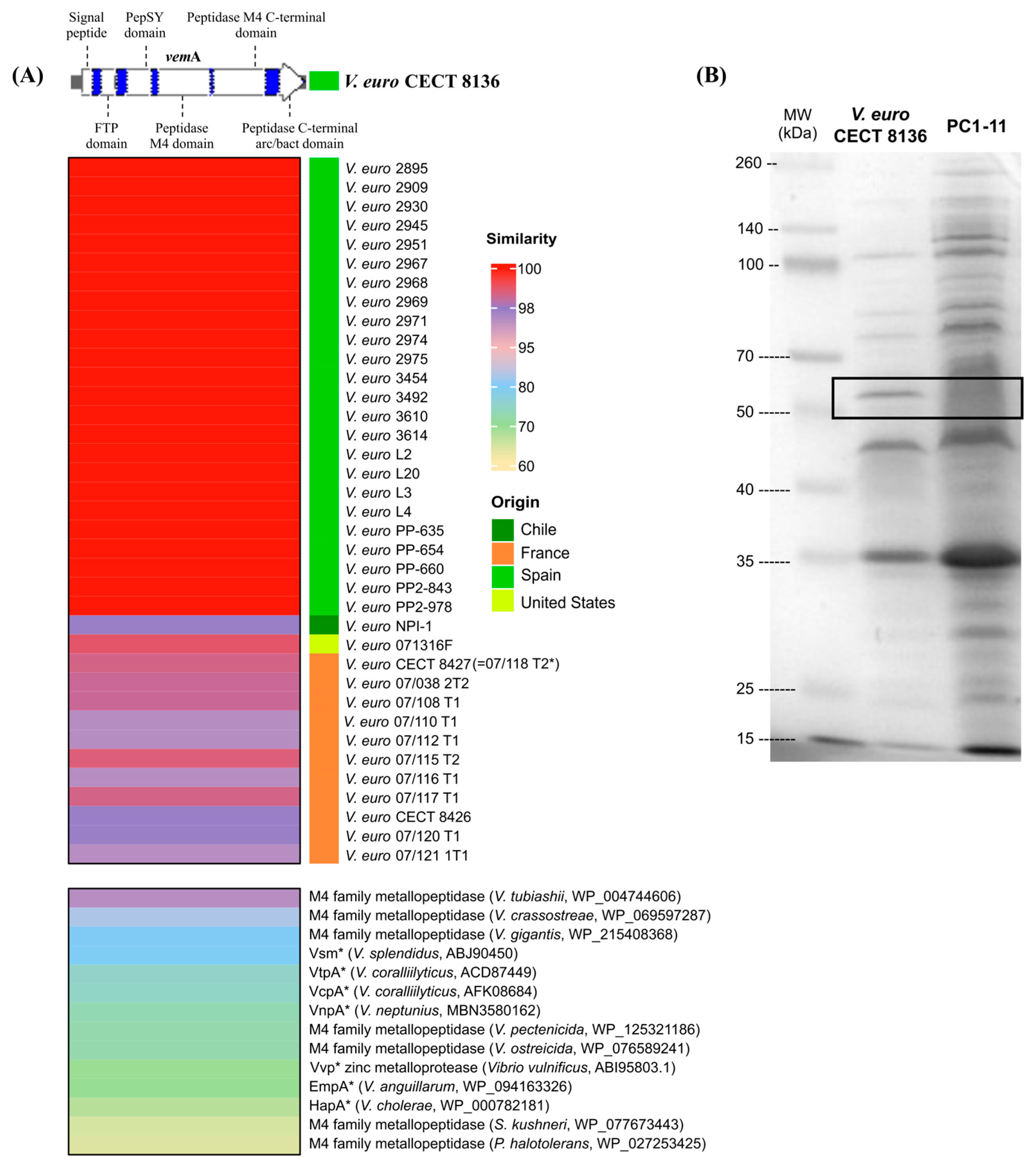
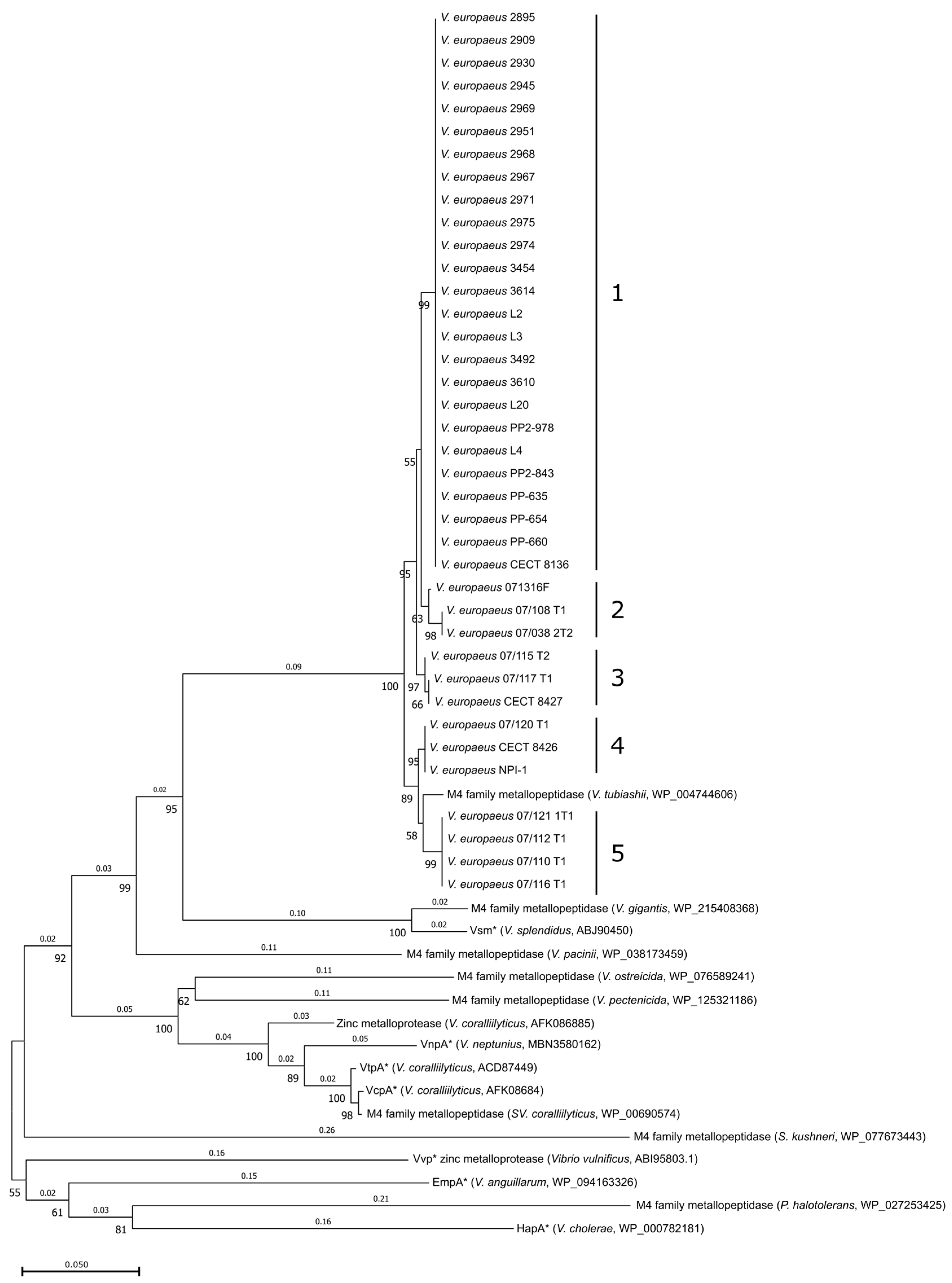
3.1. VemA Is a Vibriolysin-like Protein, Highly Conserved in V. europaeus with Homologues in Other Pathogenic Vibrio Species

Locus WP_069668927 (1824 bp) from the V. europaeus CECT 8136 genome was designated as vemA (V. europaeus metalloprotease A) and encodes for VemA, a vibriolysin belonging to the M4 family of metallopeptidases and gluzincins subfamily [33]. VemA (608 aa; Figure 1A) showed the typical structure of vibriolysins [33], they are synthesized as a pro-peptide precursor containing a signal peptide signature in the N-terminal region (1–75 bp) and four conserved regions: (i) an FTP (fungalysin/thermolysin pro-peptide) domain (164–281 bp); (ii) a PepSY domain (349–569 bp); (iii) an M4 neutral protease which includes two M4 domains, such as peptidase M4 domain (622–1050 bp) and peptidase M4 C-terminal domain (1057–1490 bp); and (iv) a C-terminal pre-peptidase domain (1582–1775 bp). The mature forms of VemA contain the conservative region with M4 neutral proteases, which is also known as the catalytic domain [33]. Interestingly, VemA is classified as a vibriolysin-like protein within the clade II (VLP-II), which includes vibriolysins encoded by other opportunist pathogens, such as V. coralliilyticus [33].
VemA was encoded by all V. europaeus strains (n = 38 genomes/strains; Table S1) showing a high similarity (>97%) with the reference strain CECT8136 by BLASTp at the intraspecific level (Figure 1A). Phylogenetic analysis (Figure 2) indicated that Spanish isolates (n = 24 strains) constituted a monophyletic group (cluster #1; bootstrap value = 99; homology = 100%). However, the remaining strains were split among four additional clusters: cluster #2 (two French strains closely related with the American strain 07136F; bootstrap value = 63; homology = 99.01–98.52%); cluster #3 (French strains; bootstrap value = 97; homology = 98.85–98.68%); cluster #4 (two French strains, including the strain 07/118 T2 = CECT 8126 studied by Mersni-Achour et al. [13], were closely related with the Chilean strain NPI-1; bootstrap value = 95; homology = 97.85%); and cluster #5 (four French strains closely related with V. tubiashii; bootstrap value = 58; homology = 97.03%).
The homology search with other bacterial species revealed VemA as a homolog of other zinc-metalloproteases described in bivalve pathogens, such as V. tubiashii, V. crassostreae, V. splendidus, V. coralliilyticus, V. neptunius, V. pectenicida and V. ostreicida (Figure 1A and Figure 2; Table S3). In addition, some of those proteins have been reported as virulence factors in V. splendidus (Vsm = 79.57% similarity), V. coralliilyticus (VtpA = 75.16% and VcpA = 74.84% similarity), V. neptunius (VnpA = 72.95% similarity) or V. cholerae (HapA = 67.38% similarity).

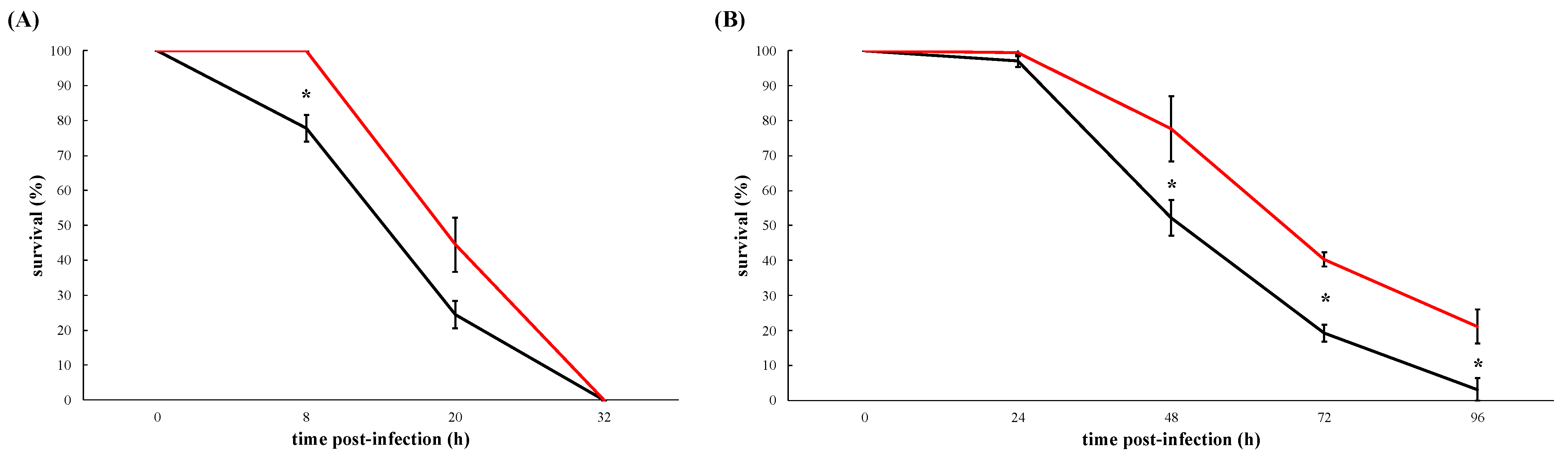
3.2. VemA Is an Extracellular Protein Necessary for the Full Virulence of V. europaeus Although It Is Not a Major Virulence Factor

An in-frame deletion mutant, designated as PC1-11 (Table 1), was produced from V. europaeus CECT 8136 to study the role of the vemA gene in infection challenges using Manila clam juveniles and larvae. Interestingly, a polyacrylamide gel used to gain insight into the VemA protein and the analysis of the ECPs between the wildtype and PC1-11 showed a protein band of ~66 kDa corresponding to VemA (Figure 1B).
In juveniles, the infection due to the wildtype (V. europaeus CECT 8136) caused mortalities faster than the ΔvemA mutant (wildtype vs. PC1-11): 22% vs. 0% after 8 h (p = 0.034); 76% vs. 56% after 20 h (p = 0.44); and 100% in both cases after 32 h post-infection (Figure 3A).
Results from virulence challenges performed on larvae followed the same mortality dynamic than juveniles (Figure 3B). Larval mortalities due to the wildtype were ~20% higher than PC1-11 (wildtype vs. PC1-11): 48% vs. 22% after 48 h (p = 0.05); 81% vs. 60% after 72 h (p = 0.05) and 97% vs. 79% after 96 h (p = 0.05) post-infection (Figure 2B).
Our results showed that VemA is an extracellular protein and in absence of the vemA gene mass mortalities of larvae and juveniles are slowed down although finally those mortalities are unavoidable reaching high mortality rates: 100% for juveniles and 80% for larvae (Figure 2A,B). These results demonstrate that VemA is not a major virulence factor although in its presence V. europaeus kills larvae and juveniles faster, specially at the beginning of the infection.

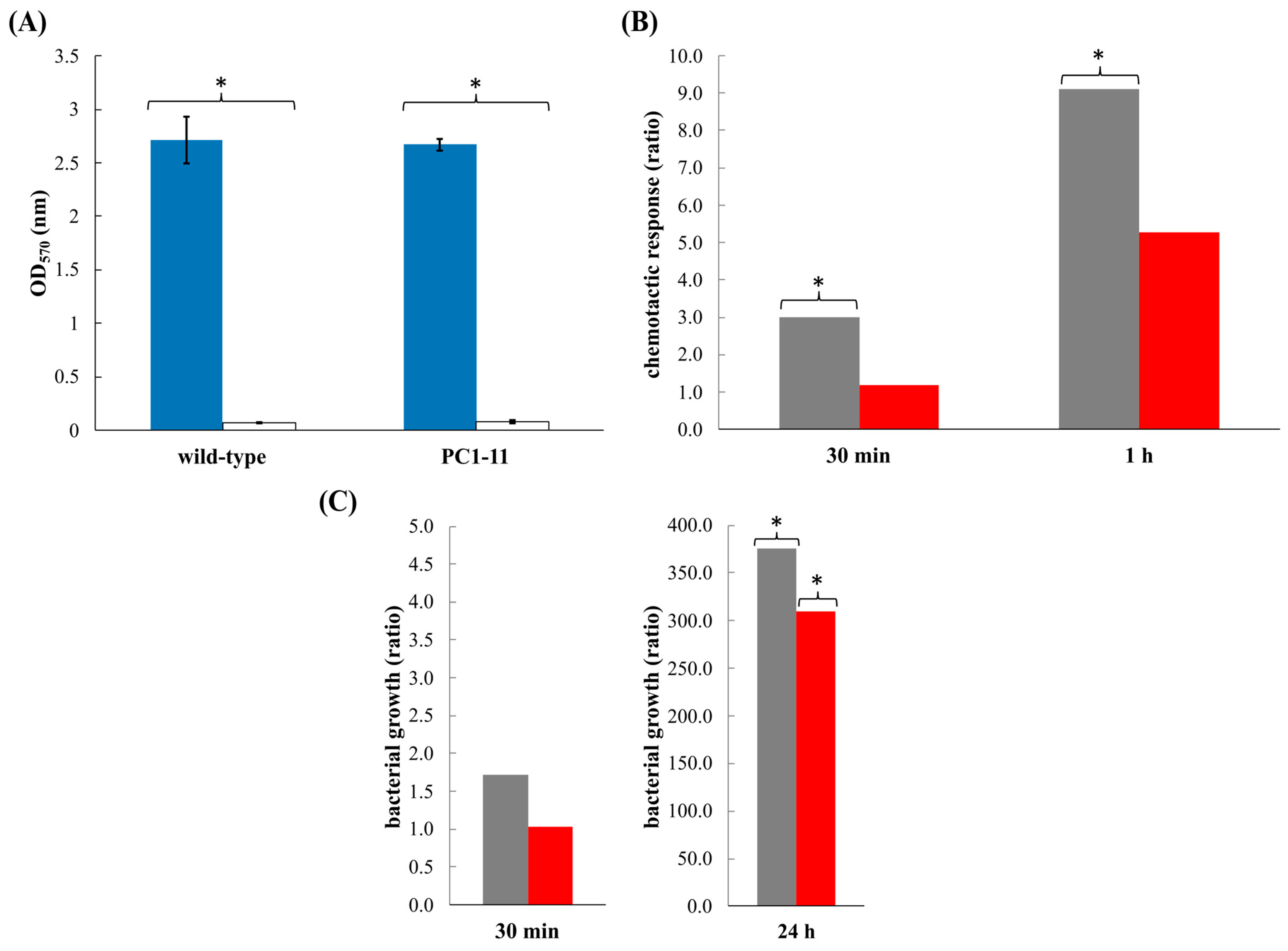
3.3. V. europaeus Forms Biofilms on Manila Clam Mucus

Body mucus (protein concentration = 2.3 μg mL−1) from Manila clams did not show any bactericidal effect on solid media in V. europaeus wildtype or PC1-1. Thus, we have evaluated the adherence by means of a bacterial biofilm formation over a surface impregnated with Manila clam mucus (Figure 4A). Interestingly, wildtype and PC1-11 exhibited a similar attachment pattern to bivalve mucus and to polystyrene, in both cases being significantly higher towards mucus than polystyrene (p = 0 for wildtype and PC1-11) (Figure 4A). These results demonstrate the ability of V. europaeus to form biofilms on bivalve mucus.

3.4. V. europaeus Is Attracted by Mucus and VemA Participates in Bacterial Chemotaxis

The wildtype strain was significantly attracted to bivalve mucus, approximately three times more after 30 min (p = 0.042) and ~9× after 1 h (p = 0.014) in relation to the control in the absence of mucus (Figure 4B). PC1-11 exhibited a decreased chemoattraction towards bivalve mucus after 30 min (p = 0.348) and 1 h (p = 0.122) in relation to the control (Figure 4B). Those results suggest that VemA plays a role in the host’s colonization attracted by the mucus.

3.5. VemA Promotes the Proliferation of V. europaeus over the Mucus Matrix

Wild-type and PC1-11 were able to grow over the mucus matrix (Figure 4C). As we observed previously by chemotaxis, the bacterial proliferation of PC1-11 was similar to the negative control after 30 min (p = 0.904) and only wildtype showed a ratio higher than 1 after 30 min (p = 0.517) (Figure 4C). Bacterial growth was significant higher (300×) for wildtype (p = 0.017) and PC1-11 (p = 0.004) than the negative controls after 24 h (Figure 4C). Although similarly, it is important to note that bacterial proliferation was slightly lower for PC1-11 (ratio = 309.3) than wildtype (ratio = 375.7).

4. Discussion

The role of M4 metalloproteases such as vibriolysins in Vibrio virulence is not clear [33]. For instance, pathogenic microorganisms, such as V. cholerae, V. vulnificus, V. anguillarum or V. neptunius, use vibriolysins as their main virulence factor [22,34,35,36]. Our results have demonstrated that vemA is a core gene highly conserved among all V. europaeus strains and even in other related species as the closest relative V. tubiashii [4,14]. Interestingly, VemA is a homolog of other zinc-metalloproteases encoded by non-pathogenic species, such as V. gigantis (Figure 1A), suggesting other functions not related to virulence [33]. In fact, VemA is classified as a vibriolysin-like protein belonging to clade II (VLP–II), which includes opportunist pathogens such as V. coralliilyticus and non-pathogenic species such V. pacinii [33].
The first part of this study was to elucidate the contribution degree of VemA as a major virulence factor (or not) in the virulence of V. europaeus towards Manila clam juveniles and larvae. Previous results described VemA as the major toxicity factor when ECPs secreted by V. europaeus 07/118 T2 (=CECT 8126; Figure 1A) were inoculated with Pacific oyster larvae, making VemA responsible of the 70% mortality (other proteins were required to obtain a full virulence) [13]. However, we have demonstrated in in vivo challenges that VemA is not a major virulence factor when live cells (ΔvemA and wildtype) are used instead ECPs because mass mortalities were unavoidable in all cases.
Divergences between the results obtained when live cells (mutants and wildtype) were used rather than ECPs in in vivo experimental challenges were also described for other metalloproteases homologous to VemA. For instance, Vsm, secreted by the bivalve pathogen V. splendidus, was described as the major toxicity factor by comparisons between the ECPs secreted by the defective mutant Δvsm and the wildtype. In contrast, when a live mutant (Δvsm) was injected into oysters the mortality rates were similar to the wildtype, promoting mass mortalities in both cases [23,37]. Binese et al. [37] found differential expression of different proteins detected in wildtype and Δvsm mutant ECPs: the Δvsm secreted a predicted protein of unknown function (VS2864) and an additional metalloprotease of the M6 family (VSA1062) while wildtype ECPs contained an unknown protein (VSA576) and three spots corresponding to Vsm. Same divergences were reported for other bivalve pathogens, such as V. coralliilyticus. First, Sussman et al. [20] identified a zinc-metalloprotease (VcpA) secreted by V. coralliilyticus P1 and demonstrated its key role in virulence by testing ECPs from mutants (ΔvcpA) vs. wildtype. However, the VcpA contribution to V. coralliilyticus virulence was different when live cells were used, and similar high-mortality rates were detected due to wildtype and the mutants (ΔvcpA) [38]. In this case, the ΔvcpA mutant showed a higher haemolytic activity and secreted 18 proteins not secreted by the wildtype, including four types of metalloproteases, a chitinase, a haemolysing-related protein RbmC, the Hcp protein and 12 hypothetical proteins [38]. Altogether, these studies indicate a diverse virulence repertoire that possibly enables both Vibrio species to be efficient animal pathogens [37,38]. Our hypothesis is that other additional factors may have a significant contribution to the degree of virulence of V. europaeus that might compensate for some of the functions fulfilled by the product of the deleted vemA gene. Thus, the deletion of a virulence factor, such as a VemA, can be compensated for by the upregulation of other proteins by live bacteria in an in vivo challenge. This must be addressed by proteomic analyses in further studies.
The second part of this study was to elucidate the role of VemA in virulence. A dramatic characteristic of vibriosis due to pathogenic Vibrio spp., including V. europaeus, is rapid proliferation of the bacterial pathogen inside the animal, and thus its sudden and fatal massive mortalities in bivalve aquacultures [4,39]. This negatively limits the application and efficacy of specific treatments once disease has been detected [4,39]. Our results showed that the infection process was faster when Manila clam larvae and juveniles were infected with the wildtype rather than the ΔvemA mutant. Despite VemA not being a major virulence factor, it plays a role in virulence at the beginning of the infection and thus affecting the “sudden and fatal” feature of vibriosis. To address this, we focused on the mucus because this is the first line of defence against microorganisms [2]. Different metalloproteases participate in chemotaxis such as the metalloprotease Vvp secreted by V. vulnificus towards eel mucus [32]. Interestingly the transcription of the metalloprotease EmpA by the fish pathogen V. anguillarum was induced by mucus [40]. In V. cholerae the haemagglutinin protease HapA is an important virulence factor attributed to multiple pathogenic activities, including degradation of mucus barriers in human intestines [41,42,43]. In V. coralliilyticus the zinc-metalloprotease VcpB, that causes photoinactivation of coral endosymbionts and coral tissue lesions, was one of the most significantly and strongly upregulated genes in coral mucus at 10 and 60 min [44]. Our results revealed that V. europaeus was able to resist the bactericidal action of mucus and display a chemotaxis ability to colonize the body mucus of clams by forming biofilms. Interestingly, it has been reported that vibriolysins have a synergistic effect with collagenase or haemagglutinin in tissue degradation [45,46]. In fact, VemA contains a C-terminal pro-peptide domain which can help protease to cohere substrate causing the enhancement of hydrolysis efficiency [47,48]. In conclusion, the overall results suggest that VemA, although it is not a major virulence factor, plays a role in the colonization of the Manila clam mucus, and thus boosts the infection process as we observed in virulence challenge experiments.

Supplementary Materials

The following supporting information can be downloaded at: https://www.mdpi.com/article/10.3390/microorganisms10122475/s1, Table S1. V. europaeus strains/genomes used in this study; Table S2. Primers used in this study; Table S3. Identities (%) obtained from interspecific homology search; Figure S1. Map of the recombinant plasmid pPC1.

Author Contributions

Conceptualization, J.D., A.E.T. and J.L.B.; methodology, C.M., S.R. and A.V.; writing—original draft preparation, J.D. and C.M.; writing—review and editing, J.D.; funding acquisition, J.D. and A.E.T. All authors have read and agreed to the published version of the manuscript.

Funding

This research was supported by grants IJDC2019–041314–I and PID2020–120503RA–I00 funded by MCIN/AEI/ 10.13039/501100011033 and grant ED431C 2022/23 funded by Xunta de Galicia (Spain).

Data Availability Statement

Not applicable.

Conflicts of Interest

The authors declare no conflict of interest.

References

  1. Naylor, R.L.; Hardy, R.W.; Buschmann, A.H.; Bush, S.R.; Cao, L.; Klinger, D.H.; Little, D.C.; Lubchenco, J.; Shumway, S.E.; Troell, M. A 20-year retrospective review of global aquaculture. Nature 2021, 591, 551–563. [Google Scholar] [CrossRef] [PubMed]
  2. Brousseau, D.J.; Braun, P.C.; Harper-Leatherman, A.S.; Sullivan, E.; Baglivo, J.A. Antimicrobial activity in the pallial cavity fluids of the oyster Crassostrea virginica (Gmelin) from a highly impacted harbor in western long island sound. J. Shellfish Res. 2014, 33, 719–725. [Google Scholar] [CrossRef]
  3. Braun, P.C.; Brousseau, D.J.; Lecleir, G.R. Microbial inhibition by bacteria isolated from pallial cavity fluids and associated mucus of the Eastern oyster Crassostrea virginica (Gmelin). J. Shellfish Res. 2019, 33, 565–572. [Google Scholar] [CrossRef]
  4. Dubert, J.; Barja, J.L.; Romalde, J.L. New insights into pathogenic vibrios affecting bivalves in hatcheries: Present and future prospects. Front. Microbiol. 2017, 8, 762. [Google Scholar] [CrossRef] [PubMed] [Green Version]
  5. Zanella, C.; Mosca, F.; Mariani, F.; Franci, G.; Folliero, V.; Galdiero, M.; Tiscar, P.G.; Galdiero, M. Microbial diseases of bivalve mollusks: Infections, immunology and antimicrobial defense. Mar. Drugs 2017, 15, 182. [Google Scholar] [CrossRef]
  6. Prado, S.; Romalde, J.L.; Montes, J.; Barja, J.L. Pathogenic bacteria isolated from disease outbreaks in shellfish hatcheries. First description of Vibrio neptunius as an oyster pathogen. Dis. Aquat. Organ. 2005, 67, 209–215. [Google Scholar]
  7. Saulnier, D.; De Decker, S.; Haffner, P.; Cobret, L.; Robert, M.; Garcia, C. A large-scale epidemiological study to identify bacteria pathogenic to Pacific Oyster Crassostrea gigas and correlation between virulence and metalloprotease-like activity. Microb. Ecol. 2010, 59, 787–798. [Google Scholar] [CrossRef] [Green Version]
  8. Travers, M.A.; Mersni-Achour, R.; Haffner, P.; Tourbiez, D.; Cassone, A.L.; Morga, B.; Doghri, I.; Garcia, C.; Renault, T.; Fruitier-Arnaudin, I.; et al. First description of French, V. tubiashii strains pathogenic to mollusk: I. Characterization of isolates and detection during mortality events. J. Invertebr. Pathol. 2014, 123, 38–48. [Google Scholar] [CrossRef] [Green Version]
  9. Prado, S.; Dubert, J.; Barja, J.L. Characterization of pathogenic vibrios isolated from bivalve hatcheries in Galicia, NW Atlantic coast of Spain. Description of Vibrio tubiashii subsp. europaensis subsp. nov. Syst. Appl. Microbiol. 2015, 38, 26–29. [Google Scholar] [CrossRef]
  10. Dubert, J.; Aranda-Burgos, J.A.; Ojea, J.; Barja, J.L.; Prado, S. Mortality event involving larvae of the carpet shell clam Ruditapes decussatus in a hatchery: Isolation of the pathogen Vibrio tubiashii subsp. europaeus. J. Fish Dis. 2017, 9, 1185–1193. [Google Scholar] [CrossRef]
  11. Rojas, R.; Blanco-Hortas, A.; Kehlet-Delgado, H.; Lema, A.; Miranda, C.D.; Romero, J.; Martinez, P.; Barja, J.L.; Dubert, J. First description outside Europe of the emergent pathogen Vibrio europaeus in shellfish aquaculture. J. Invertebr. Pathol. 2021, 180, 107542. [Google Scholar] [CrossRef] [PubMed]
  12. Mersni-Achour, R.; Imbert-Auvray, N.; Huet, V.; Cheikh, Y.B.; Faury, N.; Doghri, I.; Rouatbi, S.; Bordenave, S.; Travers, M.A.; Saulnier, D.; et al. First description of French, V. tubiashii strains pathogenic to mollusk: II. Characterization of properties of the proteolytic fraction of extracellular products. J. Invertebr. Pathol. 2014, 123, 49–59. [Google Scholar] [CrossRef] [PubMed] [Green Version]
  13. Mersni-Achour, R.; Cheikh, Y.B.; Pichereau, V.; Doghri, I.; Etien, C.; Dégremont, L.; Saulnier, D.; Fruitier-Arnaudin, I.; Travers, M.A. Factors other than metalloprotease are required for full virulence of French Vibrio tubiashii isolates in oyster larvae. Microbiology 2015, 161, 997–1007. [Google Scholar] [CrossRef] [PubMed]
  14. Dubert, J.; Romalde, J.L.; Spinard, E.J.; Nelson, D.R.; Gomez-Chiarri, M.; Barja, J.L. Reclassification of the larval pathogen for marine bivalves Vibrio tubiashii subsp. europaeus as Vibrio europaeus sp. nov. Int. J. Syst. Evol. Microbiol. 2016, 66, 4791–4796. [Google Scholar] [CrossRef]
  15. Lodeiros, C.; Bolinches, J.; Dopazo, C.P.; Toranzo, A.E. Bacillary necrosis in hatcheries of Ostrea edulis in Spain. Aquaculture 1987, 65, 15–29. [Google Scholar] [CrossRef]
  16. Dubert, J.; da Costa, F.; Aranda-Burgos, J.A.; Martínez-Patiño, D.; Prado, S.; Barja, J.L. Beneficial effects of carpet shell clam (Ruditapes decussatus) depuration during short periods of conditioning in shellfish hatchery: Role of the temperature and phytoplankton on reduction and diversity of vibrios. Aquaculture 2016, 459, 65–72. [Google Scholar] [CrossRef]
  17. Casadevall, A.; Pirofski, L.A. Virulence factors and their mechanisms of action: The view from a damage-response framework. J. Water Health 2009, 7, S2–S18. [Google Scholar] [CrossRef]
  18. Spinard, E.J.; Dubert, J.; Nelson, D.R.; Gomez-Chiarri, M.; Barja, J.L. Draft genome sequence of the emerging bivalve pathogen Vibrio tubiashii subsp. europaeus. Genome Announc. 2016, 4, e00625-16. [Google Scholar] [CrossRef] [Green Version]
  19. Hasegawa, H.; Lind, E.J.; Boin, M.A.; Häse, C.C. The extracellular metalloprotease of Vibrio tubiashii is a major virulence factor for Pacific Oyster (Crassostrea gigas) larvae. Appl. Environ. Microbiol. 2008, 74, 4101–4110. [Google Scholar] [CrossRef] [Green Version]
  20. Sussman, M.; Mieog, J.C.; Doyle, J.; Victor, S.; Willis, B.L.; Bourne, D.G. Vibrio zinc-metalloprotease causes photoinactivation of coral endosymbionts and coral tissue lesions. PLoS ONE 2009, 4, e4511. [Google Scholar] [CrossRef] [Green Version]
  21. Labreuche, Y.; Le Roux, F.; Henry, J.; Zatylny, C.; Huvet, A.; Lambert, C.; Soudant, P.; Mazel, D.; Nicolas, J.L. Vibrio aestuarianus zinc metalloprotease causes lethality in the Pacific oyster Ostrea edulis and impairs the host cellular immune defenses. Fish Shellfish Immunol. 2010, 29, 753–758. [Google Scholar] [CrossRef] [PubMed]
  22. Galvis, F.; Barja, J.L.; Lemos, M.L.; Balado, M. The vibriolysin-like protease VnpA and the vollagenase ColA are required for full virulence of the bivalve mollusks pathogen Vibrio neptunius. Antibiotics 2021, 10, 391. [Google Scholar] [CrossRef] [PubMed]
  23. Le Roux, F.; Binesse, J.; Saulnier, D.; Mazel, D. Construction of a Vibrio splendidus mutant lacking the metalloprotease gene vsm by use of a novel counterselectable suicide vector. Appl. Environ. Microbiol. 2006, 73, 777–784. [Google Scholar] [CrossRef] [PubMed] [Green Version]
  24. Beaz-Hidalgo, R.; Cleenwerck, I.; Balboa, S.; Prado, S.; De Vos, P.; Romalde, J.L. Vibrio breoganii sp. nov., a non-motile, alginolytic, marine bacterium within the Vibrio halioticoli clade. Int. J. Syst. Evol. Microbiol. 2009, 59, 1589–1594. [Google Scholar] [CrossRef] [PubMed]
  25. Val, M.E.; Skovgaard, O.; Ducos-Galand, M.; Bland, M.J.; Mazel, D. Genome engineering in Vibrio cholerae: A feasible approach to address biological issues. PLoS Genet. 2012, 8, e1002472. [Google Scholar] [CrossRef] [Green Version]
  26. Gu, Z.; Eils, R.; Schlesner, M. Complex heatmaps reveal patterns and correlations in multidimensional genomic data. Bioinformatics 2016, 32, 2847–2849. [Google Scholar] [CrossRef] [Green Version]
  27. Tamura, K.; Stecher, G.; Kumar, S. MEGA11: Molecular Evolutionary Genetics Analysis version 11. Mol. Biol. Evol. 2021, 38, 3022–3027. [Google Scholar] [CrossRef]
  28. Thompson, J.D.; Higgins, D.G.; Gibson, T.J. CLUSTAL W: Improving the sensitivity of progressive multiple sequence alignment through sequence weighting, position-specific gap penalties and weight matrix choice. Nucleic Acids Res. 1994, 22, 4673–4680. [Google Scholar] [CrossRef] [Green Version]
  29. Hussain, F.A.; Dubert, J.; Elsherbini, J.; Murphy, M.; Vaninsberghe, D.; Arevalo, P.; Kauffman, K.; Rodino-Janeiro, B.K.; Gavin, H.; Gomez, A.; et al. Rapid evolutionary turnover of mobile genetic elements drives bacterial resistance to phages. Science 2021, 374, 488–492. [Google Scholar] [CrossRef]
  30. Dubert, J.; Romalde, J.L.; Prado, S.; Barja, J.L. Vibrio bivalvicida sp. nov., a novel larval pathogen for bivalve molluscs reared in a hatchery. Syst. Appl. Microbiol. 2015, 39, 8–13. [Google Scholar] [CrossRef]
  31. Terceti, M.S.; Vences, A.; Matanza, X.M.; Barca, A.V.; Noia, M.; Lisboa, J.; Dos Santos, N.M.S.; do Vale, A.; Osorio, C.R. The RstAB system impacts virulence, motility, cell morphology, penicillin tolerance and production of type II secretion system-dependent factors in the fish and human pathogen Photobacterium damselae subsp. damselae. Front. Microbiol. 2019, 10, 897. [Google Scholar] [CrossRef] [PubMed]
  32. Valiente, E.; Lee, C.T.; Hor, L.I.; Fouz, B.; Amaro, C. Role of the metalloprotease Vvp and the virulence plasmid pR99 of Vibrio vulnificus serovar E in surface colonization and fish virulence. Environ. Microbiol. 2008, 10, 328–333. [Google Scholar] [CrossRef] [PubMed]
  33. Huang, J.F.; Zeng, B.Q.; Liu, D.; Wu, R.B.; Zhang, J.; Liao, B.Q.; He, H.L.; Bian, F. Classification and structural insight into vibriolysin-like proteases of Vibrio pathogenicity. Microb. Pathog. 2018, 117, 335–340. [Google Scholar] [CrossRef] [PubMed]
  34. Chang, A.K.; Kirn, H.Y.; Park, J.E.; Acharya, P.; Park, I.S.; Yoon, S.M.; You, H.J.; Hahm, K.S.; Park, J.K.; Lee, J.S. Vibrio vulnificus secretes a broad-specificity metalloprotease capable of interfering with blood homeostasis through prothrombin activation and fibrinolysis. J. Bacteriol. 2005, 187, 6909–6916. [Google Scholar] [CrossRef] [PubMed] [Green Version]
  35. Frans, I.; Michiels, C.W.; Bossier, P.; Willems, K.A.; Lievens, B.; Rediers, H. Vibrio anguillarum as a fish pathogen: Virulence factors, diagnosis and prevention. J. Fish Dis. 2011, 34, 643–661. [Google Scholar] [CrossRef]
  36. Liao, F.; Pang, B.; Fu, X.; Xu, W.; Kan, B.; Jing, H.; Gu, W. The complete genomic analysis of an imported Vibrio cholerae from Myanmar in southwest China. Infect. Genet. Evol. 2016, 44, 272–277. [Google Scholar] [CrossRef]
  37. Binesse, J.; Delsert, C.; Saulnier, D.; Champomier-Vergès, M.-C.; Zagorec, M.; Munier-Lehmann, H.; Mazel, D.; Le Roux, F. Metalloprotease Vsm is the major determinant of toxicity for extracellular products of Vibrio splendidus. Appl. Environ. Microbiol. 2008, 74, 7108–7117. [Google Scholar] [CrossRef] [Green Version]
  38. De O Santos, E.; Alves, N.; Dias, G.M.; Mazotto, A.M.; Vermelho, A.; Vora, G.J.; Wilson, B.; Beltran, V.H.; Bourne, D.G.; Le Roux, F.; et al. Genomic and proteomic analyses of the coral pathogen Vibrio coralliilyticus reveal a diverse virulence repertoire. ISME J. 2011, 5, 1471–1483. [Google Scholar] [CrossRef]
  39. Dubert, J.; Nelson, D.R.; Spinard, E.J.; Kessner, L.; Gomez-Chiarri, M.; DaCosta, F.; Prado, S.; Barja, J.L. Following the infection process of vibriosis in Manila clam (Ruditapes philippinarum) larvae through GFP-tagged pathogenic Vibrio species. J. Invertebr. Pathol. 2016, 133, 27–33. [Google Scholar] [CrossRef] [Green Version]
  40. Denkin, S.M.; Nelson, D.R. Induction of protease activity in Vibrio anguillarum by gastrointestinal mucus. Appl. Environ. Microbiol. 1999, 65, 3555–3560. [Google Scholar] [CrossRef] [Green Version]
  41. Booth, B.A.; Boesman-Finkelstein, M.; Finkelstein, R.A. Vibrio cholerae soluble hemagglutinin/protease is a metalloenzyme. Infect. Immun. 1983, 42, 639–644. [Google Scholar] [CrossRef] [PubMed]
  42. Hase, C.; Finkelstein, R.A. Bacterial extracellular zinc-containing metalloproteases. Microbiol. Rev. 1993, 57, 823–837. [Google Scholar] [CrossRef] [PubMed]
  43. Benitez, J.A.; Silva, A.J. Vibrio cholerae hemagglutinin (HA)/protease: An extracellular metalloprotease with multiple pathogenic activities. Toxicon 2016, 115, 55–62. [Google Scholar] [CrossRef] [PubMed] [Green Version]
  44. Gao, C.; Garren, M.; Penn, K.; Fernandez, V.I.; Seymour, J.R.; Thompson, J.R.; Raina, J.B.; Stocker, R. Coral mucus rapidly induces chemokinesis and genome-wide transcriptional shifts toward early pathogenesis in a bacterial coral pathogen. ISME J. 2021, 15, 3668–3682. [Google Scholar] [CrossRef]
  45. Bisharat, N.; Cohen, D.I.; Maiden, M.C.; Crook, D.W.; Peto, T.; Harding, R.M. The evolution of genetic structure in the marine pathogen, Vibrio vulnificus. Infect. Genet. Evol. 2007, 7, 685–693. [Google Scholar] [CrossRef]
  46. Hasan, N.A.; Grim, C.J.; Lipp, E.K.; Rivera, I.N.G.; Chun, J.; Haley, B.J.; Taviani, E.; Choi, S.Y.; Hoq, M.; Munk, C.; et al. Deep-sea hydrothermal vent bacteria related to human pathogenic Vibrio species. Proc. Natl. Acad. Sci. USA 2015, 112, E2813–E2819. [Google Scholar] [CrossRef] [Green Version]
  47. Huang, J.; Wu, C.; Liu, D.; Yang, X.; Wu, R.; Zhang, J.; Ma, C.; He, H. C-terminal domains of bacterial proteases: Structure, function and the biotechnological applications. J. Appl. Microbiol. 2017, 122, 12–22. [Google Scholar] [CrossRef] [Green Version]
  48. Huang, J.F.; Wu, R.B.; Wu, C.L.; Liu, D.; Zhang, J.; Liao, B.Q.; Lei, M.; Xiao, X.; Ma, C.B.; He, H.L. New method of detecting hydrophobic interaction between C-terminal binding domain and bio-macromolecules. J. Biotechnol. 2018, 265, 101–108. [Google Scholar] [CrossRef]
Figure 1. (A) Conserved domains and homology of the (B) extracellular VemA protein secreted by V. europaeus CECT8136. (A) Homology matches included were >60% and ranged between 63.95% (Photobacterium halotolerans) and 100% using the strain CECT8136 as reference. Asterisks indicate homologous genes in which its role in virulence was studied and published. (B) VemA protein is highlighted within a black square.
Microorganisms 10 02475 g001
Figure 2. Phylogenetic tree of the VemA protein and its homologous proteins. Phylogenetic tree based on the homologous proteins obtained by the NJ method. Horizontal branch lengths are proportional to evolutionary divergence (showed on each branch). Bootstrap support (≥50%) from 1000 replicates appears next to the corresponding branch. Intraspecific clusters are designated as 1–5. Asterisks indicate homologous genes in which its role in virulence was studied and published.
Microorganisms 10 02475 g002
Figure 3. Survival rates after challenge Manila clam juveniles (A) and larvae (B) with V. europaeus CECT 8136 (black curves) and ΔvemA mutant (PC1-11, red curves). Values are a mean of three replicates and SD is displayed by error bars. Mortalities in negative controls inoculated with V. breoganii C5.5 were not detected in juveniles after 80 h (A) and below 5% for larvae (B) (data not shown). Asterisks indicate significant differences.
Microorganisms 10 02475 g003
Figure 4. (A) Biofilm formation, (B) chemotaxis and (C) bacterial growth on body mucus by V. europaeus CECT 8136 ((B,C) grey columns) and PC1-11 mutant ((B,C) red columns). (A) Values are a mean of eight replicates and SD is displayed on the columns. Results showed the biofilm formation in the presence of mucus (blue columns) or in its absence (white columns). (B) The data shown are means of three independent replicates and the chemotactic responses are expressed as the ratio between the number of bacteria in mucus capillaries and the number of bacteria in control capillaries only in the presence of chemotaxis buffer after 30 min and 1 h. (C) The data shown are means of two independent replicates and the bacterial proliferation is expressed as the ratio between the number of bacteria in mucus after incubation and the number of bacteria at the beginning of the experiment (time 0). Asterisks indicate significant differences.
Microorganisms 10 02475 g004
Table 1. List of plasmids and bacterial strains used in this study.
Bacterial Strains and PlasmidsDescriptionReference
CECT 8136V. europaeus wildtype strainDubert et al. [14]
PC1-11CECT 8136 ΔvemAThis study
C5.5Vibrio breoganiiBeaz-Hidalgo et al. [24]
E. coli Π3813B462 ΔthyA::(erm-pir-116) (EryR)Le Roux et al. [23]
E. coli β3914β2163 gyrA462 zei-298::Tn10 (KnR EryR)Le Roux et al. [23]
pSW7848TpSW23T::araC-PBADccdB (CmR)Val et al. [25]
pPC1pSW7848T flanked by two homologous regions upstream/downstream vemAThis study
Publisher’s Note: MDPI stays neutral with regard to jurisdictional claims in published maps and institutional affiliations.

