Article

Deletion of the Viral Thymidine Kinase in a Meq-Deleted Recombinant Marek’s Disease Virus Reduces Lymphoid Atrophy but Is Less Protective

1
United States Department of Agriculture, Agricultural Research Service, U.S. National Poultry Research Center, Avian Disease and Oncology Laboratory, East Lansing, MI 48823, USA
2
Department of Veterinary Medicine, Federal University of Agriculture Abeokuta, Abeokuta 111101, Nigeria
*
Author to whom correspondence should be addressed.
Microorganisms 2022, 10(1), 7; https://doi.org/10.3390/microorganisms10010007
Submission received: 14 November 2021 / Revised: 17 December 2021 / Accepted: 18 December 2021 / Published: 22 December 2021
(This article belongs to the Special Issue Marek’s Disease Virus)

Abstract

:
Marek’s disease (MD) is a ubiquitous disease of domesticated chickens and its etiologic agent is the Gallid alphaherpesvirus 2 (GaHV-2), also known as Marek’s disease virus (MDV). MD is currently controlled by vaccination using live attenuated strains of MDV (e.g., CVI988/Rispens), non-pathogenic serotypes of MDV (GaHV-3), or non-pathogenic strains of the related Melagrid alphaherpesvirus 1 (MeHV-1). One attractive strategy for the production of new vaccine strains is a recombinant MDV attenuated by the deletion of the major viral oncogene meq. However, meq-deleted variants of MDV cause atrophy of the bursa and thymus in maternal antibody-negative chickens, and the resulting immunosuppression makes them unsuitable. Herein we detail our attempt to mitigate the lymphoid atrophy caused by meq-deleted MDV by further attenuation of the virus through ablation of the viral thymidine kinase (tk) gene. We demonstrate that ablation of the viral tk from the meq-deleted virus rMd5B40/Δmeq resulted in a virus attenuated for replication in vitro and which spared chickens from atrophy of the lymphoid organs in vivo. When the rMd5B40/Δmeqtk/GFP was used as a vaccine it was protective against challenge with the vv+MDV strain 686, but the protection was less than that provided by the CVI988/Rispens vaccine.

1. Introduction

The Gallid alphaherpesvirus 2 (genus Mardivirus, subfamily Alphaherpesvirinae, family Herpesviridae) also known as Marek’s disease virus (MDV) is the etiologic agent of Marek’s disease (MD) in chickens [1]. In poultry MDV causes an acute and highly contagious lymphoproliferative disease and the lifecycle of MDV in its host has been extensively characterized and reviewed [2]. According to the “Cornell model” the initial infection occurs when shed desquamated feather follicle epithelial cells hosting MDV are engulfed by macrophages in the lungs of the host [3,4]. Early lytic infection is initially detected in B and T cells (although B cells are dispensable for the infection cycle, see [5]), while later infection is characterized by the amplification of recruited, activated and subsequently transformed CD4+ CD25+ T cells [6]. Clinical manifestations of MD include inflammation of peripheral nerve tracts resulting in gait abnormalities and paralysis, atrophy of the lymphoid organs (bursa and thymus) with subsequent immunosuppression, and the formation of visceral tumors derived from transformed CD4+ T cells [7]. MDV establishes a latent state by approximately 21 days post-infection, during which the MDV genome can be detected integrated into the chromosomes of transformed CD4+ CD25+ T cells [8]. The resulting life-long infection can be reactivated from latency to a productive lytic state from latently infected and transformed CD4+ T cells [9].
Due to MDV’s highly contagious nature, its ability to persist in the rearing environment and the high degree of morbidity and mortality associated with MD it is one of the most significant pathogens of poultry in the United States and worldwide. In commercial flocks, MD is controlled by universal vaccination in ovo. Currently the CVI988/Rispens vaccine is the best available option for control of MD and is most commonly administered alone or in combination with the heterologous virus MeHV-1/HVT (MDV serotype 3) [10]. Broiler chickens are frequently vaccinated with either HVT or with multi-valent preparations consisting of both HVT and SB-1 [11].
All live attenuated virus vaccines used for protection against MD, including the CVI988/Rispens vaccine strain, are non-sterilizing vaccines (sometimes referred to as “leaky” or “imperfect”) [11]. These vaccines prevent MDV-induced tumorigenesis, reduce viral load during subsequent infection with MDV field strains and prevent the development of overt clinical pathology but they do not prevent infection, viral replication in the host, viral shedding, or horizontal transmission of either the vaccine strain or of other MDV field isolates [11,12]. Imperfect vaccination may be an important driver of viral evolution toward more virulent strains [11]. History has demonstrated that over time new and increasingly virulent strains of MDV will emerge and “break” the existing protection [13,14]. The possibility that universal vaccination against MD is driving viral evolution towards more virulent field isolates and MDV’s history of increasing virulence over time both provide impetus to the search for more protective vaccine options [13,15]. An ideal MD vaccine candidate would not only prevent overt pathology from new and more-virulent field isolates of MDV but would also spare the lymphoid organs from atrophy in maternal antibody-negative chickens.
Several groups have attempted to create new and more protective vaccine strains from MDV by ablation of its major viral oncogene, meq (so named because of its location on the MDV EcoRI Q fragment, [16]). meq is an oncogene unique to MDV and exists in two copies in the MDV genome. For example, in the genome of the very virulent (vv) MDV strain Md5 meq exists in two copies, designated MDV005 and MDV076, one each in the internal repeat long (IRL) and the terminal repeat long (TRL) genome segments [17]. Its gene product is the oncoprotein Meq and was initially identified as a basic leucine zipper (bZIP)-like protein with homology to the fos/jun family of nuclear-localized oncogenes [16]. Meq is a nuclear phosphoprotein of 339 amino acids [18], and is able to participate as a dimerization partner with other bZIP region-containing proteins [19]. As a homodimer, Meq functions as a transcriptional repressor and can operate upon both its own promoter sequence and upon the MDV origin of replication [19]. The Meq/Jun heterodimer acts as a transcriptional activator for these same sequences and is also capable of binding to control elements for host genes as well [19]. Both homodimerization and heterodimerization with the Jun oncoprotein are necessary for transformation of host cells [20]. In addition to heterodimerization with the c-Jun transcription factor, Meq has been shown to interact with other cellular proteins, including the histone deacetylases 1 and 2 (HDAC1 and 2) and the multifunctional protein and “guardian of the genome” p53 [21,22]. It is reasonable to expect that additional Meq interaction partners will be identified. MDV mutants lacking the meq gene are non-tumorigenic and for this reason several groups have experimented with meq-ablated mutants as live attenuated vaccine candidates [23,24,25]. Although meq-ablated MDV recombinants are generally protective in these same studies, they have consistently caused atrophy of the thymus and bursa when used as vaccine strains in chickens negative for anti-MDV maternally derived antibodies. We reasoned (as have others) that further attenuation of a meq-deleted very virulent pathotype (vv) strain of MDV, the rMd5B40 strain (Md5 harboring the B40 bacterial artificial chromosome B40, see [26]) might render it protective yet benign with respect to atrophy of the thymus and bursa. In these experiments we examined an MDV strain, rMD5B40, ablated for the viral thymidine kinase (tk) as a possible solution to the problem of lymphoid atrophy caused by the rMd5B40/Δmeq virus.
In the alphaherpesviruses the viral thymidine kinase (tk) is a non-essential virulence factor and has frequently been a target of ablation for the purpose of creating live attenuated virus vaccines (e.g., [27]; reviewed in [28]). Although the entire sequence of the herpesvirus tk is not highly conserved across the Herpesviridae, there are six highly conserved subregions within the gene that are conserved [28]. In MDV (as well as in herpes simplex viruses 1 and 2, [29]) the thymidine kinase is encoded by the UL23 gene [17]. In the alphaherpesviruses the viral tk is a polynucleotide kinase able to phosphorylate both deoxythymidine and deoxycytidine [28], but at present the full substrate range of the MDV tk has not been characterized. Ablation of the viral tk from other alphaherpesviruses (e.g., see [27]) is a common strategy for viral attenuation, and viral attenuation by this method has also been demonstrated in a variety of other double-stranded DNA viruses in vitro (e.g., the Asfarviridae, [30]; the Poxviridae [31]). Ablation of the viral tk has also been shown to reduce virulence in vivo in animal models of herpesvirus virulence [28]. In our experiments we used the recombinant MDV rMd5B40 strain from which both copies of meq have been deleted [32] as a backbone vector for further modification. We hypothesized that ablation of the MDV tk gene would result in a virus attenuated for replication efficiency and that the reduced replication would spare the bursa and thymus from damage and atrophy. We ablated the viral tk by insertion of sequence encoding the fluorescent reporter mTag green fluorescent protein (mTagGFP), a variant of GFP with enhanced stability and a faster maturation time. The recombinant viruses used in this study, rMd5B40/Δmeq and rMd5B40/Δmeqtk/GFP, were characterized for their replication potential both in vitro and in vivo, and for their vaccinal protective potential in vivo against the very virulent plus (vv+) strain MDV 686.

2. Materials and Methods

2.1. Research Animals

The animals used in these experiments were F1 crosses between two specific pathogen-free (SPF) MDV-susceptible chicken lines, 15I5 males (MD resistant) and 71 females (MD susceptible), all of which were negative for maternally derived antibodies against MDV. Chickens were housed in Horsfall–Bauer isolators and were monitored daily. All bird experiments were approved by the ADOL Institutional Animal Care and Use Committee; approval no. 2020-07.

2.2. Cells and Viruses

Virus propagation and titration were done in cultured secondary duck embryonic fibroblast (DEF) cells which were derived from SPF Khaki-Campbell ducks. Cells were cultured in medium composed of equal parts Leibovitz medium (L4386–10 L, SIGMA, St. Louis, MO, USA) and McCoy’s 5A medium (M4892–10 L, SIGMA), supplemented with 10% premium grade heat-inactivated FBS (Atlanta Biologicals, Flowery Branch, GA, USA) except after viral inoculation for titration, when 2% FBS was used.

Ablation of the Viral Thymidine Kinase

The starting backbone virus for the construction of the rMd5B40/Δmeqtk/eGFP virus was the rMd5B40/Δmeq recombinant, which was deleted for both copies of meq and has been described previously [26,32]. We used the mTagGFP as a fluorescent reporter in the construction of the rMd5B40/Δmeqtk/GFP virus. Using the unique BsteII endonuclease recognition sites withing the mTagGFP ORF of the pCMV6-AC-mTagGFP plasmid (Origene catalog number PS100010), the kanamycin ORF was inserted into the mTagGFP ORF and the correct orientation of the insert was verified by sequencing through the mTagGFP ORF (sequencing data not shown). Primers with termini homologous to the TK/UL23 ORF were then used to amplify the modified plasmid, resulting in an mTagGFP ORF interrupted with a kanamycin ORF with termini homologous to the rMd5 tk gene. Verification of the insertion and its orientation was done by PCR fragment size (Figure S1). Subsequent to clonal isolation of successful recombinant viruses, two-step red-mediated mutagenesis [33] was used to insert the kanamycin-interrupted mTagGFP sequence into the viral tk locus. The sw105 strain of E. coli harboring the pBAD-I-SceI arabinose-inducible SceI homing endonuclease was used to excise the kanamycin ORF, and recombinants in which the kanamycin ORF was successfully excised were selected by negative selection for kanamycin resistance, resulting in a viral mutant in which the functional mTagGFP had replaced the viral tk gene. The ablation of the viral tk was verified by PCR (Figure S2) and subsequent Sanger sequencing (data not shown). The resulting recombinant viruses were maintained and titrated on monolayers of secondary DEFs. The primer sequences used in the construction of the rMd5B40/Δmeqtk/GFP virus used in these experiments are detailed in the Supplementary Table S1.

2.3. Assessment of Recombinant MDVs In Vitro

In vitro replication kinetics of recombinant MDV strains rMd5B40 (wild type), rMd5B40/Δmeq (parental) and rMd5B40/Δmeqtk/GFP were compared for their replication kinetics. MDV rMd5B40 or each of the recombinant viruses were separately plated on monolayers of secondary DEFs and the degree of viral replication assessed by quantification of MDV glycoprotein B mRNA by quantitative polymerase chain reaction (refer to Section 2.4 for details of the gB/GAPDH qPCR), where chicken GAPDH mRNA quantity was used as a normalizer. Monolayers were inoculated at a multiplicity of infection of 0.1, and data collected at 2, 3 and 5 days post inoculation.

2.4. Quantification of Viremia Produced by Recombinant MDVs

At each of two sampling times (day 6 and day 13 post inoculation), ten chickens per treatment were randomly selected for whole blood sampling, from which peripheral blood lymphocytes (PBLs) were isolated. Viral DNA was isolated from PBLs using a Puregene DNA isolation kit (Gentra System, Minneapolis, MN, USA) followed by analysis of viral genome content by a quantitative multiplex real-time polymerase chain reaction (qPCR). MDV glycoprotein B was used as an indicator of viral abundance and GAPDH was used as a normalizer. Glycoprotein B was detected with the primers 5′-CGGTGGCTTTTCTAGGTTCG-3′ (F), 5′-CCAGTGGGTTCAACCGTGA-3′ (R) and the probe 5′FAM-CATTTTCGCGGCGGTTCTAGACGG-3′-BHQ. GAPDH was detected with the primers 5′-CAACGGTGACAGCCATTCCT-3′ (F), 5′-ATGGTCGTTCAGTGCAATGC-3′ (R) and the probe 5′-Vic -CCTTTGATGCGGGTGCT-BHQ-1-3′. For all qPCR reactions the TaqMan Fast Advanced Master Mix (Applied Biosystems) was used. Data is presented as the ratio of MDV glycoprotein B to GAPDH.

2.5. Assessment of Lymphoid Atrophy Caused by Recombinant MDVs

Chickens were challenged at day of hatch with 2000 plaque-forming units (pfu) of either the CVI988/Rispens (our negative control for lymphoid atrophy), rMd5B40/Δmeq, or rMd5B40/Δmeqtk/GFP viruses. Body and lymphoid organ weights were determined at the 6- and 13-day time points and for analysis and the lymphoid organ weights were expressed as a proportion of the total body weight to control for animal-to-animal variation. The reported data were produced in two independent replicate experiments and pooled for analysis.

2.6. Assessment of Protection Provided by rMDVs

Chickens were vaccinated at day of hatch with 2000 pfu of the appropriate recombinant MDV strain or the CVI988/Rispens vaccine strain (a highly protective vaccine strain used as a positive control) to assess each recombinant virus for vaccinal protection. At 5 days post-hatch, all groups (except the no vaccination control/negative control) were challenged with 500 pfu of the vv+ strain MDV686. Animals were either euthanized as the humane endpoint was reached or continued to approximately 60 days post-challenge. All animals were necropsied at the time of death to assess tumors and peripheral nerve enlargement, which are diagnostic for the presence of MD. Each group began the experiment with a total of 15 chickens in each HB isolator. The reported data was produced in two replicate experiments and pooled for analysis.

2.7. Significance Level and Statistical Analysis of Data

The p = 0.05 level was selected as the threshold for statistical significance before experiments were initiated. All statistical analysis and visualization of data was done with GraphPad Prism (v8.0) from GraphPad Software (San Diego, CA, USA, www.graphpad.com). For viral growth kinetics and for comparison of viremias in vivo the Welch’s-corrected unpaired t test was used to compare each time point to the controls. For the weights of animals and lymphoid organs we used the non-parametric Mann--Whitney U test to compare the means of the control and experimental groups. For survival (Kaplan–Meier) curve analysis we used the Mantel–Cox Chi-square test to determine p values. The survival and protection studies were done as two independent replicate experiments and the results were pooled for analysis.

3. Results

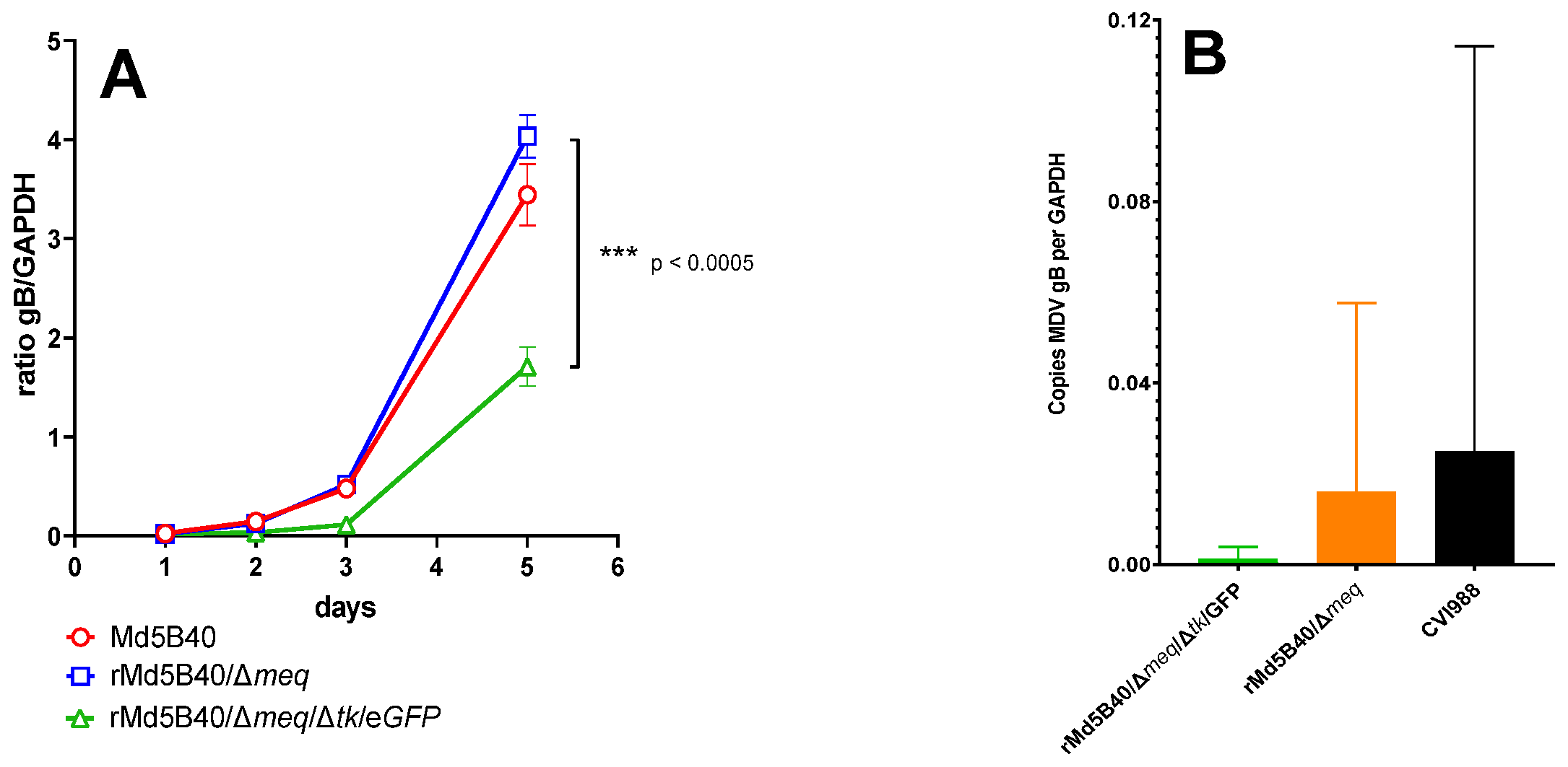
3.1. Ablation of the Viral tk from the rMd5B40/Δmeq Virus Attenuates Replication In Vitro

The wild-type MDV rMd5B40, the rMd5B40/Δmeq and the rMd5B40/Δmeqtk/GFP were compared for growth kinetics in vitro on monolayers of secondary DEF cells. Figure 1A shows that over the 5-day experiment both the MDV rMd5B40 and the rMd5B40/Δmeq viruses accumulated approximately the same quantity of viral glycoprotein B transcripts as measured by qPCR and normalized to the amount of GAPDH mRNA. Significantly less viral glycoprotein B transcript was detected in the rMd5B40/Δmeqtk/GFP-inoculated cultures than in the Md5B40 cultures at days 2, 3 and 5. By day 5, the rMd5B40/Δmeqtk/GFP virus had accumulated approximately half (p < 0.01) of the glycoprotein B message when compared to the rMd5B40 and the rMd5B40/Δmeq viruses. Replication of the Δtk recombinants was likewise reduced in vivo. Figure 1B shows that at 6 days post inoculation, when inoculated into day-of-hatch chicks, both of the Δtk recombinants produced less viremia than the CVI988 vaccine strain or the rMd5B40/Δmeq recombinant. Unfortunately high group variability in the rMd5B40/Δmeq and CVI988 groups prevents us from saying definitively that viral replication in vivo was also attenuated. We therefore suggest that ablation of the MDV tk results in a replication-attenuated virus in vitro.

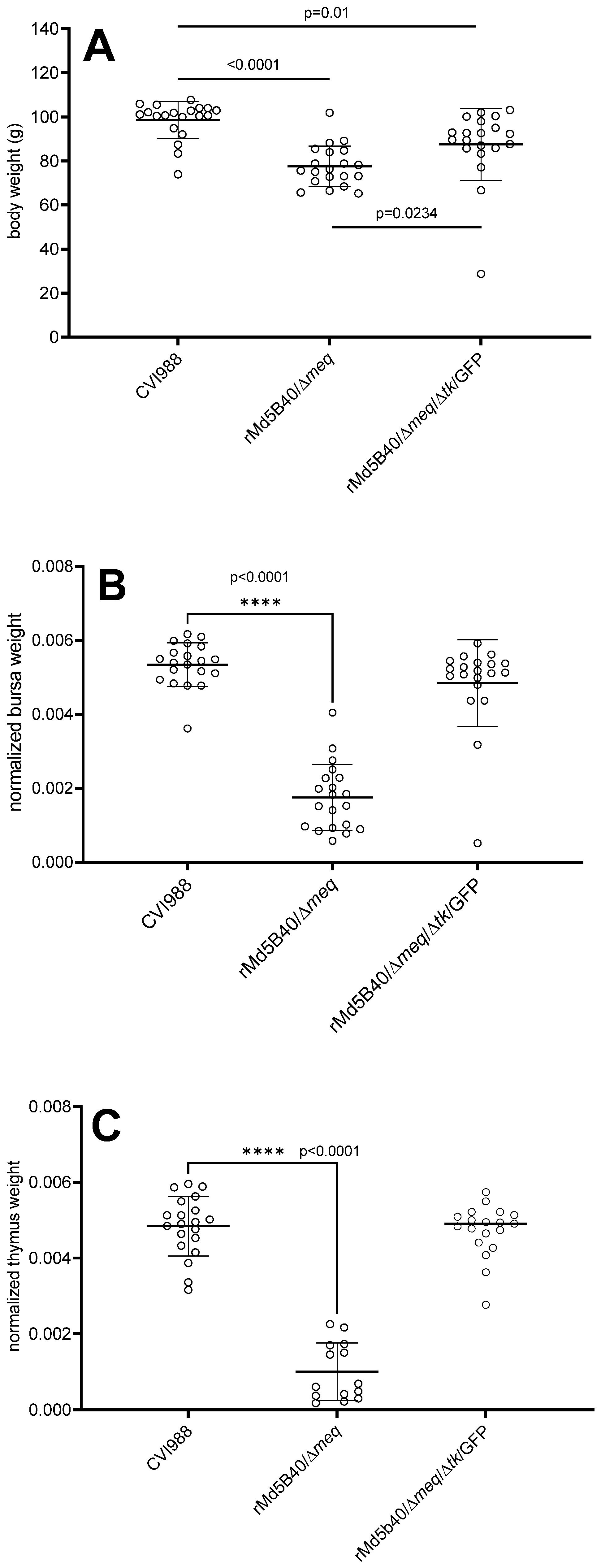
3.2. Attenuation of the rMd5B40/Δmeq Virus by Ablation of the Viral tk Spared Chickens from Lymphoid Atrophy

The rMd5B40/Δmeqtk/GFP virus was compared to both the parental rMd5B40/Δmeq virus and the CVI988/Rispens virus with respect to body weight and to determine the degree of lymphoid atrophy produced when used alone in maternal antibody-negative chickens. The CVI988/Rispens is an important comparator because it is the industry-leading vaccine at the time of our study. We examined the 13-day post-inoculation time point because viral replication should be readily detectable yet not reduced by entry into the lytic phase. As shown in Figure 2A–C, by 13 days post-inoculation clear differences among the groups could be discerned. Figure 2A shows that when compared to the CVI988/Rispens-inoculated controls, the rMd5B40/Δmeq virus and the rMd5B40/Δmeqtk/GFP virus both caused statistically significant reductions in body weight at 13 days post-inoculation. The rMd5B40/Δmeq virus caused the most severe reduction in body weight, a 21% reduction (p < 0.0001) when compared to the CVI988-vaccinated controls. In the rMd5B40/Δmeqtk/GFP-inoculated group the reduction in body weight was less pronounced (11% reduction, p = 0.0011). Notably, the rMd5B40/Δmeqtk/GFP-inoculated group (mean body weight 87.5 g) experienced less body-weight loss than did the rMd5B40/Δmeq-inoculated group (77.6 g), a small but significant (p = 0.0234) improvement. As for lymphoid atrophy, Figure 2B,C show that the rMd5B40/Δmeqtk/GFP-inoculated group (chicks experienced no significant reduction in lymphoid organ weight, and that only rMd5B40/Δmeq virus produced significant atrophy of the thymus (79%, Figure 2B) and bursa (67%, Figure 2C), as anticipated. This data supports our hypothesis that ablation of the tk gene from the rMd5B40/Δmeq virus curtails its propensity to cause atrophy of the lymphoid organs.

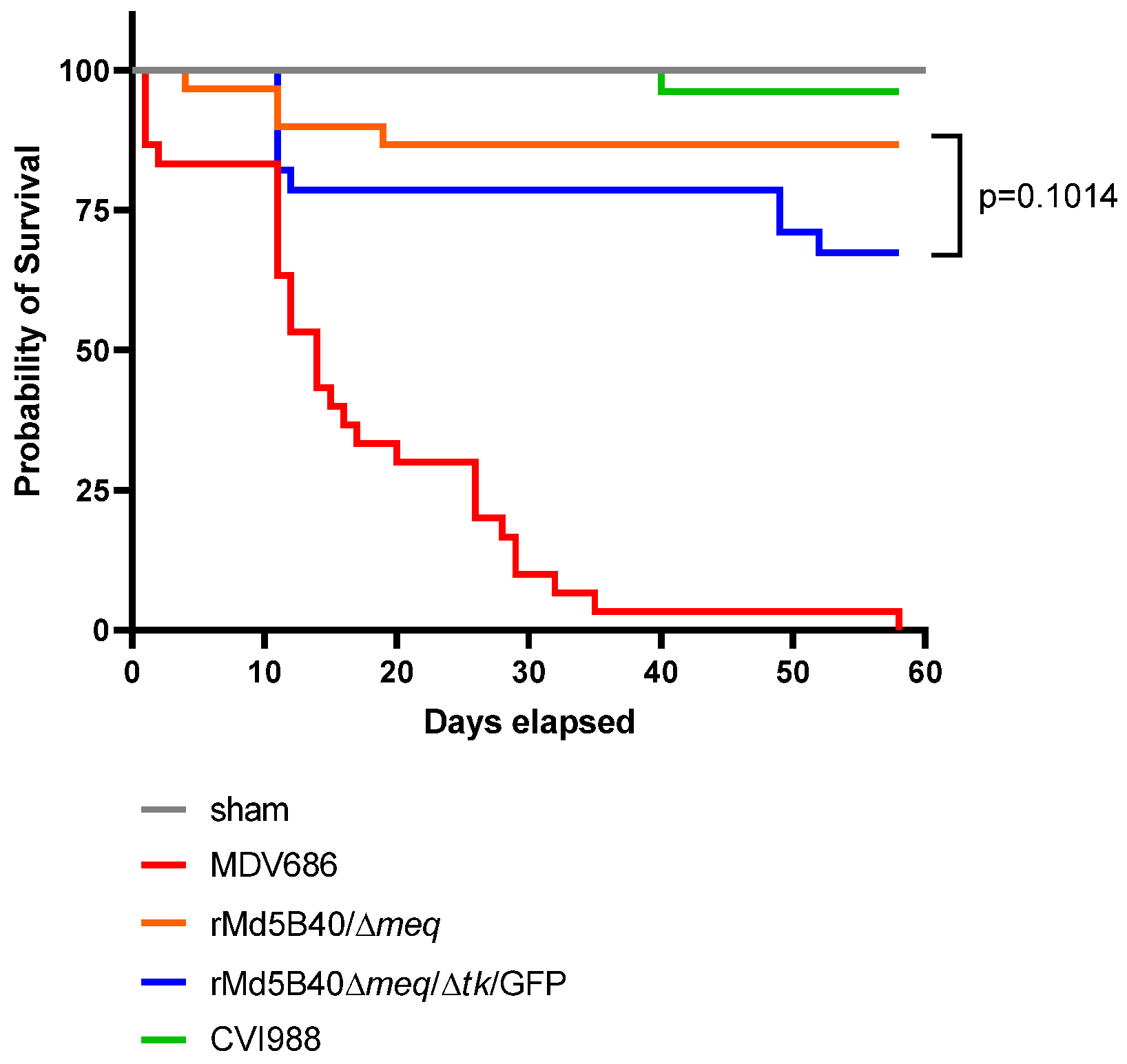
3.3. The rMd5B40/Δmeq/Δtk/GFP Viruse Displayed Reduced Protection against MD, against the Development of Visceral Tumors and against Mortality When Challenged with a vv+ MD

Table 1 shows that, compared with the group in which the rMd5B40/Δmeq virus was used as a vaccine and subsequently challenged with MDV686 (10% MD+), the rMd5B40/Δmeqtk/GFP virus was less protective against the development of the visceral tumors, which are diagnostic of MD (32.1% MD+). The recombinant virus, in which the viral tk was ablated, performed significantly worse than did the CVI988 virus and the parental rMd5B40/Δmeq virus.
Figure 3 illustrates the protective ability of the recombinants. The Kaplan–Meier survival curve shows that in the absence of protection (sham group, gray line), the MDV686 (red line) was indeed lethal over the course of the experiment, with 100% mortality by day 60 post challenge and a median survival time of 14 days. Also, the CVI988/Rispens strain (green line) performed very well, with all but one animal surviving until the end of the study. The best performer of the recombinant viruses was the rMd5B40/Δmeq virus (orange line), which was not significantly different from the CVI988/Rispens strain. Although the rMd5B40/Δmeq and rMd5B40/Δmeqtk/GFP (blue line) were not statistically different (p = 0.1014), they were clearly trending towards a significant difference in which the rMd5B40/Δmeqtk/GFP recombinant virus provided less protection than did the rMd5B40/Δmeq recombinant. For this reason, we must consider the rMd5B40/Δmeqtk/GFP virus to be slightly less protective than the rMd5B40/Δmeq virus, although the difference did not reach statistical significance.

4. Discussion

The rMd5B40/Δmeq viruses made and tested by several groups (ours and others, see [23]) have been highly protective as vaccines against vv+ MDV challenge, yet have produced an unacceptable amount of lymphoid organ atrophy. We attempted to attenuate the rMd5B40/Δmeq virus by ablation of the viral thymidine kinase. This is a common strategy to attenuate herpesviruses. Our hypothesis was that the lymphoid atrophy caused by the rMd5B40/Δmeq recombinant could be alleviated by reducing viral replication in vivo. We engineered a rMd5B40/Δmeqtk/GFP virus by ablation of the viral tk from rMd5B40/Δmeq. The ablation of the viral tk resulted in a virus that was significantly attenuated for replication in vitro (see Figure 1). When tested in vivo, the rMd5B40/Δmeqtk/GFP recombinant trended towards less viral replication when measured at 6 days post-inoculation, but high variability in the rMd5B40/Δmeq-inoculated group precludes the determination of a significant difference between these two groups. When tested in vivo alone (i.e., without a subsequent challenge) we verified that the rMd5B40/Δmeq virus did indeed cause lymphoid atrophy, in agreement with previous reports [23]. This data fits well with previous data in which deletion of the viral tk has resulted in viruses attenuated for replication [24].
As hypothesized, the ablation of the viral tk from the rMd5B40/Δmeq recombinant virus did reduce atrophy to the thymus and bursa by an amount sufficient to make it statistically indistinguishable from the CVI988-inoculated controls by 13 days post-inoculation, although there was a very small but significant amount of body weight loss in the rMd5B40/Δmeqtk/GFP group. This loss was significantly smaller than was experienced by the rMd5B40/Δmeq-inoculated group, however. The tk-deleted recombinant rMd5B40/Δmeqtk/GFP completely spared the inoculated animals from atrophy of the thymus and bursa. From these results we conclude that ablation of the viral tk does indeed attenuate the atrophy caused by the rMd5B40/Δmeq virus, although these experiments do not address the mechanism of this reduction. While this is an encouraging finding, for the rMd5B40/Δmeqtk/GFP virus to be useful as a potential vaccine it must retain all of the protective capacity of the rMd5B40/Δmeq recombinant, which it did not. Table 1 shows that animals vaccinated with the rMd5B40/Δmeq recombinant and then challenged with the vv+ MDV686 are highly protected but that animals vaccinated with the rMd5B40/Δmeqtk/GFP recombinant and challenged with MDV686 experience visceral tumor formation in the majority of the tested animals (23/32). For protection against mortality, the rMd5B40/Δmeqtk/GFP recombinant was indistinguishable from the rMd5B40/Δmeq recombinant (p = 0.1014) but the data for this group trended towards lower group survival when used as a vaccine against the vv+ pathotype MDV686 strain. From this data, combined with our data which shows that the rMd5B40/Δmeqtk/GFP caused a small amount of body weight loss compared with the existing best-in-class vaccine (CVI988/Rispens) it is clear that the most protective of our recombinant viruses, the rMd5B40/Δmeqtk/GFP, is not superior in these experiments to the CVI988/Rispens vaccine strain and would, therefore, not be an improvement compared with the currently available CVI988/Rispens.
One possible interpretation of these results is that attenuation of replication in the meq-deleted MDV also reduces its protective ability when used as a vaccine, an observation which has been made before [25,34]. Additionally, a very recent paper [35] has demonstrated that ablation of the viral IL-8 analog (vIL8) in a meq-deleted background vv+ virus (MDV686BAC) resulted in a vaccine strain that is at least as protective as the CVI988/Rispens strain and that causes no detectable atrophy of either the bursa or thymus when tested alone. The same study also noted a reduction in viral replication of the meq- and vIl8-deleted mutant in vivo. This is a clear indication that attenuation mechanisms which merely reduce viral replication in vitro or in vivo cannot be expected to result in superior vaccine strains for protection against MD. Rather, the way forward is likely to be through targeted changes in the immunomodulation capabilities of the virus. If this strategy is borne out in other herpesviruses (and other viruses in general) then its application will allow the production of safe and efficacious vaccines for any number of communicable diseases.

Supplementary Materials

The following supporting information can be downloaded at: https://www.mdpi.com/article/10.3390/microorganisms10010007/s1, Figure S1: Gel electrophoresis of isolates screened for confirmation. Figure S2: Verification of the insertion and orientation of the GFP marker into the tk gene. Table S1: Primers used.

Author Contributions

Concept and experimental design, S.J.C., J.R.D. and M.H.; investigation and laboratory manipulations E.B.O. and J.K.M.; writing—original draft preparation, S.J.C.; writing—review and editing, S.J.C., E.B.O., J.R.D. and M.H. All authors have read and agreed to the published version of the manuscript.

Funding

This research received no external funding.

Institutional Review Board Statement

The study was conducted according to the guidelines of the Declaration of Helsinki, and approved by the Institutional Review Board (or Ethics Committee) of Michigan State University Health and Safety (protocol code #SAFETY0000269, approved 3 April 2019).

Informed Consent Statement

Not applicable.

Data Availability Statement

The data presented in this study are available within this paper.

Acknowledgments

We would like to thank Lonnie Milam for his excellent technical assistance, as well as our animal caretaking staff for their commitment to animal welfare. Mention of trade names or commercial products in this publication is solely for the purpose of providing specific information and does not imply recommendation or endorsement of the U.S. Department of Agriculture.

Conflicts of Interest

The authors declare no conflict of interest. The funders had no role in the design of the study; in the collection, analyses, or interpretation of data; in the writing of the manuscript, or in the decision to publish the results.

References

  1. Osterrieder, N.; Kamil, J.P.; Schumacher, D.; Tischer, B.K.; Trapp, S. Marek’s Disease Virus: From Miasma to Model. Nat. Rev. Microbiol. 2006, 4, 283–294. [Google Scholar] [CrossRef]
  2. Bertzbach, L.D.; Conradie, A.M.; You, Y.; Kaufer, B.B. Latest Insights into Marek’s Disease Virus Pathogenesis and Tumorigenesis. Cancers 2020, 12, 647. [Google Scholar] [CrossRef] [Green Version]
  3. Calnek, B.W. Pathogenesis of Marek’s Disease Virus Infection. In Marek’s Disease; Hirai, K., Ed.; Current Topics in Microbiology and Immunology; Springer: Berlin/Heidelberg, Germany, 2001; Volume 255. [Google Scholar] [CrossRef]
  4. Jarosinski, K.W. Marek’s Disease Virus Late Protein Expression in Feather Follicle Epithelial Cells as Early as 8 Days Postinfection. Avian Dis. 2012, 56, 725–731. [Google Scholar] [CrossRef] [PubMed]
  5. Bertzbach, L.D.; Laparidou, M.; Härtle, S.; Etches, R.J.; Kaspers, B.; Schusser, B.; Kaufer, B.B. Unraveling the Role of B Cells in the Pathogenesis of an Oncogenic Avian Herpesvirus. Proc. Natl. Acad. Sci. USA 2018, 115, 11603–11607. [Google Scholar] [CrossRef] [Green Version]
  6. Engel, A.T.; Selvaraj, R.K.; Kamil, J.P.; Osterrieder, N.; Kaufer, B.B. Marek’s Disease Viral Interleukin-8 Promotes Lymphoma Formation through Targeted Recruitment of B Cells and CD4+ CD25+ T Cells. J. Virol. 2012, 86, 8536–8545. [Google Scholar] [CrossRef] [PubMed] [Green Version]
  7. Boodhoo, N.; Gurung, A.; Sharif, S.; Behboudi, S. Marek’s Disease in Chickens: A Review with Focus on Immunology. Veter. Res. 2016, 47, 119. [Google Scholar] [CrossRef] [PubMed] [Green Version]
  8. Robinson, C.M.; Cheng, H.H.; Delany, M.E. Temporal Kinetics of Marek’s Disease Herpesvirus: Integration Occurs Early after Infection in Both B and T Cells. Cytogenet. Genome Res. 2014, 144, 142–154. [Google Scholar] [CrossRef] [PubMed]
  9. Parcells, M.; Arumugaswami, V.; Prigge, J.; Pandya, K.; Dienglewicz, R. Marek’s Disease Virus Reactivation from Latency: Changes in Gene Expression at the Origin of Replication. Poult. Sci. 2003, 82, 893–898. [Google Scholar] [CrossRef]
  10. Rispens, B.H.; van Vloten, H.; Mastenbroek, N.; Maas, H.J.; Schat, K.A. Control of Marek’s disease in the Netherlands. I. Isolation of an avirulent Marek’s disease virus (strain CVI 988) and its use in laboratory vaccination trials. Avian Dis. 1972, 16, 108–125. [Google Scholar] [CrossRef]
  11. Read, A.F.; Baigent, S.J.; Powers, C.; Kgosana, L.B.; Blackwell, L.; Smith, L.P.; Kennedy, D.A.; Walkden-Brown, S.W.; Nair, V.K. Imperfect Vaccination Can Enhance the Transmission of Highly Virulent Pathogens. PLoS Biol. 2015, 13, e1002198. [Google Scholar] [CrossRef]
  12. Bailey, R.I.; Cheng, H.H.; Chase-Topping, M.; Mays, J.K.; Anacleto, O.; Dunn, J.R.; Doeschl-Wilson, A. Pathogen transmission from vaccinated hosts can cause dose-dependent reduction in virulence. PLoS Biol. 2020, 18, e3000619. [Google Scholar] [CrossRef] [Green Version]
  13. Witter, R.L. Increased Virulence of Marek’s Disease Virus Field Isolates. Avian Dis. 1997, 41, 149. [Google Scholar] [CrossRef] [PubMed]
  14. Trimpert, J.; Groenke, N.; Jenckel, M.; He, S.; Kunec, D.; Szpara, M.L.; Spatz, S.J.; Osterrieder, N.; McMahon, D.P. A Phylogenomic Analysis of Marek’s Disease Virus Reveals Independent Paths to Virulence in Eurasia and North America. Evol. Appl. 2017, 10, 1091–1101. [Google Scholar] [CrossRef] [PubMed] [Green Version]
  15. Witter, R.L. The Changing Landscape of Marek’s Disease. Avian Pathol. 1998, 27, S46–S53. [Google Scholar] [CrossRef]
  16. Jones, D.; Lee, L.; Liu, J.L.; Kung, H.J.; Tillotson, J.K. Marek Disease Virus Encodes a Basic-Leucine Zipper Gene Resembling the Fos/Jun Oncogenes That Is Highly Expressed in Lymphoblastoid Tumors. Proc. Natl. Acad. Sci. USA 1992, 89, 4042–4046. [Google Scholar] [CrossRef] [PubMed] [Green Version]
  17. Tulman, E.R.; Afonso, C.L.; Lu, Z.; Zsak, L.; Rock, D.L.; Kutish, G.F. The Genome of a Very Virulent Marek’s Disease Virus. J. Virol. 2000, 74, 7980–7988. [Google Scholar] [CrossRef] [PubMed] [Green Version]
  18. Suchodolski, P.F.; Izumiya, Y.; Lupiani, B.; Ajithdoss, D.K.; Gilad, O.; Lee, L.F.; Kung, H.-J.; Reddy, S.M. Homodimerization of Marek’s Disease Virus-Encoded Meq Protein Is Not Sufficient for Transformation of Lymphocytes in Chickens. J. Virol. 2008, 83, 859–869. [Google Scholar] [CrossRef] [PubMed] [Green Version]
  19. Levy, A.M.; Izumiya, Y.; Brunovskis, P.; Xia, L.; Parcells, M.S.; Reddy, S.M.; Lee, L.; Chen, H.-W.; Kung, H.-J. Characterization of the Chromosomal Binding Sites and Dimerization Partners of the Viral Oncoprotein Meq in Marek’s Disease Virus-Transformed T Cells. J. Virol. 2003, 77, 12841–12851. [Google Scholar] [CrossRef] [Green Version]
  20. Suchodolski, P.F.; Izumiya, Y.; Lupiani, B.; Ajithdoss, D.K.; Lee, L.F.; Kung, H.-J.; Reddy, S.M. Both Homo and Heterodimers of Marek’s Disease Virus Encoded Meq Protein Contribute to Transformation of Lymphocytes in Chickens. Virology 2010, 399, 312–321. [Google Scholar] [CrossRef] [Green Version]
  21. Levy, A.M.; Gilad, O.; Xia, L.; Izumiya, Y.; Choi, J.; Tsalenko, A.; Yakhini, Z.; Witter, R.; Lee, L.; Cardona, C.J.; et al. Marek’s Disease Virus Meq Transforms Chicken Cells via the v-Jun Transcriptional Cascade: A Converging Transforming Pathway for Avian Oncoviruses. Proc. Natl. Acad. Sci. USA 2005, 102, 14831–14836. [Google Scholar] [CrossRef] [Green Version]
  22. Deng, X.; Li, X.; Shen, Y.; Qiu, Y.; Shi, Z.; Shao, D.; Jin, Y.; Chen, H.; Ding, C.; Li, L.; et al. The Meq Oncoprotein of Marek’s Disease Virus Interacts with P53 and Inhibits Its Transcriptional and Apoptotic Activities. Virol. J. 2010, 7, 348. [Google Scholar] [CrossRef] [Green Version]
  23. Lupiani, B.; Lee, L.F.; Cui, X.; Gimeno, I.; Anderson, A.; Morgan, R.W.; Silva, R.F.; Witter, R.L.; Kung, H.-J.; Reddy, S.M. Marek’s Disease Virus-Encoded Meq Gene Is Involved in Transformation of Lymphocytes but Is Dispensable for Replication. Proc. Natl. Acad. Sci. USA 2004, 101, 11815–11820. [Google Scholar] [CrossRef] [PubMed] [Green Version]
  24. Zhang, M.; Fu, S.; Deng, M.; Xie, Q.; Xu, H.; Liu, Z.; Hu, C.; Chen, H.; Guo, A. Attenuation of Bovine Herpesvirus Type 1 by Deletion of Its Glycoprotein G and Tk Genes and Protection against Virulent Viral Challenge. Vaccine 2011, 29, 8943–8950. [Google Scholar] [CrossRef]
  25. Sun, P.; Cui, N.; Liu, L.; Su, S.; Cheng, Z.; Chen, R.; Li, Y.; Cui, Z. Attenuation of a Recombinant Marek’s Disease Virus Lacking the Meq Oncogene and Evaluation on Its Immune Efficacy against Marek’s Disease Virus. Poult. Sci. 2020, 99, 1939–1945. [Google Scholar] [CrossRef]
  26. Niikura, M.; Kim, T.; Silva, R.F.; Dodgson, J.; Cheng, H.H. Virulent Marek’s Disease Virus Generated from Infectious Bacterial Artificial Chromosome Clones with Complete DNA Sequence and the Implication of Viral Genetic Homogeneity in Pathogenesis. J. Gen. Virol. 2010, 92, 598–607. [Google Scholar] [CrossRef] [PubMed]
  27. Noll, J.C.G.; Joshi, L.R.; do Nascimento, G.M.; Fernandes, M.H.V.; Sharma, B.; Flores, E.F.; Diel, D.G. Deletion of the Thymidine Kinase Gene Attenuates Caprine Alphaherpesvirus 1 in Goats. Veter. Microbiol. 2019, 237, 108370. [Google Scholar] [CrossRef] [PubMed]
  28. Xie, Y.; Wu, L.; Wang, M.; Cheng, A.; Yang, Q.; Wu, Y.; Jia, R.; Zhu, D.; Zhao, X.; Chen, S.; et al. Alpha-Herpesvirus Thymidine Kinase Genes Mediate Viral Virulence and Are Potential Therapeutic Targets. Front. Microbiol. 2019, 10, 941. [Google Scholar] [CrossRef] [Green Version]
  29. Kokoris, M.S.; Black, M.E. Characterization of Herpes Simplex Virus Type 1 Thymidine Kinase Mutants Engineered for Improved Ganciclovir or Acyclovir Activity. Protein Sci. 2002, 11, 2267–2272. [Google Scholar] [CrossRef] [Green Version]
  30. Sanford, B.; Holinka, L.G.; O’Donnell, V.; Krug, P.W.; Carlson, J.; Alfano, M.; Carrillo, C.; Wu, P.; Lowe, A.; Risatti, G.R.; et al. Deletion of the Thymidine Kinase Gene Induces Complete Attenuation of the Georgia Isolate of African Swine Fever Virus. Virus Res. 2016, 213, 165–171. [Google Scholar] [CrossRef] [PubMed]
  31. Deng, L.; Fan, J.; Ding, Y.; Zhang, J.; Zhou, B.; Zhang, Y.; Huang, B. Oncolytic Efficacy of Thymidine Kinase-Deleted Vaccinia Virus Strain Guang9. Oncotarget 2014, 5, 40533–40543. [Google Scholar] [CrossRef] [PubMed] [Green Version]
  32. Silva, R.F.; Dunn, J.R.; Cheng, H.H.; Niikura, M. A MEQ-Deleted Marek’s Disease Virus Cloned as a Bacterial Artificial Chromosome Is a Highly Efficacious Vaccine. Avian Dis. 2010, 54, 862–869. [Google Scholar] [CrossRef] [PubMed]
  33. Karstentischer, B.; von Einem, J.; Kaufer, B.; Osterrieder, N. Two-Step Red-Mediated Recombination for Versatile High-Efficiency Markerless DNA Manipulation in Escherichia coli. Biotechniques 2006, 40, 191–197. [Google Scholar] [CrossRef] [PubMed]
  34. Cadore, G.C.; Weiss, M.; Anziliero, D.; Brum, M.C.S.; Weiblen, R.; Flores, E.F. A Thymidine Kinase-Deleted Bovine Herpesvirus 5 Establishes Latent Infection but Reactivates Poorly in a Sheep Model. Pesquisa Veterinária Brasileira 2013, 33, 331–338. [Google Scholar] [CrossRef]
  35. Liao, Y.; Reddy, S.M.; Khan, O.A.; Sun, A.; Lupiani, B. A Novel Effective and Safe Vaccine for Prevention of Marek’s Disease Caused by Infection with a Very Virulent Plus (Vv+) Marek’s Disease Virus. Vaccines 2021, 9, 159. [Google Scholar] [CrossRef] [PubMed]
Figure 1. Ablation of the tk gene in MDV rMd5B40 attenuates replication both in vitro and in vivo. (A) When viral replication was assayed in vitro, the rMd5B40Δmeq/Δtk/eGFP variant accumulated significantly less glycoprotein B transcripts than either the wild-type virus or the rMd5B40/Δmeq. (B) In vivo, the Δtk recombinant also trended towards less viremia 6 days after inoculation at day-of-hatch, but the differences between these groups and the rMd5B40/Δmeq parental were not statistically significant (data not shown). For panel A data were compared using unpaired t-tests, (***) indicates a statistically significant difference between groups. For B, data were compared using ordinary ANOVA analysis.
Microorganisms 10 00007 g001
Figure 2. Ablation of the tk gene from the rMd5B40/Δmeq virus mitigates atrophy of the lymphoid organs. 13-day old chickens (A) body weight is significantly reduced with inoculation of the recombinant viruses compared to the CVI988 control, but much less in the tk-deleted recombinant. The (B) bursa weights and (C) thymus weights were normalized to body mass. In each case only the meq-ablated recombinant causes detectable atrophy of the lymphoid organs. Each recombinant virus-inoculated group is compared to the CVI988 controls. Data were compared using unpaired t-tests, (****) indicates a statistically significant difference between groups.
Microorganisms 10 00007 g002
Figure 3. Ablation of the viral tk reduces protection against mortality when challenged by a vv+ MDV. The rMd5B40/Δmeqtk/GFP (blue line) was not significantly less protective than the parental rMd5B40/Δmeq (orange line) in which the tk was intact, although survival trended lower in this construct (p = 0.1014, Log-rank Mantel-C). As anticipated, the CVI988 vaccine was extremely protective (green line), while unvaccinated animals were highly susceptible to MDV686 in the absence of protection (red line).
Microorganisms 10 00007 g003
Table 1. Ablation of the viral tk reduces protection against the development of the visceral tumors diagnostic of MD. Birds were scored for the presence of visceral tumors at necropsy.
Vaccine StrainChallenge StrainMD+/Total
noneMDV68625/30
CVI988/RispensMDV68601/30
rMd5B40/ΔmeqMDV68603/30
rMd5B40/Δmeq/tk/GFPMDV68623/32
Publisher’s Note: MDPI stays neutral with regard to jurisdictional claims in published maps and institutional affiliations.

