
Antibacterial Potential of Essential Oils and Silver Nanoparticles against Multidrug-Resistant Staphylococcus pseudintermedius Isolates
 , , , , ,
  ,
,  and
and
Abstract
:1. Introduction
2. Materials and Methods
2.1. Bacterial Strains, Identification, and Culture Conditions
2.2. Determination of Antimicrobial Profiles
2.3. Synthesis and Characterization of Silver Nanoparticles
2.4. Characterization of Essential Oils
2.5. Minimum Inhibitory Concentration (MIC) and Minimum Bactericidal Concentration (MBC) of AgNPs and EOs
3. Results
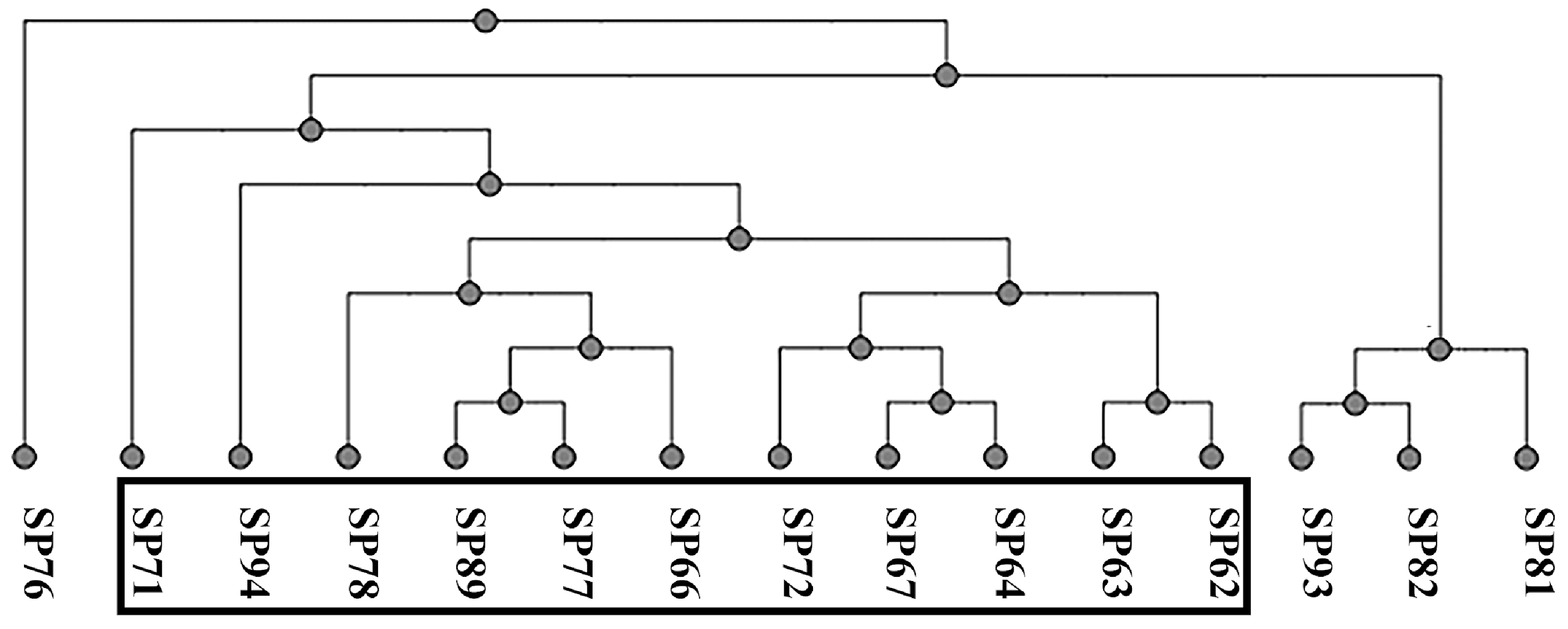
3.1. Phenotypic and Molecular Profiling of Antibiotic Resistance
3.2. Characterization of Antimicrobial Molecules
3.2.1. AgNPs
3.2.2. EOs
3.3. Antibacterial Activity of EOs and NPs
4. Discussion
5. Conclusions
Supplementary Materials
Author Contributions
Funding
Institutional Review Board Statement
Informed Consent Statement
Data Availability Statement
Acknowledgments
Conflicts of Interest
References
- Somayaji, R.; Priyantha, M.A.R.; Rubin, J.E.; Church, D. Human Infections Due to Staphylococcus Pseudintermedius, an Emerging Zoonosis of Canine Origin: Report of 24 Cases. Diagn. Microbiol. Infect. Dis. 2016, 85, 471–476. [Google Scholar] [CrossRef]
- Ference, E.H.; Danielian, A.; Kim, H.W.; Yoo, F.; Kuan, E.C.; Suh, J.D. Zoonotic Staphylococcus Pseudintermedius Sinonasal Infections: Risk Factors and Resistance Patterns. Int. Forum Allergy Rhinol. 2019, 9, 724–729. [Google Scholar] [CrossRef]
- Kmieciak, W.; Szewczyk, E.M. Are Zoonotic Staphylococcus Pseudintermedius Strains a Growing Threat for Humans? Folia Microbiol. 2018, 63, 743–747. [Google Scholar] [CrossRef] [PubMed]
- Meroni, G.; Filipe, J.F.S.; Drago, L.; Martino, P.A. Investigation on Antibiotic-Resistance, Biofilm Formation and Virulence Factors in Multi Drug Resistant and Non Multi Drug Resistant Staphylococcus Pseudintermedius. Microorganisms 2019, 7, 702. [Google Scholar] [CrossRef]
- Meroni, G.; Filipe, J.F.S.; Martino, P.A. In Vitro Antibacterial Activity of Biological-Derived Silver Nanoparticles: Preliminary Data. Vet. Sci. 2020, 7, 12. [Google Scholar] [CrossRef]
- Rubin, J.E.; Chirino-Trejo, M. Prevalence, Sites of Colonization, and Antimicrobial Resistance Among Staphylococcus Pseudintermedius Isolated from Healthy Dogs in Saskatoon, Canada. J. Vet. Diagn. Investig. 2011, 23, 351–354. [Google Scholar] [CrossRef] [PubMed]
- Small, C.; Beatty, N.; Helou, G.E.; Small, C.; Beatty, N.; Helou, G.E. Staphylococcus Pseudintermedius Bacteremia in a Lung Transplant Recipient Exposed to Domestic Pets. Cureus 2021, 13, e14895. [Google Scholar] [CrossRef] [PubMed]
- Asleh, M.; Feinstein, Y.; Lazar, I.; Rokney, A.; Baum, M.; Sagi, O.; Leibovitz, E.; Danino, D. Severe Pneumonia Caused by Methicillin-Resistant Staphylococcus Pseudintermedius in an Oncology Patient: Case Report and Literature Review. Microb. Drug Resist. Larchmt. N 2022, 28, 222–228. [Google Scholar] [CrossRef]
- Blondeau, L.D.; Rubin, J.E.; Deneer, H.; Kanthan, R.; Sanche, S.; Beshard, N.; Mpofu, C.; Blondeau, J.M. Bacteremia with Staphylococcus Pseudintermedius in a 4 Month Old Pediatric Oncology Patient. J. Chemother. 2020, 32, 260–262. [Google Scholar] [CrossRef]
- Chuang, C.-Y.; Yang, Y.-L.; Hsueh, P.-R.; Lee, P.-I. Catheter-Related Bacteremia Caused by Staphylococcus Pseudintermedius Refractory to Antibiotic-Lock Therapy in a Hemophilic Child with Dog Exposure. J. Clin. Microbiol. 2010, 48, 1497–1498. [Google Scholar] [CrossRef]
- Paul, N.; Moodley, A.; Ghibaudo, G.; Guardabassi, L. Carriage of Methicillin-Resistant Staphylococcus Pseudintermedius in Small Animal Veterinarians: Indirect Evidence of Zoonotic Transmission. Zoonoses Public Health 2011, 58, 533–539. [Google Scholar] [CrossRef]
- Moses, I.B.; Santos, F.F.; Gales, A.C. Human Colonization and Infection by Staphylococcus Pseudintermedius: An Emerging and Underestimated Zoonotic Pathogen. Microorganisms 2023, 11, 581. [Google Scholar] [CrossRef]
- Álvarez-Martínez, F.J.; Barrajón-Catalán, E.; Herranz-López, M.; Micol, V. Antibacterial Plant Compounds, Extracts and Essential Oils: An Updated Review on Their Effects and Putative Mechanisms of Action. Phytomedicine Int. J. Phytother. Phytopharm. 2021, 90, 153626. [Google Scholar] [CrossRef]
- Murugaiyan, J.; Kumar, P.A.; Rao, G.S.; Iskandar, K.; Hawser, S.; Hays, J.P.; Mohsen, Y.; Adukkadukkam, S.; Awuah, W.A.; Jose, R.A.M.; et al. Progress in Alternative Strategies to Combat Antimicrobial Resistance: Focus on Antibiotics. Antibiotics 2022, 11, 200. [Google Scholar] [CrossRef]
- Meroni, G.; Cardin, E.; Rendina, C.; Herrera Millar, V.R.; Soares Filipe, J.F.; Martino, P.A. In Vitro Efficacy of Essential Oils from Melaleuca alternifolia and Rosmarinus officinalis, Manuka Honey-Based Gel, and Propolis as Antibacterial Agents against Canine Staphylococcus Pseudintermedius Strains. Antibiotics 2020, 9, 344. [Google Scholar] [CrossRef]
- Dhifi, W.; Bellili, S.; Jazi, S.; Bahloul, N.; Mnif, W. Essential Oils’ Chemical Characterization and Investigation of Some Biological Activities: A Critical Review. Medicines 2016, 3, 25. [Google Scholar] [CrossRef]
- Sandner, G.; Heckmann, M.; Weghuber, J. Immunomodulatory Activities of Selected Essential Oils. Biomolecules 2020, 10, 1139. [Google Scholar] [CrossRef]
- Farrar, A.J.; Farrar, F.C. Clinical Aromatherapy. Nurs. Clin. N. Am. 2020, 55, 489–504. [Google Scholar] [CrossRef] [PubMed]
- Veiskaramian, A.; Gholami, M.; Yarahmadi, S.; Amanolahi Baharvand, P.; Birjandi, M. Effect of Aromatherapy with Melissa Essential Oil on Stress and Hemodynamic Parameters in Acute Coronary Syndrome Patients: A Clinical Trial in the Emergency Department. Complement. Ther. Clin. Pract. 2021, 44, 101436. [Google Scholar] [CrossRef] [PubMed]
- Szewczuk, M.A.; Zych, S.; Oster, N.; Karakulska, J. Activity of Patchouli and Tea Tree Essential Oils against Staphylococci Isolated from Pyoderma in Dogs and Their Synergistic Potential with Gentamicin and Enrofloxacin. Animals 2023, 13, 1279. [Google Scholar] [CrossRef] [PubMed]
- Duangkaew, L.; Larsuprom, L.; Lekcharoensuk, C.; Chen, C. Effect of a Mixture of Essential Oils and a Plant-Based Extract for the Management of Localized Superficial Pyoderma in Dogs: An Open-Label Clinical Trial. Thai J. Vet. Med. 2017, 47, 513–522. [Google Scholar] [CrossRef]
- Aiemsaard, J.; Aiyaranoi, K.; Thongkham, E.; Borlace, G.; Senaphan, K. In Vivo Efficacy of Clove Essential Oil Spray Formulation on Canine Superficial Pyoderma. Songklanakarin J. Sci. Technol. 2022, 44, 308–315. [Google Scholar] [CrossRef]
- Patterson, J.E.; McElmeel, L.; Wiederhold, N.P. In Vitro Activity of Essential Oils Against Gram-Positive and Gram-Negative Clinical Isolates, Including Carbapenem-Resistant Enterobacteriaceae. Open Forum Infect. Dis. 2019, 6, ofz502. [Google Scholar] [CrossRef]
- Hajibonabi, A.; Yekani, M.; Sharifi, S.; Nahad, J.S.; Dizaj, S.M.; Memar, M.Y. Antimicrobial Activity of Nanoformulations of Carvacrol and Thymol: New Trend and Applications. OpenNano 2023, 13, 100170. [Google Scholar] [CrossRef]
- Wang, X.; Tian, L.; Fu, J.; Liao, S.; Yang, S.; Jia, X.; Gong, G. Evaluation of the Membrane Damage Mechanism of Thymol against Bacillus Cereus and Its Application in the Preservation of Skim Milk. Food Control 2022, 131, 108435. [Google Scholar] [CrossRef]
- Mączka, W.; Twardawska, M.; Grabarczyk, M.; Wińska, K. Carvacrol—A Natural Phenolic Compound with Antimicrobial Properties. Antibiotics 2023, 12, 824. [Google Scholar] [CrossRef]
- Malik, S.; Muhammad, K.; Waheed, Y. Nanotechnology: A Revolution in Modern Industry. Molecules 2023, 28, 661. [Google Scholar] [CrossRef] [PubMed]
- Gherasim, O.; Puiu, R.A.; Bîrcă, A.C.; Burdușel, A.-C.; Grumezescu, A.M. An Updated Review on Silver Nanoparticles in Biomedicine. Nanomaterials 2020, 10, 2318. [Google Scholar] [CrossRef] [PubMed]
- Ong, W.T.J.; Nyam, K.L. Evaluation of Silver Nanoparticles in Cosmeceutical and Potential Biosafety Complications. Saudi J. Biol. Sci. 2022, 29, 2085–2094. [Google Scholar] [CrossRef] [PubMed]
- Sapino, S.; Chindamo, G.; Chirio, D.; Morel, S.; Peira, E.; Vercelli, C.; Gallarate, M. Nanocarriers in Veterinary Medicine: A Challenge for Improving Osteosarcoma Conventional Treatments. Nanomaterials 2022, 12, 4501. [Google Scholar] [CrossRef] [PubMed]
- Singh, M.; Thakur, V.; Kumar, V.; Raj, M.; Gupta, S.; Devi, N.; Upadhyay, S.K.; Macho, M.; Banerjee, A.; Ewe, D.; et al. Silver Nanoparticles and Its Mechanistic Insight for Chronic Wound Healing: Review on Recent Progress. Molecules 2022, 27, 5587. [Google Scholar] [CrossRef]
- Löfdahl, A.; Jern, A.; Flyman, S.; Kåredal, M.; Karlsson, H.L.; Larsson-Callerfelt, A.-K. Silver Nanoparticles Alter Cell Viability Ex Vivo and in Vitro and Induce Proinflammatory Effects in Human Lung Fibroblasts. Nanomaterials 2020, 10, 1868. [Google Scholar] [CrossRef]
- Franková, J.; Pivodová, V.; Vágnerová, H.; Juráňová, J.; Ulrichová, J. Effects of Silver Nanoparticles on Primary Cell Cultures of Fibroblasts and Keratinocytes in a Wound-Healing Model. J. Appl. Biomater. Funct. Mater. 2016, 14, 137–142. [Google Scholar] [CrossRef]
- Sasaki, T.; Tsubakishita, S.; Tanaka, Y.; Sakusabe, A.; Ohtsuka, M.; Hirotaki, S.; Kawakami, T.; Fukata, T.; Hiramatsu, K. Multiplex-PCR Method for Species Identification of Coagulase-Positive Staphylococci. J. Clin. Microbiol. 2010, 48, 765–769. [Google Scholar] [CrossRef]
- Bannoehr, J.; Franco, A.; Iurescia, M.; Battisti, A.; Fitzgerald, J.R. Molecular Diagnostic Identification of Staphylococcus pseudintermedius. J. Clin. Microbiol. 2009, 47, 469–471. [Google Scholar] [CrossRef]
- CLSI; Dolinsky, A.L.; Ohiro, R.K.; Fan, W.; Xiao, C.; Wu, F. National Committee for Clinical Laboratory Standards. 2000. Performance Standard for Antimicrobial Susceptibility Testing. Document M100–S10. J. Int. Med. Res. 2017, 46, 18. [Google Scholar] [CrossRef]
- Yerragopu, P.S.; Hiregoudar, S.; Nidoni, U.; Ramappa, K.T.; Sreenivas, A.; Doddagoudar, S. Chemical Synthesis of Silver Nanoparticles Using Tri-Sodium Citrate, Stability Study and Their Characterization. Int. Res. J. Pure Appl. Chem. 2020, 21, 37–50. [Google Scholar] [CrossRef]
- Garzoli, S.; Masci, V.L.; Caradonna, V.; Tiezzi, A.; Giacomello, P.; Ovidi, E. Liquid and Vapor Phase of Four Conifer-Derived Essential Oils: Comparison of Chemical Compositions and Antimicrobial and Antioxidant Properties. Pharmaceuticals 2021, 14, 134. [Google Scholar] [CrossRef] [PubMed]
- Taglienti, A.; Donati, L.; Ferretti, L.; Tomassoli, L.; Sapienza, F.; Sabatino, M.; Di Massimo, G.; Fiorentino, S.; Vecchiarelli, V.; Nota, P.; et al. In Vivo Antiphytoviral Activity of Essential Oils and Hydrosols from Origanum vulgare, Thymus vulgaris, and Rosmarinus officinalis to Control Zucchini Yellow Mosaic Virus and Tomato Leaf Curl New Delhi Virus in Cucurbita pepo L. Front. Microbiol. 2022, 13, 840893. [Google Scholar] [CrossRef] [PubMed]
- Tsugawa, H.; Cajka, T.; Kind, T.; Ma, Y.; Higgins, B.; Ikeda, K.; Kanazawa, M.; VanderGheynst, J.; Fiehn, O.; Arita, M. MS-DIAL: Data Independent MS/MS Deconvolution for Comprehensive Metabolome Analysis. Nat. Methods 2015, 12, 523–526. [Google Scholar] [CrossRef] [PubMed]
- Ferrer, L.; García-Fonticoba, R.; Pérez, D.; Viñes, J.; Fàbregas, N.; Madroñero, S.; Meroni, G.; Martino, P.A.; Martínez, S.; Maté, M.L.; et al. Whole Genome Sequencing and de Novo Assembly of Staphylococcus Pseudintermedius: A Pangenome Approach to Unravelling Pathogenesis of Canine Pyoderma. Vet. Dermatol. 2021, 32, 654–663. [Google Scholar] [CrossRef]
- Nocera, F.P.; Meroni, G.; Fiorito, F.; De Martino, L.; Martino, P.A. Occurrence and Antimicrobial Susceptibility Patterns of Canine Staphylococcus Pseudintermedius Strains Isolated from Two Different Italian University Veterinary Hospitals. Vet. Ital. 2020, 56, 263–269. [Google Scholar] [CrossRef]
- Meenu, M.; Padhan, B.; Patel, M.; Patel, R.; Xu, B. Antibacterial Activity of Essential oils from Different Parts of Plants against Salmonella and Listeria spp. Food Chem. 2023, 404, 134723. [Google Scholar] [CrossRef]
- Wang, X.; Shen, Y.; Thakur, K.; Han, J.; Zhang, J.-G.; Hu, F.; Wei, Z.-J. Antibacterial Activity and Mechanism of Ginger Essential Oil against Escherichia Coli and Staphylococcus Aureus. Molecules 2020, 25, 3955. [Google Scholar] [CrossRef]
- Munekata, P.E.S.; Pateiro, M.; Rodríguez-Lázaro, D.; Domínguez, R.; Zhong, J.; Lorenzo, J.M. The Role of Essential Oils against Pathogenic Escherichia coli in Food Products. Microorganisms 2020, 8, 924. [Google Scholar] [CrossRef]
- Yang, S.-K.; Yusoff, K.; Ajat, M.; Thomas, W.; Abushelaibi, A.; Akseer, R.; Lim, S.-H.E.; Lai, K.-S. Disruption of KPC-Producing Klebsiella Pneumoniae Membrane via Induction of Oxidative Stress by Cinnamon Bark (Cinnamomum verum J. Presl) Essential Oil. PLoS ONE 2019, 14, e0214326. [Google Scholar] [CrossRef] [PubMed]
- Ghafari, O.; Sharifi, A.; Ahmadi, A.; Nayeri Fasaei, B. Antibacterial and Anti-PmrA Activity of Plant Essential Oils against Fluoroquinolone-Resistant Streptococcus Pneumoniae Clinical Isolates. Lett. Appl. Microbiol. 2018, 67, 564–569. [Google Scholar] [CrossRef] [PubMed]
- Bajpai, V.K.; Baek, K.-H.; Kang, S.C. Control of Salmonella in Foods by Using Essential Oils: A Review. Food Res. Int. 2012, 45, 722–734. [Google Scholar] [CrossRef]
- Zouhir, A.; Jridi, T.; Nefzi, A.; Ben Hamida, J.; Sebei, K. Inhibition of Methicillin-Resistant Staphylococcus Aureus (MRSA) by Antimicrobial Peptides (AMPs) and Plant Essential Oils. Pharm. Biol. 2016, 54, 3136–3150. [Google Scholar] [CrossRef] [PubMed]
- Lima, C.O.; Barreto, H.M.; Lima, E.d.O.; de Souza, E.L.; de Siqueira Júnior, J.P. Antimicrobial Effect of the Essential Oil from Rosmarinus officinalis L. against Staphylococcus Pseudintermedius Isolated from Dogs. Rev. Bras. Biociênc. 2013, 11, 280–283. [Google Scholar]
- Ait-Ouazzou, A.; Lorán, S.; Bakkali, M.; Laglaoui, A.; Rota, C.; Herrera, A.; Pagán, R.; Conchello, P. Chemical Composition and Antimicrobial Activity of Essential Oils of Thymus algeriensis, Eucalyptus globulus and Rosmarinus officinalis from Morocco. J. Sci. Food Agric. 2011, 91, 2643–2651. [Google Scholar] [CrossRef]
- Mekonnen, A.; Yitayew, B.; Tesema, A.; Taddese, S. In Vitro Antimicrobial Activity of Essential Oil of Thymus schimperi, Matricaria chamomilla, Eucalyptus globulus, and Rosmarinus officinalis. Int. J. Microbiol. 2016, 2016, 9545693. [Google Scholar] [CrossRef] [PubMed]
- Panizzi, L.; Flamini, G.; Cioni, P.L.; Morelli, I. Composition and Antimicrobial Properties of Essential Oils of Four Mediterranean Lamiaceae. J. Ethnopharmacol. 1993, 39, 167–170. [Google Scholar] [CrossRef]
- Fu, Y.; Zu, Y.; Chen, L.; Shi, X.; Wang, Z.; Sun, S.; Efferth, T. Antimicrobial Activity of Clove and Rosemary Essential Oils Alone and in Combination. Phytother. Res. PTR 2007, 21, 989–994. [Google Scholar] [CrossRef] [PubMed]
- Nocera, F.P.; Mancini, S.; Najar, B.; Bertelloni, F.; Pistelli, L.; De Filippis, A.; Fiorito, F.; De Martino, L.; Fratini, F. Antimicrobial Activity of Some Essential Oils against Methicillin-Susceptible and Methicillin-Resistant Staphylococcus Pseudintermedius-Associated Pyoderma in Dogs. Anim. Open Access J. 2020, 10, 1782. [Google Scholar] [CrossRef] [PubMed]
- Padalia, R.C.; Verma, R.S.; Chauhan, A.; Goswami, P.; Singh, V.R.; Verma, S.K.; Singh, N.; Kurmi, A.; Darokar, M.P.; Saikia, D. P-Menthenols Chemotype of Cymbopogon Distans from India: Composition, Antibacterial and Antifungal Activity of the Essential Oil against Pathogens. J. Essent. Oil Res. 2018, 30, 40–46. [Google Scholar] [CrossRef]
- El-Massry, K.F.; El-Ghorab, A.H.; Shaaban, H.A.; Shibamoto, T. Chemical Compositions and Antioxidant/Antimicrobial Activities of Various Samples Prepared from Schinus Terebinthifolius Leaves Cultivated in Egypt. J. Agric. Food Chem. 2009, 57, 5265–5270. [Google Scholar] [CrossRef] [PubMed]
- Santos, F.M.; Pinto, J.E.B.P.; Bertolucci, S.K.V.; Alvarenga, A.A.; Alves, M.N.; Duarte, M.C.T.; Sartoratto, A. Chemical Composition and Antimicrobial Activity of the Essential Oil from the Leaves and Flowers of Aloysia Gratissima. Rev. Bras. Plantas Med. 2013, 15, 583–588. [Google Scholar] [CrossRef]
- Martins, R.L.; Simões, R.C.; Rabelo, É.d.M.; Farias, A.L.F.; Rodrigues, A.B.L.; da S. Ramos, R.; Fernandes, J.B.; da S. Santos, L.; Almeida, S.S.M.d.S. Chemical Composition, an Antioxidant, Cytotoxic and Microbiological Activity of the Essential Oil from the Leaves of Aeollanthus Suaveolens Mart. Ex Spreng. PLoS ONE 2016, 11, e0166684. [Google Scholar] [CrossRef]
- Demir, H.; Kalaycı, S. Chemical Composition and Antimicrobial Activity of Essential Oils of Ocimum basilicum Var. Album (L.) Benth, Lavandula Angustifolia Subsp. Angustifolia, Melissa Officinalis Belonging to Lamiaceae Family. J. Food Sci. Eng. 2017, 7. [Google Scholar] [CrossRef]
- Ambrosio, C.M.S.; Ikeda, N.Y.; Miano, A.C.; Saldaña, E.; Moreno, A.M.; Stashenko, E.; Contreras-Castillo, C.J.; Da Gloria, E.M. Unraveling the Selective Antibacterial Activity and Chemical Composition of Citrus Essential Oils. Sci. Rep. 2019, 9, 17719. [Google Scholar] [CrossRef] [PubMed]


| Antibiotics | R (%) | I (%) | S (%) | |||||||||||||||||||
| OX5 | R | R | R | S | R | R | R | R | R | R | R | S | R | R | S | R | R | R | R | 100.0 | 0 | 0.0 |
| AMC30 | R | R | R | R | R | R | R | R | R | R | R | S | R | R | S | R | R | R | R | 89.5 | 0 | 10.5 |
| AMO30 | R | R | R | R | R | R | R | R | R | R | R | R | R | R | R | R | R | R | R | 100 | 0 | 0 |
| CAR100 | R | R | R | R | R | R | R | R | R | R | R | R | R | R | R | R | R | R | R | 100 | 0 | 0 |
| CL30 | R | R | R | R | R | R | R | R | R | R | R | S | R | R | S | S | R | S | R | 78.9 | 0 | 21.1 |
| CVN30 | R | R | R | R | R | R | R | R | R | R | R | R | R | R | R | R | R | R | R | 100 | 0 | 0 |
| EFT30 | R | R | R | R | R | R | R | R | R | R | R | R | R | R | R | R | R | R | R | 100 | 0 | 0 |
| CRO30 | R | R | R | S | R | R | R | R | R | R | R | I | R | R | I | I | R | R | R | 78.9 | 15.8 | 5.3 |
| DA10 | R | R | R | R | R | R | R | R | R | R | R | R | R | R | R | R | R | R | R | 100 | 0 | 0 |
| LC-SP15 | R | I | R | R | R | R | I | R | R | R | R | R | R | R | R | R | R | R | R | 89.5 | 10.5 | 0 |
| DO30 | S | R | R | S | R | R | R | I | S | S | S | R | R | R | R | S | S | S | S | 47.4 | 5.3 | 47.4 |
| ENR5 | R | R | R | R | R | R | R | R | R | R | R | S | R | R | S | S | R | I | R | 78.9 | 5.3 | 15.8 |
| MAR5 | R | S | R | S | R | R | R | R | R | R | R | S | R | R | S | S | R | S | R | 68.4 | 0 | 31.6 |
| PRA5 | R | R | R | R | R | R | R | R | R | R | R | R | R | R | S | R | R | R | R | 94.7 | 0 | 5.3 |
| AK30 | S | S | S | S | S | S | S | S | S | S | S | S | S | S | S | S | S | S | S | 0 | 0 | 100 |
| CN30 | R | R | S | I | I | S | S | S | R | I | R | S | R | R | R | S | R | S | R | 47.4 | 15.8 | 36.8 |
| N30 | R | R | R | R | I | I | R | R | R | I | R | I | R | R | R | R | R | R | R | 78.9 | 21.1 | 0 |
| K30 | R | R | R | R | R | R | R | R | R | R | R | R | R | R | R | R | R | R | R | 100 | 0 | 0 |
| TOB10 | R | I | R | R | S | S | R | R | R | R | R | S | R | R | R | S | R | S | R | 68.4 | 5.3 | 26.3 |
| RD30 | S | S | S | S | S | S | S | S | S | S | S | S | S | S | S | S | S | S | S | 0 | 0 | 100 |
| AZM15 | R | R | R | R | R | R | R | R | R | R | R | R | R | R | R | R | R | R | R | 100 | 0 | 0 |
| E30 | R | R | R | R | R | R | R | R | R | R | R | R | R | R | R | R | R | R | R | 100 | 0 | 0 |
| # 1 | TS 2 | LRI 3 | AR 4 | AB 5 | GI 6 | RO 7 | |
|---|---|---|---|---|---|---|---|
| % TIC | |||||||
| 1 | α-Sinensal | 80.2 | 855 | 1.91 ± 0.06 | tr | - | - |
| 2 | Ethanol, 2,2′-oxybis- | 87.7 | 967 | 11.0 ± 5.01 | 5.08 ± 4.67 | 5.19 ± 0.35 | 8.25 ± 0.17 |
| 3 | Methyl DL Leucate | 82.2 | 968 | 3.01 ± 1.14 | 2.63 ± 1.38 | 2.14 ± 1.09 | 2.73 ± 0.84 |
| 4 | Acetic acid, 2-ethylbutyl ester | 82.9 | 976 | 0.28 ± 0.05 | tr | tr | tr |
| 5 | Pseudolimonene | 91.0 | 994 | 0.17 ± 0.04 | 2.92 ± 5.43 | 4.10 ± 5.59 | 6.54 ± 4.69 |
| 6 | Nonane, 3-methyl- | 74.2 | 995 | 0.11 ± 0.02 | - | tr | tr |
| 7 | α-Phellandrene | 94.8 | 996 | 0.22 ± 0.04 | tr | 4.24 ± 5.53 | 0.37 ± 0.29 |
| 8 | 3-Butenoic acid, 3-methyl-, trimethylsilyl ester | 80.0 | 1004 | 2.35 ± 0.55 | 0.18 ± 0.33 | 0.34 ± 0.17 | 0.26 ± 0.27 |
| 9 | α-Terpinene | 93.0 | 1008 | tr | 8.16 ± 7.78 | 0.82 ± 2.09 | 5.21 ± 4.19 |
| 10 | 4-Carene | 93.8 | 1009 | tr | 5.87 ± 5.13 | 1.50 ± 1.99 | 2.76 ± 3.69 |
| 11 | 2-Carene | 93.6 | 1009 | tr | 3.57 ± 4.19 | 1.03 ± 1.43 | 2.67 ± 0.20 |
| 12 | α-Tesabinenene | 91.2 | 1014 | tr | 8.16 ± 7.78 | 0.82 ± 2.09 | 5.21 ± 4.19 |
| 13 | β-cis-Ocimene | 94.4 | 1022 | 0.63 ± 0.24 | 2.94 ± 5.14 | 0.30 ± 0.80 | 5.54 ± 7.41 |
| 14 | 3-Carene | 93.2 | 1029 | tr | tr | tr | 0.58 ± 0.67 |
| 15 | β-Terpinene | 92.8 | 1036 | tr | 3.19 ± 4.80 | 0.27 ± 0.72 | 5.81 ± 7.58 |
| 16 | Crithmene | 94.6 | 1054 | 0.74 ± 0.30 | tr | 2.55 ± 3.69 | 0.30 ± 0.44 |
| 17 | Benzene, butyl- | 85.4 | 1056 | tr | 0.69 ± 0.60 | 0.95 ± 1.32 | 0.19 ± 0.26 |
| 18 | 4-Thujanol | 92.1 | 1065 | 0.47 ± 0.02 | - | 0.15 ± 0.22 | - |
| 19 | Terpinolene | 95.5 | 1080 | tr | 0.10 ± 0.09 | 5.90 ± 8.21 | 0.13 ± 0.18 |
| 20 | 1,6-Octadien-3-ol, 3,7-dimethyl- | 85.1 | 1094 | - | 0.32 ± 0.30 | 6.12 ± 8.51 | 2.00 ± 2.61 |
| 21 | Benzene, 1-ethyl-2,3-dimethyl- | 92.0 | 1096 | tr | 0.14 ± 0.14 | 0.35 ± 0.87 | tr |
| 22 | Benzene, 1,2,4,5-tetramethyl- | 89.8 | 1101 | tr | 0.15 ± 0.14 | 0.40 ± 1.01 | tr |
| 23 | 5,6-dehydrocamphor | 81.6 | 1104 | 0.22 ± 0.09 | 0.15 ± 0.27 | 3.19 ± 8.42 | 0.10 ± 0.14 |
| 24 | p-Mentha-1,3,8-triene | 92.3 | 1106 | tr | tr | 0.37 ± 1.03 | tr |
| 25 | Allo-Ocimene | 88.0 | 1113 | tr | 0.24 ± 0.44 | 0.15 ± 0.21 | 0.37 ± 0.57 |
| 26 | Disulfide, methyl (methylthio)methyl | 72.4 | 1119 | 4.12 ± 0.49 | tr | tr | - |
| 27 | (Z)-p-Menthen-2-en-1-ol | 90.8 | 1125 | 36.14 ± 0.43 | 0.15 ± 0.25 | 0.41 ± 1.06 | 7.22 ± 5.89 |
| 28 | Terpineol, cis-β- | 87.8 | 1133 | 0.98 ± 0.01 | 0.25 ± 0.23 | 0.11 ± 0.14 | 8.10 ± 6.36 |
| 29 | (E)-p-Menthen-2-en-1-ol | 93.5 | 1137 | tr | 6.10 ± 7.95 | 5.96 ± 8.29 | 3.67 ± 0.62 |
| 30 | Benzene, 1-ethenyl-4-methoxy- | 86.8 | 1158 | tr | tr | 0.20 ± 0.44 | 0.36 ± 0.44 |
| 31 | Ethanone, 1-(2-methylphenyl)- | 91.8 | 1176 | tr | 0.33 ± 0.64 | 0.59 ± 1.59 | 5.47 ± 8.30 |
| 32 | γ-Terpineol | 90.4 | 1182 | tr | tr | tr | 0.18 ± 0.23 |
| 33 | Isopentyloxyethyl acetate | 82.1 | 1184 | tr | 0.26 ± 0.49 | tr | tr |
| 34 | α-Terpineol | 88.7 | 1191 | tr | - | 0.19 ± 0.46 | 0.49 ± 0.43 |
| 35 | (L)-α-Terpineol | 88.6 | 1191 | tr | - | 1.66 ± 3.94 | 0.40 ± 0.26 |
| 36 | Ethanone, 1-(3-methylphenyl)- | 92.9 | 1192 | tr | 0.31 ± 0.60 | 0.73 ± 1.58 | tr |
| 37 | Phenol, 2,4,6-trimethyl- | 90.7 | 1202 | tr | - | 1.61 ± 4.47 | 0.20 ± 0.31 |
| 38 | (1S,4S)-Dihydrocarvone | 84.0 | 1202 | tr | 0.24 ± 0.46 | tr | - |
| 39 | Undecanal | 82.6 | 1290 | tr | - | 0.11 ± 0.29 | - |
| 40 | Citronellyl Acetate | 84.5 | 1336 | tr | 0.10 ± 0.09 | tr | tr |
| 41 | 2,3-Dimethyldodecane | 80.7 | 1346 | tr | tr | 0.10 ± 0.19 | 0.20 ± 0.32 |
| 42 | (E,Z)-jasmone | 81.4 | 1349 | tr | tr | 0.29 ± 0.62 | tr |
| 43 | Tridecane, 7-methyl- | 83.3 | 1353 | tr | tr | 0.19 ± 0.19 | 0.23 ± 0.34 |
| 44 | α-Longipinene | 95.6 | 1360 | 0.21 ± 0.05 | tr | 0.44 ± 1.20 | tr |
| 45 | Pentanoic acid, heptyl ester | 86.0 | 1364 | tr | tr | 0.14 ± 0.40 | tr |
| 46 | Cyclosativene | 93.2 | 1367 | 0.13 ± 0.00 | 0.83 ± 1.60 | 0.49 ± 1.36 | tr |
| 47 | 2-Octenal, 2-butyl | 87.7 | 1367 | 1.60 ± 0.05 | 0.20 ± 0.35 | 0.15 ± 0.23 | 0.11 ± 0.15 |
| 48 | Copaene | 86.6 | 1367 | tr | 0.17 ± 0.24 | 0.58 ± 1.59 | tr |
| 49 | α-Cubebene | 97.8 | 1374 | tr | 0.90 ± 1.71 | 0.48 ± 1.29 | tr |
| 50 | Di-epi-α-cedrene | 95.9 | 1384 | tr | 0.88 ± 1.56 | 0.18 ± 0.49 | tr |
| 51 | α-Cedrene | 95.6 | 1384 | tr | tr | 0.19 ± 0.53 | 0.47 ± 0.76 |
| 52 | β-Cubebene | 95.9 | 1394 | tr | tr | 0.22 ± 0.59 | 0.56 ± 0.74 |
| 53 | Acetic acid, decyl ester | 90.8 | 1397 | 0.42 ± 0.02 | 1.93 ± 3.45 | tr | 0.40 ± 0.61 |
| 54 | 2H-Pyran-2-one, 6-pentyl- | 84.9 | 1408 | tr | 2.96 ± 5.53 | tr | 2.74 ± 4.38 |
| 55 | β-Patchoulene | 90.8 | 1408 | tr | 0.14 ± 0.17 | 0.20 ± 0.50 | tr |
| 56 | Terpinyl propionate | 87.5 | 1418 | tr | 2.67 ± 5.10 | tr | - |
| 57 | Lynalyl butyrate | 90.3 | 1429 | tr | tr | tr | 0.84 ± 1.28 |
| 58 | β-Gurjunene | 95.4 | 1429 | tr | tr | 0.44 ± 1.20 | tr |
| 59 | β-Santalene | 84.1 | 1432 | tr | 0.32 ± 0.39 | tr | tr |
| 60 | β-Selinene | 86.5 | 1432 | tr | 0.37 ± 0.64 | tr | - |
| 61 | α-Caryophyllene | 91.0 | 1443 | tr | 0.24 ± 0.22 | tr | 0.13 ± 0.17 |
| 62 | Spathulenol | 89.6 | 1444 | tr | tr | 0.46 ± 1.26 | tr |
| 63 | α Himachalene | 94.4 | 1450 | tr | 0.50 ± 0.66 | - | tr |
| 64 | Acetophenone, 4′-hydroxy- | 79.5 | 1453 | tr | 0.41 ± 0.72 | - | tr |
| 65 | β-Humulene | 92.9 | 1458 | tr | 0.52 ± 0.71 | tr | tr |
| 66 | α-Santalene | 87.8 | 1459 | 0.42 ± 0.1 | 3.71 ± 5.86 | 0.16 ± 0.16 | 0.97 ± 1.47 |
| 67 | 11-Dodecenol | 81.9 | 1461 | tr | 0.56 ± 0.74 | tr | 0.13 ± 0.21 |
| 68 | γ-Gurjunene | 86.1 | 1470 | tr | 0.39 ± 0.73 | tr | 0.12 ± 0.12 |
| 69 | Butanoic acid, 3-methyl-, 1-ethenyl-1,5-dimethyl-4-hexenyl ester | 87.5 | 1471 | 20.78 ± 0.66 | 3.74 ± 6.58 | 0.25 ± 0.40 | 1.59 ± 1.32 |
| 70 | (E)-Isoeugenol | 82.3 | 1474 | tr | 0.15 ± 0.19 | 0.12 ± 0.29 | 0.12 ± 0.18 |
| 71 | β-Chamigrene | 95.1 | 1475 | tr | 0.20 ± 0.29 | 0.10 ± 0.27 | 1.42 ± 2.23 |
| 72 | 10-Dodecenol | 85.2 | 1479 | tr | 1.02 ± 1.74 | tr | 0.24 ± 0.34 |
| 73 | β-Guaiene | 88.5 | 1484 | tr | 0.25 ± 0.43 | 0.14 ± 0.25 | 1.52 ± 1.96 |
| 74 | γ-Cadinene | 85.8 | 1488 | tr | tr | 0.14 ± 0.24 | 0.66 ± 0.99 |
| 75 | α-Bisabolene | 85.1 | 1494 | tr | 0.76 ± 1.00 | tr | 0.64 ± 1.01 |
| 76 | Valencene (isomer R) | 89.1 | 1495 | tr | 0.33 ± 0.38 | tr | 0.56 ± 0.84 |
| 77 | Valencene (isomer S) | 94.4 | 1497 | tr | 0.77 ± 0.81 | tr | tr |
| 78 | β-Bisabolene | 83.2 | 1500 | tr | 0.64 ± 1.18 | tr | tr |
| 79 | 2,4-Dodecadienal, (E,E)- | 79.8 | 1502 | 0.34 ± 0.00 | 0.26 ± 0.37 | tr | tr |
| 80 | (Z,E)-α-Farnesene | 82.4 | 1505 | tr | 0.52 ± 0.49 | 0.41 ± 1.08 | tr |
| 81 | α-Muurolene | 91.3 | 1508 | tr | tr | 2.09 ± 5.77 | tr |
| 82 | δ-Guaiene | 88.5 | 1508 | tr | 0.14 ± 0.19 | tr | tr |
| 83 | Epizonarene | 82.1 | 1522 | tr | tr | 2.21 ± 6.14 | tr |
| 84 | Sesquiphellandrene | 84.4 | 1522 | 0.21 ± 0.04 | 0.80 ± 1.34 | 0.12 ± 0.32 | tr |
| 85 | (+)-Sativene | 85.6 | 1523 | tr | 1.63 ± 2.51 | - | 5.71 ± 9.07 |
| 86 | Hedycaryol | 85.8 | 1528 | tr | tr | tr | 0.56 ± 0.88 |
| 87 | Isocadiene | 95.2 | 1534 | tr | tr | 2.72 ± 7.53 | tr |
| 88 | Butanoic acid, 3,7-dimethyl-6-octenyl ester | 90.9 | 1536 | tr | 1.08 ± 1.07 | tr | 0.14 ± 0.08 |
| 89 | Eudesma-3,7(11)-diene | 89.6 | 1538 | tr | tr | 2.83 ± 7.41 | tr |
| 90 | β-Himachalene | 73.0 | 1561 | tr | 0.12 ± 0.19 | 0.58 ± 0.79 | 0.25 ± 0.38 |
| 91 | Nerolidol | 83.7 | 1567 | tr | 0.28 ± 0.51 | tr | 2.00 ± 3.16 |
| 92 | Caryophyllene oxide | 87.8 | 1576 | tr | 0.36 ± 0.58 | 0.16 ± 0.20 | tr |
| 93 | β-Elemenone | 81.2 | 1578 | tr | 0.51 ± 0.91 | 0.22 ± 0.48 | tr |
| 94 | Carotol | 85.9 | 1583 | - | 0.11 ± 0.11 | 1.13 ± 3.16 | tr |
| 95 | Boronia butenal | 84.4 | 1586 | tr | 2.63 ± 4.99 | tr | tr |
| 96 | Germacrene B | 89.0 | 1589 | tr | 0.28 ± 0.25 | 0.20 ± 0.31 | tr |
| 97 | Dodecan-1-yl acetate | 90.3 | 1590 | 0.39 ± 0.01 | 0.11 ± 0.10 | 0.20 ± 0.35 | tr |
| 98 | Guaiol | 88.4 | 1597 | tr | 0.11 ± 0.10 | 3.98 ± 9.16 | 0.10 ± 0.07 |
| 99 | Cedrenol | 85.0 | 1607 | 1.17 ± 0.55 | 1.19 ± 1.76 | 0.27 ± 0.72 | tr |
| 100 | α-Eudesmol | 79.0 | 1607 | tr | 0.14 ± 0.14 | 0.28 ± 0.76 | tr |
| 101 | Cubenol | 75.5 | 1611 | 0.20 ± 0.01 | 0.48 ± 0.29 | 0.20 ± 0.54 | 0.24 ± 0.34 |
| 102 | Hinesol | 85.0 | 1620 | tr | 0.37 ± 0.58 | 3.55 ± 9.82 | 0.53 ± 0.83 |
| 103 | Ledol | 81.3 | 1620 | 0.30 ± 0.01 | 0.45 ± 0.42 | 0.52 ± 1.44 | tr |
| 104 | γ-Eudesmol | 88.4 | 1625 | 0.10 ± 0.00 | tr | 3.88 ± 5.41 | tr |
| 105 | β-Homocyclocitral | 77.5 | 1627 | - | 0.31 ± 0.47 | - | - |
| 106 | τ-Cadinol | 92.7 | 1642 | tr | 0.32 ± 0.60 | 3.67 ± 9.02 | tr |
| 107 | Geranyl valerate | 79.7 | 1648 | 2.49 ± 0.09 | tr | tr | tr |
| 108 | Blumenol C | 70.4 | 1673 | tr | 1.60 ± 3.05 | tr | tr |
| 109 | 2(1H)-Quinolinone, 1-methyl- | 82.0 | 1673 | tr | tr | 3.53 ± 9.77 | 0.19 ± 0.30 |
| 110 | δ-Cadinol | 82.3 | 1678 | tr | tr | 0.54 ± 0.87 | - |
| 111 | Phenol, 3-methyl-5-(1-methylethyl)-, methylcarbamate | 90.7 83.6 | 1693 | tr | 0.72 ± 1.17 | tr | 0.17 ± 0.27 |
| 112 | Cedren-13-ol, 8- | 84.8 | 1692 | 0.22 ± 0.01 | 0.22 ± 0.40 | tr | - |
| 113 | (Z,E) Farnesyl acetate | 83.5 | 1699 | 1.04 ± 0.23 | 0.51 ± 0.50 | tr | tr |
| 114 | 1-Heptadecene | 71.8 | 1711 | tr | 0.18 ± 0.22 | tr | tr |
| 115 | (E,E) Farnesal | 83.4 | 1718 | tr | 0.32 ± 0.59 | - | - |
| 116 | Solavetivone | 73.4 | 1817 | tr | 0.18 ± 0.34 | - | tr |
| 117 | (E,E) Farnesyl acetate | 85.4 | 1833 | tr | 1.11 ± 2.09 | - | tr |
| 118 | Rimuene | 82.3 | 1905 | tr | tr | 0.35 ± 0.93 | - |
| 119 | Kaur-16-ene, (8-β,13-β)- | 82.3 | 2015 | tr | 0.15 ± 0.19 | tr | tr |
| 120 | Epimanool | 79.6 | 2019 | 2.25 ± 0.07 | tr | 0.10 ± 0.27 | tr |
| 121 | (E,E) Farnesyl lactone | 86.8 | 1920 | 0.21 ± 0.08 | - | tr | - |
| 122 | Farnesylacetone | 85.0 | 1926 | 0.29 ± 0.07 | 1.70 ± 3.26 | - | tr |
| 123 | Geranyllinalool | 89.2 | 2025 | tr | 0.38 ± 0.44 | tr | tr |
| 124 | Kaur-16-ene | 86.7 | 2046 | 1.56 ± 0.34 | 1.03 ± 1.79 | 0.10 ± 0.27 | tr |
| Class of components | |||||||
| Monoterpenoids | 40.55 | 39.68 | 39.56 | 60.17 | |||
| Monoterpene hydrocarbons | 1.76 | 26.92 | 21.66 | 29.91 | |||
| Oxygenated monoterpenes | 38.79 | 12.76 | 17.90 | 30.26 | |||
| Diterpenoids | 3.78 | 1.56 | 0.55 | - | |||
| Diterpene hydrocarbons | 1.53 | 1.18 | 0.45 | - | |||
| Oxygenated diterpenes | 2.25 | 0.38 | 0.10 | - | |||
| Sesquiterpenoids | 6.96 | 23.54 | 30.64 | 17.73 | |||
| Sesquiterpene hydrocarbons | 0.34 | 10.97 | 8.66 | 12.41 | |||
| Oxygenated sesquiterpenes | 6.62 | 12.57 | 21.98 | 5.32 | |||
| Others | 45.39 | 33.31 | 21.00 | 20.75 | |||
| 96.68 | 98.09 | 91.75 | 98.65 | ||||
| Strains | NPs | AB | AB:NPs | AR | AR:NPs | GI | GI:NPs | RO | RO:NPs |
|---|---|---|---|---|---|---|---|---|---|
| 35 | 1:128 | 1:128 | 1:1024 | 1:256 | 1:512 | 1:256 | 1:512 | 1:64 | 1:512 |
| 39 | 1:256 | 1:128 | 1:1024 | 1:128 | 1:512 | 1:256 | 1:512 | na | 1:512 |
| 45 | 1:256 | 1:64 | 1:1024 | 1:32 | 1:1024 | 1:128 | 1:1024 | 1:64 | 1:1024 |
| 46 | 1:256 | 1:512 | 1:2048 | 1:128 | 1:1024 | 1:32 | 1:1024 | na | 1:1024 |
| 62 | 1:256 | 1:2048 | 1:2048 | 1:64 | 1:2048 | 1:512 | 1:1024 | 1:256 | 1:1024 |
| 63 | 1:512 | 1:2048 | 1:2048 | 1:64 | 1:2048 | 1:512 | 1:1024 | 1:128 | 1:1024 |
| 64 | 1:256 | 1:512 | 1:1024 | 1:64 | 1:1024 | 1:128 | 1:2048 | 1:64 | 1:1024 |
| 66 | 1:256 | 1:2048 | 1:2048 | 1:64 | 1:2048 | 1:256 | 1:2048 | 1:128 | 1:512 |
| 67 | 1:128 | 1:1024 | 1:1024 | 1:256 | 1:1024 | 1:256 | 1:1024 | 1:128 | 1:1024 |
| 71 | 1:128 | 1:2048 | 1:2048 | 1:512 | 1:1024 | 1:128 | 1:1024 | 1:64 | 1:1024 |
| 72 | 1:256 | 1:2048 | 1:512 | 1:128 | 1:512 | 1:128 | 1:512 | 1:128 | 1:1024 |
| 76 | 1:128 | 1:128 | 1:256 | 1:2048 | 1:1024 | 1:256 | 1:1024 | 1:128 | 1:256 |
| 77 | 1:128 | 1:512 | 1:1024 | 1:32 | 1:512 | 1:512 | 1:512 | 1:256 | 1:256 |
| 78 | 1:128 | 1:256 | 1:2048 | 1:64 | 1:1024 | 1:256 | 1:1024 | 1:64 | 1:256 |
| 81 | 1:256 | 1:1024 | 1:1024 | 1:128 | 1:512 | 1:512 | 1:1024 | 1:256 | 1:1024 |
| 82 | 1:128 | 1:1024 | 1:1024 | 1:64 | 1:1024 | 1:256 | 1:1024 | 1:256 | 1:1024 |
| 89 | 1:256 | 1:1024 | 1:1024 | 1:128 | 1:1024 | 1:128 | 1:2048 | 1:64 | 1:1024 |
| 93 | 1:256 | 1:1024 | 1:2048 | 1:64 | 1:2048 | 1:128 | 1:1024 | 1:64 | 1:1024 |
| 94 | 1:256 | 1:512 | 1:1024 | 1:64 | 1:512 | 1:64 | 1:1024 | 1:64 | 1:1024 |
| Strains | NPs | AB | AB:NPs | AR | AR:NPs | GI | GI:NPs | RO | RO:NPs |
|---|---|---|---|---|---|---|---|---|---|
| 35 | 1:4 | 1:4 | 1:256 | 1:4 | 1:256 | 1:4 | 1:128 | 1:4 | 1:128 |
| 39 | 1:8 | 1:8 | 1:256 | 1:128 | 1:256 | 1:64 | 1:256 | na | 1:256 |
| 45 | 1:4 | 1:8 | 1:2048 | na | 1:1024 | 1:4 | 1:512 | 1:4 | 1:512 |
| 46 | 1:8 | 1:16 | 1:2048 | 1:4 | 1:1024 | 1:4 | 1:512 | na | 1:512 |
| 62 | 1:4 | 1:64 | 1:2048 | 1:32 | 1:512 | 1:64 | 1:512 | 1:4 | 1:512 |
| 63 | 1:8 | na | 1:1024 | 1:32 | 1:2048 | 1:128 | 1:1024 | 1:16 | 1:512 |
| 64 | 1:8 | 1:32 | 1:512 | 1:4 | 1:1024 | 1:16 | 1:2048 | 1:8 | 1:512 |
| 66 | 1:4 | 1:32 | 1:2048 | 1:8 | 1:2048 | 1:32 | 1:2048 | 1:8 | 1:512 |
| 67 | 1:32 | 1:512 | 1:1024 | 1:128 | 1:1024 | 1:16 | 1:1024 | 1:8 | 1:512 |
| 71 | 1:32 | 1:128 | 1:1024 | 1:16 | 1:1024 | 1:32 | 1:1024 | 1:4 | 1:1024 |
| 72 | 1:16 | 1:32 | 1:256 | 1:16 | 1:512 | 1:32 | 1:512 | 1:8 | 1:1024 |
| 76 | 1:16 | 1:1024 | 1:256 | 1:64 | 1:1024 | 1:64 | 1:512 | 1:8 | 1:128 |
| 77 | 1:8 | 1:32 | 1:1024 | 1:8 | 1:512 | 1:32 | 1:512 | 1:8 | 1:256 |
| 78 | 1:16 | 1:128 | 1:512 | 1:8 | 1:512 | 1:32 | 1:512 | 1:8 | 1:256 |
| 81 | 1:64 | 1:512 | 1:1024 | 1:64 | 1:1024 | 1:128 | 1:1024 | 1:32 | 1:512 |
| 82 | 1:32 | 1:128 | 1:256 | 1:16 | 1:512 | 1:32 | 1:512 | 1:16 | 1:1024 |
| 89 | 1:4 | 1:8 | 1:2048 | 1:4 | 1:512 | 1:16 | 1:512 | 1:8 | 1:512 |
| 93 | 1:8 | 1:64 | 1:1024 | 1:8 | 1:512 | 1:32 | 1:512 | 1:16 | 1:256 |
| 94 | 1:8 | 1:32 | 1:1024 | 1:4 | 1:512 | 1:32 | 1:256 | 1:8 | 1:256 |
Disclaimer/Publisher’s Note: The statements, opinions and data contained in all publications are solely those of the individual author(s) and contributor(s) and not of MDPI and/or the editor(s). MDPI and/or the editor(s) disclaim responsibility for any injury to people or property resulting from any ideas, methods, instructions or products referred to in the content. |
© 2024 by the authors. Licensee MDPI, Basel, Switzerland. This article is an open access article distributed under the terms and conditions of the Creative Commons Attribution (CC BY) license (https://creativecommons.org/licenses/by/4.0/).
Share and Cite
Meroni, G.; Laterza, G.; Tsikopoulos, A.; Tsikopoulos, K.; Vitalini, S.; Scaglia, B.; Iriti, M.; Bonizzi, L.; Martino, P.A.; Soggiu, A. Antibacterial Potential of Essential Oils and Silver Nanoparticles against Multidrug-Resistant Staphylococcus pseudintermedius Isolates. Pathogens 2024, 13, 156. https://doi.org/10.3390/pathogens13020156
Meroni G, Laterza G, Tsikopoulos A, Tsikopoulos K, Vitalini S, Scaglia B, Iriti M, Bonizzi L, Martino PA, Soggiu A. Antibacterial Potential of Essential Oils and Silver Nanoparticles against Multidrug-Resistant Staphylococcus pseudintermedius Isolates. Pathogens. 2024; 13(2):156. https://doi.org/10.3390/pathogens13020156
Chicago/Turabian StyleMeroni, Gabriele, Giulia Laterza, Alexios Tsikopoulos, Konstantinos Tsikopoulos, Sara Vitalini, Barbara Scaglia, Marcello Iriti, Luigi Bonizzi, Piera Anna Martino, and Alessio Soggiu. 2024. "Antibacterial Potential of Essential Oils and Silver Nanoparticles against Multidrug-Resistant Staphylococcus pseudintermedius Isolates" Pathogens 13, no. 2: 156. https://doi.org/10.3390/pathogens13020156