Review

Unrevealing the Mystery of Latent Leishmaniasis: What Cells Can Host Leishmania?

by
Andrea Valigurová
1,*,† and
Iva Kolářová
2,*,†
1
Department of Botany and Zoology, Faculty of Science, Masaryk University, Kotlářská 2, 611 37 Brno, Czech Republic
2
Department of Parasitology, Faculty of Science, Charles University, Albertov 6, 128 44 Prague, Czech Republic
*
Authors to whom correspondence should be addressed.
These authors contributed equally to the work.
Pathogens 2023, 12(2), 246; https://doi.org/10.3390/pathogens12020246
Submission received: 24 December 2022 / Revised: 31 January 2023 / Accepted: 1 February 2023 / Published: 3 February 2023
(This article belongs to the Special Issue 10th Anniversary of Pathogens—Feature Papers)

Abstract

:
Leishmania spp. (Kinetoplastida) are unicellular parasites causing leishmaniases, neglected tropical diseases of medical and veterinary importance. In the vertebrate host, Leishmania parasites multiply intracellularly in professional phagocytes, such as monocytes and macrophages. However, their close relative with intracellular development—Trypanosoma cruzi—can unlock even non-professional phagocytes. Since Leishmania and T. cruzi have similar organelle equipment, is it possible that Leishmania can invade and even proliferate in cells other than the professional phagocytes? Additionally, could these cells play a role in the long-term persistence of Leishmania in the host, even in cured individuals? In this review, we provide (i) an overview of non-canonical Leishmania host cells and (ii) an insight into the strategies that Leishmania may use to enter them. Many studies point to fibroblasts as already established host cells that are important in latent leishmaniasis and disease epidemiology, as they support Leishmania transformation into amastigotes and even their multiplication. To invade them, Leishmania causes damage to their plasma membrane and exploits the subsequent repair mechanism via lysosome-triggered endocytosis. Unrevealing the interactions between Leishmania and its non-canonical host cells may shed light on the persistence of these parasites in vertebrate hosts, a way to control latent leishmaniasis.

1. Background

Leishmania (Kinetoplastida: Trypanosomatidae) can be considered a highly successful unicellular parasite that specialises in the phagocytic cells of the mammalian immune system. Infection is initiated by vector-delivered flagellated promastigotes that are engulfed by the mammalian host cells within the skin tissue, where they transform and replicate as round intracellular amastigotes, typically within monocytes and macrophages. The infection is then maintained by replicating amastigotes capable of infecting other host cells. The established amastigote infection manifests as leishmaniasis, a spectrum of diseases with clinical outcomes dependent on multiple factors, including parasite species and virulence, host genetic background, and immune status [1].
Clinical symptoms can usually be detected once the parasite is established in the mammalian host cells; however, some of infected individuals remain asymptomatic with subclinical infection [2]. Moreover, Leishmania parasites can persist within host tissues even after treatment or self-healing [2,3,4]. In all these scenarios, the latent persistence of Leishmania parasites poses a threat to the patient, as they may reactivate during immunosuppression [2]. Therefore, the key questions are how do Leishmania parasites persist in mammalian tissues and where do they replicate?
According to textbook knowledge, Leishmania is an intracellular parasite that can only enter professional phagocytic cells. Lacking the tools, such as an apical complex, that help apicomplexan parasites (e.g., Plasmodium or Toxoplasma) to actively penetrate the host cell, Leishmania helps itself by binding opsonins to its surface, being nicely buttered to persuade the phagocyte for phagocytosis [5]. The secret of the Leishmania parasites survival success may lie in the range of suitable host cells, since monocytes and macrophages are not the only mammalian cells that can host these parasites. In addition to these canonical host cells, there is increasing evidence that Leishmania amastigotes can reside in other cell types [6,7], including fibroblasts and epithelial cells (e.g., [7,8,9,10,11,12,13,14,15]). The invasion of cells other than professional phagocytes can affect the dynamics of Leishmania infection in two different ways. First, it can provide the parasite with a safe temporary shelter for the parasite to evade host immunity before reaching monocytes/macrophages as the primary host cells. Second, these cells may also serve as a reservoir for leishmaniasis recrudescence [8,9]. The presence of latent Leishmania stages in cells such as fibroblasts, adipocytes, or adipose-tissue mesenchymal stem cells may explain the ability of this parasite to survive for decades after self-healing or treatment. The risk of Leishmania reactivation due to immunosuppression should be kept in mind, especially for patients in endemic areas undergoing transplantation or co-infected with HIV [2,16].
In this review, we provide (i) an overview of non-canonical Leishmania host cells and (ii) an insight into the strategies used by Leishmania parasites to invade the host cell.

2. Professional Phagocytes as the Leishmania Primary Host Cells

Professional phagocytes of the mammalian immune system (e.g., neutrophils, macrophages, monocytes, dendritic cells, eosinophils, and mast cells) play an essential role in homeostasis and inflammation, enabling pathogen clearance and host tissue healing [17,18]. They are equipped with phagocytic receptors (Toll-like receptors, complement receptors, Fc-gamma receptors, scavenger receptors, etc.) and compounds involved in intracellular killing. The phagocyted material is enclosed in a vesicle called a phagosome, which is intended to fuse with lysosomes for the hydrolytic degradation of the phagosome contents. If the phagosome contains a pathogenic cargo, it can also be eliminated via oxidative burst and the production of reactive nitrogen species, including nitric oxide (NO) as a potent leishmanicidal molecule [1].
Depending on the phase of Leishmania infection, different professional phagocytes are involved in Leishmania survival and host defence. Immediately after transmission, Leishmania parasites have been found in tissue-resident macrophages and, within a few hours post infection, also in neutrophils [6]. These two cell types are the first host cells for Leishmania; however, the promastigotes appear to parasitise them without apparent multiplication [6]. Tissue-resident macrophages and neutrophils thus rather provide a temporary shelter for promastigotes during the first hours of infection [6,19,20].
In contrast, tissue-resident mast cells are also capable of engulfing Leishmania promastigotes, and even supporting their transformation and multiplication [21]. However, the exact role of mast cells in leishmaniasis depends on the infecting Leishmania species and the genetic background of the host [22,23].
Later on, within a few days, Leishmania parasites infect other myeloid cells such as inflammatory monocytes, monocyte-derived dendritic cells, and also eosinophils [6]. Eosinophils and dendritic cells participate in the local inflammatory response, which also shapes the onset of adaptive anti-Leishmania immunity [23,24]. In addition, the increased migration of infected dendritic cells may contribute to parasite dissemination and the visceralisation of leishmaniasis [25].
During the subsequent chronic phase of infection, neutrophils infected with amastigotes are able to support Leishmania multiplication. It appears that neutrophils do not support Leishmania transformation from promastigote to amastigote, but once infected with the amastigote form, Leishmania is able to multiply there, as has been shown for L. amazonensis [26,27].
Multiple cell invasion mechanisms have been attributed to Leishmania to invade professional phagocytes. Passive entry into macrophages appears to be based on the ligand-receptor mediated interactions between the promastigote and macrophage surface molecules [5]. Contrary to textbook knowledge, Leishmania promastigotes are also able to enter the host cell by an active process using the host cell plasma membrane repair mechanism [8,28,29]. The details of the passive and active entry of Leishmania into the professional phagocytes are discussed in the following two subchapters.

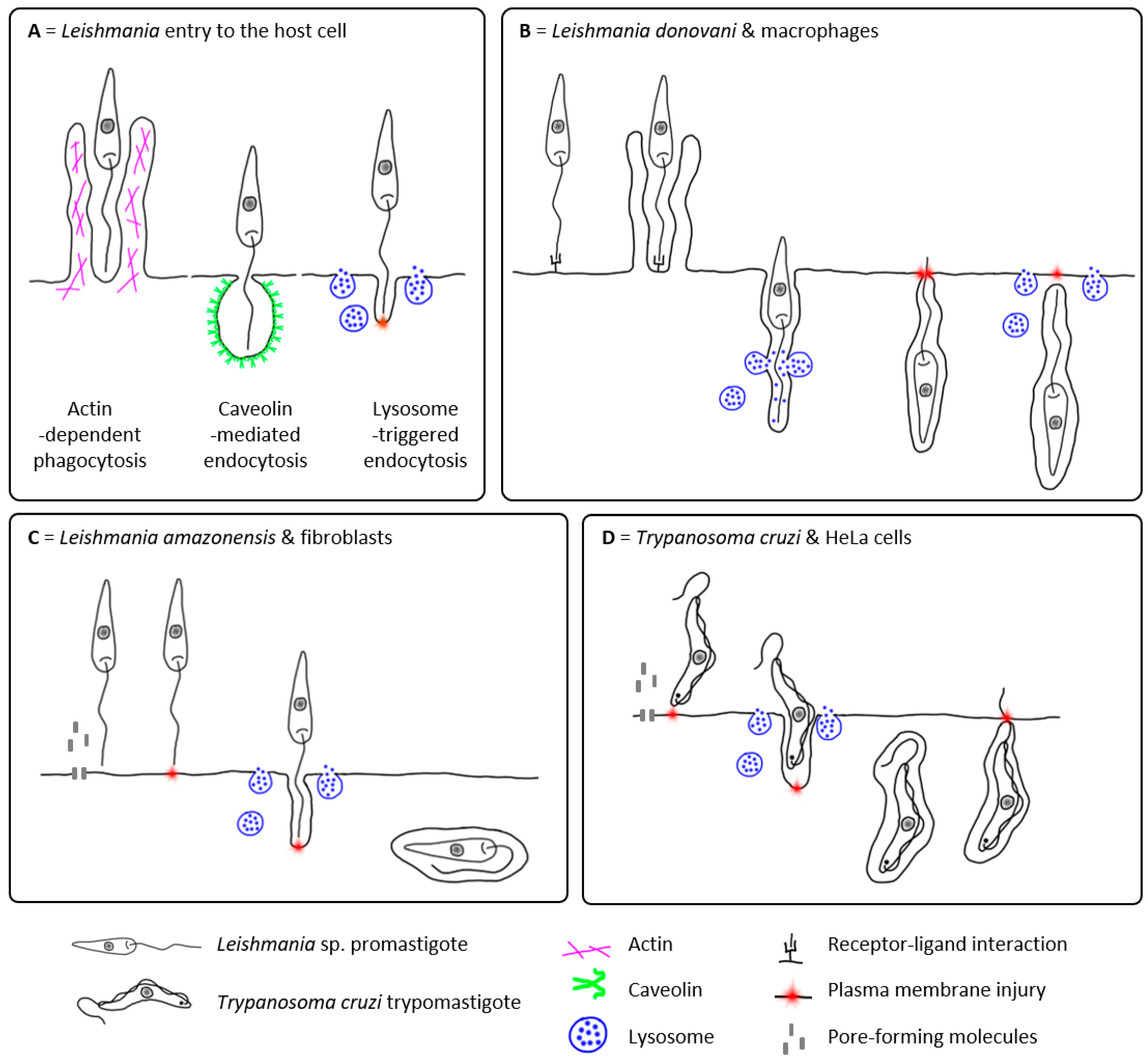
2.1. Passive Entry of Leishmania into the Host Cell

Passive host cell entry is a widely accepted mode of Leishmania internalisation. Leishmania was previously thought to be completely dependent on the phagocytic activity of its host cell, facilitating the phagocytosis by the deliberate binding of host cell opsonins. Passive entry of Leishmania into the professional phagocytes, such as macrophages and monocytes, is generally considered to be a receptor-mediated phagocytosis involving molecules such as Leishmania ligands, host cell receptors, and optionally also host-derived opsonins (reviewed in detail, e.g., in [5,30,31]).
However, phagocytosis is only one of the several processes by which the cell takes up extracellular material. In general, the process of endocytosis can be divided into four basic categories: (i) actin-mediated endocytosis (including phagocytosis and micropinocytosis), (ii) caveolin-dependent endocytosis, (iii) clathrin-dependent endocytosis, and (iv) clathrin/caveolin-independent endocytic pathways [32]. Several studies have shown that Leishmania is more likely to be internalised by caveolin-dependent endocytosis (Figure 1A) [33,34]. It is mediated by caveolins, integral membrane proteins that preferentially oligomerise in cholesterol-rich lipid rafts to form the membrane invagination. Through the caveolin-dependent endocytosis, the uptake of extracellular material can also be mediated by specific receptors [32,33]. Caveolin-mediated internalisation of Leishmania promastigotes has been observed in murine macrophages for L. chagasi and L. donovani [33,34,35,36,37]. Furthermore, caveolin-coated phagosomes showed delayed fusion with lysosomes, allowing the promastigote to transform into the amastigote form, which is better adapted to survive in acidified phagolysosomes [35]. However, this delay has only been observed in virulent metacyclic promastigotes [36,37]. Engulfment of avirulent or serum-opsonised promastigotes [36,37] or amastigotes [33,35] appears to be caveolin-independent, showing no delay in phagosome fusion with lysosomes [35]. Although this lack of the delay is fatal for promastigotes, amastigotes are, on the other hand, already adapted to the phagolysosomal microenvironment and can survive and proliferate there [33].
Clathrin-dependent endocytosis does not appear to play a role in the uptake of Leishmania promastigotes [34].

2.2. Active Entry of Leishmania into the Host Cell?

Surprisingly, Leishmania may also be able to enter host cells actively. The flagellar motility of promastigotes enables them to actively participate in phagocytic uptake by the macrophage (Figure 1B) [29]. This is most likely made possible by the polarised phagocytosis of Leishmania promastigotes induced by the interaction between their flagellar tip and the invaded cell, leading to the formation of pseudopodia. Pseudopodia begin at the tip of the parasite’s flagellum and extend towards its cell body [29]. The incessant activity of the promastigote flagellum leads to the reorientation of the parasite within the macrophage, with the flagellum pointing towards the host cell periphery. The oscillations of Leishmania parasites may even cause local damage to the host cell plasma membrane, leading to the Ca2+-dependent recruitment of host lysosomes to the site of the parasite invasion and their subsequent exocytosis, which is involved in the host cell plasma membrane repair process, as also observed in Trypanosoma cruzi invading HeLa cells (Figure 1D) [5,29,38].
The exocytosis of lysosomes during the T. cruzi internalisation occurs by their fusion with the host plasma membrane adjacent to the parasite, inducing the release of acid sphingomyelinase (ASM) and the production of ceramide by the hydrolysis of membrane sphingomyelin. The production of ceramide in the outer leaflet of the membrane induces endocytosis of the injured membrane, which the parasite uses for its own internalisation into the newly formed lysosomal endosomes [5,38]. In contrast to T. cruzi, Leishmania-mediated lysosome exocytosis has been reported to occur after the parasite uptake by the macrophage during its intracellular oscillating movement (Figure 1B) [29]. Since its close relative T. cruzi uses lysosome exocytosis for the invasion process itself [5,38,39], could Leishmania use a similar strategy to invade cells other than professional phagocytes?

3. Other Potential Host Cells of Leishmania

There is increasing evidence that Leishmania can invade or even survive and multiply in other cells that are not considered to be professional phagocytes. To enter these cells, Leishmania can use both passive as well as active strategies (Figure 1A).
Although the phagocytic process is more commonly associated with professional phagocytes, it has been shown that almost all cells in the human body are capable of phagocytosis (including skin fibroblasts) and caveolin-dependent endocytosis (including adipocytes, endothelial, and muscle cells) [18,40,41]. Entry into host cells by a process similar to classical phagocytosis has been documented, for example, in Chinese hamster ovary cell lines co-incubated with L. amazonensis amastigotes [42].
However, Leishmania may also be actively involved in the entry process into the non-canonical host cells, including but not limited to fibroblasts, adipocytes, myofibres or epidermal cells, such as pigmented cells (e.g., [8,43,44,45,46]). Since it is well documented that its close relative T. cruzi uses lysosome exocytosis to invade the host cell, including the non-professional phagocytic cells [5,38,39], it is likely that Leishmania could use a similar strategy [8,28].
While in macrophages, Leishmania-mediated lysosome exocytosis has only been reported after the parasite uptake (Figure 1B) [29]; in different host cells, it can facilitate the invasion process itself (Figure 1C) [8,28]. This appears to be the case at least for L. amazonensis entry into the fibroblasts [8]. The colocalisation of L. amazonensis promastigotes that have half-entered a fibroblast with the LAMP marker suggests that lysosome recruitment does indeed occur concomitantly with parasite invasion and that lysosomes donate their membrane to form the nascent parasitophorous vacuole [8]. Two hypotheses have been proposed for the L. amazonensis initiation of the host cell plasma membrane damage leading to lysosome exocytosis and the subsequent endocytosis in these host cells [8]: (i) parasite flagellar motility towards the host cell plasma membrane, causing mechanical damage (as in T. cruzi [38]) and/or (ii) the secretion of cytolytic molecules (such as pore-forming cytolysins) leading to plasma membrane permeabilisation [47,48].
Other possible strategies that Leishmania parasites might use to invade non-professional phagocytes should be verified in the future. It is even possible that the promastigote flagellum itself plays a much more important role in invasion than previously thought. According to older in vitro observations, the flagellum could serve as an anchor inside the invaded cell, by which the parasite pulls itself inside when penetrating cells with limited phagocytic capacity [43]. In fact, Leishmania parasites appear to possess several mechanisms that would allow them to enter their host cells, and it is likely that these mechanisms may alternate or even complement each other depending on the type of the cell attacked and the infecting Leishmania species.
It is obvious that Leishmania parasites have tools to invade even the non-canonical host cells that have been neglected as players in the dynamics of Leishmania infection and its persistence. These cells are listed in Table 1 and include, besides other professional phagocytes (mast cells, dendritic cells, eosinophils, and histiocyte-like cell lines), mainly non-professional phagocytic cells, such as lymphocytes, fibroblasts, adipocytes, epithelial and endothelial cells, mesenchymal stem cells, myocytes, and keratinocytes.
To collect the data for Table 1, we adapted the tables of Rittig and Bogdan (2000) [7] and Chang and Fish (2017) [49], which we further expanded and updated using the following strategy. A literature search of relevant articles was conducted between December 2022 and January 2023 using databases such as Web of Science and PubMed. The search was performed independently by both authors using combinations of keywords, such as this Boolean string: “Leishmania* AND (fibroblast* OR adipo* OR fat OR “epithelial cell*” OR epithel* OR myocyte* OR muscle* OR myofibre OR “stem cell*” OR keratinocyte* OR “endothelial cell*” OR endothel*) AND (multiplicat* OR transform* OR uptake OR internali* OR phagocyto* OR intracellular* OR amastigote*)”. The search was not restricted by the year of publication. The retrieved articles were selected based on the eligibility criteria including, but not limited to: an original research article, full text access, detailed description of methodology, host cells of vertebrate origin, and clear statement of results. Exceptionally, secondary citations were used for research articles with a unique output, but not accessible in the full text version. References in selected articles were also evaluated. The Leishmania species names listed in Table 1 correspond to the names provided in the original research articles, as older papers, in particular, did not use more accurate molecular techniques for species identification, and it is therefore not possible to complement them with current taxonomy and nomenclature.
The data listed in Table 1 indicate that the outcome of Leishmania internalisation is likely to depend on the host and Leishmania species, the infecting form of Leishmania (promastigote vs. amastigote), and also the origin of the host cell or tissue. This table only includes vertebrate host cells; however, some Leishmania species have also been found to invade insect cell lines [50,51,52]. Of particular interest is that the cell lines prepared from mosquitoes (Aedes aegypti) or sand flies (Lutzomyia spinicrassa) and mainly containing cell types with epithelial and fibroblast appearance, also support the internalisation of Leishmania promastigotes, together with their transformation into the amastigote form [50,51] and even their multiplication [52].
Table 1. Non-canonical host cells of Leishmania parasites.
Host Cell/OriginLeishmania SpeciesMain OutcomeDetection MethodReference
PROFESSIONAL PHAGOCYTES
Dendritic cells (DCs)
Langerhans cells from mouse skin L. major PMsNo or low PMs uptakeLM/Diff-Quik, TEM, FL/AO+EtBr[53,54,55]
L. major AMsAMs uptake and internalisation, no or weak multiplicationLM/Diff-Quik, TEM, FL/AO+EtBr, ICC[54,56,57]
Mouse lymph node DCs ♦○L. major AMsPresence of AMs LM/G, IHC[56,58,59]
Mouse spleen DCs L. major PMs/AMs, L. m. mexicana PMsPMs/AMs uptakeLM/G[59,60]
Mouse bone marrow DCs L. major PMs/AMs, L. mexicana PMs, L. amazonensis AMs/PMsPMs/AMs uptake in all; multiplication reported only in L. amazonensisLM/Diff-Quik, ICC, FC[61,62,63,64]
L. infantum PMs/PMs (CFSE)PMs uptake, transformation into AMsLM/G, FC/CFSE-PMs[65]
Human immature monocyte-derived DCsL. amazonensis, L. braziliensis, L. infantum PMsPMs uptake, internalisationCLSM/Dapi[25]
L. donovani PMsPMs uptake, transformation into AMsLM/MGG[66]
Mast cells (MCs)
Mouse peritoneal MCs L. tropica, L. donovani PMs (CFSE)PMs uptake in L. tropica, but not in L. donovaniFC+CLSM/CFSE-PMs[22,67]
Mouse bone marrow MCs L. major, L. infantum PMsPMs uptake, transformation into AMs, multiplication leading to cell lysis and AMs releaseLM/MGG[21]
Eosinophils
Human peripheral eosinophils L. donovani PMsPMs uptake and killing after 2 h p.i.LM/Diff-Quik[68]
L. donovani AMsAMs uptake, not efficient killingLM, TEM[69]
Rat peritoneal eosinophils L. major PMsPMs uptake and killingLM/MGG, ICC[70]
Rat peritoneal eosinophils L. m. amazonensis PMs/AMsPMs/AMs uptake and killingTEM[71]
Mouse eosinophils in skin lesion ♦○L. m. mexicana AMsAMs uptake, not efficient killing TEM[72]
Histiocyte-like cells[7]
Sticker dog sarcoma 503 cells L. donovani, L. mexicana, L. m. mexicana, L. braziliensis, L. b. pifanoi, L. t. major PMs/AMsPMs/AMs uptake, multiplication, continuous passagesLM/G, TEM[73,74,75,76,77,78]
L. m. mexicana PMsPMs uptake, transformation into AMs, multiplication after day 3 p.i., transformation into PMsLM/G, TEM[43]
L. adleri, L. hoogstraali, L. agamae PMsLow PMs uptake, transformation into AMsLM/G[43]
NON-PROFESSIONAL PHAGOCYTES
Lymphocytes
Human B (Daudi) and T (HUT78) cells L. donovani PMs/AMsPMs/AMs uptake, PMs transformation into AMs, viability up to 2 weeks after infection with AMsLM/G, TEM[79]
Fibrocytes
Mouse peripheral blood fibrocytes L. amazonensis PMsPMs uptake, transformation into AMs, low multiplication, clearance by 72 h p.i.LM/G, FL/Dapi, TEM, SEM[80]
Fibroblasts
Canine skin fibroblasts ♦□Leishmania sp.Presence of AMsTEM, LM/HE, G, PAS[81]
L. donovaniPresence of AMsIHC[82]
Human skin fibroblasts ♦□L. tropicaPresence of AMsLM/G, TEM[83,84]
Leishmania sp. (cutaneous)Presence of AMsTEM[85]
Human skin fibroblasts L. amazonensis PMsPMs uptake, transformation into AMs, multiplicationTEM[14]
L. m. amazonensis AMsAMs uptake, multiplication, killing of AMs by day 8 p.i.LM/G, TEM, ICC[12]
Leishmania sp. (mucocutaneous), L. donovani PMsPMs uptake in Leishmania sp. (not in L. donovani), transformation into AMs, no or low multiplication, decline during a 3-week period p.i.LM, TEM, SEM[86]
Human foreskin fibroblasts L. donovani PMsPMs uptake, transformation into AMs, no multiplication, viability up to day 14 p.i.LM, TEM, SEM[87]
L. major PMs (SPIONs)PMs uptakeLM/SPIONs-PMs+Prussian blue, TEM[88]
L. major PMs (AO, Dil)PMs uptakeFL/AO-PMs, Dil-PMs[89]
Mouse skin fibroblasts ♦○L. amazonensis PMsAMs presenceLM/HE, Lennert’s G[90]
Mouse skin fibroblasts L. major PMs/AMsPMs/AMs uptakeICC[9]
L. amazonensis PMsPMs uptake and killing of PMs after day 3 p.i.LM/G, TEM,
FL/Dapi
[91]
L. infantum, L. mexicana PMsPMs uptake, transformation into AMs, multiplicationLM/G, TEM[10]
Hamster skin fibroblasts L. infantum, L. mexicana PMsPMs uptake, transformation into AMs, multiplicationLM/G, TEM[10]
Rat skin fibroblasts L. infantum, L. mexicana PMsPMs uptake, transformation into AMs, no multiplicationLM/G, TEM[10]
Human fibroblasts in lymph node ♦□Leishmania sp.AMs presenceLM/G[92]
Mouse fibroblasts in lymph node L. major PMs/AMsPMs/AMs uptakeTEM, ICC[9]
Draining lymph nodes of healed mice (presumably fibroblasts) ♦○L. major PMsPresence of AMs, parasite survival or limited killingIHC[9]
Mouse embryonic fibroblasts L. donovani PMsPMs uptake, transformation into AMs, efficient host defence via IFN-inducible guanylate binding proteinsLM/G, CLSM/Dapi[93]
L. amazonensis PMs (RFP)PMs uptake, transformation into AMs, multiplicationLM/HE, TEM, CLSM+FC/RFP-PMs [8]
L. major PMsPMs uptakeCLSM/Dapi[94]
Mouse tumour fibroblasts (L cells) L. amazonensis, L. major AMs (GFP)AMs uptake, internalisation (low in L. major), multiplication (not in L. major)CLSM/GFP-AMs[15]
Fibroblasts from embryonic chick brain L. donovani AMsAMs uptake, viability up to day 17 p.i., transformation into PMsLM/G[11]
Fibroblast-like cells from embryonic chick muscle L. donovani/presumably AMsAMs uptake, no multiplication, degeneration after day 20 p.i.LM/HE[95]
Mouse perineurial cells ♦○L. amazonensis PMsPresence of AMsTEM[96]
Adipocytes
Mouse brown and white adipose tissue ♦○L. infantum PMsPMs uptake, viable AMs present for up to 40 weeks p.i.IHC, qPCR[46]
Mouse adipocytes derived from primary pre-adipocytes from subcutaneous white adipose tissue L. infantum PMs (GFP)PMs uptake (further progress not reported)TEM, qPCR, CLSM/GFP-AMs[46]
Human adipocytes derived from adipose tissue primary progenitor cells L. infantum PMs (GFP)PMs uptake (further progress not reported)qPCR, CLSM/GFP-AMs[46]
Mouse adipocytes differentiated in vitro from 3T3-L1 fibroblasts L. amazonensis, L. braziliensis PMs/AMs PMs/AMs uptake, PMs transformation into AMs, viability up to 144 h p.i. and ability to transform into PMsLM/G, FL/Dapi, TEM[45]
L. amazonensis AMs (GFP)AMs uptake, viability up to 144 h p.i.FL/GFP-AMs[45]
Epithelial cells
Human epithelial cells of eccrine sweat gland (HIV patient) ♦□Leishmania sp., L. infantumAMs presenceLM/HE[97,98]
Human retinal pigmented epithelial cells (ARPE-19) L. amazonensis PMsPMs uptake, internalisationLM/G, IHC, TEM[99]
Human amnion epithelium L. donovani, L. b. pifanoi PMsPMs uptake, transformation into AMs, clearance by day 29–32 p.i.LM/G[100]
L. donovan PMsPMs uptake, transformation into AMs, multiplication (not clear whether PMS or AMs)LM/G[101]
A549 (human adenocarcinomic alveolar basal epithelium) cells L. donovani PMsPMs uptake, transformation into AMs, efficient defence via IFN-inducible guanylate binding proteinsLM/G, CLSM/Dapi[93]
HeLa (human cervix carcinoma) cells L. t. major PMsPMs uptake, transformation into AMs, multiplication, destruction of host cells after day 3LM[102]
L. donovani PMsPMs uptake, transformation into AMs, decline after 5 h p.i.LM/G[103]
LLC-MK2 (rhesus monkey kidney epithelium) cells L. donovani AMsAMs uptake, multiplicationLM/G[104]
Vero (monkey kidney) cells L. chagasi, L. braziliensis PMsPMs uptake, transformation into AMs, multiplicationLM/G, TEM[105,106]
Chinese hamster ovary cells L. amazonensis AMsAMs uptake, multiplicationIHC, TEM[42]
C. burnetii-infected Vero cells L. amazonensis AMsAMs uptake, multiplication LM, TEM[107]
C. burnetii-infected Chinese hamster ovary cells L. amazonensis AMsAMs uptake, multiplicationLM, TEM, CLSM/PI[107,108]
Mesenchymal stem cells (MSCs)
Mouse bone marrow MSCs ♦○❖L. infantum PMs PMs uptake, transformation into AMsLM/G, ICC, FC[109]
Human adipose tissue MSCs L. donovani, L. infantum, L. major,
L. tropica PMs
PMs uptake, transformation into AMs but AMs present only at day 1 p.i.; at day 7, 14, 21, and 28 only PMs detectedLM/G, microcapillary culture method, PCR[16]
Myocytes
Canine skeletal/smooth muscles ♦□L. infantum, Leishmania sp.Presence of AMs within myofibresLM/HE, IHC[110,111]
Mouse skeletal muscles ♦○L. amazonensis AMsPresence of AMs within myofibresLM/HE[112]
Turtle heart cells L. m. mexicana, L. adleri, L. hoogstraali PMsPMs uptake (lower in L. adleri and L. hoogstraali), transformation into AMs (further progress not reported)TEM (L. mexicana only), LM/G[43]
Endothelial cells
Human endothelial cells of blood vessels ♦□L. donovani, Leishmania sp.Presence of AMsLM [81,113,114]
Human endothelial cells of capillaries ♦○L. tropica PMsPresence of AMsLM[115]
Human microvascular endothelial (HMEC-1) cell line L. infantum PMsNo uptake of PMsLM/G[116]
Keratinocytes
Human keratinocytes (HIV patient) ♦□L. infantumAMs presenceLM/HE[98]
Human keratinocytes L. infantum, L. major PMsPMs uptake, transformation into AMs at low levels, no multiplicationLM/G, CLSM/Dapi[117]
Unidentified cells in primary cultures
Hamster kidney cells L. braziliensis, L. donovani PMsPMs uptake, transformation into AMs, multiplication (not in L. donovani)LM/G[118,119], as cited in [7,49]
Chicken embryo muscles ♦○L. t. major PMsPMs uptake, transformation into AMs, multiplication, destruction of host cells after day 3LM[102]
Abbreviations: AMs—amastigotes, AO—acridine orange, CFSE—carboxyfluorescein N-succinimidyl ester, CLSM—confocal laser scanning microscopy, DCs—dendritic cells, EtBr—ethidium bromide, FC—flow cytometry, FL—fluorescence microscopy, G—Giemsa, GFP—green fluorescent protein, HE—haematoxylin-eosin, ICC—immunocytochemistry, IHC—immunohistochemistry, LM—light microscopy, MCs—mast cells, MGG—May–Grünwald–Giemsa, MSCs—mesenchymal stems cells, PAS—periodic acid-Schiff, PCR—polymerase chain reaction, PI—propidium iodide, PMs—promastigotes, p.i.—post inoculation, qPCR—quantitative polymerase chain reaction, RFP—red fluorescent protein, SEM—scanning electron microscopy, SPIONs—superparamagnetic iron oxide nanoparticles, TEM—transmission electron microscopy. Symbols: —in vitro, —in vivo, —clinical case, —experimental infection. Note: Leishmania species names correspond to the names as provided in the original research articles.
In the following subchapters, we have focused on studies using host cells that are as close as possible to their natural state, since cancerous or mutated cells (such as those used for immortalised cell lines) may have altered metabolism that may also affect Leishmania entry, survival, and multiplication.

3.1. Fibroblasts

Fibroblasts may play a neglected role in latent leishmaniasis and the disease epidemiology because, in some host-parasite combinations, they can support intracellular parasite survival, transformation into amastigotes, and even amastigote multiplication, resulting in viable progeny capable of transforming back to promastigotes [8].
Several in vitro and in vivo studies have reported that fibroblasts harbour Leishmania amastigotes (Table 1) [8,9,10,11,14,81,83,84,85,86,87]. Conflicting results have been observed regarding the survival of L. amazonensis in fibroblasts, ranging from limited survival of parasites (e.g., [12,91]) to their successful multiplication [8,10,14,15] (Table 1). Co-incubation of enucleated fibroblasts (cytoplasts, cell nucleus artificially removed in vitro) with L. amazonensis revealed that the parasitophorous vacuole biogenesis and parasite multiplication are independent of the host cell nucleus, and showed these cells to be a promising model for studies focusing on the role of the host cell nucleus during the parasite-host interactions (in particular, the modulation of the gene expression) [15]. The amastigotes of L. amazonensis have also been detected in the perineurial cells (=epithelioid myofibroblasts) of BALB/c mice with experimental cutaneous leishmaniasis [96]. It is interesting that L. mexicana and L. infantum were able to multiply in mouse and hamster skin fibroblasts, but not in skin fibroblasts from rats [10]. In contrast, L. donovani does not appear to be capable of long-term survival and multiplication in fibroblasts in any of the host species tested—human [87], mouse [93], nor chicken [11,95] (Table 1).
Ultrastructural and immunohistochemical analysis of skin biopsies from dogs with naturally acquired leishmaniasis revealed the presence of free and occasionally vacuole-enclosed amastigotes within fibroblasts, while some amastigotes in close contact with the host plasma membrane appear to damage it, indicating that amastigotes also have an active invasion potential [81]. The authors speculated that the source of these amastigotes infecting fibroblasts in the deeper layers of the skin were necrotic macrophages. Similarly, L. donovani amastigotes have been seen surrounded by a closely applied membrane of human foreskin fibroblasts cultured in vitro [87].
The potential involvement of fibroblasts in the pathogenesis of cutaneous leishmaniasis is of particular interest, as recent studies indicate their role in Leishmania immune evasion strategies [13]. Fibroblasts are one of the most abundant cells at the site of transmission and are important producers of macrophage- and neutrophil-attracting chemokines. Fibroblasts also interact directly with macrophages during wound healing and can move by diapedesis, thus being capable of Leishmania dissemination [8]. The relatively long lifespan of fibroblasts and their limited ability to eliminate invaders could lead to the persistence of infection [7]. In addition, fibroblasts are capable of phagocytosis but have a limited ability to control the Leishmania infection through NO production [9]. Indeed, in healed mice, approximately 40% of L. major amastigotes were found in skin and draining lymph node fibroblasts, indicating that they may serve as a safe shelter and a site of potential recrudescence [9,58]. In latent leishmaniasis, the balance may be maintained by neighbouring macrophages producing enough NO to destroy Leishmania amastigotes within the fibroblasts [9].
Similar to professional phagocytes, the entry of Leishmania into fibroblasts could be either passive or active (Figure 1A). An older in vitro study, supported by transmission electron microscopic visualisation, claimed that the infection of fibroblasts by L. braziliensis promastigotes occurs via the parasite-induced phagocytosis, when the parasites enter fibroblasts with their flagellar end through pseudopodia-like formations on the host cell surface [86]. The engulfed promastigotes settled in vacuoles that did not fuse with secondary lysosomes and transformed into amastigotes. Uptake is likely to be receptor-mediated, as skin fibroblasts cocultured with L. amazonensis promastigotes showed an increased expression of the mannose receptor during the early stages of infection, possibly binding mannosylated ligands on the promastigote surface [91]. This modulation of fibroblast mannose receptor expression was reversed concomitantly with the loss of parasite viability, indicating that the presence of viable parasites is required to maintain it [91].
However, the active entry of Leishmania into the fibroblasts has also been reported (Figure 1). It has been shown that L. amazonensis induces its entry into host fibroblasts by damaging the fibroblast plasma membrane, leading to the internalisation of promastigotes during the subsequent membrane repair process (Figure 1C) [8]. The invasion process is independent of host actin remodelling (i.e., this process is not a form of induced phagocytosis) and involves Ca2+-dependent recruitment/exocytosis of host lysosomes to repair the plasma membrane. During this process Leishmania actively induces lysosome-triggered endocytosis, a cell invasion mechanism based on the transient permeabilisation of the host cell plasma membrane [8]. Similar to T. cruzi interactions with HeLa cells [38], this invasion process has only been observed with the viable metacyclic stage of the parasite; neither dead metacyclic nor the procyclic promastigotes (the developmental stage of Leishmania found only in the vector) were engulfed by fibroblasts [8]. On the other hand, parasite-unrelated membrane injury promotes internalisation of L. amazonensis promastigotes [8] as well as T. cruzi trypomastigotes [38].

3.2. Adipocytes

Adipose tissue has also been postulated as a potential reservoir for intracellular pathogens with the ability to induce disease relapses [46]. Indeed, recent studies have confirmed the ability of Leishmania promastigotes to infect adipocytes in vitro and in vivo [45,46]. Adipose tissue could serve as a perfect reservoir for Leishmania, especially considering that this tissue niche is also used by its relatives—extracellular T. brucei [120] and intracellular T. cruzi [121]. Although their survival strategies are not entirely the same as those of Leishmania, we may still find some parallels that can inspire future research. Trypanosoma brucei has been shown to accumulate in the adipose tissue of mice early after infection [120]. These adipose tissue extracellular T. brucei forms, which are transcriptionally distinct from bloodstream forms, can replicate and are capable of infecting a naïve host [120]. Moreover, trypomastigotes of T. cruzi invade human and mouse adipocytes and transform into amastigotes during the acute phase of infection [121]. Although replication in adipocytes has not yet been directly observed in Leishmania, L. amazonensis, and L. brasiliensis amastigotes recovered from infected cells retain the ability to differentiate into replicative promastigotes [45], and recovered L. infantum amastigotes were infectious to another host [46].

3.3. Mesenchymal Stems Cells

Mesenchymal stems cells (MSCs) residing in bone marrow could also provide a perfect protective niche for Leishmania parasites and support their persistence in the host organism. Among other factors, these cells are (i) capable of self-renewal and have low-reactive oxygen species properties (ideal for long-term parasite viability), (ii) do not normally express MHC Class II on their surface and their MHC Class I molecules do not trigger effector functions of cytotoxic T-cells, and (iii) express potent drug efflux pumps (probably enabling Leishmania drug evasion) [109]. Indeed, L. infantum promastigotes successfully infected the CD271+/Sca1+ bone marrow MSCs of C57BL/6 mice and transformed into amastigotes in both in vivo and in vitro settings [109]. Moreover, several Leishmania species, including agents of both the cutaneous and visceral leishmaniases (Table 1), have been shown to persist for some time in an inactive form in cultures of adipose tissue-derived MSCs [16]. As stem cells generally remain dormant in the absence of an exogenous stimulus, they may represent ideal reservoir host cells for Leishmania [16]. The mechanism of invasion of mesenchymal stem cells remains to be elucidated, although phagocytic properties have already been reported for adipose tissue MSCs [16].

3.4. Myocytes

Muscle cells are highly parasitised by T. cruzi [122] but are overlooked in Leishmania studies. It is therefore of particular interest that L. infantum amastigotes have been detected in the muscle biopsies from dogs with obvious muscle damage, causing a progressive polymyositis affecting the masticatory and skeletal muscles [110]. Moreover, another study reported canine leishmaniasis associated with myositis of adnexal, extraocular and intraocular smooth and striated muscles that were parasitised by Leishmania amastigotes [111].
In addition, there are in vitro studies reporting the internalisation and replication of several Leishmania species in muscle cells of different origins [43,102]. The ability of Leishmania amastigotes to invade muscle fibres was also confirmed by an in vivo experimental study comparing the muscle infection by L. amazonensis in two mouse strains with a different susceptibility to leishmaniasis—susceptible BALB/c mice and resistant C3H.He mice [112]. While the BALB/c mice showed an intense inflammatory infiltrate between the amastigote-infected myofibres, followed by a total muscle destruction at day 90 p.i., the C3H.He mice showed only a mild inflammatory infiltrate without intracellular amastigotes, followed by a muscle repair process [112].
In BALB/c mice experimentally inoculated with L. major promastigotes, we also occasionally observed the presence of amastigote-like structures within muscle fibres (Figure 2, unpublished results). However, these observations require confirmation by transmission electron microscopy or immunolabelling.
The mechanism of Leishmania entry into muscle cells remains to be elucidated. While older studies hypothesised the ability of promastigotes to actively penetrate target cells through the motility of their flagellum acting as an anchor [43], others speculate that Leishmania parasites could penetrate myocytes, rich on fucose-mannose ligands, using the fucose-mannose receptor [112]. Parasite-unrelated injury to muscle cells may facilitate Leishmania uptake via lysosome-triggered endocytosis during repair of the damaged plasma membrane, as has been shown for L. amazonensis and fibroblasts [8].

3.5. Endothelial Cells

Endothelial cell parasitism by Leishmania remains unclear, as only a few studies (some of them older) with conflicting results are currently available [113,115,116,123,124]. However, similar to T. cruzi [122], it is likely that at least some Leishmania species could be able to infect endothelial cells.
While a recent in vitro analysis of a human microvascular endothelial cell line (HMEC-1) co-incubated with L. infantum promastigotes reported the absence of internalised parasites [116], the intracellular localisation of Leishmania has been reported from endothelial cells lining the blood-vessels of the kidney, liver, and colon in visceral leishmaniasis [81,113,114,123]. However, it should be noted that the accurate localisation of amastigotes in routine histopathology (usually used as the sole detection method in older studies) is challenging and may be less sensitive than immunolabelling, as demonstrated in canine cutaneous leishmaniasis [125]. For example, the histopathological examination of a human subcutaneous nodule after experimental inoculation with L. tropica promastigotes revealed the abundant presence of amastigotes within the endothelial cells lining the capillaries near the centre of the lesion [115], but another study reported that L. braziliensis parasites were more likely to be attached to the wall of dermal blood vessels and free in the capillary lumen [124]. Accordingly, the L-SIGN receptor, specifically expressed in liver sinusoidal endothelial cells, acts as a receptor for viscerotropic L. infantum (but not dermotropic L. pifanoi), resulting in the strong binding of amastigotes to the cultured endothelial cells; however, no invasion of these cells was reported [126].

3.6. Keratinocytes

As one of the most abundant epidermal cell types and an important source of immunomodulatory signals in the skin, keratinocytes may play a key role in the early stages of insect-borne diseases, including leishmaniasis. These cells appear to be unsuitable for Leishmania replication, the function of keratinocytes being more immunomodulatory. Although human keratinocytes were shown to internalise L. infantum or L. major promastigotes in vitro at low levels, they did not allow efficient amastigote multiplication [117]. Accordingly, several in vivo or ex vivo studies reported the absence of keratinocytes parasitised by Leishmania [55,96,127]. Nevertheless, keratinocytes exposed to extracellular L. infantum and L. major parasites have been shown to alter their transcriptional signatures and appear to be stimulated to release factors that influence monocyte infection [117]. The authors hypothesised that the pro-inflammatory response of keratinocytes induced by L. infantum may limit the local survival of the parasite in the skin environment and thus promote its dissemination, whereas the ‘silent’ interaction of L. major with keratinocytes may increase its ability to survive locally, leading to cutaneous leishmaniasis [117]. It has therefore been proposed that keratinocytes may initiate or suppress the pro-inflammatory response at the site of infection, thereby influencing tissue pathology [117].

4. Conclusions and Perspectives

Leishmania well-recognised primary host cells are professional phagocytes, macrophages, and monocytes, but other cell types might also be infected with Leishmania parasites. The presence of L. major transcripts has been shown to be associated with multiple cell types at the site of infection. In addition to the well-known host cells of the myeloid lineage (macrophages, inflammatory monocytes, neutrophils, and dendritic cells), they are also found in endothelial and epithelial cells, fibroblasts, keratinocytes, chondrocytes, and myoblasts [128]. However, the key requirement for the host cell—to support Leishmania survival and multiplication—has only been proven for a few of them (e.g., [8,10,46]).
The most studied non-canonical Leishmania host cells are fibroblasts [8,9,10,11,12,13,14,15,81,82,83,84,85,86,87,88,89,90,91,92,93,94,95,96]. They can migrate within the skin tissue [8], potentially allowing Leishmania to spread from the site of transmission, thereby disseminating the infection and enhancing the possibility of being engulfed by the vector during blood feeding. Due to their relatively long lifespan and low leishmanicidal activity, fibroblasts may serve as an ideal reservoir host cell in latent cutaneous leishmaniasis [9]. In visceral leishmaniasis, adipocytes may also play this role [45,46]. The putative reservoir host cells for latent infection should ideally support the intracellular survival of Leishmania for a prolonged period, ideally also facilitating amastigote replication. If the cell cannot support Leishmania replication, the amastigote may leave the reservoir host cell, multiply in monocytes or macrophages, and find safe shelter in another reservoir host cell. Such a scenario may be possible since Leishmania parasites are not dormant during latent leishmaniasis but are continuously replicating [129], being under the tight immune control of macrophage-derived NO [58].
The promiscuity of Leishmania with respect to host cells has been demonstrated using the cell lines of invertebrate origin [50,51,52], showing the ability of Leishmania parasites to also infect—under artificial conditions—the cells of the insect vector [51], where they naturally occur only extracellularly [130,131]. In humans, such artificial conditions could be induced, for example, by HIV immunosuppression, leading to an unusual and rare localisation of Leishmania within sweat gland epithelial cells in the host dermis [97,98]. To our knowledge, such localisation has not been observed in immunocompetent individuals (Table 1). Moreover, the hidden promiscuity of Leishmania parasites may have unexpected therapeutic implications, affecting the screening of new drug candidates. The widely used in vitro testing on promastigotes (vector-derived developmental stage) [132] may not reveal the full complexity of amastigote presence in different tissues/cells during the mammalian host infection. Pharmacokinetics and pharmacodynamics would be better evaluated in the context of multiple Leishmania host cells.
The ability of Leishmania to invade different cell types may also affect the epidemiology of leishmaniasis, being another factor to be consider during the transmission from the mammalian host to the insect vector. Leishmania is able to persist in the uninflamed skin of the mammalian host, while preserving its infectious potential for sand fly vectors [133]. As these parasites can gradually accumulate in the skin, even in clinically healthy hosts, and remain infectious to their insect vectors [133], it is necessary, at least in endemic areas, to monitor not only cured patients but also potential reservoirs, such as dogs or asymptomatic humans.
Leishmania internalisation into the host cell is well described in professional phagocytes, but the mechanisms in non-canonical host cells are less well understood. The mode of Leishmania entry into the host cells, of whatever type, appears to be multifactorial, depending on the host cell type, Leishmania developmental stage (promastigotes vs. amastigotes), Leishmania virulence, as well as on the immune status of the host (e.g., the presence of anti-Leishmania antibodies as opsonising agents). The Leishmania internalisation could be receptor-mediated (e.g., [5,30,31,91]), where Leishmania appears to be passively engulfed, or actively initiated by Leishmania promastigote by wounding the host cell plasma membrane [8], e.g., via the movement of its flagellar tip [29,43]. At least three possible entry pathways have been described (Figure 1): (i) actin-dependent phagocytosis, (ii) caveolin-mediated endocytosis, and (iii) lysosome-triggered endocytosis associated with the host cell plasma–membrane repair mechanism [7,8,28,32,33,34,36,37].
Undoubtedly, more studies are needed to reveal all the details of the interactions between Leishmania and its various host cells in the hope of finding a way to better control leishmaniasis, including its latent form.

Author Contributions

Conceptualisation, A.V. and I.K.; writing—original draft preparation, A.V. and I.K.; writing—review and editing, A.V. and I.K. All authors have read and agreed to the published version of the manuscript.

Funding

I.K. was financially supported by ERD Funds (project CePaViP, grant No. CZ.02.1.01/0.0/0.0/16_019/0000759).

Data Availability Statement

No new data were created or analysed in this study. Data sharing is not applicable to this article.

Conflicts of Interest

The authors declare no conflict of interest. The funders had no role in the design of the study; in the collection, analyses, or interpretation of data; in the writing of the manuscript; or in the decision to publish the results.

Abbreviations

AMsAmastigotes
AOAcridine orange
ASMAcid sphingomyelinase
CFSECarboxyfluorescein N-succinimidyl ester
CLSMConfocal laser scanning microscopy
DCsDendritic cells
EtBrEthidium bromide
FCFlow cytometry
FLFluorescence microscopy
GGiemsa
GFPGreen fluorescent protein
HEHaematoxylin-eosin
HMEC-1Human microvascular endothelial cell line
ICCImmunocytochemistry
IHCImmunohistochemistry
LAMPLysosomal membrane-associated protein
LMLight microscopy
L-SIGNLiver/lymph node-specific ICAM-3 grabbing nonintegrin
MCsMast cells
MGGMay–Grünwald–Giemsa
MSCsMesenchymal stems cells
NONitric oxide
PASPeriodic acid-Schiff
PCRPolymerase chain reaction
PIPropidium iodide
PMsPromastigotes
p.i.Post inoculation
qPCRQuantitative polymerase chain reaction
RFPRed fluorescent protein
SEMScanning electron microscopy
SPIONsSuperparamagnetic iron oxide nanoparticles
TEMTransmission electron microscopy

References

  1. Kolářová, I.; Valigurová, A. Hide-and-seek: A game played between parasitic protists and their hosts. Microorganisms 2021, 9, 2434. [Google Scholar] [CrossRef]
  2. Conceição-Silva, F.; Morgado, F.N. Leishmania spp-host interaction: There is always an onset, but is there an end? Front. Cell. Infect. Microbiol. 2019, 9, 330. [Google Scholar] [CrossRef] [PubMed]
  3. Morgado, F.N.; Schubach, A.; Vasconcellos, E.; Azeredo-Coutinho, R.B.; Valete-Rosalino, C.M.; Quintella, L.P.; Santos, G.; Salgueiro, M.; Palmeiro, M.R.; Conceição-Silva, F. Signs of an in situ inflammatory reaction in scars of human American tegumentary leishmaniasis. Parasite Immunol. 2010, 32, 285–295. [Google Scholar] [CrossRef] [PubMed]
  4. Martínez-Valencia, A.J.; Daza-Rivera, C.F.; Rosales-Chilama, M.; Cossio, A.; Casadiego Rincón, E.J.; Desai, M.M.; Saravia, N.G.; Gómez, M.A. Clinical and parasitological factors in parasite persistence after treatment and clinical cure of cutaneous leishmaniasis. PLoS Negl. Trop. Dis. 2017, 11, e0005713. [Google Scholar] [CrossRef]
  5. Walker, D.M.; Oghumu, S.; Gupta, G.; McGwire, B.S.; Drew, M.E.; Satoskar, A.R. Mechanisms of cellular invasion by intracellular parasites. Cell. Mol. Life Sci. 2014, 71, 1245–1263. [Google Scholar] [CrossRef]
  6. Chaves, M.M.; Lee, S.H.; Kamenyeva, O.; Ghosh, K.; Peters, N.C.; Sacks, D. The role of dermis resident macrophages and their interaction with neutrophils in the early establishment of Leishmania major infection transmitted by sand fly bite. PLoS Pathog. 2020, 16, e1008674. [Google Scholar] [CrossRef]
  7. Rittig, M.G.; Bogdan, C. Leishmania-host-cell interaction: Complexities and alternative views. Parasitol. Today 2000, 16, 292–297. [Google Scholar] [CrossRef]
  8. Cavalcante-Costa, V.S.; Costa-Reginaldo, M.; Queiroz-Oliveira, T.; Oliveira, A.C.S.; Couto, N.F.; Dos Anjos, D.O.; Lima-Santos, J.; Andrade, L.O.; Horta, M.F.; Castro-Gomes, T. Leishmania amazonensis hijacks host cell lysosomes involved in plasma membrane repair to induce invasion in fibroblasts. J. Cell. Sci. 2019, 132, jcs226183. [Google Scholar] [CrossRef]
  9. Bogdan, C.; Donhauser, N.; Döring, R.; Röllinghoff, M.; Diefenbach, A.; Rittig, M.G. Fibroblasts as host cells in latent leishmaniosis. J. Exp. Med. 2000, 191, 2121–2130. [Google Scholar] [CrossRef]
  10. Minero, M.A.; Chinchilla, M.; Guerrero, O.M.; Castro, A. Infection of skin fibroblasts in animals with different levels of sensitivity to Leishmania infantum and Leishmania mexicana (Kinetoplastida: Trypanosomatidae). Rev. Biol. Trop. 2004, 52, 261–267. [Google Scholar] [CrossRef] [Green Version]
  11. Holbrook, T.W.; Palczuk, N.C. Leishmania in the chick embryo. IV. Effects of embryo age and hatching, and behavior of L. donovani in cultures of chick fibroblasts. Exp. Parasitol. 1975, 37, 398–404. [Google Scholar] [CrossRef]
  12. Dedet, J.P.; Ryter, A.; Vogt, E.; Hosli, P.; Da Silva, L.P. Uptake and killing of Leishmania mexicana amazonensis amastigotes by human skin fibroblasts. Ann. Trop. Med. Parasitol. 1983, 77, 35–44. [Google Scholar] [CrossRef] [PubMed]
  13. Kaye, P.; Scott, P. Leishmaniasis: Complexity at the host-pathogen interface. Nat. Rev. Microbiol. 2011, 9, 604–615. [Google Scholar] [CrossRef] [PubMed]
  14. Corte-Real, S.; Santos, C.B.; Meirelles, M.N. Differential expression of the plasma membrane Mg2+ ATPase and Ca2+ ATPase activity during adhesion and interiorization of Leishmania amazonensis in fibroblasts in vitro. J. Submicrosc. Cytol. Pathol. 1995, 27, 359–366. [Google Scholar] [PubMed]
  15. Orikaza, C.M.; Pessoa, C.C.; Paladino, F.V.; Florentino, P.T.V.; Barbiéri, C.L.; Goto, H.; Ramos-Sanchez, E.M.; Silveira, J.F.D.; Rabinovitch, M.; Mortara, R.A.; et al. Dual host-intracellular parasite transcriptome of enucleated cells hosting Leishmania amazonensis: Control of half-life of host cell transcripts by the parasite. Infect. Immun. 2020, 88, e00261-20. [Google Scholar] [CrossRef] [PubMed]
  16. Allahverdiyev, A.M.; Bagirova, M.; Elcicek, S.; Koc, R.C.; Baydar, S.Y.; Findikli, N.; Oztel, O.N. Adipose tissue-derived mesenchymal stem cells as a new host cell in latent leishmaniasis. Am. J. Trop. Med. Hyg. 2011, 85, 535–539. [Google Scholar] [CrossRef]
  17. Carneiro, M.B.; Peters, N.C. The paradox of a phagosomal lifestyle: How innate host cell-Leishmania amazonensis interactions lead to a progressive chronic disease. Front. Immunol. 2021, 12, 728848. [Google Scholar] [CrossRef]
  18. Rabinovitch, M. Professional and non-professional phagocytes: An introduction. Trends Cell Biol. 1995, 5, 85–87. [Google Scholar] [CrossRef]
  19. Laskay, T.; van Zandbergen, G.; Solbach, W. Neutrophil granulocytes as host cells and transport vehicles for intracellular pathogens: Apoptosis as infection-promoting factor. Immunobiology 2008, 213, 183–191. [Google Scholar] [CrossRef]
  20. van Zandbergen, G.; Klinger, M.; Mueller, A.; Dannenberg, S.; Gebert, A.; Solbach, W.; Laskay, T. Cutting edge: Neutrophil granulocyte serves as a vector for Leishmania entry into macrophages. J. Immunol. 2004, 173, 6521–6525. [Google Scholar] [CrossRef] [Green Version]
  21. Bidri, M.; Vouldoukis, I.; Mossalayi, M.D.; Debré, P.; Guillosson, J.J.; Mazier, D.; Arock, M. Evidence for direct interaction between mast cells and Leishmania parasites. Parasite Immunol. 1997, 19, 475–483. [Google Scholar] [CrossRef] [PubMed]
  22. Naqvi, N.; Srivastava, R.; Selvapandiyan, A.; Puri, N. Host mast cells in leishmaniasis: Friend or foe? Trends Parasitol. 2020, 36, 952–956. [Google Scholar] [CrossRef] [PubMed]
  23. Rodríguez, N.E.; Wilson, M.E. Eosinophils and mast cells in leishmaniasis. Immunol. Res. 2014, 59, 129–141. [Google Scholar] [CrossRef] [PubMed]
  24. Martínez-López, M.; Soto, M.; Iborra, S.; Sancho, D. Leishmania hijacks myeloid cells for immune escape. Front. Microbiol. 2018, 9, 883. [Google Scholar] [CrossRef] [PubMed]
  25. Rebouças, A.; Silva, T.S.; Medina, L.S.; Paredes, B.D.; Aragão, L.S.; Souza, B.S.F.; Borges, V.M.; Schriefer, A.; Veras, P.S.T.; Brodskyn, C.I.; et al. Leishmania-induced dendritic cell migration and its potential contribution to parasite dissemination. Microorganisms 2021, 9, 1268. [Google Scholar] [CrossRef] [PubMed]
  26. Hurrell, B.P.; Beaumann, M.; Heyde, S.; Regli, I.B.; Müller, A.J.; Tacchini-Cottier, F. Frontline Science: Leishmania mexicana amastigotes can replicate within neutrophils. J. Leukoc. Biol. 2017, 102, 1187–1198. [Google Scholar] [CrossRef]
  27. Passelli, K.; Billion, O.; Tacchini-Cottier, F. The impact of neutrophil recruitment to the skin on the pathology induced by Leishmania infection. Front. Immunol. 2021, 12, 649348. [Google Scholar] [CrossRef]
  28. Andrade, L.O. Chapter Nine—Plasma membrane repair involvement in parasitic and other pathogen infections. In Current Topics in Membranes; Andrade, L.O., Ed.; Academic Press: Cambridge, MA, USA, 2019; Volume 84, pp. 217–238. [Google Scholar] [CrossRef]
  29. Forestier, C.-L.; Machu, C.; Loussert, C.; Pescher, P.; Späth, G.F. Imaging host cell-Leishmania interaction dynamics implicates parasite motility, lysosome recruitment, and host cell wounding in the infection process. Cell Host Microbe 2011, 9, 319–330. [Google Scholar] [CrossRef]
  30. Kima, P.E. The amastigote forms of Leishmania are experts at exploiting host cell processes to establish infection and persist. Int. J. Parasitol. 2007, 37, 1087–1096. [Google Scholar] [CrossRef] [Green Version]
  31. Ueno, N.; Wilson, M.E. Receptor-mediated phagocytosis of Leishmania: Implications for intracellular survival. Trends Parasitol. 2012, 28, 335–344. [Google Scholar] [CrossRef]
  32. Mayor, S.; Pagano, R.E. Pathways of clathrin-independent endocytosis. Nat. Rev. Mol. Cell Biol. 2007, 8, 603–612. [Google Scholar] [CrossRef] [PubMed]
  33. Machado, F.S.; Rodriguez, N.E.; Adesse, D.; Garzoni, L.R.; Esper, L.; Lisanti, M.P.; Burk, R.D.; Albanese, C.; Van Doorslaer, K.; Weiss, L.M.; et al. Recent developments in the interactions between caveolin and pathogens. In Caveolins and Caveolae. Advances in Experimental Medicine and Biology; Jasmin, J.F., Frank, P.G., Lisanti, M.P., Eds.; Springer: New York, NY, USA, 2012; Volume 729, pp. 65–82. [Google Scholar] [CrossRef]
  34. Kumar, G.A.; Karmakar, J.; Mandal, C.; Chattopadhyay, A. Leishmania donovani internalizes into host cells via caveolin-mediated endocytosis. Sci. Rep. 2019, 9, 12636. [Google Scholar] [CrossRef] [PubMed]
  35. Rodríguez, N.E.; Gaur Dixit, U.; Allen, L.-A.H.; Wilson, M.E. Stage-specific pathways of Leishmania infantum chagasi entry and phagosome maturation in macrophages. PLoS ONE 2011, 6, e19000. [Google Scholar] [CrossRef] [PubMed]
  36. Ueno, N.; Bratt, C.L.; Rodriguez, N.E.; Wilson, M.E. Differences in human macrophage receptor usage, lysosomal fusion kinetics and survival between logarithmic and metacyclic Leishmania infantum chagasi promastigotes. Cell. Microbiol. 2009, 11, 1827–1841. [Google Scholar] [CrossRef]
  37. Rodríguez, N.E.; Gaur, U.; Wilson, M.E. Role of caveolae in Leishmania chagasi phagocytosis and intracellular survival in macrophages. Cell. Microbiol. 2006, 8, 1106–1120. [Google Scholar] [CrossRef]
  38. Fernandes, M.C.; Cortez, M.; Flannery, A.R.; Tam, C.; Mortara, R.A.; Andrews, N.W. Trypanosoma cruzi subverts the sphingomyelinase-mediated plasma membrane repair pathway for cell invasion. J. Exp. Med. 2011, 208, 909–921. [Google Scholar] [CrossRef]
  39. Andrews, N.W. Lysosomes and the plasma membrane: Trypanosomes reveal a secret relationship. J. Cell Biol. 2002, 158, 389–394. [Google Scholar] [CrossRef]
  40. Seeberg, J.C.; Loibl, M.; Moser, F.; Schwegler, M.; Büttner-Herold, M.; Daniel, C.; Engel, F.B.; Hartmann, A.; Schlötzer-Schrehardt, U.; Goppelt-Struebe, M.; et al. Non-professional phagocytosis: A general feature of normal tissue cells. Sci. Rep. 2019, 9, 11875. [Google Scholar] [CrossRef] [Green Version]
  41. Williams, T.M.; Lisanti, M.P. The caveolin proteins. Genome Biol. 2004, 5, 214. [Google Scholar] [CrossRef]
  42. Morehead, J.; Coppens, I.; Andrews, N.W. Opsonization modulates Rac-1 activation during cell entry by Leishmania amazonensis. Infect. Immun. 2002, 70, 4571–4580. [Google Scholar] [CrossRef]
  43. Lewis, D.H. Infection of tissue culture cells of low phagocytic ability by Leishmania mexicana mexicana. Ann. Trop. Med. Parasitol. 1974, 68, 327–336. [Google Scholar] [CrossRef] [PubMed]
  44. Lainson, R.; Strangways-Dixon, J. Leishmania mexicana: The epidemiology of dermal leishmaniasis in British Honduras. Trans. R. Soc. Trop. Med. Hyg. 1963, 57, 242–265. [Google Scholar] [CrossRef] [PubMed]
  45. Mendes, B.; Minori, K.; Consonni, S.R.; Andrews, N.W.; Miguel, D.C. Causative agents of American tegumentary leishmaniasis are able to infect 3T3-L1 adipocytes in vitro. Front. Cell. Infect. Microbiol. 2022, 12, 824494. [Google Scholar] [CrossRef] [PubMed]
  46. Schwing, A.; Pisani, D.F.; Pomares, C.; Majoor, A.; Lacas-Gervais, S.; Jager, J.; Lemichez, E.; Marty, P.; Boyer, L.; Michel, G. Identification of adipocytes as target cells for Leishmania infantum parasites. Sci. Rep. 2021, 11, 21275. [Google Scholar] [CrossRef]
  47. Noronha, F.S.M.; Cruz, J.S.; Beirão, P.S.L.; Horta, M.F. Macrophage damage by Leishmania amazonensis cytolysin: Evidence of pore formation on cell membrane. Infect. Immun. 2000, 68, 4578–4584. [Google Scholar] [CrossRef]
  48. Castro-Gomes, T.; Almeida-Campos, F.R.; Calzavara-Silva, C.E.; da Silva, R.A.; Frézard, F.; Horta, M.F. Membrane binding requirements for the cytolytic activity of Leishmania amazonensis leishporin. FEBS Lett. 2009, 583, 3209–3214. [Google Scholar] [CrossRef]
  49. Chang, K.P.; Fish, W.R. Leishmania. In In Vitro Cultivation of Protozoan Parasites; Jenson, P., Ed.; CRC Press: Boca Raton, FL, USA, 2018; pp. 11–153. [Google Scholar] [CrossRef]
  50. Miranda, A.A.; Sarmiento, L.; Caldas, M.L.; Zapata, C.; Bello, F.J. Morphology and cytochemistry of Aedes aegypti’s cell cultures (Diptera: Culicidae) and susceptibility to Leishmania panamensis (Kinetoplastida: Trypanosomatidae). Rev. Biol. Trop. 2008, 56, 447–458. [Google Scholar]
  51. Zapata Lesmes, A.C.; Cárdenas Castro, E.; Bello, F. Characterization of cell cultures derived from Lutzomyia spinicrassa (Diptera: Psychodidae) and their susceptibility to infection with Leishmania (Viannia) braziliensis. Med. Sci. Monit. 2005, 11, BR457–BR464. [Google Scholar]
  52. Dedet, J.P.; Gaudin, O.G. Leishmania donovani multiplication in a cell line of Aedes albopictus. Trans. R. Soc. Trop. Med. Hyg. 1976, 70, 535–536. [Google Scholar] [CrossRef]
  53. Locksley, R.M.; Heinzel, F.P.; Fankhauser, J.E.; Nelson, C.S.; Sadick, M.D. Cutaneous host defense in leishmaniasis: Interaction of isolated dermal macrophages and epidermal Langerhans cells with the insect-stage promastigote. Infect. Immun. 1988, 56, 336–342. [Google Scholar] [CrossRef]
  54. von Stebut, E.; Belkaid, Y.; Jakob, T.; Sacks, D.L.; Udey, M.C. Uptake of Leishmania major amastigotes results in activation and interleukin 12 release from murine skin–derived dendritic cells: Implications for the initiation of anti-Leishmania immunity. J. Exp. Med. 1998, 188, 1547–1552. [Google Scholar] [CrossRef] [PubMed]
  55. Mbow, M.L.; DeKrey, G.K.; Titus, R.G. Leishmania major induces differential expression of costimulatory molecules on mouse epidermal cells. Eur. J. Immunol. 2001, 31, 1400–1409. [Google Scholar] [CrossRef] [PubMed]
  56. Moll, H.; Flohé, S.; Röllinghoff, M. Dendritic cells in Leishmania major-immune mice harbor persistent parasites and mediate an antigen-specific T cell immune response. Eur. J. Immunol. 1995, 25, 693–699. [Google Scholar] [CrossRef] [PubMed]
  57. Blank, C.; Fuchs, H.; Rappersberger, K.; Röllinghoff, M.; Moll, H. Parasitism of epidermal Langerhans cells in experimental cutaneous leishmaniasis with Leishmania major. J. Infect. Dis. 1993, 167, 418–425. [Google Scholar] [CrossRef]
  58. Stenger, S.; Donhauser, N.; Thüring, H.; Röllinghoff, M.; Bogdan, C. Reactivation of latent leishmaniasis by inhibition of inducible nitric oxide synthase. J. Exp. Med. 1996, 183, 1501–1514. [Google Scholar] [CrossRef]
  59. Williams, R.O. Invasion of murine dendritic cells by Leishmania major and L. mexicana mexicana. J. Parasitol. 1988, 74, 186–187. [Google Scholar] [CrossRef]
  60. Henri, S.; Curtis, J.; Hochrein, H.; Vremec, D.; Shortman, K.; Handman, E. Hierarchy of susceptibility of dendritic cell subsets to infection by Leishmania major: Inverse relationship to interleukin-12 production. Infect. Immun. 2002, 70, 3874–3880. [Google Scholar] [CrossRef]
  61. Contreras, I.; Estrada, J.A.; Guak, H.; Martel, C.; Borjian, A.; Ralph, B.; Shio, M.T.; Fournier, S.; Krawczyk, C.M.; Olivier, M. Impact of Leishmania mexicana infection on dendritic cell signaling and functions. PLoS Negl. Trop. Dis. 2014, 8, e3202. [Google Scholar] [CrossRef]
  62. Boggiatto, P.M.; Jie, F.; Ghosh, M.; Gibson-Corley, K.N.; Ramer-Tait, A.E.; Jones, D.E.; Petersen, C.A. Altered dendritic cell phenotype in response to Leishmania amazonensis amastigote infection is mediated by MAP kinase, ERK. Am. J. Pathol. 2009, 174, 1818–1826. [Google Scholar] [CrossRef] [Green Version]
  63. Prina, E.; Abdi, S.Z.; Lebastard, M.; Perret, E.; Winter, N.; Antoine, J.-C. Dendritic cells as host cells for the promastigote and amastigote stages of Leishmania amazonensis: The role of opsonins in parasite uptake and dendritic cell maturation. J. Cell Sci. 2004, 117, 315–325. [Google Scholar] [CrossRef]
  64. Xin, L.; Li, K.; Soong, L. Down-regulation of dendritic cell signaling pathways by Leishmania amazonensis amastigotes. Mol. Immunol. 2008, 45, 3371–3382. [Google Scholar] [CrossRef]
  65. Margaroni, M.; Agallou, M.; Vasilakaki, A.; Karagkouni, D.; Skoufos, G.; Hatzigeorgiou, A.G.; Karagouni, E. Transcriptional profiling of Leishmania infantum infected dendritic cells: Insights into the role of immunometabolism in host-parasite interaction. Microorganisms 2022, 10, 1271. [Google Scholar] [CrossRef] [PubMed]
  66. Donaghy, L.; Cabillic, F.; Corlu, A.; Rostan, O.; Toutirais, O.; Guguen-Guillouzo, C.; Guiguen, C.; Gangneux, J.P. Immunostimulatory properties of dendritic cells after Leishmania donovani infection using an in vitro model of liver microenvironment. PLoS Negl. Trop. Dis. 2010, 4, e703. [Google Scholar] [CrossRef]
  67. Naqvi, N.; Ahuja, K.; Selvapandiyan, A.; Dey, R.; Nakhasi, H.; Puri, N. Role of mast cells in clearance of Leishmania through extracellular trap formation. Sci. Rep. 2017, 7, 13240. [Google Scholar] [CrossRef]
  68. Pearson, R.D.; Uydess, I.L.; Chapman, S.W.; Steigbigel, R.T. Interaction of human eosinophils with Leishmania donovani. Ann. Trop. Med. Parasitol. 1987, 81, 735–739. [Google Scholar] [CrossRef] [PubMed]
  69. Chang, K.-P. Leishmanicidal mechanisms of human polymorphonuclear phagocytes. Am. J. Trop. Med. Hyg. 1981, 30, 322–333. [Google Scholar] [CrossRef] [PubMed]
  70. Oliveira, S.H.; Fonseca, S.G.; Romão, P.R.; Figueiredo, F.; Ferreira, S.H.; Cunha, F.Q. Microbicidal activity of eosinophils is associated with activation of the arginine-NO pathway. Parasite Immunol. 1998, 20, 405–412. [Google Scholar] [CrossRef]
  71. Pimenta, P.F.; Dos Santos, M.A.; De Souza, W. Fine structure and cytochemistry of the interaction between Leishmania mexicana amazonensis and rat neutrophils and eosinophils. J. Submicrosc. Cytol. 1987, 19, 387–395. [Google Scholar]
  72. Grimaldi, G.J.; Soares, M.J.; Moriearty, P.L. Tissue eosinophilia and Leishmania mexicana mexicana eosinophil interactions in murine cutaneous leishmaniasis. Parasite Immunol. 1984, 6, 397–408. [Google Scholar] [CrossRef]
  73. Lamy, L.; Samso, A.; Lamy, H. Installation, multiplication et entretien d’une souche de Leishmania donovani en culture cellulaire. Bull. Soc. Path. Exot. 1964, 57, 16–21. [Google Scholar]
  74. Frothingham, T.E.; Lehtimaki, E. Prolonged growth of Leishmania species in cell culture. J. Parasitol. 1969, 55, 196–199. [Google Scholar] [CrossRef]
  75. Akiyama, H.J.; McQuillen, N.K. Interaction and transformation of Leishmania donovani within in vitro cultured cells: An electron microscopical study. Am. J. Trop. Med. Hyg. 1972, 21, 873–879. [Google Scholar] [CrossRef] [PubMed]
  76. Lamy, L.H. La transformation réciproque des formes mastigotes et amastigotes de Leishmania et son déterminisme en présence de cellules vivantes in vitro. Ann. Inst. Pasteur. 1969, 117, 545–555. [Google Scholar]
  77. Lamy, L.H.; Fromentin, H.; Lamy, H. Comparison, perte et récupération du pouvoir infectieux par des Leishmania en l’absence et en présence de cellules vivantes. Protistologica 1971, 7, 435–437. [Google Scholar]
  78. Mattock, N.M.; Peters, W. The experimental chemotherapy of leishmaniasis. I: Techniques for the study of drug action in tissue culture. Ann. Trop. Med. Parasitol. 1975, 69, 349–357. [Google Scholar] [CrossRef]
  79. Manna, P.P.; Basu, A.; Saha, A.; Hassan, M.Q.; Mukherjee, S.; Majumdar, S.; Adhya, S.; Bandyopadhyay, S. Leishmania donovani infects lymphocyte cell lines in vitro. Curr. Sci. 1997, 73, 610–614. [Google Scholar]
  80. Macedo-Silva, R.M.; dos Santos, C.D.P.; Diniz, V.A.; de Carvalho, J.J.; Guerra, C.; Corte-Real, S. Peripheral blood fibrocytes: New information to explain the dynamics of Leishmania infection. Mem. Inst. Oswaldo Cruz 2014, 109, 61–69. [Google Scholar] [CrossRef] [PubMed]
  81. Rodríguez, J.H.; Mozos, E.; Méndez, A.; Pérez, J.; Gómez-Villamandos, J.C. Leishmania infection of canine skin fibroblasts in vivo. Vet. Pathol. 1996, 33, 469–473. [Google Scholar] [CrossRef]
  82. Ferrer, L.; Rabanal, R.M.; Domingo, M.; Ramos, J.A.; Fondevila, D. Identification of Leishmania donovani amastigotes in canine tissues by immunoperoxidase staining. Res. Vet. Sci. 1988, 44, 194–196. [Google Scholar] [CrossRef]
  83. Dabiri, S.; Hayes, M.M.M.; Meymandi, S.S.; Basiri, M.; Soleimani, F.; Mousavi, M.R.A. Cytologic features of “dry-type” cutaneous leishmaniasis. Diagn. Cytopathol. 1998, 19, 182–185. [Google Scholar] [CrossRef]
  84. el-Shoura, S.M.; Tallab, T.M.; Bahamdan, K.A. Human cutaneous leishmaniasis: Ultrastructural interactions between the inflammatory cells and Leishman bodies in the skin lesions. Parasite 1996, 3, 229–236. [Google Scholar] [CrossRef]
  85. el-Shoura, S.M.; Sheikha, A.K.; Bahamdan, K.A.; Tallab, T.M.; Hassounah, O.A. Visceral and cutaneous leishmaniasis comparative ultrastructure of host-parasite interactions. J. Egypt. Soc. Parasitol. 1995, 25, 861–876. [Google Scholar] [PubMed]
  86. Chang, K.P. Leishmania infection of human skin fibroblasts in vitro: Absence of phagolysosomal fusion after induced phagocytosis of promastigotes, and their intracellular transformation. Am. J. Trop. Med. Hyg. 1978, 27, 1084–1096. [Google Scholar] [CrossRef] [PubMed]
  87. Schwartzman, J.D.; Pearson, R.D. The interaction of Leishmania donovani promastigotes and human fibroblasts in vitro. Am. J. Trop. Med. Hyg. 1985, 34, 850–855. [Google Scholar] [CrossRef] [PubMed]
  88. Yektaeian, N.; Zare, S.; Radfar, A.H.; Hatam, G. Superparamagnetic iron oxide-labeled Leishmania major can be traced in fibroblasts. J. Parasitol. Res. 2023, 2023, 7628912. [Google Scholar] [CrossRef]
  89. Yektaeian, N.; Mehrabani, D.; Sepaskhah, M.; Zare, S.; Jamhiri, I.; Hatam, G. Lipophilic tracer Dil and fluorescence labeling of acridine orange used for Leishmania major tracing in the fibroblast cells. Heliyon 2019, 5, e03073. [Google Scholar] [CrossRef]
  90. de Oliveira Cardoso, F.; da Silva Freitas de Souza, C.; Gonçalves Mendes, V.; Abreu-Silva, A.L.; da Costa, S.C.G.; da Silva Calabrese, K. Immunopathological studies of Leishmania amazonensis infection in resistant and in susceptible mice. J. Infect. Dis. 2010, 201, 1933–1940. [Google Scholar] [CrossRef]
  91. Hespanhol, R.C.; Soeiro, M.d.N.C.; Meuser, M.B.; Meirelles, M.d.N.S.L.; Côrte-Real, S. The expression of mannose receptors in skin fibroblast and their involvement in Leishmania (L.) amazonensis invasion. J. Histochem. Cytochem. 2005, 53, 35–44. [Google Scholar] [CrossRef]
  92. Daneshbod, Y.; Daneshbod, K.; Khademi, B.; Negahban, S.; Bedayat, G.R. New cytologic clues in localized Leishmania lymphadenitis. Acta Cytol. 2007, 51, 699–710. [Google Scholar] [CrossRef]
  93. Haldar, A.K.; Nigam, U.; Yamamoto, M.; Coers, J.; Goyal, N. Guanylate binding proteins restrict Leishmania donovani growth in nonphagocytic cells independent of parasitophorous vacuolar targeting. MBio 2020, 11, e01464-20. [Google Scholar] [CrossRef]
  94. Hallé, M.; Gomez, M.A.; Stuible, M.; Shimizu, H.; McMaster, W.R.; Olivier, M.; Tremblay, M.L. The Leishmania surface protease GP63 cleaves multiple intracellular proteins and actively participates in p38 mitogen-activated protein kinase inactivation. J. Biol. Chem. 2009, 284, 6893–6908. [Google Scholar] [CrossRef] [PubMed]
  95. Pai, H.C.; Hu, C.H. Attempts to grow Leishmania donovani in tissue cultures. Proc. Soc. Exp. Biol. Med. 1941, 46, 606–608. [Google Scholar] [CrossRef]
  96. Vasconcellos, C.; Sotto, M.N. Experimental cutaneous leishmaniasis: Transmission electron microscopy of the inoculation site. Int. J. Exp. Pathol. 1997, 78, 81–89. [Google Scholar] [CrossRef] [PubMed]
  97. Ara, M.; Maillo, C.; Peon, G.; Clavel, A.; Cuesta, J.; Grasa, M.P.; Carapeto, F.J. Visceral leishmaniasis with cutaneous lesions in a patient infected with human immunodeficiency virus. Br. J. Dermatol. 1998, 139, 114–117. [Google Scholar] [CrossRef] [PubMed]
  98. Perrin, C.; Taillan, B.; Hofman, P.; Mondain, V.; Lefichoux, Y.; Michiels, J.F. Atypical cutaneous histological features of visceral leishmaniasis in acquired immunodeficiency syndrome. Am. J. Dermatopathol. 1995, 17, 145–150. [Google Scholar] [CrossRef]
  99. Calabrese, K.D.; Silva, L.D.; Carvalho, L.O.P.; Hardoim, D.D.; da Silva-Almeida, M.; Mortara, R.A.; de Souza, C.D.F. Infection of retinal epithelial cells with L. amazonensis impacts in extracellular matrix proteins. Parasitol. Res. 2011, 109, 727–736. [Google Scholar] [CrossRef]
  100. Frothingham, T.E.; Lehtimaki, E. Leishmania in primary cultures of human amniotic cells. Am. J. Trop. Med. Hyg. 1967, 16, 658–664. [Google Scholar] [CrossRef]
  101. Belle, E.A. Cultivation of Leishmania donovani in human amnion epithelial cell tissue cultures: A preliminary report. Can. Med. Assoc. J. 1958, 79, 726–728. [Google Scholar]
  102. Degtiareva, S.M.; Zasukhin, D.N. Cultivation of the causative agent of cutaneous leishmaniasis of the desert type in tissue culture. Med. Parazitol(Mosk). 1959, 28, 706–710. [Google Scholar]
  103. Miller, H.C. Invasion of Cultured Cells by Leptomonads of Leishmania donovani. Master’s Thesis, Michigan State University of Agriculture and Applied Science, East Lansing, MI, USA, 1966. [Google Scholar]
  104. Herman, R. Acriflavin-induced dyskinetoplastic Leishmania donovani grown in monkey kidney cell culture. J. Protozool. 1968, 15, 35–44. [Google Scholar] [CrossRef]
  105. Pessotti, J.H.; Zaverucha Do Valle, T.; Corte-Real, S.; Gonçalves Da Costa, S.C. Interaction of Leishmania (L.) chagasi with the Vero cell line. Parasite 2004, 11, 99–102. [Google Scholar] [CrossRef] [PubMed]
  106. Walton, B.C.; Brooks, W.H.; Arjona, I. Serodiagnosis of American leishmaniasis by indirect fluorescent antibody test. Am. J. Trop. Med. Hyg. 1972, 21, 296–299. [Google Scholar] [CrossRef] [PubMed]
  107. Veras, P.S.; Moulia, C.; Dauguet, C.; Tunis, C.T.; Thibon, M.; Rabinovitch, M. Entry and survival of Leishmania amazonensis amastigotes within phagolysosome-like vacuoles that shelter Coxiella burnetii in Chinese hamster ovary cells. Infect. Immun. 1995, 63, 3502–3506. [Google Scholar] [CrossRef] [PubMed]
  108. Veras, P.S.; de Chastellier, C.; Moreau, M.F.; Villiers, V.; Thibon, M.; Mattei, D.; Rabinovitch, M. Fusion between large phagocytic vesicles: Targeting of yeast and other particulates to phagolysosomes that shelter the bacterium Coxiella burnetii or the protozoan Leishmania amazonensis in Chinese hamster ovary cells. J. Cell Sci. 1994, 107, 3065–3076. [Google Scholar] [CrossRef] [PubMed]
  109. Lopes, C.S.; Daifalla, N.; Das, B.; Dias da Silva, V.; Campos-Neto, A. CD271+ mesenchymal stem cells as a possible infectious niche for Leishmania infantum. PLoS ONE 2016, 11, e0162927. [Google Scholar] [CrossRef]
  110. Vamvakidis, C.D.; Koutinas, A.E.; Saridomichelakis, M.; Kanakoudis, G.; Georgiadis, G. Masticatory and skeletal muscle myositis in canine leishmaniasis (Leishmania infantum). Vet. Rec. 2000, 146, 698–703. [Google Scholar] [CrossRef]
  111. Naranjo, C.; Fondevila, D.; Leiva, M.; Roura, X.; Peña, T. Detection of Leishmania spp. and associated inflammation in ocular-associated smooth and striated muscles in dogs with patent leishmaniosis. Vet. Ophthalmol. 2010, 13, 139–143. [Google Scholar] [CrossRef]
  112. Silva-Almeida, M.; Carvalho, L.O.P.; Abreu-Silva, A.L.; d’Escoffier, L.N.; Calabrese, K.S. Leishmania (Leishmania) amazonensis infection: Muscular involvement in BALB/c and C3H.HeN mice. Exp. Parasitol. 2010, 124, 315–318. [Google Scholar] [CrossRef]
  113. Jarallah, H.M. Pathological effects of Leishmania donovani promastigotes on liver and spleen of experimentally infected BALB/c mice. Med. J. Baby. 2016, 13, 134–140. [Google Scholar]
  114. Piekarski, G. Protozoen. In Medizinische Parasitologie in Tafeln; Springer: Berlin, Heidelberg, 1987; pp. 5–115. [Google Scholar]
  115. Adler, S. A Note on the histopathology of a case of experimental cutaneous leishmaniasis. Ann. Trop. Med. Parasitol. 1926, 20, 407–410. [Google Scholar] [CrossRef]
  116. D’Alessandro, S.; Parapini, S.; Corbett, Y.; Frigerio, R.; Delbue, S.; Modenese, A.; Gramiccia, M.; Ferrante, P.; Taramelli, D.; Basilico, N. Leishmania promastigotes enhance neutrophil recruitment through the production of CXCL8 by endothelial cells. Pathogens 2021, 10, 1380. [Google Scholar] [CrossRef] [PubMed]
  117. Scorza, B.M.; Wacker, M.A.; Messingham, K.; Kim, P.; Klingelhutz, A.; Fairley, J.; Wilson, M.E. Differential activation of human keratinocytes by Leishmania species causing localized or disseminated disease. J. Investig. Dermatol. 2017, 137, 2149–2156. [Google Scholar] [CrossRef] [PubMed]
  118. Lelijveld, J.; Atanasiu, P. Multiplication de Leishmania brasiliensis sur culture cellulaire de rein de hamster. Ann. Inst. Pasteur (Paris) 1966, 110, 788–791. [Google Scholar] [PubMed]
  119. Lupaşco, G.; Bossie, A.; Dincoulesco, M.; Epurean, E.; Profeta, A. Cultivation and cytopathogenic activity of L. donovani in tissue cultures. Arch. Roum. Pathol. Exp. Microbiol. 1968, 27, 641–650. [Google Scholar] [PubMed]
  120. Trindade, S.; Rijo-Ferreira, F.; Carvalho, T.; Pinto-Neves, D.; Guegan, F.; Aresta-Branco, F.; Bento, F.; Young, S.A.; Pinto, A.; Van Den Abbeele, J.; et al. Trypanosoma brucei parasites occupy and functionally adapt to the adipose tissue in mice. Cell Host Microbe 2016, 19, 837–848. [Google Scholar] [CrossRef]
  121. Ferreira, A.V.; Segatto, M.; Menezes, Z.; Macedo, A.M.; Gelape, C.; de Oliveira Andrade, L.; Nagajyothi, F.; Scherer, P.E.; Teixeira, M.M.; Tanowitz, H.B. Evidence for Trypanosoma cruzi in adipose tissue in human chronic Chagas disease. Microbes Infect. 2011, 13, 1002–1005. [Google Scholar] [CrossRef]
  122. Costales, J.A.; Daily, J.P.; Burleigh, B.A. Cytokine-dependent and-independent gene expression changes and cell cycle block revealed in Trypanosoma cruzi-infected host cells by comparative mRNA profiling. BMC Genom. 2009, 10, 252. [Google Scholar] [CrossRef] [Green Version]
  123. Perry, H.M. Some Observations on the occurrence of Leishmania in the intestinal tissues in Indian kala-azar; on the pathological changes occasioned by their presence, and on their possible significance in this situation. Proc. R. Soc. Med. 1923, 16, 1–8. [Google Scholar] [CrossRef]
  124. Lugo-Yarbuh, A.; Valera, M.; Alarcón, M.; Moreno, E.; Premoli-Percoco, G.; Colasante, C. Detection of Leishmania (Viannia) braziliensis in vascular endothelium lesions of patients with localized cutaneous leishmaniasis. Investig. Clin. 2003, 44, 61–76. [Google Scholar]
  125. dos Santos, I.B.; Tortelly, R.; Quintella, L.P.; de Fátima Madeira, M.; Monteiro de Miranda, L.H.; Borges Figueiredo, F.; Carvalhaes de Oliveira Rde, V.; Pacheco Schubach, T.M. Higher sensitivity of immunohistochemistry for bona fide diagnosis of dog Leishmania (Viannia) braziliensis-driven American tegumentary leishmaniasis: Description of an optimized immunohistochemistry method. Trans. R. Soc. Trop. Med. Hyg. 2015, 109, 469–476. [Google Scholar] [CrossRef]
  126. Caparrós, E.; Serrano, D.; Puig-Kröger, A.; Riol, L.; Lasala, F.; Martinez, I.; Vidal-Vanaclocha, F.; Delgado, R.; Rodríguez-Fernández, J.L.; Rivas, L.; et al. Role of the C-type lectins DC-SIGN and L-SIGN in Leishmania interaction with host phagocytes. Immunobiology 2005, 210, 185–193. [Google Scholar] [CrossRef] [PubMed]
  127. Elhassan, A.M.; Gaafar, A.; Theander, T.G. Antigen-presenting cells in human cutaneous leishmaniasis due to Leishmania major. Clin. Exp. Immunol. 2008, 99, 445–453. [Google Scholar] [CrossRef] [PubMed]
  128. Venugopal, G.; Bird, J.T.; Washam, C.L.; Roys, H.; Bowlin, A.; Byrum, S.D.; Weinkopff, T. In vivo transcriptional analysis of mice infected with Leishmania major unveils cellular heterogeneity and altered transcriptomic profiling at single-cell resolution. PLoS Negl. Trop. Dis. 2022, 16, e0010518. [Google Scholar] [CrossRef]
  129. Mandell, M.A.; Beverley, S.M. Continual renewal and replication of persistent Leishmania major parasites in concomitantly immune hosts. Proc. Natl. Acad. Sci. USA 2017, 114, E801–E810. [Google Scholar] [CrossRef] [PubMed]
  130. Dostálová, A.; Volf, P. Leishmania development in sand flies: Parasite-vector interactions overview. Parasit. Vectors 2012, 5, 276. [Google Scholar] [CrossRef]
  131. Cecílio, P.; Cordeiro-da-Silva, A.; Oliveira, F. Sand flies: Basic information on the vectors of leishmaniasis and their interactions with Leishmania parasites. Commun. Biol. 2022, 5, 305. [Google Scholar] [CrossRef] [PubMed]
  132. Robles-Loaiza, A.A.; Pinos-Tamayo, E.A.; Mendes, B.; Teixeira, C.; Alves, C.; Gomes, P.; Almeida, J.R. Peptides to tackle leishmaniasis: Current status and future directions. Int. J. Mol. Sci. 2021, 22, 4400. [Google Scholar] [CrossRef]
  133. Arumugam, S.; Scorza, B.M.; Petersen, C. Visceral leishmaniasis and the skin: Dermal parasite transmission to sand flies. Pathogens 2022, 11, 610. [Google Scholar] [CrossRef] [PubMed]
Figure 1. Interactions of Leishmania sp. and Trypanosoma cruzi with the host cell. Three possible entry pathways of Leishmania promastigotes into the host cell as discussed in this review (A) and models of Leishmania sp. and Trypanosoma cruzi interactions with different host cells involving lysosome-triggered endocytosis (BD). (A) The most widely accepted models of Leishmania promastigote entry into the host cell are actin-dependent phagocytosis (magenta) and caveolin-mediated endocytosis (green). In the newly proposed model of lysosome-triggered endocytosis (blue), Leishmania cause injury (red) to the host cell plasma membrane and exploit the subsequent repair mechanism based on the lysosome exocytosis, which facilitates the endocytosis of the damaged plasma membrane together with the Leishmania promastigotes. (B) Leishmania donovani metacyclic promastigotes preferentially enter primary bone marrow-derived murine macrophages via the flagellar tip, presumably in a receptor-ligand mediated pathway. During the internalisation, lysosomes fuse with the forming phagosome. Prior to complete engulfment, the motile promastigote inside the incomplete parasitophorous vacuole reorients the flagellar tip towards the macrophage plasma membrane, in some cases even protruding out of the host cell. During this phase, the flagellar motility causes damage to the plasma membrane leading to lysosome exocytosis, followed by increased endocytosis. Complete internalisation is accompanied by the loss of promastigote motility and the phagolysosome is located close to the host cell nucleus. Lysosome exocytosis during the later phase of promastigote internalisation appears to promote host cell survival rather than the host cell invasion process itself (as proposed in [29]). (C) Leishmania amazonensis metacyclic promastigotes enter murine fibroblasts (mouse embryonic cell line) via a non-phagocytic pathway dependent on lysosome exocytosis. Prior to internalisation, promastigotes induce host cell membrane injury by an unknown mechanism, probably involving flagellar motility and/or Leishmania-derived pore-forming cytolysins. Membrane damage and the associated increase of intracellular Ca2+ lead to lysosome exocytosis, followed by increased endocytosis. After internalisation, Leishmania is enclosed in ceramide-rich endocytic vacuoles. In contrast to macrophages, the lysosome exocytosis facilitates the host cell invasion process itself (as proposed in [8]). (D) Trypanosoma cruzi trypomastigotes enter epithelial HeLa cells via a non-phagocytic pathway dependent on lysosome exocytosis. Prior to internalisation, trypomastigotes cause injury to the host cell membrane by an unknown mechanism, probably involving parasite motility and/or Trypanosoma-derived pore-forming toxins. Membrane damage and the associated increase in intracellular Ca2+ lead to lysosome exocytosis. Acid shingomyelinase released from lysosomes hydrolyses sphingomyelin on the outer membrane leaflet to ceramide, leading to increased ceramide-driven endocytosis of the injured plasma membrane together with trypomastigotes. After internalisation, T. cruzi is found in ceramide-rich endocytic vacuoles. In some cases, engulfed trypomastigotes move towards the host cell plasma membrane, protruding their flagella out of the host cell. During this event, the flagellar motility causes additional damage to the plasma membrane, leading to increased endocytosis. Lysosome exocytosis facilitates the host cell invasion process itself and supports host cell survival by restoring membrane integrity (as proposed in [38]).
Pathogens 12 00246 g001
Figure 2. Putative involvement of myocytes in cutaneous leishmaniasis shown in sectioned ear pinna of a female BALB/c mouse infected with Leishmania major. Histological sections stained with green Masson’s trichrome. (A) Leishmania lesion in the ear pinna. (B) Detailed view showing infected macrophages (some of which are encircled) and muscle cells with putative amastigotes (arrowhead and the inset). Asterisk—lesion with massive cell infiltration, arrowhead—putative amastigotes, c—cartilage, e—epidermis, f—hair follicle, m—muscle, white circles—infected macrophages. The involvement of myocytes in cutaneous leishmaniosis requires further confirmation using more specific detection methods.
Pathogens 12 00246 g002
Disclaimer/Publisher’s Note: The statements, opinions and data contained in all publications are solely those of the individual author(s) and contributor(s) and not of MDPI and/or the editor(s). MDPI and/or the editor(s) disclaim responsibility for any injury to people or property resulting from any ideas, methods, instructions or products referred to in the content.

