Article

Compositional Differences of Meconium Microbiomes of Preterm and Term Infants, and Infants That Developed Necrotizing Enterocolitis or Feeding Intolerance

1
Department of Pediatrics, Seoul St. Mary’s Hospital, Catholic University, Seoul 06591, Republic of Korea
2
Vaccine Bio Research Institute, College of Medicine, The Catholic University of Korea, Seoul 06591, Republic of Korea
3
Department of Radiology, Seoul St. Mary’s Hospital, College of Medicine, The Catholic University of Korea, Seoul 06591, Republic of Korea
*
Author to whom correspondence should be addressed.
Pathogens 2023, 12(1), 55; https://doi.org/10.3390/pathogens12010055
Submission received: 31 October 2022 / Revised: 23 December 2022 / Accepted: 27 December 2022 / Published: 29 December 2022
(This article belongs to the Special Issue The Role of the Gut Microbiome in Health and Disease)

Abstract

:
The primary aim of this study was to investigate the compositional differences of the first passed meconium microbiome in preterm and term infants, and the secondary aim was to compare the meconium microbiomes of preterm and term infants that later developed necrotizing enterocolitis (NEC)/Feeding intolerance (FI) compared to those that did not develop NEC/FI. During the study period, a total of 108 preterm and term newborns' first passed meconium occurring within 72 hours of birth were collected and microbiome analyzed. Meconium microbiomes showed a disruption in the percentages of the core microbiome constituents in both the phylum and genus levels in infants born < 30 weeks of gestational age (GA) compared to those born ≥ 30 weeks of GA. In the phylum level, Bacteroidetes and Firmicutes, and in the genus level, Prevotella and Bacteroides, were predominant, with Prevotella accounting for 20–30% of the relative abundance. As GA increased, a significant increase in the relative abundance of Bacteroidetes (P for trend < 0.001) and decrease in Proteobacteria (P for trend = 0.049) was observed in the phylum level. In the genus level, as GA increased, Prevotella (P for trend < 0.001) and Bacteroides (P for trend = 0.002) increased significantly, whereas Enterococcus (P for trend = 0.020) decreased. Compared to the control group, the meconium of infants that later developed NEC/FI had significantly lower alpha diversities but similar beta-diversities. Furthermore, the NEC/FI group showed a significantly lower abundance of Bacteroidetes (P < 0.001), and higher abundance of Firmicutes (P = 0.034). To conclude, differences were observed in the composition of the first passed meconium in preterm and term infants that later develop NEC/FI compared to those that did not.

1. Introduction

In the last few decades, improvements in the survival rate of preterm infants have been observed [1]. The management and prevention of complications arising from preterm birth and immature organs, such as necrotizing enterocolitis (NEC), feeding intolerance (FI) and sepsis, are crucial to the developmental outcome of these infants. Prematurity is a consistent independent risk factor of NEC, and NEC incidence is known to increase with decreasing birth weight and gestational age (GA) [2,3]. However, prematurity itself cannot entirely explain the pathogenesis of NEC/FI because some infants born at higher GA may also manifest NEC/FI. Due to the multifactorial and complex etiology, the pathogenesis of NEC has remained elusive and controversial until now [4,5,6].
The intestinal microbiota performs several functions, including maturation of the immune system, regulating immune response, protection against the invasion of opportunistic pathogens, and mediating hormonal regulation [7]. Prior to birth, infants are isolated from exposure to microorganisms in the environment [8], and fetal meconium has been reported to be without any detectable microbiota before birth [9]. Other studies suggest that the environment in utero is not sterile, and therefore the first passed meconium may somewhat mirror the in utero microbial environment [10,11]. Nevertheless, perinatal exposures to microbes lead to transient or long-term colonization. Therefore, perinatal events such as the method of delivery affect the types of microbes that infants are exposed to, resulting in an establishment of different types of gut microbiomes [12,13,14].
The preterm gut microbiome undergoes early disruption before achieving bacterial maturation. Gestational age at birth, representing the timing of first microbial exposure, is known to be significantly associated with the microbial composition including biomass and diversity of meconium in preterm infants [14,15]. In fact, according to a study examining the relationship of NEC to GA, events occurring at or soon after birth was found to be important in NEC development [16]. The development of NEC highly involves gut mucosal immunity, comprising intestinal cell signaling and gut microbiota [17]. The immature intestine of premature infants may be in a hyperactive state with increased inflammatory responses and impairment of intestinal perfusion [18]. Thus, the timing and state of the preterm infants’ gut can influence the composition of the gut microbiome leading to the development of NEC.
In order to understand how the composition of the gut microbiome in the immediate perinatal period may affect the clinical course of preterm and term infants, the primary aim of this study was to investigate the composition of the first passed meconium microbiome in Korean preterm and term infants. The secondary aim was to compare the meconium microbiomes of preterm and term infants that later developed NEC/FI versus those that did not develop NEC/FI.

2. Materials and Methods

2.1. Study Population and Sample Collection

This was a prospective study carried out at a neonatal intensive care unit (NICU) of a tertiary referral university hospital located in Seoul, Korea. Preterm and term infants admitted to the NICU during May 2021 to February 2022 were included as study participants. The inclusion criteria were as follows: (1) first meconium stool sample obtained within 72 hours of birth during the study period, (2) admitted to the NICU regardless of the GA at birth, (3) transferred to the NICU immediately after birth for any symptoms requiring intensive care, and (4) obtained informed consent from the legal guardians. Exclusion criteria were as follows: (1) transferred to the NICU after being admitted to the nursery or another hospital, and (2) first meconium stool passed 72 hours or more after birth.
Immediately upon passage, 1–2 mL (maximum 3 mL) of the infant’s first passed meconium was collected and stored at −20 °C. The specimens were then transferred to a deep freezer set at −80 °C until DNA was extracted for microbiome analyses. The clinical course and progression of the infants included in the study were surveyed until death or discharge. Demographic and clinical data, including GA, birth weight, history of sepsis, and postnatal hospital outcomes were obtained. The patients were divided into two groups: NEC/FI group comprised 26 (24.1%) infants that were diagnosed with NEC/FI during the course of prospective monitoring, and the control group consisting of 82 infants (75.9%) that did not present with any GI (gastrointestinal) symptoms during the NICU admission period.
The study was approved by the Ethics Committee of Seoul St. Mary’s Hospital, The Catholic University of Seoul, Korea (IRB no. KC21TISI0329). All legal guardians of the study participants gave approval and signed informed consent forms for study participation.

2.2. Fecal Microbiome Analysis; DNA Extraction and Amplification

DNA was extracted using TIANLONG ®-nucleic acid extraction kit (for stool DNA/RNA extraction) (TianLong Science and Technology Co., Xi’an, China) then determined concentration and purity using nanodrop spectrophotometer. The target region (V3-V4) was amplified using PCRBIO VeriFi Mix (PCR Biosystems®, London, UK) and 16S-amplicon primer (Macrogen®, Seoul, Republic of Korea) at 95 °C for 3 min hot start followed by 25 cycles of 95 °C for 30 s and 55 °C for 30 s, 72 °C for 30 s with a final elongation step of 72 °C for 5 min. The V3-V4 target region reverse primer (5'TCGTCGGCAGCGTCAGATGTGTATAAGAGACAGCCTACGGGNGGCW-GCAG3’) and forward primer (5'GTCTCGTGGGCTCGGAGATGTGTATAAGAGACAGGACT-ACHVGGGTATCTAATCC3’) were used according to the guideline ‘Preparing 16S Ribosomal RNA Gene Amplicons for the Illumina MiSeq System’ [19].
The amplified DNA subsequently were purified automatically in Nucleic Acid Extractor (TianLong Science and Technology Co.) using MagListo™ PCR/Gel Purification Kit (Bioneer®, Daejon, Republic of Korea), then the size and concentration were determined using Qsep100 (Bioptic®, Changzhou City, China). Purified samples were then standardized to 8 ng per reaction (total reaction volume of 20 µL) and Index PCR was performed to attach in dual indices and Illumina sequencing using PCRBIO VeriFi Mix (PCR Biosystems®, London, UK) and Nextera ® Index kit V2 Set A (Illumina®, San Diego, CA, USA) at 95 °C for 3 min hotstart followed by 8 cycles of 95 °C for 30s and 55 °C for 30 s, 72 °C for 30 s with a final elongation step of 72 °C for 5 min.
After indexing, the final library was cleaned up using MagListo™ PCR/Gel Purification Kit (Bioneer®) before quantification. DNA size and concentration were again measured using Qsep100 (Bioptic®) and Qbit flex fluorometer (Invitrogen®). After Qbit and Qsep measurements were completed, the mixing volume value was calculated then the PCR product was pooled into a microtube. After mixing, the final pooled library concentration and size were checked using Qbit and Qsep. The counts per reaction for each microbiome species was recorded.

2.3. Sequencing and Data Analysis

Libraries were prepared using MiSeq ® Reagent Kit V3 600 cycles kit (Illumina®), pooled libraries were denatured with NaOH 0.2 N, then diluted with hybridization buffer before sequencing, PhiX was used as an internal control for each run. DNA were pooled and sequenced on an Illumina Miseq platform according to the manufacturer’s standard instruction, sequencing data of bacterial variability sites (V3-V4) were analyzed with 16S metagenomics app. Analysis was performed on Quantitative Insights into Microbial Ecology 2 (QIIME2; version 2020.2 and 2020.6) [20]. DNA reads under 200 bp were omitted from the taxonomic analysis. Reads were then demultiplexed and denoised with DADA2 [21]. Denoised reads were trimmed at 15 and truncated at 260, and chimeric reads were filtered out, resulting in a total of 3,994,640 processed reads ready for further analyses. R package decontam (version 1.8.0) was used to filter out environmental contaminants from each sample type [22].

2.4. Definitions

NEC was defined as stage II or above, according to the modified Bell’s staging classification grade [23], which includes one or more of the following clinical signs: bilious, gastric aspirator emesis, abdominal distention, or occult or gross blood in stool. This classification also includes one or more of the following radiographic findings: pneumatosis intestinalis, hepatobiliary gas, or pneumoperitoneum. Therapeutic decisions were based on clinical staging. FI was defined as persistent gastric aspirates of >50% of the feed volume with or without increased abdominal girth in the absence of culture-positive sepsis or radiographic evidence of NEC for 48 hours [24], more than 3 times a day which did not allow the advancement of feeding > 10–20 mL/kg/day. Respiratory distress syndrome (RDS) was diagnosed based on both clinical and radiographic findings. Bronchopulmonary dysplasia was defined as use of oxygen ≥ 0.21 at 36 weeks.
Preterm infants were defined as infants born < 37 weeks of gestational age, late preterm as GA 34 to <37 weeks, moderately preterm as GA 32 to <34 weeks, very preterm as GA 28 to <32 weeks, and extremely preterm as GA < 28 weeks [25]. Small for gestational age was defined as a neonate born with birthweights below the <10th percentile for gestational age.

2.5. Statistical Analysis

For descriptive statistics with demographic data and concentration and continuous variables were presented as means and standard deviation (SD) while categorical variables were presented as percentages and frequencies. For inferential statistics, continuous variables were compared using the t test, one-way ANOVA or Wilcoxon rank sum test depending on the normality of the variable being tested. More specifically, we compared the microbiome concentrations in the NEC group versus controls using the student’s t test (or Welch–Satterthwaite t test when the variance in the two groups was unequal). The logistic regression analysis was used to find the P for trend. The alpha diversity was calculated using Shannon’s diversity index, and beta diversity was plotted using Principal Coordinate Analysis of Bray–Curtis dissimilarity. Differences in categorical variables are compared using the chi-square or Fisher’s exact test. A univariate and multivariate analyses was carried out on all possible perinatal factors associated with NEC/FI. A p value of <0.05 was considered statistically significant.

3. Results

3.1. Clinical Characteristics

During the one-year study period, a total 108 preterm and term infants born between GA 22 weeks to 40 weeks were included in the study. The mean GA was 34.7 (standard deviation [SD] ± 4.2) and mean birth weight was 2,305.9g (SD ± 863.0). A total 83.3% (n = 90/108) of the patients were born via caesarean section, and 88.9% (n = 96/108) were male (Table 1). A total 58.3% (n = 63/108) were preterm infants with a mean GA 31.6 weeks (SD ± 3.7) and birth weight 1,807.4 (SD ± 773.0) g. A total 34.9% (n = 22/63) of preterm infants and 8.9% (n = 4/45) term infants were diagnosed with NEC/FI (P = 0.003) (Table 1).

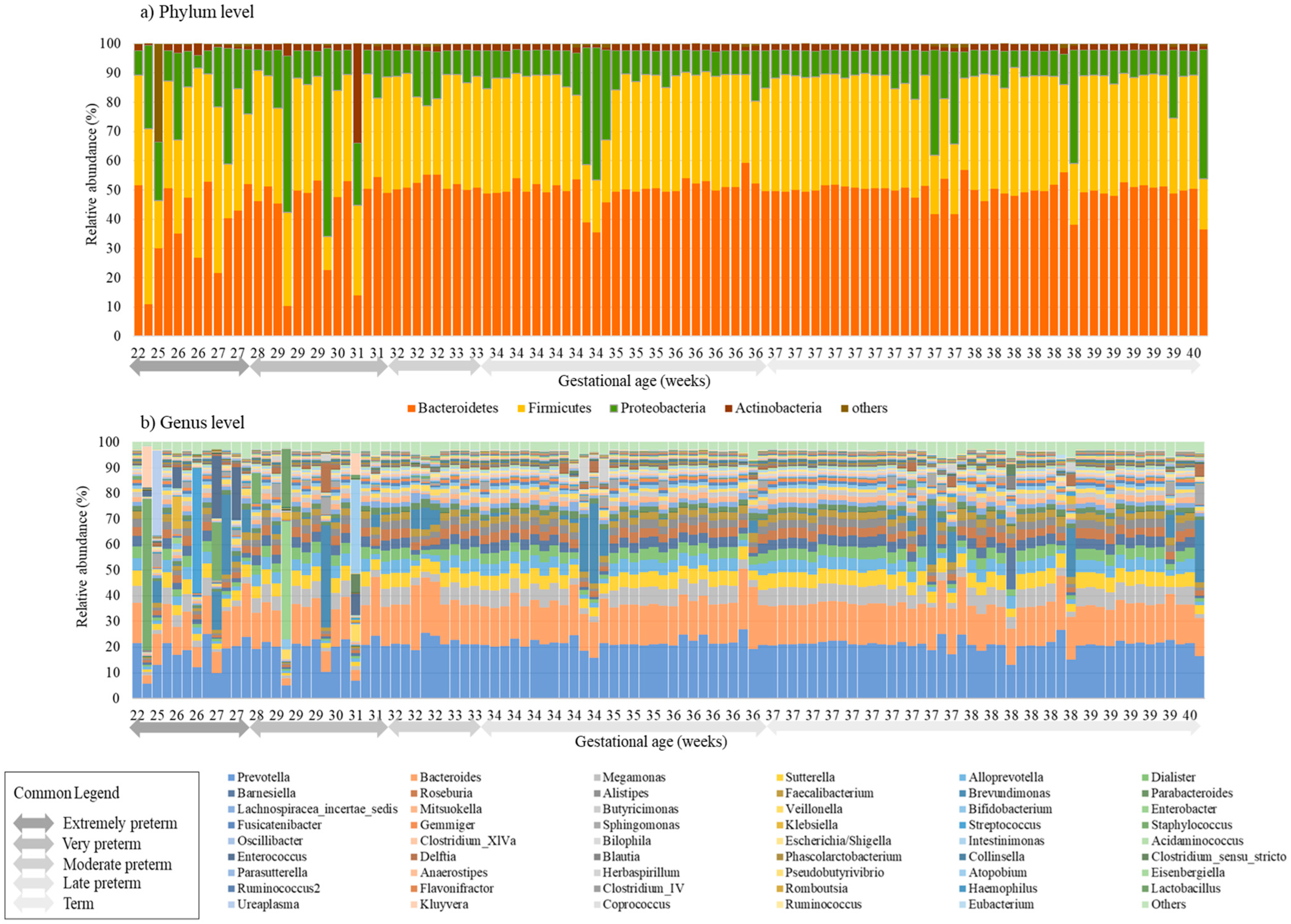
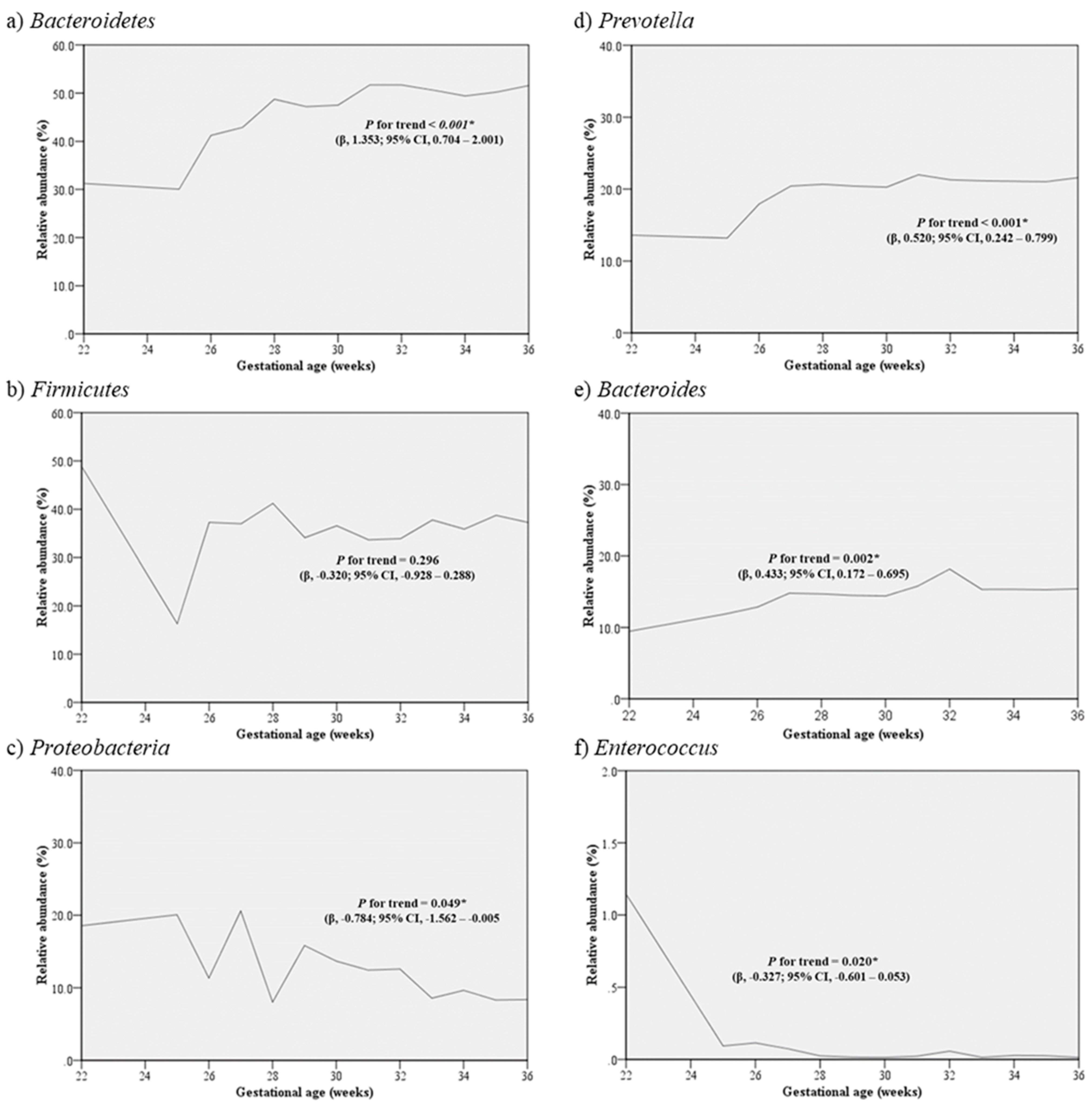
3.2. Distribution of Microbiota according to Gestational Age

Compared to the meconium microbiomes of infants born after the 30th week of gestation, those born prior to 30 weeks showed a disruption in the percentages of the core microbiome in both the phylum and genus levels (Figure 1). Bacteroidetes and Firmicutes were dominant in the first meconium microbiomes (Figure 1a), and in the genus level, Prevotella and Bacteroides were the most predominant species, with Prevotella’s relative abundance accounting for 20–30% of the gut microbiome. Furthermore, Bacteriodes composed the next large proportion, of up to ~20% of the microbiome. Bifidobacterium was not significantly different according to GA (Figure 1b).
In an analysis including only preterm infants born prior to 37 weeks of GA (n = 63), an analysis of the relationship between GA and relative abundance of the phyla Bacteroidetes in the meconium increased significantly as GA increased (P for trend < 0.001), and Proteobacteria decreased significantly as GA increased (P for trend = 0.049). In the genus level, as gestational age increased, Prevotella (P for trend < 0.001) and Bacteroides (P for trend = 0.002) increased significantly as GA increased, whereas Enterococcus (P for trend = 0.020) decreased as GA increased (Figure 2).

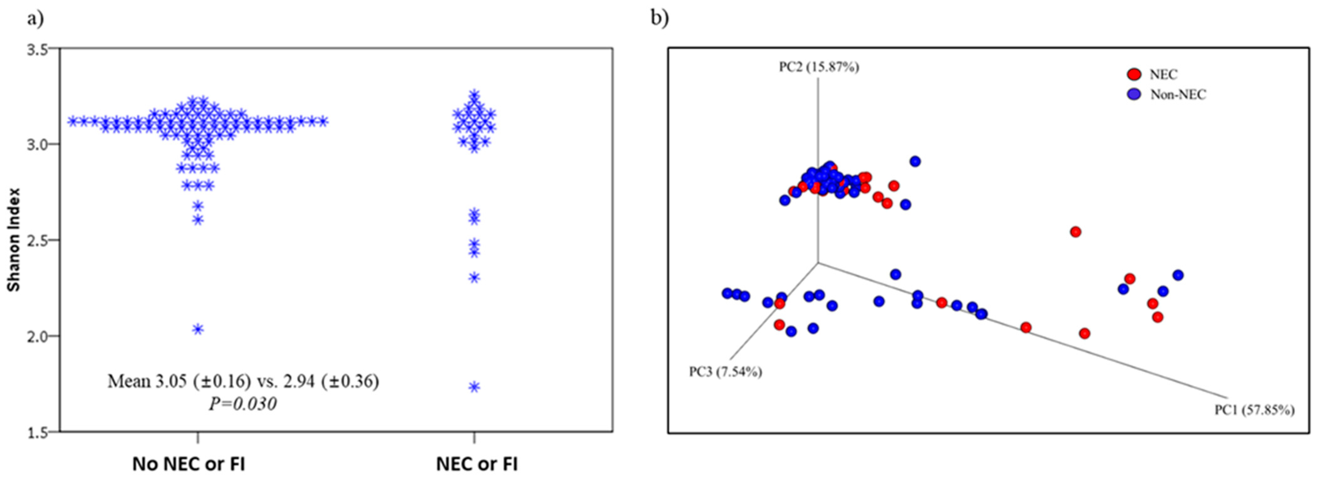
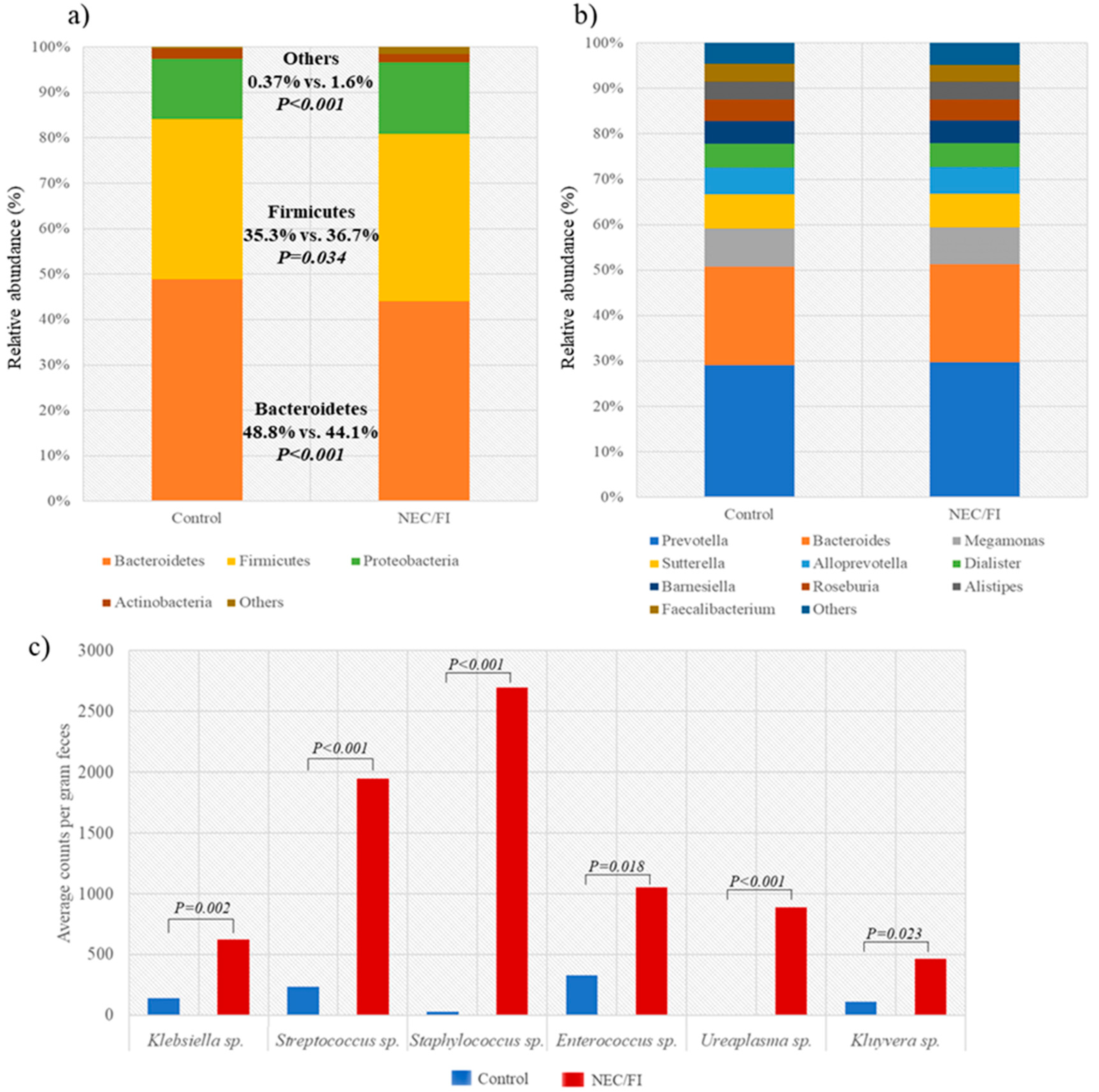
3.3. Comparison of Microbiomes and Clinical Outcomes between NEC/FI Group Versus Control Group

A total 24.1% (n = 26) were diagnosed with NEC or FI. Of these, 22 were preterm infants, and 4 were term infants. All possible perinatal and postnatal clinical factors associated with an increased risk for NEC/FI were investigated [26]. In the univariate analysis testing a total of 21 factors, GA (P < 0.001), birthweight (P < 0.001), antenatal steroid use (P = 0.003), congenital infection (P = 0.010), RDS (P = 0.002), and intraventricular hemorrhage (IVH) above grade II (P = 0.021) were factors associated with NEC/FI. However, in the multivariate analysis, after adjusting for confounding, none of these factors were independently significantly associated with NEC/FI in this cohort of patients (Table 2).
Compared to the control group, infants that had NEC/FI had significantly lower alpha diversities (Shannon Index, 3.05 vs. 2.94; P = 0.030, respectively), however, similar beta diversities (Figure 3).
The relative abundance of microbiota in the phylum level of the NEC/FI group showed a significantly lower abundance of Bacteroidetes, and higher abundance of Firmicutes and others (Figure 4a). In the genus level, there were no significant differences in the relative abundance of the 10 most dominant microbiota in the genus level (Figure 4b), however, the average counts in the NEC/FI group for the following species were significantly higher compared to the control group: Klebsiella, 624.6 [SD ± 2853.6] vs. 140.8 [SD ± 328.6], P = 0.002; Streptococcus, 1947.9 [SD ± 8891.1] vs. 237.8 [SD ± 152.8], P < 0.001; Staphylococcus, 2695.6 [SD ± 8790.8] vs. 26.8 [SD ± 95.0], P < 0.001; Enterococcus, 1053.4 [SD ± 3486.8] vs. 329.7 [SD ± 2052.6], P = 0.018; Ureaplasma, 886.5 [SD ± 4517.4] vs. 1.72 [SD ± 11.8], P < 0.001; Kluyvera, 462.1 [SD ± 2291.9] vs. 109.1 [SD ± 902.5], P = 0.023; respectively) (Figure 4c).

4. Discussion

The gut microbiota is a group of microorganisms, mainly bacteria, yeast, fungi, bacteriophages, and other viruses, as well as protozoa and archaea, which form a complex ecosystem in the human GI tract [27,28,29]. Meconium is often defined as the first stool passed within 48 h from birth. Neonatal microbiota starts diversifying quickly after birth, and compared to adults or older children, infant microbiota is known to have lower diversity as well as an unstable and highly dynamic microbiota structure [30]. This present study investigated the gut microbiome of 108 preterm and term infants' first passed meconium. The microbiomes of the meconium of preterm infants up to 30 weeks of GA showed lower alpha diversities and disruption in the percentages of the core microbiome in both the phylum and genus level, compared to infants born at a higher GA portraying a difference in the perinatal gut microbiome of preterm and term infants.
The two phyla, Bacteroidetes and Firmicutes, constituted majority of the microbiota of the neonatal meconium, and as the GA increased, a significant increase in the relative abundance of Bacteroidetes and decrease in Proteobacteria was observed. A study from Germany also showed that GA was significantly associated with the composition of meconium from very preterm infants, and the most abundant phyla included Firmicutes, Bacteriodetes, Proteobacteria, and Actinobacteria [14]. Our study showed that in the genus level, Prevotella and Bacteroides were the predominant species, with Prevotella’s relative abundance accounting for 20-30% of the gut microbiome. Whereas in healthy adults, the two main phyla—Firmicutes and Bacteroidetes—constitute over 90% of the gut microbiota, followed by Actinobacteria and Proteobacteria [31,32].
Upon delivery, infants are exposed for the first time to a variety of microbes from various sources which lead to an establishment of the infants’ gut microbiome. Recent studies have shown that the predominant early colonizers of infant’s gut are maternal fecal bacteria, mainly Bifidobacterium and Bacteroides, and Clostridium [27,33,34,35]. Studies have also demonstrated that Bacteroides are associated with increased gut diversity and faster intestine maturation and natural childbirth has been shown to be significantly related to microorganisms reflecting the mother’s vaginal flora, such as Bacteroides, Lactobacillus, and Prevotella [36,37]. In the first passed microbiome of neonates in our study, Prevotella and Bacteroides were the most prevalent, accounting for about 50% of the core microbiome. However, because 83.3% (n = 90) were born via caesarean section, Lactobacillus was not a predominant genus in the meconium of the infants in our study.
One of the most devastating gastrointestinal emergencies and a major cause of mortality in very low birth weight infants (VLBWI) is NEC. Furthermore, FI is also a frequently faced problem in many neonatal intensive care units (NICUs). Prenatal and postnatal factors, such as placental insufficiency, chorioamnionitis, gut ischemia, altered bacterial colonization, viruses, and blood transfusions, presumably disrupt the mucosal barrier, which may trigger inflammatory reactions in the immature intestines of preterm infants [4]. This study investigated whether a difference can be observed in the microbiome of meconium of infants that are eventually diagnosed with NEC. This study found that compared to infants that are discharged from the NICU without any gastrointestinal symptoms, infants that were diagnosed with NEC/FI had significantly lower alpha diversities in their first meconium. Furthermore, a significantly lower abundance of Bacteroidetes, and higher abundance of Firmicutes and other microbes were observed. Firmicutes, Streptococcaceae, and Enterococcaceae are known to predominate in adults with lower diversity gut microbiota [38]. Lower diversity gut microbiota is also known to be drivers of many diseases such as inflammatory bowel disease, acute diarrheal disease, C. difficile infection, and observed in cancer patients [38,39,40,41]. This shows that infants with NEC/FI have lower diversity dysbiosis, and this may have an important role for triggering inflammation leading to NEC/FI.
In the study on meconium microbiome and its relation to neonatal growth and head circumference catch-up in preterm infants, Polynucleobacter, Gp1, and Prevotella appeared in greater abundance in meconium of preterm infants with adequate birth weight for GA [42]. In our study, we found that the composition of Prevotella was positively correlated with GA, suggesting that the abundance of Prevotella has an important role in the growth and development of preterm infants.
The recent study conducted by Liu et al. on early gut microbiota in very low and extremely low birth weight preterm infants with feeding intolerance showed that the meconium samples of the FI group had higher proportions of γ-proteobacteria and Escherichia-Shigella and a lower proportion of Bacteroides compared with the those that did not have FI [43]. Although this was not corroborated in our study, our study found a lower proportion of Bacteroides in preterm infants with lower GA, suggesting that Bacteroides is a part of the healthy gut microbiome. Perturbation of the gut microbiota may promote overgrowth of pathobionts increasing the risk of infections and inflammation in the gut [44]. In a study on infants born via caesarean section, a high-level colonization by opportunistic pathogens was observed, including Enterococcus, Enterobacter, and Klebsiella [12]. In this study, the average counts in the NEC/FI group for the following pathobionts were significantly higher: Klebsiella, Streptococcus, Staphylococcus, Enterococcus, and Ureaplasma, and Kluyvera. The first moments of life are known to contribute to the formation of an NEC-associated microbiota [45], however, our study shows that the initiation in the formation of an NEC associated microbiota may even occur prior to events following birth.
In this study, there were six cases of placental abruption and 14 cases of PROM, meaning that these infants may have been exposed to the vaginal microbiota in utero, and subsequently affecting the results of the gut microbiome in these subjects. Due to the small number of subjects, further analyses were difficult. However, a study on 8 preterm neonates in Indonesia reported that PROM and mother’s diet influenced the meconium microbiome [46]. Further studies including a larger cohort of preterm infants are needed to make any conclusions on the impact of early exposure to vaginal microbiomes in utero and colonization and establishment of the infants’ gut microbiome.
When compared with bacteria, the healthy human gut mycobiome is lower in biomass compared to bacteriome biomass. However, the roles of mycobiomes are increasingly recognized as important, whether it be beneficial or harmful. The gut mycobiome is known to be dominated by Saccharomyces, Malassezia, and Candida [47], and these pathobionts may potentially be harmful to preterm infants [48]. Further studies are needed to characterize the composition of mycobiomes in preterm infants to better understand its role in the immature gut.
This study had a few limitations the sample size of the study groups was relatively small. However, to date, this study contains one of the largest cohort of neonates, from GA 22 to 40. Secondly, although not conclusive, studies have shown evidence of sex related difference in the gut microbiome [49]. Other studies do not support this [50]. In our study, 88.9% (n = 96) patient were male, therefore there may have been selection bias genders-wise. There is a lack of studies on the influence that gender has in the gut microbial composition of infants, and further studies are warranted. Finally, this study lacked a longitudinal analysis of changes in the gut microbiome of neonates after birth. However, the focus of our study was investigating the microbiome of the first meconium passed by the infant immediately after birth reflecting the perinatal gut microbiome of the infant.
In summary, this study showed that low diversity dysbiosis was observed in the initial gut microbiomes of infants born premature, below 30 weeks of GA, and an increasing abundance of Bacteroidetes was observed as gestation age increased. Infants with NEC/FI had significantly lower alpha diversities, and a significantly higher counts of pathobionts such as Klebsiella, Streptococcus, Staphylococcus, Enterococcus, Ureaplasma, and Kluyvera were observed. To conclude, differences were observed in the composition of the first passed meconium in preterm and term infants that later developed NEC/FI compared to those that did not. These findings fuel the need to understand disease pathogenesis and develop novel and significant microbiota for earlier therapeutic and preventative strategies for vulnerable preterm babies in further research.

Supplementary Materials

The following are available online at https://www.mdpi.com/article/10.3390/pathogens12010055/s1, Tables S1–S3.

Author Contributions

Conceptualization, Y.-A.Y., S.H.-B. and H.M.K.; methodology, H.M.K., S.H.-B., S.K., Y.-M.S., I.H.Y., M.Y.O. and S.-A.I.; validation, H.M.K., S.-A.I. and Y.-A.Y.; formal analysis, Y.-A.Y. and H.M.K.; investigation, Y.-A.Y. and H.M.K.; resources, Y.-A.Y. and H.M.K.; data curation, S.-A.I., Y.-A.Y. and H.M.K.; writing—original draft preparation, Y.-A.Y. and H.M.K.; writing—review and editing, Y.-A.Y. and H.M.K.; funding acquisition, Y.-A.Y. and H.M.K. All authors have read and agreed to the published version of the manuscript.

Funding

This work was funded by the National Research Foundation of Korea (NRF) granted by the Korean government of Ministry of Science (2022R1A2C100399611). This study was also supported by The Catholic Medical Center Research Foundation made in the program year of 2022.

Institutional Review Board Statement

The study was conducted in accordance with the Declaration of Helsinki and approved by the Institutional Review Board of Seoul St. Mary’s Hospital, The Catholic University of Korea (IRB no. KC21TISI0329).

Informed Consent Statement

Informed consent was obtained from the legal guardians of all subjects involved in the study.

Data Availability Statement

The aggregate counts of the phylum and genus level data (Supplement Materials Tables S1 and S2) and patient characteristics (Supplement Material Table S3) are partially available on the website, with the removal of any patient identifiers according to the IRB guidelines of Seoul St. Mary’s Hospital.

Acknowledgments

The authors wish to acknowledge the financial support of the Catholic Medical Center Research Foundation made in the program year 2022. We would like to thank AIBIOTICS Co., Ltd for their technical and statistical assistance.

Conflicts of Interest

The authors declare no conflict of interest.

References

  1. Chung, S.H.; Bae, C.W. Improvement in the Survival Rates of Very Low Birth Weight Infants after the Establishment of the Korean Neonatal Network: Comparison between the 2000s and 2010s. J. Korean Med. Sci. 2017, 32, 1228–1234. [Google Scholar] [CrossRef] [PubMed]
  2. Stoll, B.J. Epidemiology of necrotizing enterocolitis. Clin. Perinatol. 1994, 21, 205–218. [Google Scholar] [CrossRef] [PubMed]
  3. Clark, R.H.; Gordon, P.; Walker, W.M.; Laughon, M.; Smith, P.B.; Spitzer, A.R. Characteristics of patients who die of necrotizing enterocolitis. J. Perinatol. Off. J. Calif. Perinat. Assoc. 2012, 32, 199–204. [Google Scholar] [CrossRef] [PubMed]
  4. Schnabl, K.L.; Van Aerde, J.E.; Thomson, A.B.; Clandinin, M.T. Necrotizing enterocolitis: A multifactorial disease with no cure. World J. Gastroenterol. 2008, 14, 2142–2161. [Google Scholar] [CrossRef]
  5. Thompson, A.M.; Bizzarro, M.J. Necrotizing enterocolitis in newborns: Pathogenesis, prevention and management. Drugs 2008, 68, 1227–1238. [Google Scholar] [CrossRef]
  6. Neu, J.; Walker, W.A. Necrotizing enterocolitis. New Engl. J. Med. 2011, 364, 255–264. [Google Scholar] [CrossRef] [Green Version]
  7. Belkaid, Y.; Hand, T.W. Role of the microbiota in immunity and inflammation. Cell 2014, 157, 121–141. [Google Scholar] [CrossRef] [Green Version]
  8. Perez-Muñoz, M.E.; Arrieta, M.C.; Ramer-Tait, A.E.; Walter, J. A critical assessment of the “sterile womb” and “in utero colonization” hypotheses: Implications for research on the pioneer infant microbiome. Microbiome 2017, 5, 48. [Google Scholar] [CrossRef] [Green Version]
  9. Kennedy, K.M.; Gerlach, M.J.; Adam, T.; Heimesaat, M.M.; Rossi, L.; Surette, M.G.; Sloboda, D.M.; Braun, T. Fetal meconium does not have a detectable microbiota before birth. Nat. Microbiol. 2021, 6, 865–873. [Google Scholar] [CrossRef]
  10. Stinson, L.F.; Boyce, M.C.; Payne, M.S.; Keelan, J.A. The Not-so-Sterile Womb: Evidence That the Human Fetus Is Exposed to Bacteria Prior to Birth. Front. Microbiol. 2019, 10, 1124. [Google Scholar] [CrossRef]
  11. He, Q.; Kwok, L.Y.; Xi, X.; Zhong, Z.; Ma, T.; Xu, H.; Meng, H.; Zhao, F.; Zhang, H. The meconium microbiota shares more features with the amniotic fluid microbiota than the maternal fecal and vaginal microbiota. Gut Microbes 2020, 12, 1794266. [Google Scholar] [CrossRef] [PubMed]
  12. Shao, Y.; Forster, S.C.; Tsaliki, E.; Vervier, K.; Strang, A.; Simpson, N.; Kumar, N.; Stares, M.D.; Rodger, A.; Brocklehurst, P.; et al. Stunted microbiota and opportunistic pathogen colonization in caesarean-section birth. Nature 2019, 574, 117–121. [Google Scholar] [CrossRef] [PubMed]
  13. Dalby, M.J.; Hall, L.J. Recent advances in understanding the neonatal microbiome. F1000Research 2020, 9. [Google Scholar] [CrossRef] [PubMed]
  14. Klopp, J.; Ferretti, P.; Meyer, C.U.; Hilbert, K.; Haiß, A.; Marißen, J.; Henneke, P.; Hudalla, H.; Pirr, S.; Viemann, D.; et al. Meconium Microbiome of Very Preterm Infants across Germany. mSphere. 2022, 7, e00808-21. [Google Scholar] [CrossRef]
  15. Shi, Y.-C.; Guo, H.; Chen, J.; Sun, G.; Ren, R.-R.; Guo, M.-Z.; Peng, L.-H.; Yang, Y.-S. Initial meconium microbiome in Chinese neonates delivered naturally or by cesarean section. Sci. Rep. 2018, 8, 3255. [Google Scholar] [CrossRef] [Green Version]
  16. González-Rivera, R.; Culverhouse, R.C.; Hamvas, A.; Tarr, P.I.; Warner, B.B. The age of necrotizing enterocolitis onset: An application of Sartwell's incubation period model. J. Perinatol. Off. J. Calif. Perinat. Assoc. 2011, 31, 519–523. [Google Scholar] [CrossRef] [Green Version]
  17. Hodzic, Z.; Bolock, A.M.; Good, M. The Role of Mucosal Immunity in the Pathogenesis of Necrotizing Enterocolitis. Front. Pediatr. 2017, 5, 40. [Google Scholar] [CrossRef] [Green Version]
  18. Niño, D.F.; Sodhi, C.P.; Hackam, D.J. Necrotizing enterocolitis: New insights into pathogenesis and mechanisms. Nat. Rev. Gastroenterol. Hepatol. 2016, 13, 590–600. [Google Scholar] [CrossRef] [Green Version]
  19. 16S Metagenomic Sequencing Library Preparation. Available online: https://support.illumina.com/documents/documentation/chemistry_documentation/16s/16s-metagenomic-library-prep-guide-15044223-b.pdf (accessed on 20 July 2022).
  20. Bolyen, E.; Rideout, J.R.; Dillon, M.R.; Bokulich, N.A.; Abnet, C.C.; Al-Ghalith, G.A.; Alexander, H.; Alm, E.J.; Arumugam, M.; Asnicar, F.; et al. Reproducible, interactive, scalable and extensible microbiome data science using QIIME 2. Nat. Biotechnol. 2019, 37, 852–857. [Google Scholar] [CrossRef] [PubMed]
  21. Callahan, B.J.; McMurdie, P.J.; Rosen, M.J.; Han, A.W.; Johnson, A.J.A.; Holmes, S.P. DADA2: High-resolution sample inference from Illumina amplicon data. Nat. Methods 2016, 13, 581–583. [Google Scholar] [CrossRef]
  22. Davis, N.M.; Proctor, D.M.; Holmes, S.P.; Relman, D.A.; Callahan, B.J. Simple statistical identification and removal of contaminant sequences in marker-gene and metagenomics data. Microbiome 2018, 6, 226. [Google Scholar] [CrossRef] [PubMed] [Green Version]
  23. Bell, M.J.; Ternberg, J.L.; Feigin, R.D.; Keating, J.P.; Marshall, R.; Barton, L.; Brotherton, T. Neonatal necrotizing enterocolitis. Therapeutic decisions based upon clinical staging. Ann. Surg. 1978, 187, 1–7. [Google Scholar] [CrossRef] [PubMed]
  24. Carlos, M.A.; Babyn, P.S.; Marcon, M.A.; Moore, A.M. Changes in gastric emptying in early postnatal life. J. Pediatr. 1997, 130, 931–937. [Google Scholar] [CrossRef] [PubMed]
  25. WHO Factsheet. Preterm Birth. Available online: https://www.who.int/news-room/fact-sheets/detail/preterm-birth (accessed on 31 November 2022).
  26. Samuels, N.; van de Graaf, R.A.; de Jonge, R.C.J.; Reiss, I.K.M.; Vermeulen, M.J. Risk factors for necrotizing enterocolitis in neonates: A systematic review of prognostic studies. BMC Pediatr. 2017, 17, 105. [Google Scholar] [CrossRef] [PubMed]
  27. Bäckhed, F.; Roswall, J.; Peng, Y.; Feng, Q.; Jia, H.; Kovatcheva-Datchary, P.; Li, Y.; Xia, Y.; Xie, H.; Zhong, H.; et al. Dynamics and Stabilization of the Human Gut Microbiome during the First Year of Life. Cell Host Microbe 2015, 17, 690–703. [Google Scholar] [CrossRef] [PubMed] [Green Version]
  28. Neish, A.S. Microbes in gastrointestinal health and disease. Gastroenterology 2009, 136, 65–80. [Google Scholar] [CrossRef] [Green Version]
  29. Scarpellini, E.; Ianiro, G.; Attili, F.; Bassanelli, C.; De Santis, A.; Gasbarrini, A. The human gut microbiota and virome: Potential therapeutic implications. Dig. Liver Dis. Off. J. Ital. Soc. Gastroenterol. Ital. Assoc. Study Liver 2015, 47, 1007–1012. [Google Scholar] [CrossRef] [Green Version]
  30. Cheng, J.; Ringel-Kulka, T.; Heikamp-de Jong, I.; Ringel, Y.; Carroll, I.; de Vos, W.M.; Salojärvi, J.; Satokari, R. Discordant temporal development of bacterial phyla and the emergence of core in the fecal microbiota of young children. ISME J. 2016, 10, 1002–1014. [Google Scholar] [CrossRef] [Green Version]
  31. Jalanka-Tuovinen, J.; Salonen, A.; Nikkilä, J.; Immonen, O.; Kekkonen, R.; Lahti, L.; Palva, A.; de Vos, W.M. Intestinal microbiota in healthy adults: Temporal analysis reveals individual and common core and relation to intestinal symptoms. PloS ONE 2011, 6, e23035. [Google Scholar] [CrossRef]
  32. Claesson, M.J.; Jeffery, I.B.; Conde, S.; Power, S.E.; O’Connor, E.M.; Cusack, S.; Harris, H.M.; Coakley, M.; Lakshminarayanan, B.; O’Sullivan, O.; et al. Gut microbiota composition correlates with diet and health in the elderly. Nature 2012, 488, 178–184. [Google Scholar] [CrossRef]
  33. Koenig, J.E.; Spor, A.; Scalfone, N.; Fricker, A.D.; Stombaugh, J.; Knight, R.; Angenent, L.T.; Ley, R.E. Succession of microbial consortia in the developing infant gut microbiome. Proc. Natl. Acad. Sci. USA 2011, 108 (Suppl. 1), 4578–4585. [Google Scholar] [CrossRef] [Green Version]
  34. Asnicar, F.; Manara, S.; Zolfo, M.; Truong, D.T.; Scholz, M.; Armanini, F.; Ferretti, P.; Gorfer, V.; Pedrotti, A.; Tett, A.; et al. Studying Vertical Microbiome Transmission from Mothers to Infants by Strain-Level Metagenomic Profiling. mSystems 2017, 2, e00164-16. [Google Scholar] [CrossRef] [PubMed] [Green Version]
  35. Korpela, K.; Costea, P.; Coelho, L.P.; Kandels-Lewis, S.; Willemsen, G.; Boomsma, D.I.; Segata, N.; Bork, P. Selective maternal seeding and environment shape the human gut microbiome. Genome Res. 2018, 28, 561–568. [Google Scholar] [CrossRef] [PubMed] [Green Version]
  36. Stewart, C.J.; Ajami, N.J.; O’Brien, J.L.; Hutchinson, D.S.; Smith, D.P.; Wong, M.C.; Ross, M.C.; Lloyd, R.E.; Doddapaneni, H.; Metcalf, G.A.; et al. Temporal development of the gut microbiome in early childhood from the TEDDY study. Nature 2018, 562, 583–588. [Google Scholar] [CrossRef] [PubMed] [Green Version]
  37. Morais, J.; Marques, C.; Teixeira, D.; Durão, C.; Faria, A.; Brito, S.; Cardoso, M.; Macedo, I.; Pereira, E.; Tomé, T.; et al. Extremely preterm neonates have more Lactobacillus in meconium than very preterm neonates—The in utero microbial colonization hypothesis. Gut Microbes 2020, 12, 1785804. [Google Scholar] [CrossRef]
  38. Kriss, M.; Hazleton, K.Z.; Nusbacher, N.M.; Martin, C.G.; Lozupone, C.A. Low diversity gut microbiota dysbiosis: Drivers, functional implications and recovery. Curr. Opin. Microbiol. 2018, 44, 34–40. [Google Scholar] [CrossRef]
  39. Hamilton, M.J.; Weingarden, A.R.; Unno, T.; Khoruts, A.; Sadowsky, M.J. High-throughput DNA sequence analysis reveals stable engraftment of gut microbiota following transplantation of previously frozen fecal bacteria. Gut Microbes 2013, 4, 125–135. [Google Scholar] [CrossRef] [Green Version]
  40. Lozupone, C.A.; Stombaugh, J.; Gonzalez, A.; Ackermann, G.; Wendel, D.; Vázquez-Baeza, Y.; Jansson, J.K.; Gordon, J.I.; Knight, R. Meta-analyses of studies of the human microbiota. Genome Res. 2013, 23, 1704–1714. [Google Scholar] [CrossRef] [Green Version]
  41. Liu, C.; Frank, D.N.; Horch, M.; Chau, S.; Ir, D.; Horch, E.A.; Tretina, K.; van Besien, K.; Lozupone, C.A.; Nguyen, V.H. Associations between acute gastrointestinal GvHD and the baseline gut microbiota of allogeneic hematopoietic stem cell transplant recipients and donors. Bone Marrow Transplant. 2017, 52, 1643–1650. [Google Scholar] [CrossRef] [Green Version]
  42. Terrazzan Nutricionist, A.C.; Procianoy, R.S.; Roesch, L.F.W.; Corso, A.L.; Dobbler, P.T.; Silveira, R.C. Meconium microbiome and its relation to neonatal growth and head circumference catch-up in preterm infants. PloS ONE 2020, 15, e0238632. [Google Scholar] [CrossRef]
  43. Liu, L.; Ao, D.; Cai, X.; Huang, P.; Cai, N.; Lin, S.; Wu, B. Early gut microbiota in very low and extremely low birth weight preterm infants with feeding intolerance: A prospective case-control study. J. Microbiol. 2022, 60, 1021–1031. [Google Scholar] [CrossRef] [PubMed]
  44. Pickard, J.M.; Zeng, M.Y.; Caruso, R.; Núñez, G. Gut microbiota: Role in pathogen colonization, immune responses, and inflammatory disease. Immunol. Rev. 2017, 279, 70–89. [Google Scholar] [CrossRef]
  45. Sharif, S.; Meader, N.; Oddie, S.J.; Rojas-Reyes, M.X.; McGuire, W. Probiotics to prevent necrotising enterocolitis in very preterm or very low birth weight infants. Cochrane Database Syst Rev 2020, 10, CD005496. [Google Scholar] [CrossRef] [PubMed]
  46. Amandito, R.; Malik, A.; Rohsiswatmo, R. Metagenomic profiles of the early life microbiome of Indonesian inpatient neonates and their influence on clinical characteristics. Sci. Rep. 2022, 12, 9413. [Google Scholar] [CrossRef]
  47. Jiang, L.; Stärkel, P.; Fan, J.G.; Fouts, D.E.; Bacher, P.; Schnabl, B. The gut mycobiome: A novel player in chronic liver diseases. J. Gastroenterol. 2021, 56, 1–11. [Google Scholar] [CrossRef] [PubMed]
  48. James, S.A.; Phillips, S.; Telatin, A.; Baker, D.; Ansorge, R.; Clarke, P.; Hall, L.J.; Carding, S.R. Preterm Infants Harbour a Rapidly Changing Mycobiota That Includes Candida Pathobionts. J. Fungi 2020, 6, 273. [Google Scholar] [CrossRef]
  49. Kim, Y.S.; Unno, T.; Kim, B.Y.; Park, M.S. Sex Differences in Gut Microbiota. World J. Men’s Health 2020, 38, 48–60. [Google Scholar] [CrossRef]
  50. Lay, C.; Rigottier-Gois, L.; Holmstrøm, K.; Rajilic, M.; Vaughan, E.E.; de Vos, W.M.; Collins, M.D.; Thiel, R.; Namsolleck, P.; Blaut, M.; et al. Colonic microbiota signatures across five northern European countries. Appl. Environ. Microbiol. 2005, 71, 4153–4155. [Google Scholar] [CrossRef]
Figure 1. Distribution of (a) phylum levels and (b) genus levels according to gestational age. Microbiomes of the 1st stool samples obtained after delivery showed dysbiosis in extremely preterm babies born < 31 weeks of gestational age in both the phylum and genus levels. In the phylum level, Bacteroidetes and Firmicutes were the most dominant phyla in the first neonatal microbiomes. In the genus level, Prevotella sp. and Bacteroides sp. were the most predominant species in the meconium.
Pathogens 12 00055 g001
Figure 2. Change in the relative abundance of the phyla (a) Bacteroidetes, (b) Firmicutes, (c) Proteobacteria and genus (d) Prevotella, (e) Bacteroides, (f) Enterococcus according to gestational age in preterm infant born prior to 37 weeks of gestation (n = 63). The graph shows the median relative abundance for each gestational age.
Pathogens 12 00055 g002
Figure 3. Comparison of alpha diversities (Shannon Index) and beta diversities (Bray–Curtis dissimilarity) of infants without any gastrointestinal symptoms versus those with NEC or FI. (a) Compared to the control group, infants that had NEC/FI had significantly lower alpha diversities however, (b) no significant differences in the beta diversities.
Pathogens 12 00055 g003
Figure 4. Gut microbiome comparison between the control group versus NEC/FI group of (a) absolute abundance and types of bacteria in the phylum level between the control group and NEC/FI group showing a significantly lower abundance of Bacteroidetes, and higher abundance of Firmicutes and others. (b) In the genus level, there were no significant differences in the absolute abundance of the 10 most dominant microbiota in the genus level, however, (c) the average counts in the NEC/FI group for the following species were significantly higher: Klebsiella, Streptococcus, Staphylococcus, Enterococcus, Ureaplasma, and Kluyvera.
Pathogens 12 00055 g004
Table 1. Comparison of perinatal characteristics of preterm and term infants included in the study.
No. of Participants (%)P Value
Total
N = 108
Preterm
N = 63
Term
N = 45
GA, weeks (mean ± SD)34.7 ± 4.231.6 ± 3.737.9 ± 1.0<0.001
Birth weight, g (mean ± SD)2,305.9 ± 863.01,807.4 ± 773.03,003.8 ± 339.3<0.001
Mother DM11 (10.2)8 (12.7)3 (6.7)0.354
IVF6 (5.6)5 (7.9)1 (2.2)0.397
Male96 (88.9)60 (95.2)36 (80.0)0.026
Caesarean section90 (83.3)58 (92.1)32 (71.1)0.007
Small for gestational age6 (5.6)3 (4.8)3 (6.7)0.692
Chorioamnionitis2 (1.9)2 (3.2)00.509
PROM, hrs14 (13.0)11 (17.5)2 (4.4)0.069
Placental abruption6 (5.6)6 (9.5)00.040
Oligohydramnios10 (9.3)10 (15.9)00.005
Antenatal steroid use43 (39.8)43 (68.3)0<0.001
Congenital infection7 (6.5)7 (11.1)00.040
Need for resuscitation at birth50 (46.3)40 (63.5)10 (22.2)<0.001
NEC/FI26 (24.1)22 (34.9)4 (8.9)0.003
Late preterm, n = 28; moderately preterm, n = 10; very preterm, n = 13; extremely preterm, n = 12; Abbreviations: DM, diabetes mellitus; FI, feeding intolerance; GA, gestational age; IVF, in vivo fertilization; NEC, necrotizing enterocolitis; PROM, preterm prolonged rupture of membranes.
Table 2. Factors associated with an increased risk for NEC/FI in preterm and term infants.
UnivariateP ValueMultivariateP Value
OR95% CIOR95% CI
GA, weeks (mean ± SD)0.7160.624–0.820<0.0010.7360.491–1.1040.138
Birth weight, g (mean ± SD)0.9980.998–0.999<0.0010.9990.997–1.0010.162
Caesarean section1.7160.455–6.4700.425
PROM3.2140.971–10.6380.056
Preeclampsia0.8930.174–4.5900.892
Placental abruption3.4350.649–18.1790.147
Oligohydramnios3.6670.970–13.8660.056
IUGR3.4350.649–18.1790.147
Chorioamnionitis--0.999
Antenatal steroid use4.0681.602–10.3350.0030.3760.074–1.9130.239
Congenital infection9.5241.725–52.5930.0101.2750.179–9.0970.808
RDS4.2271.673–10.6820.0020.2070.033–1.3090.094
Massive pulmonary hemorrhage--0.999
Air leak1.2830.234–7.0430.774
Sepsis--0.998
IVH > grade II2.1871.126–4.2470.0210.5950.227–1.5600.291
PDA2.1940.346–13.9090.404
CLD--0.998
PAH3.3330.446–24.9360.241
No breast milk given enterally prior to NEC/FI0.4060.147–1.1190.081
Antibiotics use prior to NEC/FI--0.999
Abbreviations: CI, confidence interval; CLD, chronic lung disease; GA, gestational age; IUGR, intrauterine growth restriction; IVH, intraventricular hemorrhage; NEC/FI, necrotizing enterocolitis/feeding intolerance; PAH, pulmonary artery hypertension; PDA, patent ductus arteriosus; Covariates used in the multivariate analyses were as follows: Gestational age, birth weight, antenatal steroid use, congenital infection, RDS, IVH grade III and IV.
Disclaimer/Publisher’s Note: The statements, opinions and data contained in all publications are solely those of the individual author(s) and contributor(s) and not of MDPI and/or the editor(s). MDPI and/or the editor(s) disclaim responsibility for any injury to people or property resulting from any ideas, methods, instructions or products referred to in the content.

