Article

Lasiodiplodia fici sp. nov., Causing Leaf Spot on Ficus altissima in China

1
Innovative Institute for Plant Health, Zhongkai University of Agriculture and Engineering, Guangzhou 510225, China
2
Center of Excellence in Fungal Research, Mae Fah Luang University, Chiang Rai 57100, Thailand
3
School of Science, Mae Fah Luang University, Chiang Rai 57100, Thailand
4
Faculdade de Ciências, Biosystems and Integrative Sciences Institute (BioISI), Universidade de Lisboa, Campo Grande, 1749-016 Lisbon, Portugal
5
Key Laboratory of Fruit and Vegetable Green Prevention and Control in South-China, Ministry of Agriculture and Rural Affairs, Guangzhou 510225, China
*
Authors to whom correspondence should be addressed.
Pathogens 2022, 11(8), 840; https://doi.org/10.3390/pathogens11080840
Submission received: 17 June 2022 / Revised: 22 July 2022 / Accepted: 22 July 2022 / Published: 27 July 2022
(This article belongs to the Special Issue 10th Anniversary of Pathogens—Feature Papers)

Abstract

:
High temperatures and the seasonality in tropical ecosystems favours plant pathogens, which result in many fungal diseases. Among these, diseases caused by Botryosphaeriaceae species are prominent as dieback, canker and leaf spots. In this research, we isolated one leaf-spot-causing Botryosphaeriaceae species from Ficus altissima leaves, which were collected in Guangzhou, Guangdong Province, China. Isolation and identification of the pathogen were based on morphological and molecular aspects. Based on multigene phylogenetic analysis of combined internal transcribed spacer (ITS), translation elongation factor 1-α gene (tef1) and beta-tubulin gene (tub2), the fungus associated with leaf spots on F. altissima is described as Lasiodiplodia fici, a novel species. Pathogenicity assays were conducted by inoculating the fungus onto detached shoots and plants under controlled environmental conditions. The results revealed that the L. fici isolates can infect the plant tissues under stress conditions by developing disease symptoms on detached shoots within three days. However, when it was inoculated onto the leaves of the host and grown in natural conditions, the progression of the disease was slow. The putative pathogen was re-isolated, and Koch’s assumptions were satisfied. This is the first report of Lasiodiplodia species causing disease on Ficus altissima. Results from the present study will provide additional knowledge on fungal pathogens associated with forest and ornamental plant species.

1. Introduction

Fungi are one of the key organisms in forest ecosystems. They live in the ecosystems as decomposers, mutualists and pathogens while contributing to continuous nutrient recycling and affecting biodiversity [1]. However, fungal pathogens associated with forest plants are understudied [2]. Among a wide range of pathogens, opportunistic pathogens are of great concern as they act when the environmental conditions are favourable for disease development [3,4,5]. Botryosphaeriaceae species are well-known opportunistic fungal pathogens causing devastating forest diebacks for a wide range of hosts [6,7]. Diseases caused by Botryosphaeriaceae species have higher significance as most of them infect the perennial parts of the plants [7]. Sydow and Theissen [8] introduced Botryosphaeriaceae in 1918, and the taxonomic status of Botryosphaeriaceae was controversial until it was reintegrated by Schoch et al. in 2006 [9]. However, following both morpho molecular aspects, Botryosphaeriaceae consists of 23 genera and over 100 species that are confirmed with DNA sequence data [7,10,11,12,13,14].
In 1894, Ellis introduced the Lasiodiplodia [15] with L. tubericola as the type species. Index Fungorum (March 2022) lists 84 Lasiodiplodia epithets. However, there are 71 species accepted with molecular data [7]. Lasiodiplodia is a cosmopolitan genus which has an extensive host and geographical range. Species of Lasiodiplodia are pathogenic on economically important fruit crops [6,16]. The pathogenicity of these species is varied, even within the same host in different countries. Even within a country, a single species can infect several different hosts [4]. As an example, Lasiodiplodia theobromae is one of the major pathogens of grapevine dieback, and the pathogenicity of this species varies in different countries [17,18,19,20,21]. In addition, Lasiodiplodia species have already been reported to cause disease in a broad range of forest plants, including Acacia sp. [12], Eucalyptus [22] and Ficus [23].
The banyan tree (Ficus) is one of the genera of Moraceae and the only genus of the banyan Trib. Ficeae Trec [24]. This species is distributed from the southwest to the east and the south of China, while it is relatively rare in the rest of the country. This tree is a large tree reaching a height of 15–25 m; the bark is dark grey, the chest is about 50 cm in height with a wide crown and old trees often have rusty brown aerial roots [25]. The leaves are thin, tough, elongated and oval, with dark green surfaces and shiny and complete leaf arrangements [25]. The following are common characteristics of banyan trees: they come in pairs or are born with the axils of deciduous branches, they are yellow or reddish when mature, they are oblate, they have three basal bracts, they are broadly ovoid and they are persistent [25,26]. This plant is an important tropical forest tree as well as an ornamental plant (Figure 1). They are widely used as bonsai and landscaping trees [26]. Therefore, the isolation and characterisation of the diseases associated with this plant have both economic and ecological significance in China.
Banyan trees are highly susceptible to diseases and insect pests [27]. African snails, Ficus thrips, banyan grey silk moth, vermillion hair spot moth and banyan tree louse are the pests most widely associated with this host. Banyan trees are susceptible to several fungal diseases, including Anthracnose (Colletotrichum gloeosporiodes), a leaf spot caused by Cercospora spp. and Corynespora cassiicola, grey mould (Botrytis spp.), Phomopsis dieback (Diplodia spp.), Southern blight (Sclerotium rolfsii) and Verticillium wilt (Verticilliumalboatrum) [28,29].
In a sampling survey of banyan trees in Guangzhou City, Guangdong Province, China, a new leaf disease was observed. The objectives of this study were to (i) isolate and characterise the pathogen and (ii) confirmed the pathogenicity of isolated species. As result, a novel Lasiodiplodia species was identified by morphology and phylogenetic analysis based on ITS, tef1 and tub2. Pathogenicity assays were conducted to confirm the pathogenicity, and Koch’s postulates were fulfilled. A detailed description and graphical illustration of new Lasiodiplodia species are provided.

2. Results

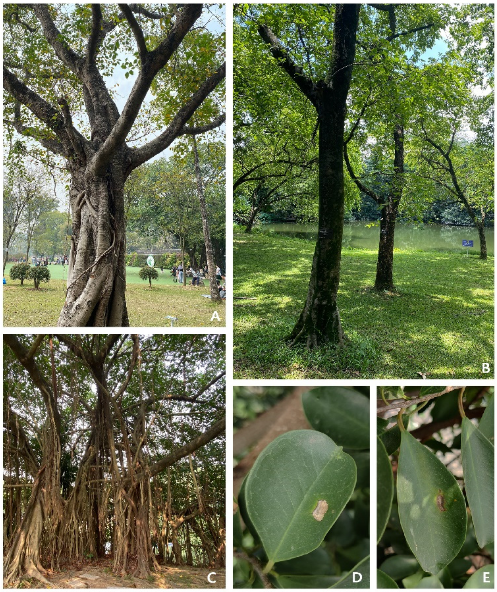
2.1. Sampling and Isolation

During our tropical fungal survey at the South China Botanical Garden in Guangzhou City, Guangdong Province, China, a leaf spot disease was observed in banyan trees. A part of the infected leaf becomes brown and shows chlorosis. Then, the leaf spot becomes wider, and the plant tissue dies. When a leaf is severely damaged, leaf tissue becomes a grey/white transparent film (Figure 1). In a total of 20 banyan trees, 17 were reported to have these symptoms. From these infected trees, 10 plants were randomly selected, and from each plant, 10 leaves were collected. Almost all the leaves had similar disease symptoms (Figure 1D,E); thus, the samples were pooled, and 10 leaves were selected for tissue isolation. Tissue isolation resulted in 15 isolates, which were identical and resembled the morphology of Lasiodiplodia species. Thus, three strains (ZHKUCC 21-0125, ZHKUCC 21-0126 and ZHKUCC 21-0127) were selected for further analyses.

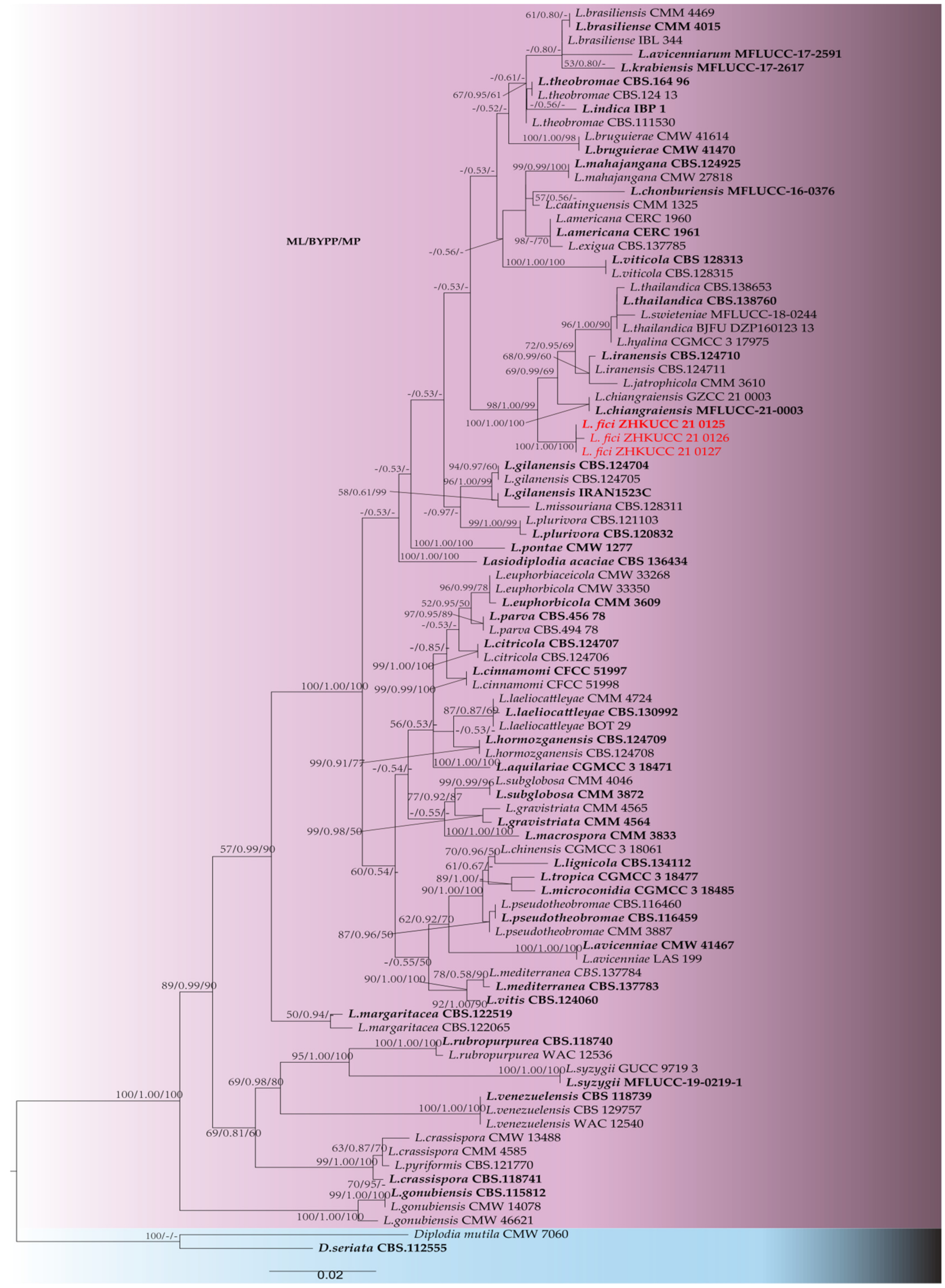
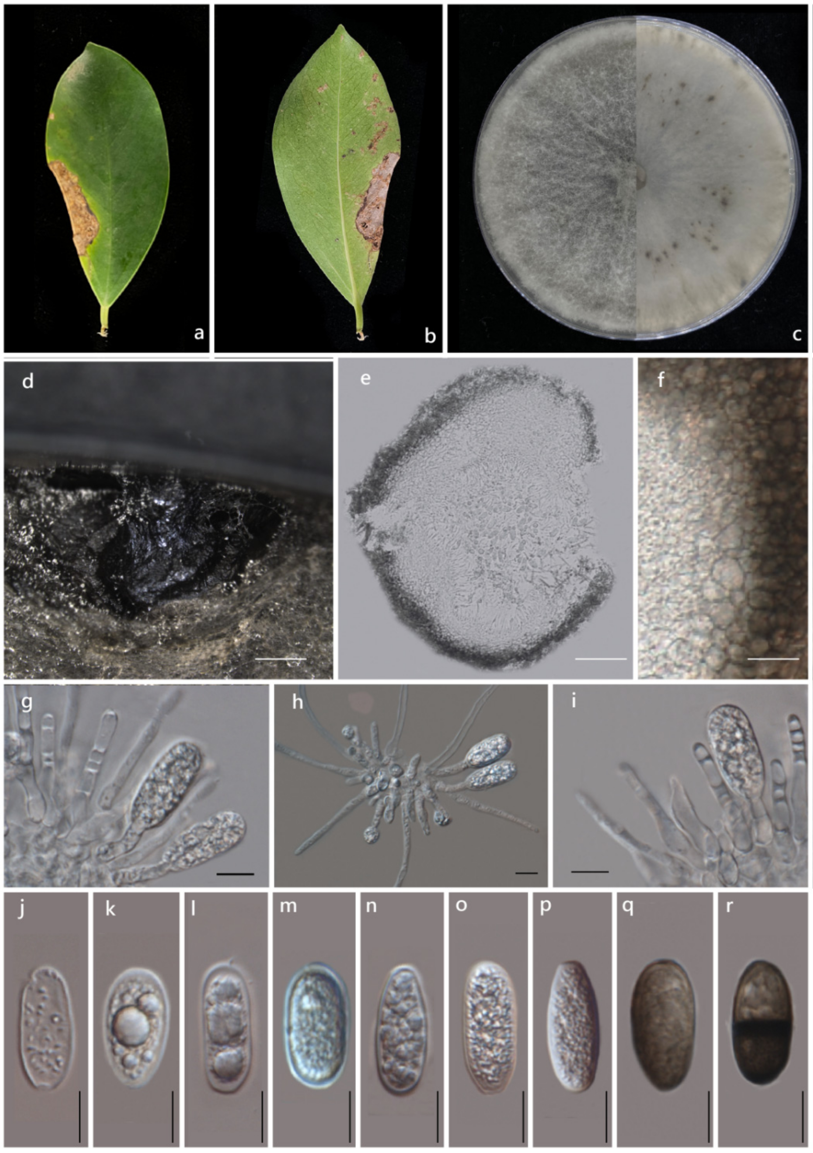
2.2. Morphological and Molecular Characterisation

Phylogenetic analyses were conducted using combined ITS, tef1 and tub2 sequence alignment of 91 Lasiodiplodia strains (including type strains), with D. mutila (CMW 7060) and Diplodia seriata (CBS 112555) as outgroup taxa. The resulting maximum likelihood (ML) tree shared a similar topology with maximum parsimony (MP) and Bayesian analysis (BYPP). The best-scoring ML tree is shown in Figure 2. The final dataset consists of 854 characters, of which 1139 characters are constants and 44 are parsimony informative. The maximal parsimonious analysis of the remaining 241 parsimonious information characters resulted in the 1000 most parsimonious trees of length 1001 steps (CI = 0.595, RI = 0.916, RC = 0.545, HI = 0.405). The best-scoring ML tree had the final likelihood value of -5342.638988. The matrix consisted of 357 distinct alignment patterns, with 7.89% of undetermined characters or gaps. Estimated base frequencies were as follows: A = 0.207301, C = 0.303614, G = 0.256996, T = 0.232089; substitution rates AC = 1.100912, AG =4.148566, AT = 1.475433, CG = 1.038445, CT = 5.970500, GT=1.000000; and gamma distribution shape parameter a = 0.195691. In the phylogenetic analysis, three isolates (ZHKUCC 21-0125, ZHKUCC 21-0126 and ZHKUCC 21-0127) from this study developed an independent clade from other known species. To confirm the novelty of isolated taxon, a strict morphological comparison was conducted, and sequence data were compared. We identified these isolates as novel Lasiodiplodia species. Species descriptions and illustrations are given below.
Lasiodiplodia fici G.Y. Xia, Manawas., M. Luo and K.D. Hyde sp. nov.
Index Fungorum number: IF 555265, Facesoffungi number: FoF 10815, (Figure 3).
Holotype: ZHKU 21-0092
Etymology: Epithet refers to the host genus from which the fungus was isolated.
Pathogenic on Ficus altissima L. leaves. Sexual morph: Not observed. Asexual morph: Conidiomata pycnidial, produced on PDA within 4–8 wk, dark brown–black, unilocular, up to 3100 μm diam, immersed in the needle tissue, globose–subglobose, ostiolate, wall composed of several layers of dark brown textura angularis. Peridium 20–50 µm, composed of thick-walled, brown-black cells of textura angularis, thin inner wall. Cylindrical, septate, unbranched, ends rounded, formed among conidiogenous cells. Conidiophores absent. Conidiogenous cells 20–30 × 10–15 µm ( x ¯ = 27 × 12 µm, n = 20), holoblastic, hyaline, smooth, thin-walled, cylindrical. Conidia 15–30 µm × 10–12 µm ( x ¯ = 22 × 11 µm, n = 50), initially hyaline, aseptate, ellipsoid–ovoid, thin-walled with granular content, rounded at apex, base round or truncate, becoming dark brown, 1 septate with longitudinal striations.
Culture characteristics: Colonies on PDA reached 7 cm diameter after five days at 28 °C. The upper view was filamentous, flat and turbid. The aerial hyphae were fluffy, dense and turned grey and black with time. The back became black.
Materials examined: China, Guangdong Province, Guangzhou City, South China Botanical Garden on the dead leaves of Ficus altissima L. (Moraceae), May 12, 2021, X. Guiyan, (ZHKU 21-0092 Holotype), living cultures ZHKUCC 21-0125 ex-type; ZHKUCC 21-0126, ZHKUCC 21-0127.
Note: Three isolates obtained in this study were phylogenetically closely related to L. iranensis, L. chiangraiensis and L. thailandica. However, isolates from this study formed a distinct linage with 100% ML, 100% MP and 1.0 BYPP support (Figure 2). Morphologically, these species can be distinguished from the dimensions of their conidia (Table 1), in which Lasiodiplodia fici developed conidia with a higher L/W ratio. In terms of the nucleotide comparison, Lasiodiplodia fici (ZHKUCC 21-0125) and Lasiodiplodia chiangraiensis (MFLUCC 21-0003) differed in one base pair (bp) in ITS, seven in tef1 and two in tub2. Lasiodiplodia fici (ZHKUCC 21-0125) and Lasiodiplodia iranensis (CBS 12471) differed in one base pair (bp) in ITS, 13 in tef1-α, and CBS 124710 did not have tub2 sequence. Lasiodiplodia fici (ZHKUCC 21-0125) and Lasiodiplodia thailandic (CPC 2279) differed in one base pair (bp) in ITS and fifteen in tef1, but tub2 of these two species are identical. Based on these polyphasic approaches, we introduced Lasiodiplodia fici as a new species.

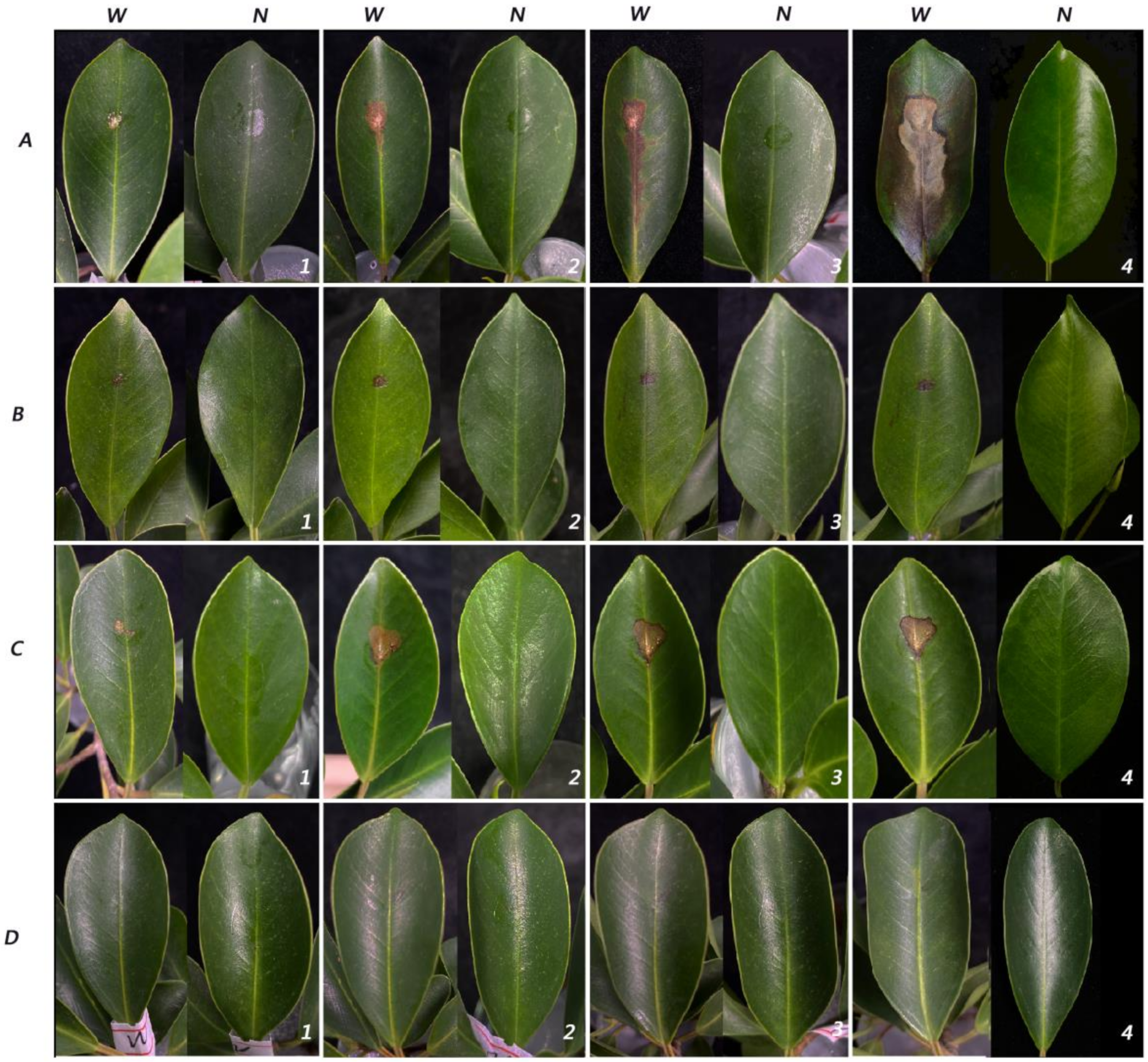
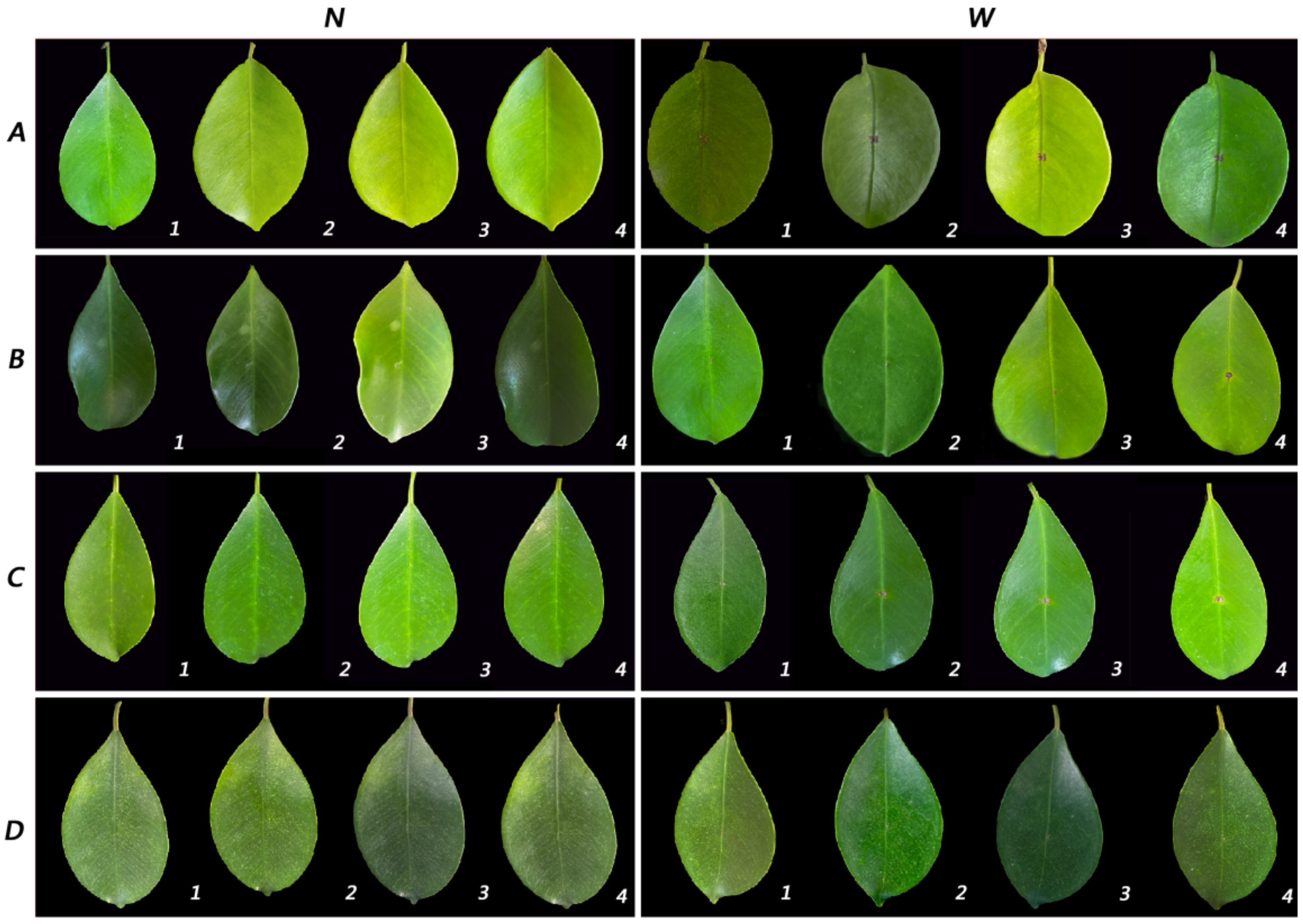
2.3. Pathogenicity Assay

In this study, sporulation of Lasiodiplodia fici was difficult to perform, even after trying different media (MEA, WA and PDA) and pine needles. Therefore, the pathogenicity test was carried out by the mycelial plug method. From actively growing cultures on PDA, mycelial plugs 50 mm in diameter and 20 mm in height were used. After 24 h, all wounded inoculated detached shoots had small lesions. Three days later, leaves fell from one infected shoot of the isolate ZHKUCC 21-0127. Then, leaf spots enlarged. Five days later, all infected leaves of three isolates had fallen off, while non-inoculated leaves did not. The disease development and progress of the isolate ZHKUCC 21-0125 were higher than the other two strains. No disease symptoms developed on any of the controls. All non-wounded samples were the same as the control leaves without any disease. Under field conditions, wounded leaves inoculated with mycelial and developed the disease slowly. After seven days of inoculation, ZHKUCC 21-0125 produced the largest lesion (Figure 4). However, disease establishment and progress were slower on plants inoculated in their natural habitat. All diseased leaf tissues were subjected to re-isolation of the pathogen, and the species was confirmed as Lasiodiplodia fici using sequence data (Figure 5).

3. Discussion

In this paper, a new Lasiodiplodia species is described from Ficus altissima in China. The fungus was shown to be pathogenic, causing leaf spots similar to the ones from which it was isolated. Correct species identification is important in plant pathology for early detection and development of management strategies [33]. Species delineation in Botryosphaeriaceae genera, such as Lasiodiplodia has been the subject of debate in recent years. Morphology and molecular phylogeny have become important criteria in this genus to resolve species [6]. In the phylogenetic analysis of this study, L. fici clusters together with Lasiodiplodi iranensis, L. chiangraiensis and L. thailandica. Together, these four species develop a distinct clade from other Lasiodiplodia species in the tree. Even though, within this cluster, these species developed independent lineages, their morphology and sequence data are very similar (Table 1). This clustering variation might be a result of the host specificity of species. Lasiodiplodia iranensis was introduced from Salvadora persica from Iran, and both L. chiangraiensis and L. thailandica were recorded from Thailand, while L. thailandica is from Mangifera indica, while L. chiangraiensis from dead wood. The new species from this study was found on Ficus altissima in China. Further studies are required to define the limits of species in Lasiodiplodia and to determine whether the four species in this clade are distinct or if they constitute variation within a single species.
Up until today, four Lasiodiplodia species have been recorded on Ficus: L. mahajangana on Ficus opposite [34], L. pseudotheobromae on Ficus racemosa [32], L. theobromae on Ficus benghalensis [35], Ficus carica [36,37,38,39], Ficus elastica [40], Ficus insipida [41], Ficus lyrata [42], Ficus nitida [35], Ficus opposita [43], Ficus sp. [37] and L. venezuelensis on Ficus insipida [41]. Moreover, 19 Botryosphaeriaceae species have been reported on Banyan trees [33]. These species are Botryosphaeria dothidea, B. fabicerciana, Diplodia seriata, Dothiorella alpina, Do. citrimurcotticola, Do. plurivora, Lasiodiplodia citricola, L. guilinensis, L. huangyanensis, L. iraniensis, L. linhaiensis, L. microconidia, L. ponkanicola, L. pseudotheobromae, L. theobromae, Neodeightonia subglobosa, Neofusicoccum parvum and Sphaeropsis linhaiensis [29]. The new species described here will be an addition to this.
In this study, we observed that L. fici becomes more aggressive and forms lesions within three days when mycelial plugs were inoculated onto the detached shoots. However, when it was inoculated on the host in its natural habitat, disease progression was slower. Thus, our results confirm the opportunistic nature of this fungal group. Isolate ZHKUCC 21-0125 was more aggressive than the other two strains, showing the variations in pathogenicity among the three strains used in this study. This suggests that Lasiodiplodia might have inter-species variation based on pathogenicity as well. Previous studies have shown that several other Botryosphaeriaceae, such as Botryosphaeria [44] and Neofusicoccum [45], as well as Lasiodiplodia [46], have high genetic diversity. However, in this study, we used the mycelial plug method, and the use of the mycelial plug method over spore suspension also leads to false-positive conclusions, in which weak pathogens can be more aggressive when supplied with a large nutrient base in the agar growth medium [4].
In the present study, one of the main problems we faced was the difficulty to sporulate this fungus. We tried different media (PDA, MEA, WA), adding pine needles to the medium as well as adding host tissues to sporulate this fungus. However, it took over three months for out observations of the fruiting bodies to confirm the morphology. This is the main reason why we did not perform pathogenicity using spore suspension, as we could not obtain spores to make 1 × 10⁶ spore suspension. In addition to that, we did not observe any type of fruiting bodies on the leaf surface in this study. Up until today, it is debatable how they become pathogenic and what their infection mechanisms are. Lasiodiplodia species do not produce appressoria or any other structures to become pathogenic on the host. Thus, most studies related to Lasiodiplodia and most of the Botryosphaeriaceae taxa showed similar observations as we did [3,4,16,20]. These species are opportunistic fungal pathogens; when the host becomes stressed due to environmental conditions, they are supposed to develop the disease. Therefore, it is necessary to have natural connections to the host tissues, such as stromata or physical injury. This is the main reason for our observations in the pathogenicity essay.
China has a rich and diverse vegetation cover; thus, identifying different fungal species associated with plants is of great significance for the ecological control of plants. Moraceae represents many ecological and economically important plants, including Ficus spp. and Mulberries [47]. However, there is a limited number of studies on microfungi associated with these hosts, especially with forest plants such as Ficus. Therefore, the new species described here will provide new insight into the disease occurrence in forest plants as well as Moraceae hosts. The new species record of Lasiodiplodia in Botryosphaeriaceae can enrich domestic resources and provide a certain database which is of great significance for enriching fungal species in China.

4. Materials and Methods

4.1. Samples Collection and Isolation

Samples were collected from the South China Botanical Garden in Guangzhou City, Guangdong Province (E 113°21′56″ N 23°11′24″). The sample was brought into the laboratory, and the pathogen was isolated using tissue dissociation [33,48,49]. The infected tissue was thoroughly cleaned with sterile water. Then, the samples were cut with sterilized scissors into small pieces (5 mm2) that included both infected and healthy tissues. After that, the leaf cuttings were washed with 75% ethanol for 30 s, 10% NaOCl for 30 s and followed by three times with sterilised water. After that, cuttings were dried on sterilized filter paper, transferred to a PDA plate and incubated for 5–7 days at 25 °C. When the mycelia appeared in the tissue samples, the purification was carried out by hyphal tip isolation. Purification was preformed 3–5 times to obtain pure cultures. All pure cultures obtained in this study and Herbarium materials (as dry cultures) were deposited in the Culture Collection and the Herbarium of Zhongkai University of Agricultural Engineering (ZHKUCC), Guangzhou City, Guangdong, China.

4.2. DNA Extraction, Amplification and Sequencing

Total genomic DNA was extracted from the sterile 7-day-old pure cultures using MagPure Plant AS Kit (Aidlab Biotechnologies Co., Ltd., Beijing, China) following the manufacturer’s protocols. Three gene regions, ITS, tef1 and tub2, were used for PCR amplification of the isolated strains. The PCR mixtures consisted of 1 μL genomic DNA, 1× NH4 reaction buffer Guangzhou Meiji Biotechnology Co., Ltd., Guangzhou, China), 2 mM MgCl2, 40 μM of each dNTP, 0.2 μM of each primer and 0.5 U Taq DNA polymerase (Guangzhou Meiji Biotechnology Co., Ltd., China), in a total volume of 12.5 μL. Each gene region ITS, tef1 and tub2 was amplified and sequenced following White et al. [12], Carbone et al. [50] and Glass et al. [51], respectively. The PCR was performed in a C1000 TouchTM thermal cycler (Guangzhou Hongtu instrument Co., Ltd., Guangzhou, China), and the positive implications were observed after 1% agarose gel electrophoresis under ultraviolet light using GelDoc XR+ (BIO-RAD, Hercules, CA, USA). All positive amplicons were sequenced by Tianyi (Guangzhou, China) Co., Ltd. Sequence data generated in this study were deposited in NCBI Genbank (Supplementary Table S1).

4.3. Phylogenetic Analysis

BioEdit v.7.0.5.2 [52] was used to check the sequence quality by checking sequence chromatograms. Then, the sequences were combined with BioEdit v.7.0.5.2. Initial species confirmation was performed using the National Centre for Biotechnology Information (NCBI) search engine GenBank BLASTn (https://blast.ncbi.nlm.nih.gov/Blast.cgi (accessed on 18 March 2022)). Relevant sequences of species belonging to Lasiodiplodia and outgroup taxa were downloaded from NCBI following Xiao [53]. Using MAFFT v. 7 webserver (http://mafft.cbrc.jp/alignment/server (accessed on 20 March 2022), sequences from this study aligned together with the downloaded sequences. Then, files were combined in ITS, tef1 and tub2 order by BioEdit v.7.0.5.2 [52]. In the present study, maximum likelihood (ML), maximum parsimony (MP) and Bayesian analysis were used in phylogenetic analyses.
Maximum parsimony analysis was performed using PAUP (phylogenetic analysis using parsimony) v.4.0b10 [54]. Here, the heuristic search was performed with tree bisection–reconnection (TBR) branch swapping and 1000 random sequence additions. In the alignment, ambiguous regions were excluded. Then, gaps were treated as missing data. Tree stability was evaluated by 1000 bootstrap replications. The most parsimonious trees were saved after branches of zero length collapsed. Tree length (TL), consistency index (CI), retention index (RI), relative consistency index (RC) and homoplasy index (HI) were calculated.
The best model of evolution was determined by MrModeltest v. 2.2 for each gene. Maximum likelihood analyses via RAxML v. 8.2.12 [55] were accomplished using RAxML-HPC2 on XSEDE v. 8.2.8 [55,56] using the CIPRES Science Gateway platform [57]. GTR + I + G evolution model was used with 1000 non-parametric bootstrapping iterations. MrBayes v.3.0b4 was [55] used for the Bayesian analyses. The Markov Chain Monte Carlo sampling (BMCMC) analysis was conducted with four simultaneous Markov chains. The MCMC heated chain was set with a “temperature” value and run for 1,000,000 generations. Trees were sampled at every 100th generation. From the 10,000 trees obtained, the first 2000, representing the burn-in phase, were discarded. To calculate posterior probabilities, the remaining 8000 trees were used. New data were submitted to the Faces of Fungi database [58] and Index Fungorum (http://www.indexfungorum.org (accessed on 20 March 2022). New species were described following Jayawardena et al. [59] and Manawasinghe et al. [60].

4.4. Pathogenicity Assay

The pathogenicity of the three isolates obtained in this study (ZHKUCC 21-0125, ZKUCC 21-0126 and ZKUCC 21-0127) was tested on healthy Ficus leaves. The inoculum was mycelial plugs. We did not use conidial suspension inoculation since our isolates did not produce enough conidia to make a 1 × 10⁶ spore suspension. We used detached shoots and plants grown in natural habitats. Healthy leaves with the same developmental stages were selected. The leaves were surface sterilized with 75% alcohol and wounded by pricking with a sterilized needle. Another set of unwounded leaves was inoculated. Mycelial plugs 5 mm diameter were applied to both wounded and non-wounded leaves and covered with parafilm. Non-colonised PDA plugs were used in the control. There were three replicates for each isolate and control. Once the disease was developed, the pathogen was re-isolated to confirm Koch’s postulates.

Supplementary Materials

The following supporting information can be downloaded at: https://www.mdpi.com/article/10.3390/pathogens11080840/s1, Table S1: GenBank accession numbers of the isolates included in the phylogenetic analysis.

Author Contributions

Conceptualization, M.L., I.S.M. and K.D.H.; methodology, G.X., I.S.M., A.J.L.P.; software, G.X. and I.S.M.; validation, A.J.L.P., I.S.M., M.L. and K.D.H.; formal analysis, G.X.; investigation, G.X.; resources, G.X.; data curation, G.X.; writing—original draft preparation, G.X.; writing—writing review and editing, A.J.L.P., I.S.M., M.L., C.Y., R.S.J. and K.D.H.; funding acquisition, M.L. and K.D.H. All authors have read and agreed to the published version of the manuscript.

Funding

This project was funded by the, High-level Talents in Zhongkai University of Agriculture and Engineering (J2201080102), Guangdong rural science and Technology Commissioner project (KTP20210313) and the Higher education teaching reform project from Zhongkai University of Agriculture and Engineering, Guangzhou, China (Construction of electronic Specimen Bank of plant diseases).

Data Availability Statement

The sequence data generated in this study are deposited in NCBI GenBank (https://www.ncbi.nlm.nih.gov/genbank). All accession numbers are given in Supplementary Table S1.

Acknowledgments

We acknowledge Shaun Pennycook, Nomenclature Editor, Mycotaxon for helping with new species names. Mei Luo would like to thank the support of the Guangdong rural science and Technology Commissioner project (KTP20210313) and the Higher education teaching reform project from Zhongkai University of Agriculture and Engineering, Guangzhou, China (Construction of electronic Specimen Bank of plant diseases). Alan J. L. Phillips acknowledges the support from UIDB/04046/2020 and UIDP/04046/2020 Centre grants from FCT, Portugal (to BioISI). Ishara S. Manawasinghe would like to acknowledge the support from Zhongkai University of Agriculture and Engineering, Guangzhou, China (KA210319288).

Conflicts of Interest

The authors declare no conflict of interest. The funders had no role in the design of the study; in the collection, analyses or interpretation of data; in the writing of the manuscript; or in the decision to publish the results.

References

  1. Brussaard, L. Biodiversity and ecosystem functioning in soil. Ambio 1997, 26, 563–570. [Google Scholar]
  2. Hyde, K.; Jeewon, R.; Chen, Y.J.; Bhunjun, B.C.; Calabon, M.S.; Jiang, H.B.; Lin, C.G.; Norphanphoun, C.; Sysouphanthong, P.; Pem, D.; et al. The numbers of fungi: Is the descriptive curve flattening? Fungal Divers. 2020, 103, 219–271. [Google Scholar] [CrossRef]
  3. Chethana, K.W.T.; Li, X.; Zhang, W.; Hyde, K.D.; Yan, J.Y. Trail of decryption of molecular research on Botryosphaeriaceae in woody plants. Phytopathol. Mediterr. 2016, 55, 147–171. [Google Scholar]
  4. Manawasinghe, I.S.; Phillips, A.J.L.; Hyde, K.D.; Chethana, K.W.T.; Zhang, W.; Zhao, W.S.; Yan, J.Y.; Li, X.H. Mycosphere Essays 14: Assessing the aggressiveness of plant pathogenic Botryosphaeriaceae. Mycosphere 2016, 7, 883–892. [Google Scholar] [CrossRef]
  5. Yan, J.Y.; Zhao, W.S.; Chen, Z.; Xing, Q.K.; Zhang, W.; Chethana, K.W.T.; Xue, M.F.; Xu, J.P.; Phillips, A.J.L.; Wang, Y.; et al. Comparative genome and transcriptome analyses reveal adaptations to opportunistic infections in woody plant degrading pathogens of Botryosphaeriaceae. DNA Res. 2018, 25, 87–102. [Google Scholar] [CrossRef] [Green Version]
  6. Phillips, A.J.L.; Alves, A.; Abdollahzadeh, J.; Slippers, B.; Wingfield, M.J.; Groenewald, J.Z.; Crous, P.W. The Botryosphaeriaceae: Genera and species known from culture. Stud. Mycol. 2006, 55, 53–63. [Google Scholar] [CrossRef] [Green Version]
  7. Wu, N.; Dissanayake, A.J.; Manawasinghe, I.S.; Rathnayaka, A.R.; Liu, J.K.; Promputtha, I.; Hyde, K.D. https://botryosphaeriales.org/, an online platform for up-to-date classification and account of taxa of Botryosphaeriales. Database 2021, 2021, baab061. [Google Scholar] [CrossRef]
  8. Theissen, F.; Sydow, H. Vorentwürfe zu den Pseudosphaeriales. Ann. Mycol. 1918, 16, 1–34. [Google Scholar]
  9. Schoch, C.L.; Shoemaker, R.A.; Seifert, K.A.; Hambleton, S.; Spatafora, J.W.; Crous, P.W. A multigene phylogeny of the Dothideomycetes using four nuclear loci. Mycologia 2006, 98, 1041–1052. [Google Scholar] [CrossRef]
  10. Slippers, B.; Crous, P.W.; Jami, F.; Groenewald, J.Z.; Wingfield, M.J. Diversity in the Botryosphaeriales: Looking back, looking forward. Fungal Biol. 2017, 121, 307–321. [Google Scholar] [CrossRef]
  11. Yang, T.; Groenewald, J.Z.; Cheewangkoon, R.; Jami, F.; Abdollahzadeh, J.; Lombard, L.; Crous, P.W. Families, genera, and species of Botryosphaeriales. Fungal Biol. 2017, 121, 322–346. [Google Scholar] [CrossRef] [PubMed]
  12. Zhang, W.; Groenewald, J.Z.; Lombard, L.; Schumacher, R.K.; Phillips, A.J.L.; Crous, P.W. Evaluating species in Botryosphaeriales. Persoonia 2021, 46, 63–115. [Google Scholar] [CrossRef]
  13. Wijayawardene, N.N.; Hyde, K.D.; Dai, D.Q.; Sánchez-García, M.; Goto, B.T.; Saxena, R.K.; Erdoğdu, M.; Selçuk, F.; Rajeshkumar, K.C.; Aptroot, A.; et al. Outline of Fungi and fungus-like taxa. Mycosphere 2021, 13, 53–453. [Google Scholar] [CrossRef]
  14. Wijayawardene, N.N.; Hyde, K.D.; Al-Ani, L.K.; Tedersoo, L.; Tedersoo, L.; Haelewaters, D.; Rajeshkumar, K.C.; Zhao, R.L.; Aptroot, A.; Leontyev, D.V.; et al. Outline of Fungi and funguslike taxa. Mycosphere 2020, 11, 1060–1456. [Google Scholar] [CrossRef]
  15. Clendenin, I. Lasiodiplodia E. & E., n. gen. Bot Gaz 1896, 21, 92. [Google Scholar]
  16. Jayawardena, R.S.; Hyde, K.D.; Jeewon, R.; Ghobad-Nejhad, M.; Wanasinghe, D.N.; Liu, N.; Phillips, A.J.; Oliveira-Filho, J.R.C.; da Silva, G.A.; Gibertoni, T.B.; et al. One stop shop II: Taxonomic update with molecular phylogeny for important phytopathogenic genera: 26–50. Fungal Divers. 2019, 94, 41–129. [Google Scholar] [CrossRef]
  17. Úrbez-Torres, J.R.; Battany, M.; Bettiga, L.J.; Gispert, C.; McGourty, G.; Roncoroni, J.; Smith, R.J.; Verdegaal, P.; Gubler, W.D. Botryosphaeriaceae species spore-trapping studies in California vineyards. Plant Dis. 2010, 94, 717–724. [Google Scholar] [CrossRef] [Green Version]
  18. Urbez-Torres, J.R. The status of Botryosphaeriaceae species infecting grapevines. Phytopatho. Mediterr. 2011, 50, S5–S45. [Google Scholar]
  19. Whitelaw-Weckert, M.A.; Rahman, L.; Appleby, L.M.; Hall, A.; Clark, A.C.; Waite, H.; Hardie, W.J. Co-infection by B otryosphaeriaceae and I lyonectria spp. fungi during propagation causes decline of young grafted grapevines. Plant Pathol. 2013, 62, 1226–1237. [Google Scholar] [CrossRef]
  20. Yan, J.Y.; Xie, Y.; Zhang, W.; Wang, Y.; Liu, J.K.; Hyde, K.D.; Seem, R.C.; Zhang, G.Z.; Wang, Z.Y.; Yao, S.W.; et al. Species of Botryosphaeriaceae involved in grapevine dieback in China. Fungal Divers. 2013, 61, 221–236. [Google Scholar] [CrossRef]
  21. Linaldeddu, B.T.; Deidda, A.; Scanu, B.; Franceschini, A.; Serra, S.; Berraf-Tebbal, A.; Zouaoui Boutiti, M.; Ben Jamâa, M.L.; Phillips, A.J. Diversity of Botryosphaeriaceae species associated with grapevine and other woody hosts in Italy, Algeria and Tunisia, with descriptions of Lasiodiplodia exigua and Lasiodiplodia mediterranea sp. nov. Fungal Divers. 2015, 71, 201–214. [Google Scholar] [CrossRef]
  22. Coutinho, T.A.; Brady, C.L.; Van der Vaart, M.; Venter, S.N.; Telechea, N.; Rolfo, M.; Perez, C.; Wingfield, M.J. A new shoot and stem disease of Eucalyptus species caused by Erwinia psidii. Australas. Plant Path. 2011, 40, 55–60. [Google Scholar] [CrossRef] [Green Version]
  23. Farr, D.F.; Rossman, A.Y.; Fungal Databases, U.S. National Fungus Collections, ARS, USDA. Available online: https://nt.ars-grin.gov/fungaldatabases (accessed on 16 May 2022).
  24. Moustafa, A.A. A threatened introduced species (Ficus benghalensis L.) in Ismailia, Egypt. In Modern Fruit Industry; IntechOpen: London, UK, 2020. [Google Scholar]
  25. Edlin, H.; Nimmo, M. The Illustrated Encyclopedia of Trees. In Salamander; Timbers and Forests of the World: London, UK, 1978; pp. 220–221. [Google Scholar]
  26. Henley, D. An Overview of Ficus for Interior Landscapes. Greenhouse 1991, 9, 62–64. [Google Scholar]
  27. Henley, R.W.; Poole, R.T. Evaluation of selected ornamental figs for interior use. In Proceedings of the Annual Meeting of the Florida State Horticulture Society, Tampa, FL, USA, 31 October–2 November 1989. [Google Scholar]
  28. Villard, C.; Larbat, R.; Munakata, R.; Hehn, A. Defence mechanisms of Ficus: Pyramiding strategies to cope with pests and pathogens. Planta 2019, 249, 617–633. [Google Scholar] [CrossRef]
  29. Alizadeh, A.; Javan-Nikkhah, M.; Nourmohammadi Nazarian, R.; Liu, F.; Zare, R.; Fotouhifar, K.B.; Stukenbrock, E.H.; Damm, U. New species of Colletotrichum from wild Poaceae and Cyperaceae plants in Iran. Mycologia 2022, 114, 89–113. [Google Scholar] [CrossRef]
  30. Abdollahzadeh, J.; Javadi, A.; Goltapeh, E.M.; Zare, R.; Phillips, A.J.L. Phylogeny and morphology of four new species of Lasiodiplodia from Iran. Persoonia 2010, 25, 1–10. [Google Scholar] [CrossRef] [Green Version]
  31. Wu, N.; Dissanayake, A.J.; Chethana, K.T.; Hyde, K.D.; Liu, J.K. Morpho-phylogenetic evidence reveals Lasiodiplodia chiangraiensis sp. nov. (Botryosphaeriaceae) associated with woody hosts in northern Thailand. Phytotaxa 2021, 508, 142–154. [Google Scholar] [CrossRef]
  32. Trakunyingcharoen, T.; Lombard, L.; Groenewald, J.Z.; Cheewangkoon, R.; To-Anun, C.; Crous, P.W. Caulicolous Botryosphaeriales from Thailand. Persoonia 2015, 34, 87–99. [Google Scholar] [CrossRef] [Green Version]
  33. Senanayake, I.C.; Jeewon, R.; Hyde, K.D.; Bhat, J.D.; Cheewangkoon, R. Taxonomy and phylogeny of Leptosillia cordylinea sp. nov. from China. Phytotaxa 2020, 435, 213–226. [Google Scholar] [CrossRef]
  34. Burgess, T.I.; Tan, Y.P.; Garnas, J.; Edwards, J.; Scarlett, K.A.; Shuttleworth, L.A.; Daniel, R.; Dann, E.K.; Parkinson, L.E.; Dinh, Q.; et al. Current status of the Botryosphaeriaceae in Australia. Australas. Plant Pathol. 2019, 48, 35–44. [Google Scholar] [CrossRef]
  35. Rehab, M.A.; Rashed, M.F.; Ammar, M.I.; El-Morsy, S.A. Dieback and sooty canker of Ficus trees in Egypt and its control. Pak. J. 2014, 17, 364–371. [Google Scholar] [CrossRef] [PubMed] [Green Version]
  36. Chen, Y.; Wei, H.; Du, G.; Zhu, L.; Song, Q.; Hu, Y.; Wang, E.; Wang, M.; Fan, X. First Report of Lasiodiplodia theobromae Causing Stem Canker on Common Fig (Ficus carica) in Zhejiang Province of China. Plant Dis. 2018, 102, 2656–2657. [Google Scholar] [CrossRef]
  37. Urtiaga, R. Indice de Enfermedadesy Desordenes en Plantas de Venezuela—Addendum; Published by the Author; 2004. Available online: https://www.researchgate.net/profile/Radames-Urtiaga/publication/315148773_INDICE_DE_ENFERMEDADES_Y_DESORDENES_EN_PLANTAS_DE_VENEZUELA/links/58cbef1fa6fdccdf531c6f8f/INDICE-DE-ENFERMEDADES-Y-DESORDENES-EN-PLANTAS-DE-VENEZUELA.pdf (accessed on 20 March 2022).
  38. Seo, Y.; Back, C.G.; Park, M.J.; Park, J.H. First Report of Lasiodiplodia theobromae Causing Canker and Dieback of Common Fig in Korea. Plant Dis. 2019, 103, 1023. [Google Scholar] [CrossRef]
  39. Çeliker, N.M.; Michailides, T.J. First report of Lasiodiplodia theobromae causing canker and shoot blight of fig in Turkey. New Dis. Rep. 2012, 25, 12. [Google Scholar] [CrossRef]
  40. Kobayashi, T. Index of Fungi Inhabiting Woody Plants in Japan; Zenkoku-Noson-Kyoiku Kyokai Publishing: Tokyo, Japan, 2007. [Google Scholar]
  41. Mohali, S.R.; Castro-Medina, F.; Úrbez-Torres, J.R.; Gubler, W.D. First report of Lasiodiplodia theobromae and L. venezuelensis associated with blue stain on Ficus insipida wood from the Natural Forest of Venezuela. Forest Pathol. 2017, 47, e12355. [Google Scholar] [CrossRef]
  42. French, A.M. California Plant Disease Host Index; California Department of Food and Agriculture, Division of Plant Industry: Sacramento, CA, USA, 1989.
  43. Sakalidis, M.L.; Hardy, G.E.S.J.; Burgess, T.I. Endophytes as potential pathogens of the baobab species Adansonia gregorii: A focus on the Botryosphaeriaceae. Fungal Ecol. 2011, 4, 1–14. [Google Scholar] [CrossRef] [Green Version]
  44. Manawasinghe, I.S.; Zhang, W.; Li, X.; Zhao, W.; Chethana, K.T.; Xu, J.; Chen, Z.; Dissanayaka, A.J.; Mugnai, L.; Úrbez-Torres, J.R.; et al. Novel microsatellite markers reveal multiple origins of Botryosphaeria dothidea causing the Chinese grapevine trunk disease. Fungal Ecol. 2018, 33, 134–142. [Google Scholar] [CrossRef]
  45. Pavlic-Zupanc, D.; Wingfield, M.J.; Boissin, E.; Slippers, B. The distribution of genetic diversity in the Neofusicoccum parvum/N. ribis complex suggests structure correlated with level of disturbance. Fungal Ecol. 2015, 13, 93–102. [Google Scholar] [CrossRef] [Green Version]
  46. Mohali, S.; Burgess, T.I.; Wingfield, M.J. Diversity and host association of the tropical tree endophyte Lasiodiplodia theobromae revealed using simple sequence repeat markers. Forest Pathol. 2005, 35, 385–396. [Google Scholar] [CrossRef]
  47. Yongkang, H. Mulberry cultivation and utilization in China. In Proceedings of the Electronic Conference on Mulberry for Animal Production (Morus 1-L), Rome, Italy; 2002. [Google Scholar]
  48. Dong, Z.Y.; Huang, Y.H.; Manawasinghe, I.S.; Wanasinghe, D.N.; Liu, J.W.; Shu, Y.-X.; Zhao, M.P.; Xiang, M.M.; Luo, M. Stagonosporopsis pogostemonis: A Novel Ascomycete Fungus Causing Leaf Spot and Stem Blight on Pogostemon cablin (Lamiaceae) in South China. Pathogens 2021, 10, 1093. [Google Scholar] [CrossRef]
  49. White, T.J.; Bruns, T.; Lee, S.; Taylor, J. Amplification and direct sequencing of fungal ribosomal RNA genes for phylogenetics. PCR Protoc. A Guide Methods Appl. 1990, 18, 315–322. [Google Scholar]
  50. Carbone, I.; Anderson, J.B.; Kohn, L.M. Patterns of descent in clonal lineages and their multilocus fingerprints are resolved with combined gene genealogies. Evolution 1999, 53, 11–21. [Google Scholar] [CrossRef] [PubMed]
  51. Glass, N.L.; Donaldson, G.C. Development of primer sets designed for use with the PCR to amplify conserved genes from filamentous ascomycetes. Appl. Environ. Microbiol. 1995, 61, 1323–1330. [Google Scholar] [CrossRef] [PubMed] [Green Version]
  52. Hall, T. BioEdit: A user-friendly biological sequence alignment editor and analysis program for Windows 95/98/NT. Nucleic Acids Symp. Ser. 1999, 41, 95–98. [Google Scholar]
  53. Xiao, X.E.; Wang, W.; Crous, P.W.; Wang, H.K.; Jiao, C.; Huang, F.; Pu, Z.X.; Zhu, Z.R.; Li, H.Y. Species of Botryosphaeriaceae associated with citrus branch diseases in China. Persoonia 2021, 47, 106–135. [Google Scholar] [CrossRef]
  54. Swofford, D.L. Phylogenetic Analysis Using Parsimony (*and Other Methods). Version 4.0b10. Mac. Vers. 2002, 56, 1776–1788. [Google Scholar] [CrossRef]
  55. Stamatakis, A. RAxML version 8: A tool for phylogenetic analysis and post-analysis of large phylogenies. Bioinformatics 2014, 30, 1312–1313. [Google Scholar] [CrossRef]
  56. Stamatakis, A.; Hoover, P.; Rougemont, J. A rapid bootstrap algorithm for the RAxML web servers. Syst. Bio. 2008, 57, 758–771. [Google Scholar] [CrossRef]
  57. Miller, M.A.; Pfeiffer, W.; Schwartz, T. Creating the CIPRES Science Gateway for inference of large phylogenetic trees. In Proceedings of the 2010 Gateway Computing Environments Workshop (GCE), New Orleans, LA, USA, 14 November 2010; pp. 1–8. [Google Scholar]
  58. Jayasiri, S.C.; Hyde, K.D.; Ariyawansa, H.A.; Bhat, J.; Buyck, B.; Cai, L.; Dai, Y.C.; Abd-Elsalam, K.A.; Ertz, D.; Hidayat, I.; et al. The faces of fungi database: Fungal names linked with morphology, molecular and human attributes. Fungal Divers. 2015, 74, 3–18. [Google Scholar] [CrossRef]
  59. Jayawardena, S.; Hyde, K.D.; de Farias, A.R.G.; Bhunjun, C.S.; Ferdinandez, H.S.; Manamgoda, D.S.; Udayanga, D.; Herath, I.S.; Thambugala, K.M.; Manawasinghe, I.S.; et al. What is a species in fungal plant pathogens? Fungal Divers. 2021, 109, 239–266. [Google Scholar] [CrossRef]
  60. Manawasinghe, I.S.; Phillips, A.J.L.; Xu, J.; Balasuriya, A.; Hyde, K.D.; tępień, L.; Harischandra, D.L.; Karunarathna, A.; Yan, J.; Weerasinghe, J.; et al. Defining a species in plant pathology: Beyond the species level. Fungal Divers. 2021, 109, 267–282. [Google Scholar] [CrossRef]
Figure 1. (AC) Ornamental banyan trees are grown in South China Botanical Garden, Guangzhou, Guangdong, China. (D,E) Leafspot symptoms in the field.
Pathogens 11 00840 g001
Figure 2. The best-scoring ML tree obtained from a heuristic search of the combined ITS, tef1 and tub2 sequence alignment of the Lasiodiplodia species. Bootstrap support values equal to or greater than 50% in MP, and ML and BYPP equal to or greater than 0.90 are shown as ML/BYPP/MP above the respective node. Diplodia mutila (CMW 7060) and Diplodia seriata (CBS 112555) are used as outgroup taxa. Ex-type strains are in bold and isolates belonging to this study are in red. Ex-type strains are bold.
Pathogens 11 00840 g002
Figure 3. Lasiodiplodia fici (ZHKU 21-0092; Holotype). (a) Upper view of an infected leaf. (b) Reverse view of an infected leave. (c) Upper and reverse view of colonies on PDA after seven days. (d) A pycnidium on PDA after 28 days. (e) Vertical section through a pycnidium. (f) Pycnidial wall. (gi) Conidiogenous cells with developing conidia. (jr) Conidia. Scale bars: (d) = 1 mm, (e) = 500 um, (f) = 15 um, (gr) = 10 um.
Pathogens 11 00840 g003
Figure 4. Pathogenicity test results of Lasiodiplodia fici inoculated to detached shoots. (A) ZHKUCC 21-0125; (B) ZHKUCC 21-0126; (C) ZHKUCC 21-0127; (D) control. (1) One day after inoculation (dpi); (2) 3 dpi; (3) 5 dpi; (4) 7 dpi. W—wounded; N—non-wounded.
Pathogens 11 00840 g004
Figure 5. Pathogenicity test results of Lasiodiplodia fici inoculated to host plant. (A) ZHKUCC 21-0125; (B) ZHKUCC 21-0126; (C) ZHKUCC 21-0127; (D); control. (1) One day after inoculation (dpi); (2) 3 dpi; (3) 5 dpi; (4) 7 dpi. W—wounded; N—non-wounded.
Pathogens 11 00840 g005
Table 1. A morphological comparison of conidial dimensions of Lasiodiplodia fici and its phylogenetically closely related species.
Conidial Dimensions (μm)L/W RatioReference
Lasiodiplodia fici17–28 × 9–142.0This study
Lasiodiplodia chiangraiensis22–27 × 13–151.9[30]
Lasiodiplodia iranensis17–23 × 11–141.6[31]
Lasiodiplodia thailandica22–25 × 13–151.7[32]
Publisher’s Note: MDPI stays neutral with regard to jurisdictional claims in published maps and institutional affiliations.

