Article

Chicken Skin Decontamination of Thermotolerant Campylobacter spp. and Hygiene Indicator Escherichia coli Assessed by Viability Real-Time PCR

1
National Reference Laboratory for Campylobacter, Department of Biological Safety, German Federal Institute for Risk Assessment (BfR), 12277 Berlin, Germany
2
National Reference Laboratory for Escherichia coli including Verotoxin-Producing E. coli, Department of Biological Safety, German Federal Institute for Risk Assessment (BfR), 12277 Berlin, Germany
*
Author to whom correspondence should be addressed.
Pathogens 2022, 11(6), 706; https://doi.org/10.3390/pathogens11060706
Submission received: 26 April 2022 / Revised: 31 May 2022 / Accepted: 10 June 2022 / Published: 18 June 2022
(This article belongs to the Collection Campylobacter Infections Collection)

Abstract

:
Thermotolerant Campylobacter spp. are fecal contaminants of chicken meat with serious implications for human health. E. coli is considered as hygiene indicator since, in contrast to Campylobacter. spp., the bacterium is generally present in the avian gut. Stress exposure may transiently cease bacterial division. Therefore, colony forming units (CFU) may underestimate the infection risk of pathogens. We developed a viability real-time PCR (v-qPCR) for the quantification of viable E. coli targeting the uidA gene, encoding β-glucuronidase, which is usually detected for phenotypic species identification. The short- and long-term effects of decontaminating chicken skin on the survival of both C. jejuni and an ESBL-producing E. coli were evaluated by CFU and v-qPCR. The results showed that freezing and storage in cool conditions are potentially underestimated by CFU but not by v-qPCR. The effect of treatment with peroxyacetic acid on survival was consistently detected by CFU and v-qPCR. v-qPCR analysis detected bacterial survival upon the application of lactic acid, which awaits further analysis. Interestingly, both bacteria showed similar kinetics of inactivation upon the application of reduction strategies, suggesting that E. coli might be a complementary hygiene indicator. We conclude that v-qPCR can improve food safety under the consideration of some limitations.

1. Introduction

Along the poultry production chain, Thermotolerant Campylobacter spp. are frequent fecal contaminants with serious implications for human health. With 220,682 confirmed human cases in 2019, Campylobacter infections remain the most common bacterial gastro-intestinal disease in the European Union [1]. C. jejuni represents the major reported species. Poultry meat products play a pivotal role as transmitters of sporadic campylobacteriosis cases in the EU [1]. Escherichia coli is often used as a hygiene indicator for fecal contamination [2,3]. Furthermore, several E. coli strains pose, in addition to Campylobacter, a risk for consumers due to their virulence and multi-drug resistance traits, complicating antimicrobial treatments not only of gastric disorders [4,5,6,7].
The viability of bacteria is commonly quantified by their capacity to produce colony forming units (CFU) on agar plates. However, under unfavorable conditions, i.e., upon stress exposure, bacteria may transiently cease cell division, while potentially maintaining infectivity [8,9,10,11,12]. In consequence, CFU may lead to an overestimation of the reduction efficiency of applied decontamination strategies.
Real-time PCR (qPCR) offers a fast, specific, and culture-independent method for bacterial quantification. When applied in combination with a DNA-intercalating agent prior to qPCR, viable/dead differentiation can be achieved in a viability qPCR (v-qPCR). The agent propidium monoazide (PMA) was shown to be passively excluded from viable bacteria while efficiently entering dead cells with compromised membranes [13,14,15]. In dead bacteria, PMA is cross-linked to DNA upon light exposure and subsequently prevents DNA amplification during qPCR.
PCR protocols exist for the detection of E. coli without or with the objective to differentiate between viable and dead bacteria. They often utilize the uidA sequence [16,17,18,19,20], encoding the enzyme β-glucuronidase, which is commonly used for phenotypic species differentiation. Also genes for serogroups such as O157 [14,21,22,23] are applied. However, for an optimal reduction in the dead cell signal, a suitable amplicon length is necessary in order to maintain efficient amplification and to optimize the chance of PMA binding within the target sequence [24].
In 2018, the European Commission set a process hygiene criterion into force, limiting the contamination level of Thermotolerant Campylobacter spp. on chicken carcasses at slaughterhouses [25]. The ultimate goal is to enhance biosecurity at the farm level and to significantly reduce fecal contamination by improving slaughter techniques. However, various decontamination strategies have been discussed, aiming to reduce pathogens in poultry abattoirs [26,27,28,29,30,31,32,33,34,35,36]. For beef, decontamination with 2–5% lactic acid (LA) is legally applied and might complement other hygiene measures [37]. Furthermore, an EFSA scientific opinion presented the option for the decontamination of chicken carcasses by 0.2% peroxyacetic acid (PAA) [38]. Besides chemical decontamination, freezing was also proposed as a reduction strategy for Thermotolerant Campylobacter spp. on chicken meat [39].
In this study, we addressed two central aspects. First, we wanted to evaluate diagnostic methods for their suitability to detect viable Campylobacter spp. This is most important after stress exposure, e.g., upon chemical decontamination. Second, we wondered whether E. coli behaves similar to Thermotolerant Campylobacter spp. concerning survival on chicken skin, indicating the “easy-to-handle” E. coli as a suitable hygiene indicator. For this purpose, we designed and characterized a novel v-qPCR that is specific for E. coli with sufficient viable/dead differentiation power. We also addressed the observation of some authors who discussed false-positive uidA signals that were attributed to residual E. coli DNA in recombinant Taq polymerases or other reagents [20,40,41]. The novel v-qPCR was applied in parallel with a previously validated v-qPCR for Thermotolerant Campylobacter spp. [42] and CFU determination. They were utilized in order to evaluate the survival of both Gram-negative bacterial fecal pathogens after the application of reduction strategies on chicken skin.

2. Results

A v-qPCR for the quantification of viable Thermotolerant Campylobacter spp. was recently validated [42]. To our knowledge, such a publicly available method, which includes a suitable target length and an internal sample process control (ISPC) is still missing for E. coli. Hence, we designed a novel v-qPCR for E. coli using a fragment of the uidA gene as target. Both methods were then applied to quantify survival in decontamination experiments using chicken skin.

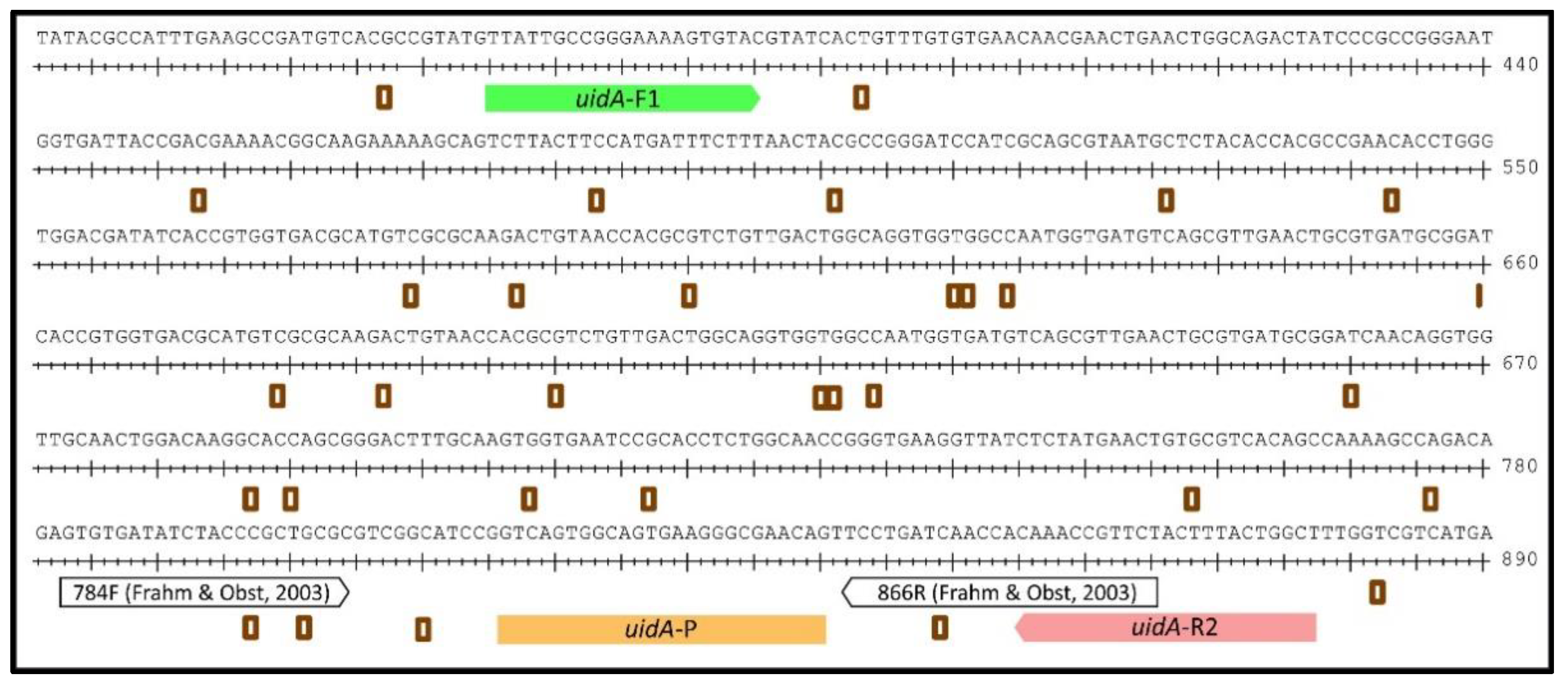
2.1. Design of a uidA-Targeting qPCR for Quantifying E. coli

For the reliable detection of E. coli, we chose a fragment of the uidA gene, encoding the β-glucuronidase, which is commonly used for the phenotypic identification of E. coli. We performed a nucleotide BLAST analysis using the uidA gene of E. coli CFT073 as a template (https://blast.ncbi.nlm.nih.gov/Blast.cgi; last accessed on 1 May 2022). The resulting homologous sequences of each E. coli taxon in NCBI and of Shigella spp. (in total 372 sequences) were used to generate a consensus sequence of uidA (Figure 1). Primer and probe annealing sites were chosen to align within the highly conserved gene segments and were verified towards the uidA sequence of our Sanger-sequenced laboratory strains, the reference strain E. coli DSM 1103, and the field strain ESBL E. coli 10714. The chosen forward primer (uidA-F1: 5′-TTATTGCCGGGAAAAGTGTAC-3′) and reverse primer (uidA-R2: 5′-AGCCAGTAAAGTAGAACGGTTTG-3′) in combination with the probe (uidA-P: 5′-JOE-CTGTTCGCCCTTCACTGCCACTGAC-BBQ-3′) resulted in the amplification of a 513 bp fragment (Figure 1).

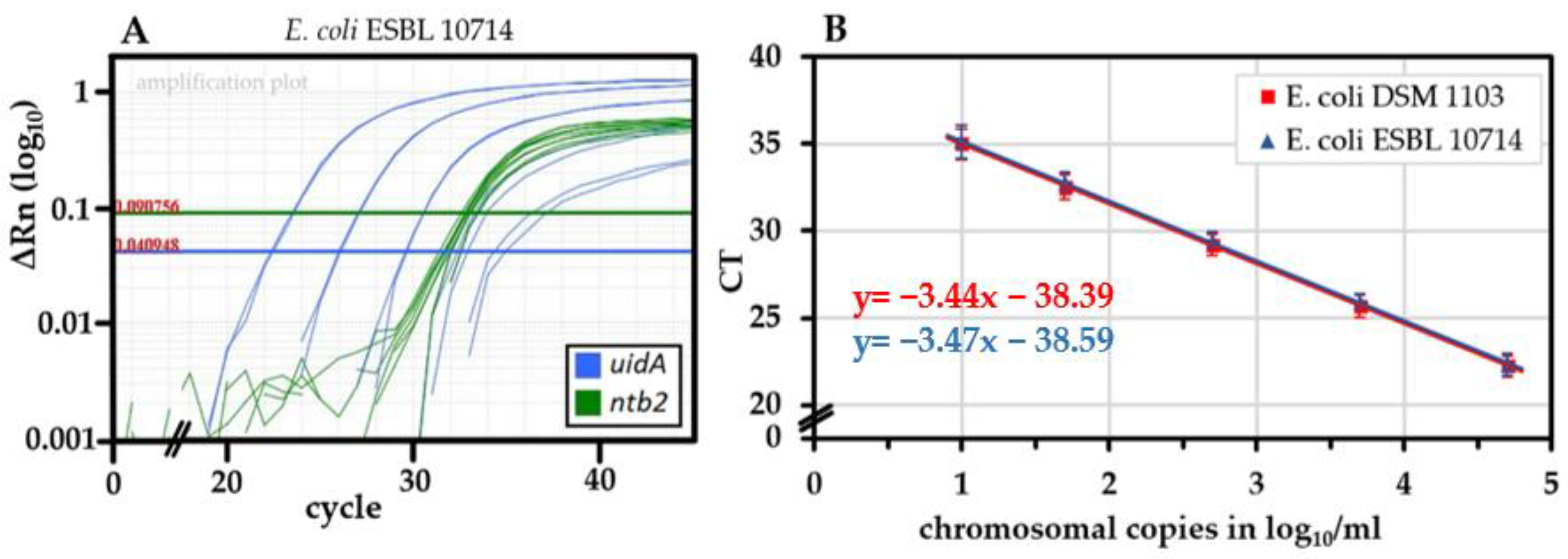
2.2. Performance of the uidA-Targeting qPCR and Absence of False Positive Signals

For quantification, we produced DNA standards from E. coli and tested the novel qPCR for performance. The E. coli standards DSM 1103 and ESBL 10714 were amplified with an average efficiency of 95.5 ± 5.7% (n = 21) and 94.7 ± 6.8% (n = 23), respectively (Figure 2A). The IPC-ntb2 [43] was included as an internal amplification control (Figure 2B).
False positive amplification signals may occur due to residual genomic DNA in the IPC-ntb2 plasmid preparation or the heterologous Taq polymerase. They might be an obstacle for the reliable performance of a uidA-targeted qPCR, as addressed previously [20,40,41]. However, no false positive signal of uidA was detected in the negative controls when 25 copies (or 1000 copies) of the amplification control were applied, indicating the absence of relevant contamination of genomic DNA in the plasmid preparation from the E. coli K12 host (Supplementary Table S2).
In addition, we compared a native Taq polymerase and several recombinant enzymes or master mixes in terms of efficiently amplifying the E. coli DNA standards and the absence of false-positive signals. The native Taq polymerase amplified both E. coli genome standards less efficiently. A reduced sensitivity towards lower E. coli copy numbers was observed (Supplementary Figure S1). Besides, the uidA channel showed no false-positive amplifications in the negative control samples using either recombinant enzymes or master mixes (Platinum Taq Polymerase, AmpliTaq Gold® DNA Polymerase in TaqMan Gene Expression Master Mix, and HotStarTaq DNA Polymerase in Quanti Tect Multiplex PCR Master Mix), even at elevated concentrations (Supplementary Table S2). In further experiments we, therefore, utilized the Platinum Taq Polymerase.

2.3. The Novel uidA-Targeted qPCR Is Sensitive and Specific for E. coli/Shigella spp.

The uidA-based qPCR for E. coli was evaluated for its sensitivity towards the uidA-positive target strains. In total, we used 150 field strains (142 E. coli) and 19 reference strains (Table 1).
E. coli field strains were selected by serotype diversity, virulence gene composition, and biochemical characteristics. The field strains were derived from different food and animal sources (Supplementary Table S1), in particular from poultry and cattle but also pigs and wild animals. The tested field strains included 54 enteropathogenic E. coli (EPEC) and 46 Shigatoxin-producing E. coli (STEC) strains (19 stx1 only, 21 stx2 only, and 6 stx1 and stx2). In total, 18 E. coli STEC field strains belonged to serotype O157:[H7].
From all 142 E. coli field strains and the 4 E. coli and 1 Shigella sonnei reference strains, positive amplification results were obtained. The novel uidA-based qPCR detected all tested E. coli/Shigella strains (100% sensitivity), which also included E. coli with a reduced or absent phenotypic ß-glucuronidase activity, cleaving MUG.
All tested strains outside the species E. coli/Shigella showed negative results (100% specificity). It also accounted for the closely related species E. albertii, E. fergusonii, and E. hermannii, which did not amplify. Hence, the uidA target, the designed oligos, and the probe appeared to be suitable for identification of E. coli and Shigella spp. strains.

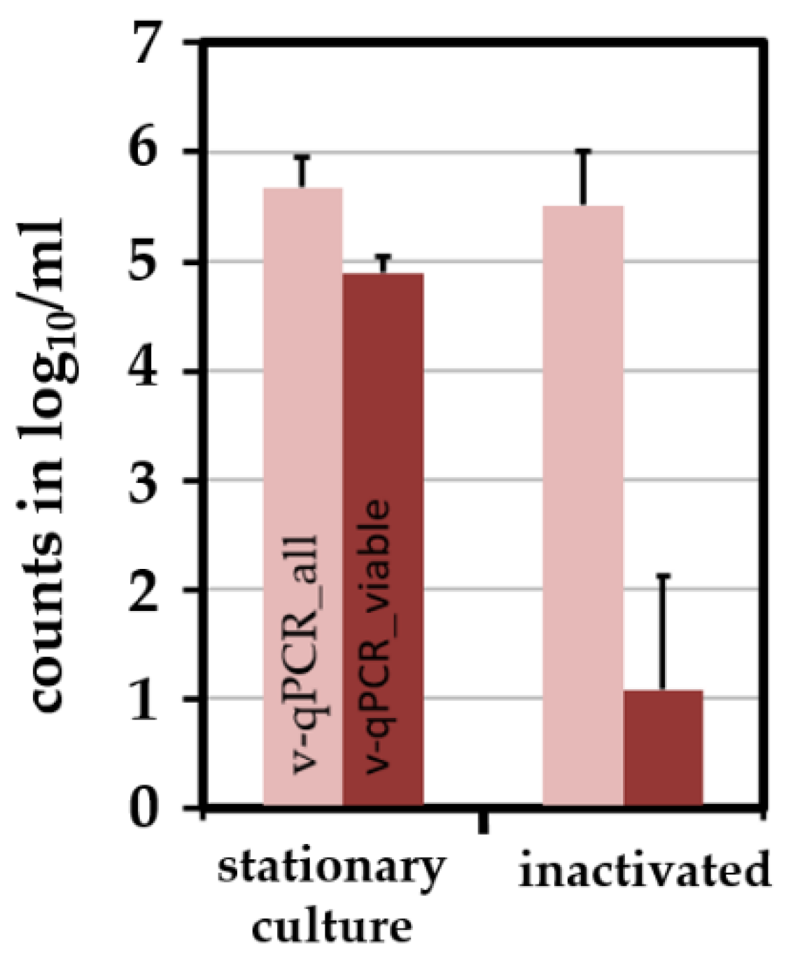
2.4. Combining the Novel qPCR with a Pre-Treatment Step Using PMA Allows the Differentiation of Viable and Dead E. coli

Viable E. coli (DSM 1103 and ESBL 10714) cells were produced by culturing bacteria in BHI until the stationary phase. E. coli were killed by 5% H2O2 under low osmotic conditions at 50 °C. The absence of CFU was checked after enrichment of at least 106 killed bacteria in BHI and subsequent streak-plating. When exposing artificially inactivated E. coli to 50 µM PMA, a reduction power of >3.5 log10/mL due to the PMA treatment was observed relative to viable bacteria, which is well-suited to differentiate viable from dead cells by v-qPCR (Figure 3).

2.5. Storage of Chicken Skin in Refrigeration and Freezing Conditions

Since chicken products are either stored in refrigeration conditions or frozen, we evaluated the cooling/freezing effect on viable C. jejuni and E. coli over time. For this purpose, we spiked Campylobacter-free chicken skin samples with both species and analyzed intact and potentially infectious units (IPIU) by v-qPCR and CFU over a period of 3 days to 3 weeks.
In skin samples stored at 4 °C, initial CFU of C. jejuni were maintained stable within 3 days and declined afterwards gradually by around 2 log10/mL to reach 3.0 ± 0.7 log10/mL after three weeks (Figure 4A, dashed blue line). E. coli CFU were stable during 7 days and declined thereafter by around 1 log10/mL to reach 3.8 ± 0.3 log10/mL after three weeks (Figure 4B, dashed blue line). In contrast to CFU, viable v-qPCR counts (IPIU) from cooled samples remained stable at approximately 5 log10/mL for both species, without any decline throughout the experiment (Figure 4, solid blue lines).
Freezing reduced the CFU of C. jejuni and E. coli after 3 days by 1.5 and 1.2 log10/mL, respectively (Figure 4, dashed orange lines). Subsequently, the CFU decreased only marginally. After 3 weeks of frozen storage, the CFU had declined by 2.1 and 1.6 log10/mL for C. jejuni and E. coli, respectively. The loss of IPIU was lower than of CFU. Freezing for three weeks reduced intact C. jejuni (IPIU) to some extent, by around 1 log10/mL. E. coli IPIU were reduced even less, by 0.4 log10/mL (Figure 4, solid orange lines). The overall DNA content remained at a constantly high level of around 5 log10/mL, indicating the presence of some dead cells (Figure 4, dotted orange lines).

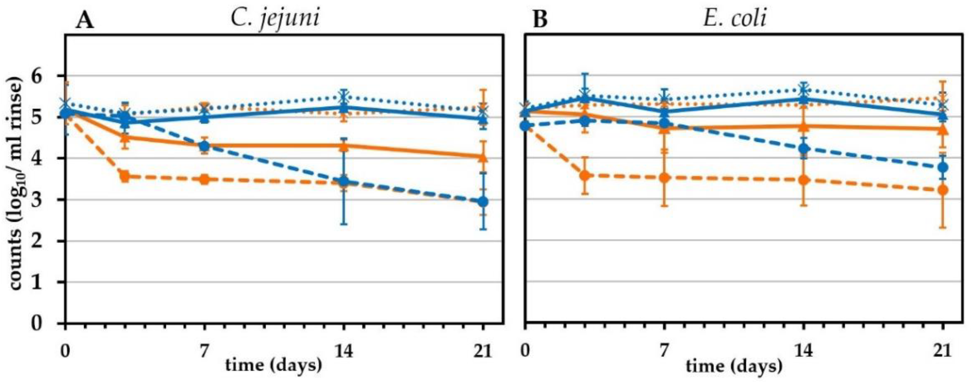
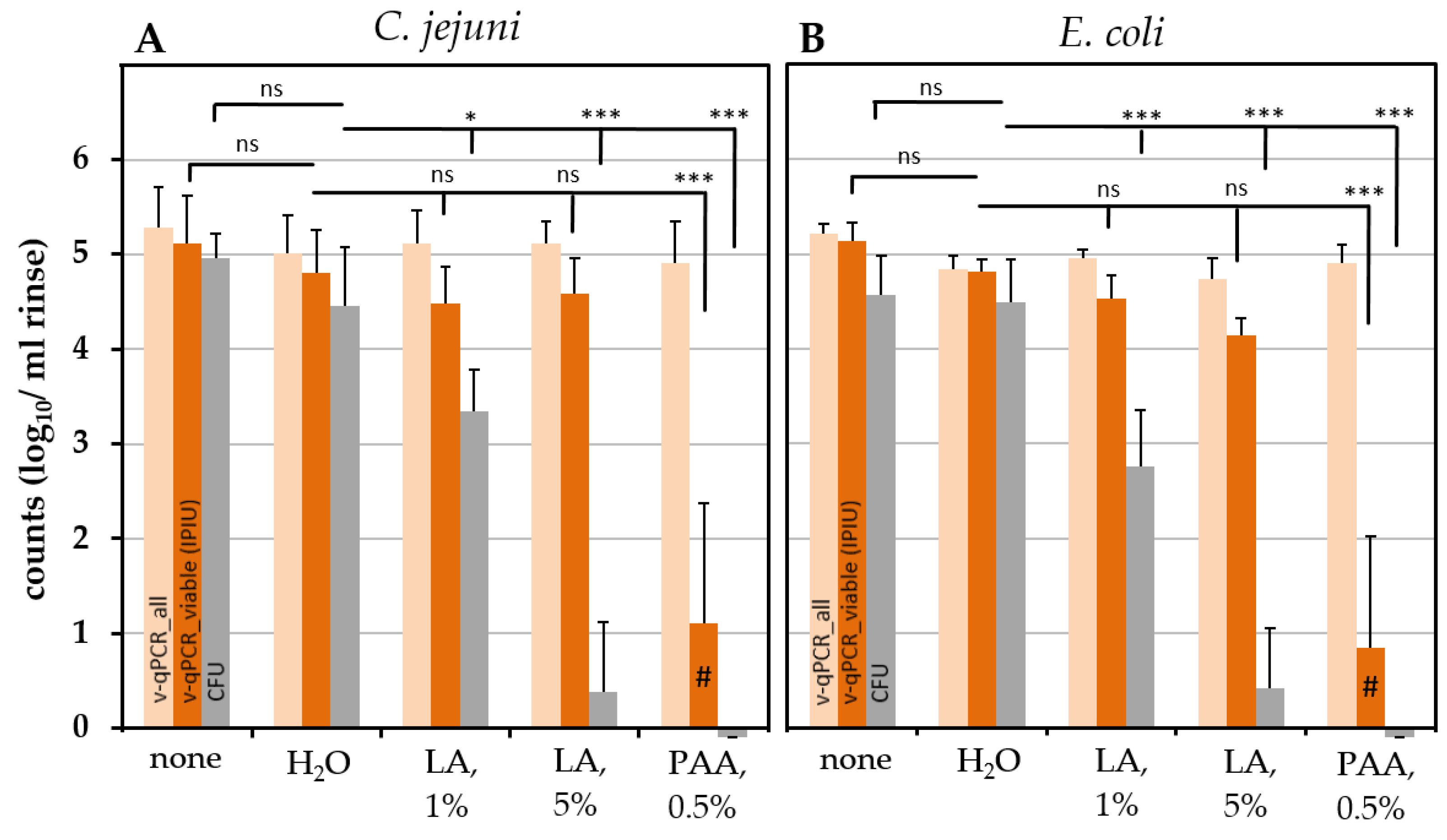
2.6. Effect of Chemical Treatment on the Survival of C. jejuni and ESBL-Producing E. coli on Chicken Skin

We quantified the effect of chemical decontamination on chicken skin that was initially spiked in parallel with C. jejuni and an ESBL-producing E. coli strain (Figure 5).
Skins were treated with the respective chemical solution for 3 min and short-term neutralized in PBS. The final rinse in PBS was analyzed for CFU and by v-qPCR. The chemical solutions PAA (0.5%) and LA (1% and 5%) were evaluated in comparison to a control in which the chemical solution was replaced by water. The initial pathogenic load was assessed by an analysis of untreated samples. The treatment of chicken skin with water for 3 min resulted in a slight but not significant decrease in the initial contamination by ≤0.5 log10/mL CFU or IPIU in E. coli and C. jejuni (Figure 6).
In both species, PAA (0.5%) caused a highly significant complete loss of CFU. Consistently, the absence of a v-qPCR signal or a low residual signal was observed, with reductions of >3.5 log10/mL upon PMA treatment. When compared with the exclusively dead cell internal sample process control (ISPC) included in each sample, this indicated the presence of a rest signal from dead cells (Figure 6).
LA significantly reduced CFU of both species compared to water-treated samples in a concentration-dependent manner. Immediate reductions of 1.1 and 1.7 log10/mL CFU were observed by treatment with 1% LA for C. jejuni and E. coli, respectively (Figure 6). When 5% LA was applied, no colonies or only single colonies were detected (≥4 log10/mL reduction). In contrast, v-qPCR signals only slightly declined upon LA treatment, with a maximum decrease of 0.7 log10/mL for E. coli by treatment with 5% LA (Figure 6).
During survival experiments with chemical treatments, the pH was measured of the decontamination solution (right before use), the neutralization bath (after use), and the chicken skin rinse (Table 2). After treatment with 1% LA (and 0.5% PAA), samples passed the neutralization bath, which remained close to neutral in pH (7.0). Consistently, the respective chicken rinses were only slightly acidified, indicating that neutralization showed a sufficient effect. Only in the case of 5% LA treatment, the neutralization bath measured an acidified pH of 4.5–5.5 after use and the actual skin rinses were measured at pH 4.0 to 5.

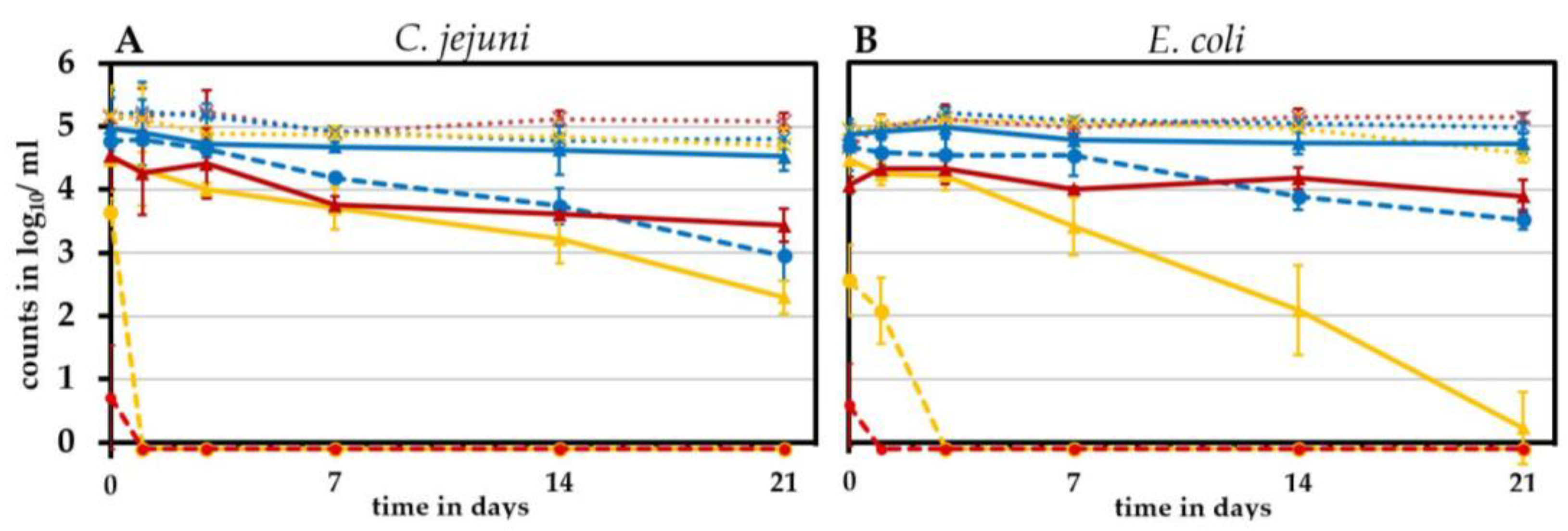
2.7. Long-Term Effects of Lactic Acid Treatment

Since we observed a discrepancy between the CFU and v-qPCR signals upon LA decontamination, we wondered how decontamination affected the CFU and the v-qPCR signals in a long-term experiment. Would CFU recover and how would the signal kinetics of the v-qPCR behave over time, i.e., was a decay of the membrane and a subsequent entry of PMA over time detectable? Therefore, we stored LA-treated samples at 4 °C for 1 day up to 3 weeks and regularly quantified CFU and the v-qPCR signal (Figure 7).
Water-treated control samples (Figure 7, in blue) resembled in pattern in the untreated cooled samples mentioned above (Figure 4). As observed before, the LA treatment led to an immediate rapid loss of CFU, but the v-qPCR signals remained initially high. A recovery of CFU was not observed within the experimental time of three weeks. Instead, we even observed a complete loss of CFU, i.e., 1% LA fully abolished CFU in C. jejuni within a day and in E. coli within three days.
The viable v-qPCR signals were reduced over time to some extent but inversely to the applied concentration of LA. The decrease in the viable v-qPCR signal was lower after treatment with 5% LA than when 1% LA was applied. In 1% LA-treated samples, the viable v-qPCR signal for E. coli was lost after 3 weeks, when it measured 2.3 log10/mL for C. jejuni. The overall DNA content (without PMA) remained stable for both species and was independent of the intervention and time point tested. These results suggested that after lactic acid treatment, membrane permeability does not indicate bacterial death, which awaits further studies.

3. Discussion

Our study was focused on two main aspects. On the one hand, we wondered whether CFU underestimates the survival of Thermotolerant Campylobacter spp. on chicken skin upon storage or chemical decontamination. On the other hand, the question was addressed as to whether E. coli can serve as a hygiene indicator to mimic Thermotolerant Campylobacter on chicken skin. The latter bacterium is much easier to diagnose for routine laboratories and might compensate for technical pitfalls in Campylobacter detection.

3.1. Establishing a Novel uidA-Based v-qPCR for the Quantification of Viable E. coli

In order to reliably quantify viable E. coli, we decided to design a novel uidA-based v-qPCR with a target fragment optimized in length. The activity of the targeted enzyme, β-glucuronidase, is also utilized in E. coli identification [44,45,46]. Yet, about 3% to 6% of E. coli strains remain phenotypically β-glucuronidase-negative [45,47], especially among E. coli O157 [48]. Among our tested E. coli field strains, cultural β-glucuronidase activity was absent in 22 strains and reduced in 12 strains when evaluated with the substrate 4-methylum-belliferyl beta-D-glucuronide (MUG) by routine testing on Fluorocult E. coli 0157:H7 agar. As expected, the uidA-based qPCR was superior since all E. coli strains tested positive. In an exclusivity and inclusivity study, we ascertained the appropriate sensitivity and specificity of the qPCR.
As usually performed in our Campylobacter-qPCR studies, we quantified using a stable DNA standard with a known chromosomal copy number [42,49,50]. This allowed absolute quantification and yielded approximately 95% amplification efficiency (Figure 2). In addition, the v-qPCR was controlled by an ISPC. The ISPC monitors a sufficient reduction in the dead cell signal. It is influenced simultaneously by matrix and potential DNA losses in each individual sample. In this study, the ISPC also guaranteed reliable crosslinking to PMA in samples with slightly decreased pH. With this method at hand, viable and dead E. coli were differentiated to a high degree since the signal of dead cells was reduced by at least 3.5 log10/mL upon PMA treatment (Figure 3). In combination with our previously developed v-qPCR for Thermotolerant Campylobacter spp. [42], we quantified the survival of both C. jejuni and an ESBL-producing E. coli field strain on chicken skin upon the application of different reduction strategies. Theoretically, the sensitivity of the novel qPCR is 100 copies per ml of chicken skin rinse since our standard curve reliably detected 10 copies per PCR reaction and 10% of the DNA was used per sample.

3.2. Effect of Cooling and Freezing

Reductions of 1–2 log10 CFU C. jejuni on chicken meat have been observed due to freezing. Therefore, freezing is suggested as a suitable reduction strategy [39]. In our study, CFU initially declined in a similar way and maintained over time at a comparable level, as observed previously [29,31,51,52,53].
Freezing and cooling were investigated by culture-independent quantification methods in the past [54]. Moreover, differences in the recovery of E. coli on selective and non-selective media were observed after the cooling of meat [55]. The results are in line with our study, with only slight CFU reductions for C. jejuni but stable numbers of intact cells detected by v-qPCR under cooling conditions. Moreover, freezing appeared to underestimate the survival of both pathogens, however, to a lower degree than cooling. Our data showed that C. jejuni and E. coli reacted similarly in physical storage conditions. Hence, cooling and freezing appeared to overestimate reduction strategies. In conclusion, the data suggested an advantage to control the efficiency of those strategies by culture-independent v-qPCR in addition to CFU.

3.3. PAA Treatment

The immediate effects of 0.5% PAA and 1% and 5% LA were evaluated in comparison to water-treated and untreated control samples by both CFU and v-qPCR. Intriguingly, the reduction effects were remarkably similar for both Gram-negative species. Water-treated versus untreated samples showed only minor insignificant reductions in both IPIU and CFU. Upon treatment with PAA, IPIU corresponded very well to CFU. PAA is a reagent with oxidizing properties [56], likely leading to loss of cell membrane integrity. It confirms a direct effect on cell membrane integrity and indicates the ultimate loss of cell viability. We conclude that v-qPCR can measure bacterial survival very well upon PAA treatment and can be applied for such samples in practice, which would overcome problems in cultivation and speed up diagnostics. Zhang et al. [57] and Nagel et al. [58] achieved 1–1.3 log10 CFU reductions compared to water-treatment on chicken parts with a short-term (~20 s) immersion in 0.12% PAA. As expected, our reductions by 0.5% PAA and 3 min of incubation were much higher, demonstrating cell membrane effects at maximal CFU-reducing conditions. However, these concentrations are not thought to be applied on the product itself, since a maximum of 0.2% PAA is recommended by EFSA for chicken products [38]. Moreover, color changes were observed on chicken skin. However, as a surface disinfectant in poultry production facilities, PAA at high concentrations might be an efficient agent for pathogen reduction. Reductions in E. coli CFU were high (>4.2 log10) upon the application of 0.5% PAA for 1 min in a surface disinfecting experiment on steel disks without food matrix components [59]. Lower but effective reductions of 1.2–1.3 log10 were found in that study in the presence of a matrix-mimicking yeast-albumin agar. Interestingly, our data showed an even higher reduction effect on both E. coli and C. jejuni by 0.5% PAA in the presence of natural matrix, as found in abattoirs. Our prolonged incubation time may partly explain the differences.

3.4. LA Treatment

CFU reductions due to LA treatment observed in previous studies are generally in line with our results. When naturally contaminated broiler carcasses were submerged in 1.5% LA during slaughter, a reduction of 1.2 log10 CFU of C. jejuni per carcass was measured [60] compared to a water control. Dipping breast pieces with skin for 10 min into 1% or 3% LA reduced C. jejuni by 1.3 and 2.0 log10 most probable number (MPN), respectively [61]. Reductions on broiler legs were lower. Anang et al. [28] studied E. coli O157:H7 re-inoculated on purchased, ethanol/heat-decontaminated chicken breast pieces. CFU were reduced with 2% LA by 1.6 or 2.4 log10/g, when treated for 10 or 30 min, respectively.
In our study, LA treatment reduced CFU either partially by 1.1 to 1.7 log10/mL (1% LA) or nearly fully (5% LA) after 3 min of dip treatment. We observed that IPIU, in contrast to CFU, remained only marginally affected in both species, upon immediate measurement. These data suggested that LA affected colony formation, but not cell membrane integrity. Theoretically, it leaves the possibility of a part of the cells entering a viable but non-culturable (VBNC) state. Hence, we wondered if, upon storage, sub-lethal injury entailed long-term effects, measurable as accelerated loss of integrity of the membrane.
Indeed, initial CFU loss increased with storage time after 1% LA treatment, until complete loss of culturability. Riedel et al. [62] applied 2.5% LA for 1 min to C. jejuni on chicken skin. Compared to water, this dipping treatment initially only slightly reduced C. jejuni by 0.7 log10 CFU/ml. However, upon additional storage for 24 h at 5 °C, CFU declined by 2.8 log10/mL significantly. Thus, our study also confirms a certain accelerated loss of viable cells over time after treatment with 1% LA. This is, as loss of integrity of the inner cell membrane necessarily implies death. However, in 5%-LA-treated chicken skins, not completely neutralised by PBS, loss of intact bacteria was lower over time.
LA is a strong acid with a relatively low pKa value [63]. It is reported to acidify the cytoplasm of neutrophilic bacteria, leading to the denaturation of cell components and the impairment of their functions [64,65]. Indeed, contact to 3% LA led to an immediate drop in internal pH and a progressive loss of CFU in C. jejuni [66]. Besides, it was suggested that LA caused permeabilization of the outer membrane of Gram-negative bacteria [67]. Therefore, LA may not have necessarily harmed the inner membrane integrity at first instance. Meanwhile, cell activity was probably strongly abolished. The recovery of the pH gradient after lactic acid exposure and incubation in nutrient broth under microaerobic conditions at 4 °C indicated the integrity of the inner membrane and the principal capacity to regain growth properties in C. jejuni [66]. The resuscitation of sublethally injured E. coli after exposure to low LA concentrations (<0.1%) was dependent on the medium composition and metal ion availability due to an enhanced oxidative stress response [68,69]. Thus, the selective agar needed for the detection of food pathogens might be suboptimal for the recovery of sublethally injured bacteria. Hence, these aspects call for clarifying the viable state after lactic acid decontamination monitored by DNA-intercalating agents alone. Alternative viability markers and, in particular, animal models may help to answer the question of whether v-qPCR had reached its limit.

4. Materials and Methods

4.1. Strains and Growth Conditions

E. coli strains DSM 1103 (DSMZ strain collection, Braunschweig, Germany) and an extended-spectrum ß-lactamase (ESBL) field strain, specifically ESBL 10714, from chicken meat were grown from a −80 °C cryoculture (MAST Group Ltd, Bootle, UK) in brain–heart infusion (BHI, Becton Dickinson, Franklin Lakes, NJ, USA) at 37 °C and 180 rpm. The preculture was subcultured by an inoculation of an initial OD600 of 0.01 and incubation until a stationary phase to an OD600 of 4.5 ± 0.5. Bacteria were diluted using phosphate-buffered saline (PBS, pH = 7.4) to an OD600 of 1, corresponding to about 9 log10 bacteria per ml, and kept at 5 ± 1 °C in a cooling rack (CoolBoxTM, Corning Inc., Corning, NY, USA) until further use.
Further reference and field strains used for inclusivity and exclusivity evaluations of the qPCR were grown either on tryptic soy agar (TSA, Merck KGaA, Darmstadt, Germany) or Columbia agar supplemented with 5% sheep blood (ColbA, Oxoid, Thermo Fisher Scientific Inc., Waltham, MA, USA). Reference strains, usually obtained as lyophilisates from strain collections, were passaged once and cryo-conserved. Our laboratories are accredited and regularly prove the identity of strains.
C. jejuni DSM 4688 (DSMZ strain collection, Braunschweig, Germany) was grown from a −80 °C cryoculture on ColbA at 42 °C in a microaerobic incubator (5% O2, 10% CO2, 85% N2; Binder GmbH, Tuttlingen, Germany) for 24 ± 4 h. The cells derived from an 18 ± 2 h subculture and were suspended in peptone water (PW, Merck KGaA, Darmstadt, Germany) to an OD600 of 0.2, corresponding to about 9 log10 bacteria per mL [13], and kept at 5 ± 1 °C in a cooling rack until further use.
CFU determination was performed according to ISO 16649-3:2015 for E. coli and ISO 10272-2:2017 for C. jejuni with the following modifications. Tryptone bile x-glucuronide agar (TBX, Oxoid, Thermo Fisher Scientific Inc., Waltham, MA, USA) was used for spread-plating E. coli, which were incubated for 24 h at 37 °C under aerobic conditions. mCCDA was used for C. jejuni and incubated for 48–72 h at 42 °C in a microaerobic atmosphere (5% O2, 10% CO2, 85% N2). The MUG activity of E. coli strains was checked on Fluorocult E. coli O157:H7 Agar (Merck KGaA, Darmstadt, Germany).

4.2. DNA Extraction and qPCR

DNA from the survival experiments was extracted using the GeneJet Genomic DNA Purification kit (Thermo Fisher Scientific Inc., Waltham, MA, USA) according to the manufacturer’s instructions and eluted in 100 µL of elution buffer. DNA for the sensitivity and specificity analysis of the uidA-based qPCR was extracted from the strains using either the GeneJet Genomic DNA Purification kit or the RTP Bacteria DNA Mini Kit (Stratec, Birkenfeld, Germany). The latter DNA was generally diluted 1:1000 in H2O before use. DNA from the samples of the survival experiments was analyzed undiluted for maximum sensitivity.
The qPCR analysis was performed on an ABI Prism 7500 (Life Technologies, Thermo Fisher Scientific Inc., Waltham, MA, USA) in duplicate for each sample. For each reaction, 10 µL of DNA eluate and 15 µL of master mix were used. The three targets, the 16S rRNA of Thermotolerant Campylobacter, the 16S rRNA of the ISPC (C. sputorum), and the uidA of E. coli, were each analyzed as duplex qPCRs, including the amplification control IPC-ntb2 [43]. The former two Campylobacter targets were performed as previously described [49].
For E. coli, the master mix contained 1× Platinum Taq buffer, 2.5 mM MgCl2, each dNTP at 0.2 mM, 0.06× ROX, the uidA-F1 and uidA-R2 each at 500 nM, oligos IPC-ntb2-fw and IPC-ntb2-re at 300 nM, the dark-quenched probes FAM-uidA-probe-BBQ and TAMRA-IPC-ntb2-probe-BBQ each at 100 nM (TIB Molbiol, Berlin, Germany), and 2 U of Platinum Taq DNA Polymerase (Invitrogen, Thermo Fisher Scientific Inc., USA). The IPC-ntb2 plasmid was added at 25 copies per reaction to the master mix. An initial denaturation and activation of the hot start polymerase activity at 95 °C for 3 min was followed by 45 cycles of denaturation at 95 °C for 30 s, annealing/elongation and detection of the fluorescent signals at 60 °C for 1 min, and an additional incubation at 72 °C for 30 s. If indicated, other Taq polymerases, such as a native Taq Polymerase from Thermus aquaticus (Invitrogen, Thermo Fisher Scientific Inc.), the AmpliTaq Gold® DNA Polymerase (TaqMan Gene Expression Master Mix, Life Technologies, Thermo Fisher Scientific Inc.), and HotStarTaq DNA Polymerase (Quanti Tect Multiplex PCR Master Mix, Qiagen, Hilden, Germany) were used according to the manufacturer’s protocol.

4.3. Preparation of Quantification Standards for qPCR

We prepared DNA quantification standards for the uidA-based qPCR from the E. coli strains DSM 1103 and E. coli ESBL 10,714 as previously described for C. jejuni [42]. In short, strains from liquid BHI cultures were pelleted by centrifugation for 5 min at 16,000× g. The DNA was extracted using the GeneJet Genomic DNA Purification kit according to the manufacturer’s manual. The DNA was quantified using the Qubit 3.0 Fluorometer (Life Technologies, Thermo Fisher Scientific Inc., USA). A volume of 40 µL, containing 26.3 ng of genomic DNA or 2,500,000 E. coli chromosomal equivalents per µL, was added to each DNA stable tube (Biomatrica, San Diego, CA, USA). After a thorough mixing of the DNA-stabilizing reagent, 2 µL aliquots were dried overnight in 0.5 mL reaction tubes under the working bench and stored at room temperature. Just before qPCR analysis, an aliquot of E. coli DNA was reconstituted in 200 µL of H2O (corresponding to 50,000 genome copies per µL). Using 10 ng/µL salmon sperm DNA (Invitrogen, Thermo Fisher Scientific Inc., USA), the reconstituted aliquot was decimally diluted to 50 copies per reaction. A final 5-fold dilution yielded 10 copies per reaction. The five different standards were applied in duplicate during each qPCR run.
For quantifying the genome equivalents of Thermotolerant Campylobacter and the ISPC, we applied similar genome standards of the strains C. jejuni NCTC 11,168 and C. sputorum DSM 5363 as described previously [49]. These quantification standards were applied as 50,000, 5000, 500, 50, and 5 copies per reaction in duplicate. The uidA gene is present as a single copy gene in the E. coli chromosome, while the 16S rRNA targets are present in the C. jejuni and C. sputorum genome as three identical copies.

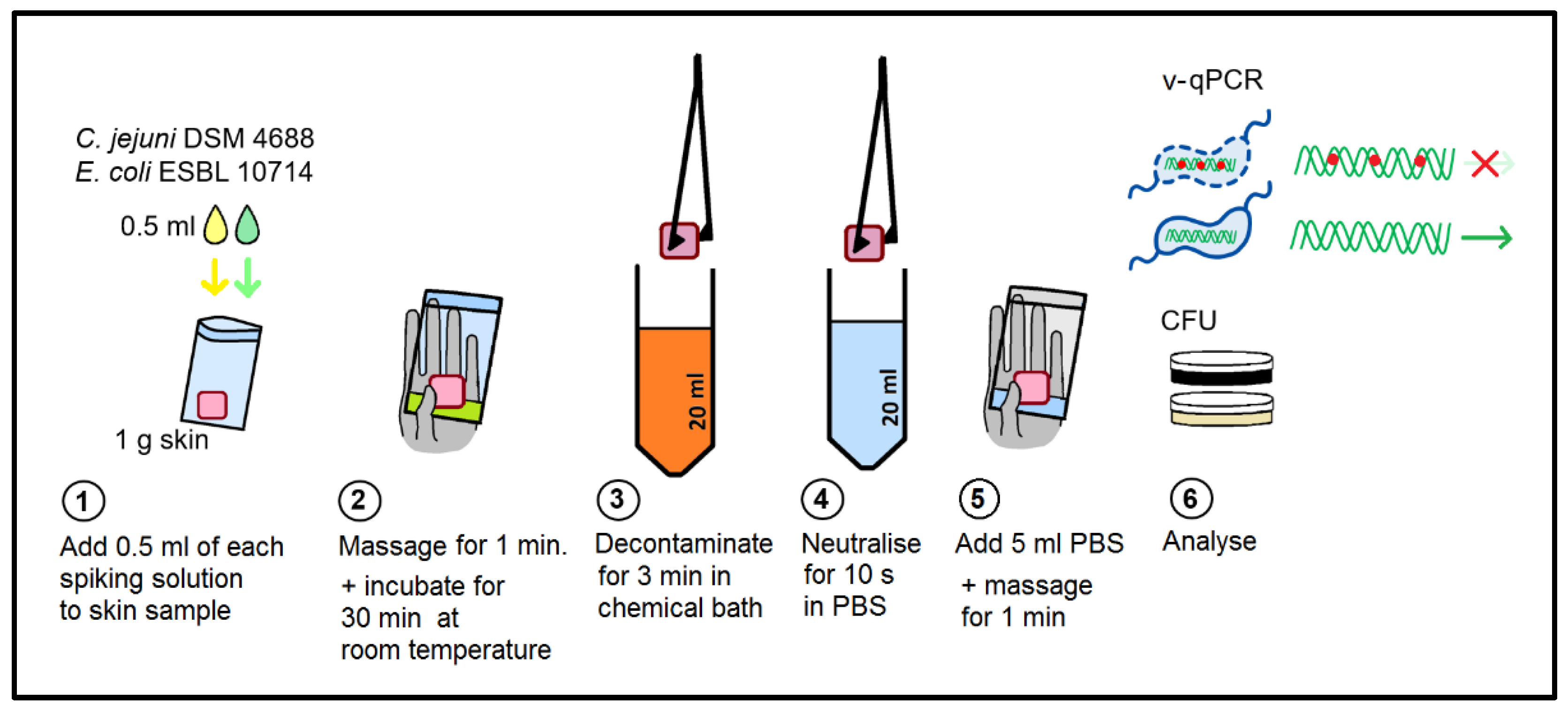
4.4. Survival Experiments with Chicken Skin

Campylobacter-free skin was produced from ROSS 308 chickens reared and slaughtered at the institute’s facilities. The absence of Campylobacter was checked by ISO 10272-1:2017 and by real-time PCR after enrichment in Bolton broth [70]. E. coli background flora was quantified on TBX agar with a mean of 3.76 ± 0.46 log10 per g of skin. Samples stored at −80 °C were thawed, rinsed twice with MilliQ water, cut to 1 ± 0.1 g portions, and stored at −20 °C until use. For each data point of the experiment (per condition and time point), a separate skin sample of 1 g was used. The CFU and v-qPCR were analyzed from the same skin sample rinse. All skins were retrieved from the same batch of slaughtered chickens. For inoculation, aliquots were placed into 9 × 12.5 cm closable plastic bags (RUBIN, Rossmann, Hannover, Germany) and kept on ice until inoculation.
For survival evaluation, 0.5 mL of each bacterial inoculation suspension of either E. coli or C. jejuni (7–7.3 log10 per ml PW) (see Section 4.1) were both applied to each 1 g skin sample. After manual massaging for one minute, the inoculated skin samples were incubated at room temperature for 30 min.
Chemical solutions were prepared just before application. The pH was measured using pH indicator strips (Merck KGaA, Darmstadt, Germany). The final solutions were as follows: 0.5% peroxyacetic acid (PAA, Wofasteril SC super 1 + 1, Kesla Pharma Wolfen GmbH, Bitterfeld-Wolfen, Germany; pH 5.0) and 1% and 5% lactic acid (LA, Sigma-Aldrich, Merck KGaA, Darmstadt, Germany, pH 2 and pH 1.5, respectively). As a water control, either Milli-Q water (Merck KGaA, Darmstadt, Germany) or tap water was used.
Each skin sample was placed into a separate 20 mL chemical solution for 3 min with an occasional inversion of the tube. Subsequently, the sample was neutralized for 10 s in 20 mL of PBS (single-use). Thereafter, 5 mL of PBS was added to the sample in a new plastic bag, which was massaged for 1 min. Rinse aliquots were taken for CFU and v-qPCR analysis.
The effects of cooling and freezing were evaluated at 5 °C and at −25 °C. The long-term effect of the LA treatment was evaluated by storing the samples at 5 °C for 1 day to 3 weeks.

4.5. Viable/Dead Differentiation by v-qPCR

For the quantification of C. jejuni, the v-qPCR was conducted as previously described [42]. In short, two 1 mL rinse aliquots were analyzed. One aliquot contained no PMA, which served for the quantification of the overall DNA content. The other aliquot contained PMA for quantifying viable bacteria (IPIU). The ISPC—C. sputorum dead cell control—allowed the monitoring of the dead cell signal reduction by PMA as well as DNA losses during DNA extraction for each sample individually. The aliquot measuring the overall DNA content received 10 µL of the C. sputorum dead cell control (ISPClow) and was centrifuged for 5 min at 16,000× g and 4 °C. After discarding the supernatant, the bacterial pellet was stored at −20 °C until DNA extraction and qPCR analysis (see Section 4.2). The aliquot measuring viable bacteria (IPIU) first received 10 µL of the concentrated ISPChigh and was pre-incubated for 10 min at 30 °C with gentle shaking at 700 rpm. Subsequently, 50 µM PMA was added, and the sample was incubated for another 15 min at 30 °C in the dark. Crosslinking occurred afterwards for 15 min using the PhAST Blue photo-activation system at 100% light intensity (GenIUL, Terrassa, Spain). Subsequent to the inactivation of the PMA-reactive azide groups by light, 10 µL of the ISPClow were added, and the sample was centrifuged for 5 min at 16,000× g and 4 °C. The bacterial pellets were stored at −20 °C. Control samples accompanied the procedure as described previously [42].
Dead E. coli were obtained as follows: A bacterial suspension of 9 log10 cells per mL (see Section 4.1) was centrifuged at 16,000× g for 5 min. The cell pellet was resuspended in 1:10 diluted PBS supplemented with 5% H2O2 at a cell density with an OD600 of 0.5 and incubated at 50 °C for 60 min. Killed bacteria were centrifuged at 16,000× g for 5 min, washed once, and resuspended in PBS to an OD600 of 0.1, corresponding to around 108 dead bacteria/mL. The absence of growth was checked by adding 10 µL of this suspension to 10 mL of BHI and incubating overnight at 37 °C with shaking at 180 rpm. The enrichment broth was subsequently streaked on TSA.

4.6. Data Handling

The qPCR data were analyzed using a pre-version of the Microsoft Excel data analysis sheet (Stingl et al. 2021) [42]. The CFU and v-qPCR outcomes were log10-transformed. The absence of a CFU or qPCR signal was defined as −0.1 log10/mL. Statistical significance was tested by one-way ANOVA followed by Tukey’s multiple comparison post hoc test. For each quantification method, intervention groups were analyzed for the statistical significance of their difference compared to the water-treated control group. Statistics were performed in Graph Pad Prism v5.01.

5. Conclusions

The current quantification of microbial contaminants in food is limited, as pathogens are underestimated when transiently inactive. Here, we designed and established an ISPC-controlled v-qPCR for E. coli and applied a previously validated respective method for Campylobacter spp. Using two selected strains, we showed that, depending on the inactivation method used, artificially killed cells can also be principally quantified by v-qPCR. Furthermore, E. coli and C. jejuni behaved similarly upon immediate quantification. The results of our study are restricted to the tested strains. Therefore, further characterization of multiple E. coli and C. spp. strains might be considered in future approaches. The method showed high viability upon lactic acid treatment and awaits further studies on the infectivity of these bacteria in animal models or by alternative viability markers. However, we conclude that for improved food safety, v-qPCR methods should complement microbial cultivation.

Supplementary Materials

The following are available online at https://www.mdpi.com/article/10.3390/pathogens11060706/s1, Figure S1: Standard amplification curves obtained with the recombinant versus native Taq polymerases; Table S1: List of field isolates tested for sensitivity and specificity of the uidA-based qPCR; Table S2: Absence of false-positive uidA signals in negative controls using different Taq Polymerases and the amplification control IPC-ntb2.

Author Contributions

Conceptualization, K.S., I.F.W.; methodology, A.G., E.S., I.F.W., M.T., K.S.; investigation, I.F.W. and M.T.; data analysis, I.F.W., A.G., E.S., K.S.; writing, I.F.W., K.S. All authors have read and agreed to the published version of the manuscript.

Funding

This study was funded by SFB 1322-580 of the German Federal Institute for Risk Assessment (BfR); I.F.W was financed by the research project EsRAM (No. 2817701614) of the German Federal Ministry of Food and Agriculture (BMEL).

Data Availability Statement

The datasets generated during and analyzed during the current study can be find in the main text and the Supplementary Materials.

Acknowledgments

We thank Stefanie Banneke from the BfR animal facility for rearing ROSS 308 chicken and Niels Bandick and Felix Reich from the BfR food technology for preparing chicken skin samples. The E. coli strain ESBL 10714 was kindly provided by Uwe Rösler and Anika Friese, Institute for Animal and Environmental Hygiene at the Freie Universität Berlin.

Conflicts of Interest

The authors declare no conflict of interest. The funders had no role in the design of the study; in the collection, analyses, or interpretation of data; in the writing of the manuscript, or in the decision to publish the results.

References

  1. European Food Safety Authority (EFSA); European Centre for Disease Prevention and Control (ECDC). The European Union One Health 2019 Zoonoses Report. EFSA J. 2021, 19, 6406. [Google Scholar] [CrossRef]
  2. EFSA Panel on Biological Hazards; Ricci, A.; Allende, A.; Bolton, D.; Chemaly, M.; Davies, R.; Fernández Escámez, P.S.; Girones, R.; Herman, L.; Koutsoumanis, K.; et al. Guidance on the requirements for the development of microbiological criteria. EFSA J. 2017, 15, e05052. [Google Scholar] [CrossRef] [PubMed] [Green Version]
  3. Barco, L.; Belluco, S.; Roccato, A.; Ricci, A. Escherichia coli and Enterobacteriaceae counts on pig and ruminant carcasses along the slaughterline, factors influencing the counts and relationship between visual faecal contamination of carcasses and counts: A review. EFSA Supporting Publ. 2014, 11, 634e. [Google Scholar] [CrossRef] [Green Version]
  4. Liu, G.; Bogaj, K.; Bortolaia, V.; Olsen, J.E.; Thomsen, L.E. Antibiotic-induced, increased conjugative transfer is common to diverse naturally occurring ESBL plasmids in Escherichia coli. Front. Microbiol. 2019, 10, 2119. [Google Scholar] [CrossRef]
  5. Garcia-Migura, L.; Hendriksen, R.S.; Fraile, L.; Aarestrup, F.M. Antimicrobial resistance of zoonotic and commensal bacteria in Europe: The missing link between consumption and resistance in veterinary medicine. Vet. Microbiol. 2014, 170, 1–9. [Google Scholar] [CrossRef]
  6. European Food Safety Authority (EFSA); European Centre for Disease Prevention Control (ECDC). The European Union summary report on antimicrobial resistance in zoonotic and indicator bacteria from humans, animals and food in 2017/2018. EFSA J. 2020, 18, e06007. [Google Scholar] [CrossRef] [Green Version]
  7. Roth, N.; Käsbohrer, A.; Mayrhofer, S.; Zitz, U.; Hofacre, C.; Domig, K.J. The application of antibiotics in broiler production and the resulting antibiotic resistance in Escherichia coli: A global overview. Poult. Sci. 2019, 98, 1791–1804. [Google Scholar] [CrossRef]
  8. Federighi, M.; Tholozan, J.L.; Cappelier, J.M.; Tissier, J.P.; Jouve, J.L. Evidence of non-coccoid viable but non-culturable Campylobacter jejuni cells in microcosm water by direct viable count, CTC-DAPI double staining, and scanning electron microscopy. Food Microbiol. 1998, 15, 539–550. [Google Scholar] [CrossRef]
  9. Baffone, W.; Casaroli, A.; Citterio, B.; Pierfelici, L.; Campana, R.; Vittoria, E.; Guaglianone, E.; Donelli, G. Campylobacter jejuni loss of culturability in aqueous microcosms and ability to resuscitate in a mouse model. Int. J. Food Microbiol. 2006, 107, 83–91. [Google Scholar] [CrossRef]
  10. Darcan, C.; Ozkanca, R.; Idil, O.; Flint, K.P. Viable but non-culturable state (VBNC) of Escherichia coli related to EnvZ under the effect of pH, starvation and osmotic stress in sea water. Pol. J. Microbiol. 2009, 58, 307–317. [Google Scholar]
  11. Pienaar, J.A.; Singh, A.; Barnard, T.G. The viable but non-culturable state in pathogenic Escherichia coli: A general review. Afr. J. Lab. Med. 2016, 5, 368. [Google Scholar] [CrossRef] [PubMed] [Green Version]
  12. Zhao, F.; Wang, Y.; An, H.; Hao, Y.; Hu, X.; Liao, X. New insights into the formation of viable but nonculturable Escherichia coli O157:H7 induced by high-pressure CO2. mBio 2016, 7. [Google Scholar] [CrossRef] [PubMed] [Green Version]
  13. Krüger, N.J.; Buhler, C.; Iwobi, A.N.; Huber, I.; Ellerbroek, L.; Appel, B.; Stingl, K. “Limits of control”—Crucial parameters for a reliable quantification of viable Campylobacter by real-time PCR. PLoS ONE 2014, 9, e88108. [Google Scholar] [CrossRef] [Green Version]
  14. Nocker, A.; Cheung, C.Y.; Camper, A.K. Comparison of propidium monoazide with ethidium monoazide for differentiation of live vs. dead bacteria by selective removal of DNA from dead cells. J. Microbiol. Methods 2006, 67, 310–320. [Google Scholar] [CrossRef] [PubMed]
  15. Josefsen, M.H.; Lofstrom, C.; Hansen, T.B.; Christensen, L.S.; Olsen, J.E.; Hoorfar, J. Rapid quantification of viable Campylobacter bacteria on chicken carcasses, using real-time PCR and propidium monoazide treatment, as a tool for quantitative risk assessment. Appl. Environ. Microbiol. 2010, 76, 5097–5104. [Google Scholar] [CrossRef] [Green Version]
  16. Bej, A.K.; DiCesare, J.L.; Haff, L.; Atlas, R.M. Detection of Escherichia coli and Shigella spp. in water by using the polymerase chain reaction and gene probes for uid. Appl. Environ. Microbiol. 1991, 57, 1013–1017. [Google Scholar] [CrossRef] [Green Version]
  17. Chern, E.C.; Brenner, K.P.; Wymer, L.; Haugland, R.A. Comparison of fecal indicator bacteria densities in marine recreational waters by qPCR. Water Qual. Expo. Health 2009, 1, 203–214. [Google Scholar] [CrossRef]
  18. Frahm, E.; Obst, U. Application of the fluorogenic probe technique (TaqMan PCR) to the detection of Enterococcus spp. and Escherichia coli in water samples. J. Microbiol. Methods 2003, 52, 123–131. [Google Scholar] [CrossRef]
  19. Heijnen, L.; Medema, G. Quantitative detection of E. coli, E. coli O157 and other shiga toxin producing E. coli in water samples using a culture method combined with real-time PCR. J. Water Health 2006, 4, 487–498. [Google Scholar] [CrossRef] [Green Version]
  20. Silkie, S.S.; Tolcher, M.P.; Nelson, K.L. Reagent decontamination to eliminate false-positives in Escherichia coli qPCR. J. Microbiol. Methods 2008, 72, 275–282. [Google Scholar] [CrossRef]
  21. Liu, Y.; Mustapha, A. Detection of viable Escherichia coli O157:H7 in ground beef by propidium monoazide real-time PCR. Int. J. Food Microbiol. 2014, 170, 48–54. [Google Scholar] [CrossRef] [PubMed]
  22. Miszczycha, S.D.; Ganet, S.; Duniere, L.; Rozand, C.; Loukiadis, E.; Thevenot-Sergentet, D. Novel real-time PCR method to detect Escherichia coli O157:H7 in raw milk cheese and raw ground meat. J. Food Prot. 2012, 75, 1373–1381. [Google Scholar] [CrossRef] [PubMed]
  23. Li, B.; Chen, J.Q. Real-time PCR methodology for selective detection of viable Escherichia coli O157:H7 cells by targeting Z3276 as a genetic marker. Appl. Environ. Microbiol. 2012, 78, 5297–5304. [Google Scholar] [CrossRef] [PubMed] [Green Version]
  24. Schnetzinger, F.; Pan, Y.; Nocker, A. Use of propidium monoazide and increased amplicon length reduce false-positive signals in quantitative PCR for bioburden analysis. Appl. Microbiol. Biotechnol. 2013, 97, 2153–2162. [Google Scholar] [CrossRef] [PubMed]
  25. European Union: European Commission. Commission Regulation (EU) 2017/1495 of 23 August 2017 amending Regulation (EC) No 2073/2005 as regards Campylobacter in broiler carcasses. Off. J. Eur. Union 2017, OJ L 218, 1–6. Available online: http://data.europa.eu/eli/reg/2017/1495/oj (accessed on 9 June 2022).
  26. Loretz, M.; Stephan, R.; Zweifel, C. Antimicrobial activity of decontamination treatments for poultry carcasses: A literature survey. Food Control 2010, 21, 791–804. [Google Scholar] [CrossRef]
  27. Musavian, H.S.; Krebs, N.H.; Nonboe, U.; Corry, J.E.; Purnell, G. Combined steam and ultrasound treatment of broilers at slaughter: A promising intervention to significantly reduce numbers of naturally occurring campylobacters on carcasses. Int. J. Food Microbiol. 2014, 176, 23–28. [Google Scholar] [CrossRef]
  28. Anang, D.M.; Rusul, G.; Bakar, J.; Ling, F.H. Effects of lactic acid and lauricidin on the survival of Listeria monocytogenes, Salmonella enteritidis and Escherichia coli O157:H7 in chicken breast stored at 4 °C. Food Control 2007, 18, 961–969. [Google Scholar] [CrossRef]
  29. Boysen, L.; Wechter, N.S.; Rosenquist, H. Effects of decontamination at varying contamination levels of Campylobacter jejuni on broiler meat. Poult. Sci. 2013, 92, 1425–1429. [Google Scholar] [CrossRef]
  30. Rajkovic, A.; Tomic, N.; Smigic, N.; Uyttendaele, M.; Ragaert, P.; Devlieghere, F. Survival of Campylobacter jejuni on raw chicken legs packed in high-oxygen or high-carbon dioxide atmosphere after the decontamination with lactic acid/sodium lactate buffer. Int. J. Food Microbiol. 2010, 140, 201–206. [Google Scholar] [CrossRef]
  31. Harrison, D.; Corry, J.E.; Tchórzewska, M.A.; Morris, V.K.; Hutchison, M.L. Freezing as an intervention to reduce the numbers of campylobacters isolated from chicken livers. Lett. Appl. Microbiol. 2013, 57, 206–213. [Google Scholar] [CrossRef] [PubMed]
  32. Bauermeister, L.J.; Bowers, J.W.; Townsend, J.C.; McKee, S.R. The microbial and quality properties of poultry carcasses treated with peracetic acid as an antimicrobial treatment. Poult. Sci. 2008, 87, 2390–2398. [Google Scholar] [CrossRef] [PubMed]
  33. Isohanni, P.M.; Lyhs, U. Use of ultraviolet irradiation to reduce Campylobacter jejuni on broiler meat. Poult. Sci. 2009, 88, 661–668. [Google Scholar] [CrossRef] [PubMed]
  34. James, C.; James, S.J.; Hannay, N.; Purnell, G.; Barbedo-Pinto, C.; Yaman, H.; Araujo, M.; Gonzalez, M.L.; Calvo, J.; Howell, M.; et al. Decontamination of poultry carcasses using steam or hot water in combination with rapid cooling, chilling or freezing of carcass surfaces. Int. J. Food Microbiol. 2007, 114, 195–203. [Google Scholar] [CrossRef] [PubMed]
  35. Chaves, B.D.; Han, I.Y.; Dawson, P.L.; Northcutt, J.K. Survival of artificially inoculated Escherichia coli and Salmonella Typhimurium on the surface of raw poultry products subjected to crust freezing. Poult. Sci. 2011, 90, 2874–2878. [Google Scholar] [CrossRef]
  36. Stefani, L.M.; Backes, R.G.; Faria, G.A.; Biffi, C.P.; de Almeida, J.M.; da Silva, H.K.; das Neves, G.B.; Langaro, A. Trimming and washing poultry carcass to reduce microbial contamination: A comparative study. Poult. Sci. 2014, 93, 3119–3122. [Google Scholar] [CrossRef]
  37. European Union: European Commission. Commission Regulation (EU) No 101/2013 of 4 February 2013 concerning the use of lactic acid to reduce microbiological surface contamination on bovine carcases. Off. J. Eur. Union 2013, OJ L 34, 1–3. Available online: http://data.europa.eu/eli/reg/2013/101/oj (accessed on 9 June 2022).
  38. EFSA Panel on Biological Hazards (BIOHAZ). Scientific Opinion on the evaluation of the safety and efficacy of peroxyacetic acid solutions for reduction of pathogens on poultry carcasses and meat. EFSA J. 2014, 12, 3599. [Google Scholar] [CrossRef] [Green Version]
  39. EFSA Panel on Biological Hazards (BIOHAZ). Scientific Opinion on Campylobacter in broiler meat production: Control options and performance objectives and/or targets at different stages of the food chain. EFSA J. 2011, 9, 2105. [Google Scholar] [CrossRef]
  40. Kibbee, R.; Linklater, N.; Ormeci, B. Eliminating false positives in a qPCR assay for the detection of the uidA gene in Escherichia coli. J. Water Health 2013, 11, 382–386. [Google Scholar] [CrossRef] [Green Version]
  41. Kibbee, R.J.; Örmeci, B. Development of a sensitive and false-positive free PMA-qPCR viability assay to quantify VBNC Escherichia coli and evaluate disinfection performance in wastewater effluent. J. Microbiol. Methods 2017, 132, 139–147. [Google Scholar] [CrossRef] [PubMed]
  42. Stingl, K.; Heise, J.; Thieck, M.; Wulsten, I.F.; Pacholewicz, E.; Iwobi, A.N.; Govindaswamy, J.; Zeller-Péronnet, V.; Scheuring, S.; Luu, H.Q.; et al. Challenging the “gold standard” of colony-forming units—Validation of a multiplex real-time PCR for quantification of viable Campylobacter spp. in meat rinses. Int. J. Food Microbiol. 2021, 359, 109417. [Google Scholar] [CrossRef] [PubMed]
  43. Anderson, A.; Pietsch, K.; Zucker, R.; Mayr, A.; Müller-Hohe, E.; Messelhäusser, U.; Sing, A.; Busch, U.; Huber, I. Validation of a duplex real-time PCR for the detection of Salmonella spp. in different food products. Food Anal. Methods 2011, 4, 259–267. [Google Scholar] [CrossRef]
  44. Damaré, J.M.; Campbell, D.F.; Johnston, R.W. Simplified direct plating method for enhanced recovery of Escherichia coli in food. J. Food Sci. 1985, 50, 1736–1737. [Google Scholar] [CrossRef]
  45. Feng, P.C.; Hartman, P.A. Fluorogenic assays for immediate confirmation of Escherichia coli. Appl. Environ. Microbiol. 1982, 43, 1320–1329. [Google Scholar] [CrossRef] [Green Version]
  46. Frampton, E.W.; Restaino, L.; Blaszko, N. Evaluation of the β-glucuronidase substrate 5-bromo-4-chloro-3-indolyl-β-d-glucuronide (X-GLUC) in a 24-h direct plating method for Escherichia coli. J. Food Prot. 1988, 51, 402–404. [Google Scholar] [CrossRef]
  47. Hansen, W.; Yourassowsky, E. Detection of beta-glucuronidase in lactose-fermenting members of the family Enterobacteriaceae and its presence in bacterial urine cultures. J. Clin. Microbiol. 1984, 20, 1177–1179. [Google Scholar] [CrossRef] [Green Version]
  48. Ratnam, S.; March, S.B.; Ahmed, R.; Bezanson, G.S.; Kasatiya, S. Characterization of Escherichia coli serotype O157:H7. J. Clin. Microbiol. 1988, 26, 2006–2012. [Google Scholar] [CrossRef] [Green Version]
  49. Pacholewicz, E.; Buhler, C.; Wulsten, I.F.; Kraushaar, B.; Luu, H.Q.; Iwobi, A.N.; Huber, I.; Stingl, K. Internal sample process control improves cultivation-independent quantification of thermotolerant Campylobacter. Food Microbiol. 2019, 78, 53–61. [Google Scholar] [CrossRef]
  50. Wulsten, I.F.; Galeev, A.; Stingl, K. Underestimated survival of Campylobacter in raw milk highlighted by viability real-time PCR and growth recovery. Front. Microbiol. 2020, 11, 1107. [Google Scholar] [CrossRef]
  51. Gill, C.O.; Harris, L.M. Survival and growth of Campylobacter fetus subsp. jejuni on meat and in cooked foods. Appl. Environ. Microbiol. 1982, 44, 259. [Google Scholar] [CrossRef] [PubMed] [Green Version]
  52. Garénaux, A.; Ritz, M.; Jugiau, F.; Rama, F.; Federighi, M.; de Jonge, R. Role of oxidative stress in C. jejuni inactivation during freeze-thaw treatment. Curr. Microbiol. 2009, 58, 134–138. [Google Scholar] [CrossRef] [PubMed]
  53. Georgsson, F.; Thornorkelsson, A.E.; Geirsdóttir, M.; Reiersen, J.; Stern, N.J. The influence of freezing and duration of storage on Campylobacter and indicator bacteria in broiler carcasses. Food Microbiol. 2006, 23, 677–683. [Google Scholar] [CrossRef] [PubMed]
  54. Duarte, A.; Botteldoorn, N.; Coucke, W.; Denayer, S.; Dierick, K.; Uyttendaele, M. Effect of exposure to stress conditions on propidium monoazide (PMA)-qPCR based Campylobacter enumeration in broiler carcass rinses. Food Microbiol. 2015, 48, 182–190. [Google Scholar] [CrossRef] [PubMed]
  55. Lazou, T.P.; Gelasakis, A.I.; Chaintoutis, S.C.; Iossifidou, E.G.; Dovas, C.I. Method-dependent implications in foodborne pathogen quantification: The case of Campylobacter coli survival on meat as comparatively assessed by colony count and viability PCR. Front. Microbiol. 2021, 12, 604933. [Google Scholar] [CrossRef]
  56. OECD Existing Chemicals Database, Peroxyacetic acid, SIDS Initial Assessment Profile, File: 79210_SIAP_final.pdf of 16 April 2008. Available online: https://hpvchemicals.oecd.org/UI/SIDS_Details.aspx?key=1f07490d-8c31-44e8-a2d6-7b261e310720&idx=0 (accessed on 11 May 2022).
  57. Zhang, L.; Garner, L.J.; Mckee, S.R.; Bilgili, S.F. Effectiveness of several antimicrobials used in a postchill decontamination tank against Salmonella and Campylobacter on broiler carcass parts. J. Food Prot. 2018, 81, 1134–1141. [Google Scholar] [CrossRef]
  58. Nagel, G.M.; Bauermeister, L.J.; Bratcher, C.L.; Singh, M.; McKee, S.R. Salmonella and Campylobacter reduction and quality characteristics of poultry carcasses treated with various antimicrobials in a post-chill immersion tank. Int. J. Food Microbiol. 2013, 165, 281–286. [Google Scholar] [CrossRef]
  59. Thomas, C.; Schönknecht, A.; Püning, C.; Alter, T.; Martin, A.; Bandick, N. Effect of peracetic acid solutions and lactic acid on microorganisms in on-line reprocessing systems for chicken slaughter plants. J. Food Prot. 2020, 83, 615–620. [Google Scholar] [CrossRef] [Green Version]
  60. Rasschaert, G.; Piessens, V.; Scheldeman, P.; Leleu, S.; Stals, A.; Herman, L.; Heyndrickx, M.; Messens, W. Efficacy of electrolyzed oxidizing water and lactic acid on the reduction of Campylobacter on naturally contaminated broiler carcasses during processing. Poult. Sci. 2013, 92, 1077–1084. [Google Scholar] [CrossRef]
  61. Coşansu, S.; Ayhan, K. Effects of lactic and acetic acid treatments on Campylobacter jejuni inoculated onto chicken leg and breast meat during storage at 4 °C and −18 °C. J. Food Processing Preserv. 2010, 34, 98–113. [Google Scholar] [CrossRef]
  62. Riedel, C.T.; Brøndsted, L.; Rosenquist, H.; Haxgart, S.N.; Christensen, B.B. Chemical decontamination of Campylobacter jejuni on chicken skin and meat. J. Food Prot. 2009, 72, 1173–1180. [Google Scholar] [CrossRef] [PubMed]
  63. OECD Existing Chemicals Database, Lactic Acid, SIDS Initial Assessment Profile, File: 50215_SIAP_final_Oct10.pdf of 20 October 2009. Available online: https://hpvchemicals.oecd.org/UI/SIDS_Details.aspx?key=7828dbad-7405-4619-afa1-9250df698ad7&idx=0 (accessed on 11 May 2022).
  64. Beales, N. Adaptation of microorganisms to cold temperatures, weak acid preservatives, low pH, and osmotic stress: A review. Compr. Rev. Food Sci. Food Saf. 2004, 3, 1–20. [Google Scholar] [CrossRef] [PubMed]
  65. Hirshfield, I.N.; Terzulli, S.; O’Byrne, C. Weak organic acids: A panoply of effects on bacteria. Sci. Prog. 2003, 86, 245–270. [Google Scholar] [CrossRef] [PubMed]
  66. Smigic, N.; Rajkovic, A.; Nielsen, D.S.; Siegumfeldt, H.; Uyttendaele, M.; Devlieghere, F.; Arneborg, N. Intracellular pH as an indicator of viability and resuscitation of Campylobacter jejuni after decontamination with lactic acid. Int. J. Food Microbiol. 2009, 135, 136–143. [Google Scholar] [CrossRef]
  67. Alakomi, H.L.; Skyttä, E.; Saarela, M.; Mattila-Sandholm, T.; Latva-Kala, K.; Helander, I.M. Lactic acid permeabilizes gram-negative bacteria by disrupting the outer membrane. Appl. Environ. Microbiol. 2000, 66, 2001–2005. [Google Scholar] [CrossRef] [Green Version]
  68. Shi, H.; Chen, Z.; Chen, D.; Kan, J. Sublethal injury and recovery of Escherichia coli O157:H7 and K-12 after exposure to lactic acid. Food Control 2017, 82, 190–195. [Google Scholar] [CrossRef]
  69. Zhang, H.C.; Zhang, R.; Shi, H. The effect of manganese and iron on mediating resuscitation of lactic acid-injured Escherichia coli. Lett. Appl. Microbiol. 2022. [Google Scholar] [CrossRef]
  70. Mayr, A.M.; Lick, S.; Bauer, J.; Thärigen, D.; Busch, U.; Huber, I. Rapid detection and differentiation of Campylobacter jejuni, Campylobacter coli, and Campylobacter lari in food, using multiplex real-time PCR. J. Food Prot. 2010, 73, 241–250. [Google Scholar] [CrossRef]
Figure 1. Consensus sequence of the uidA fragment (residue 331 to 890) after alignment of 372 E. coli and Shigella spp. sequences. The novel qPCR targets a 513 bp fragment using the oligos uidA-F1 and uidA-R2 and the probe uidA-P. Example oligo annealing sites of a published qPCR within the depicted region using an 83 bp fragment [18] are also illustrated. Brown squares indicate non-conserved positions in uidA. Numbers on the right present the position ruler in uidA.
Pathogens 11 00706 g001
Figure 2. Performance of the uidA-targeting qPCR for E. coli. (A) Example ΔRn amplification plot of the uidA target from a decimally diluted genomic DNA standard (E. coli ESBL 10714) and the IPC-ntb2 as internal amplification control. (B) Correlation between log10-transformed Ct values and chromosomal copies of DNA standards (E. coli DSM 1103, n = 21 and ESBL 10714, n = 23).
Pathogens 11 00706 g002
Figure 3. Differentiation of viable and dead E. coli adjusted to 106 log10/mL PBS, either derived from a stationary culture (n = 4) or after inactivation (n = 15).
Pathogens 11 00706 g003
Figure 4. Effect of cooling and freezing on CFU and viability by v-qPCR (IPIU). (A) C. jejuni DSM 4688; (B) E. coli ESBL 10714. Orange, −25 °C; blue, 4 °C; dashed lines, CFU; solid lines, viable bacteria (IPIU) counts; dotted lines, total DNA. Means ± standard deviations are illustrated from three independent experiments.
Pathogens 11 00706 g004
Figure 5. Experimental setup of chemical decontamination of chicken skin.
Pathogens 11 00706 g005
Figure 6. Effect of different chemical treatments of chicken skin on CFU and IPIU analyzed by v-qPCR. (A) C. jejuni DSM 4688; (B) E. coli ESBL 10714. Light orange, v-qPCR_all (total DNA); orange, v-qPCR_viable (IPIU); grey, CFU. Significance was evaluated by Tukey’s multiple comparison. Intervention groups were compared to water-treated samples for each analysis method. The means ± standard deviations are depicted from at least three independent experiments. ns, not significant (p > 0.05); * 0.05 > p > 0.01; *** p < 0.001. #, rest signal of dead cells.
Pathogens 11 00706 g006
Figure 7. Long-term effects of lactic acid (LA) treatment on cell membrane integrity (v-qPCR) and culturability (CFU) in (A) C. jejuni DSM 4688 and (B) E. coli ESBL 10714. Dashed lines, CFU; solid lines, v-qPCR_viable (IPIU); dotted lines, v-qPCR_all (total DNA). Blue, water control; yellow, 1% LA; red, 5% LA. The means ± standard deviations of at least three independent experiments are shown.
Pathogens 11 00706 g007
Table 1. Results of sensitivity and specificity characterization of the uidA-based qPCR.
Reference Strains Results Field Strains Number of Isolates Results
Escherichia coli DSM 1103positiveEscherichia coli, MUG+108positive
Escherichia coli EDL 933 positiveEscherichia coli, MUG(+)12positive
Escherichia coli DSM 498positiveEscherichia coli, MUG-22positive
Shigella sonnei DSM 5570positiveEnterobacter cloacae complex 1negative
Escherichia albertii DSM 17582negativeEscherichia albertii2negative
Escherichiafergusonii DSM 13698 negativeAeromonas jandaei1negative
Escherichia hermannii DSM 4560 negativeCitrobacter koseri1negative
Enterococcus faecalis DSM 20478negativeHafnia alvei3negative
Klebsiella pneumoniae DSM 30104negative
Proteus mirabilis DSM 4479negative
Pseudomonas aeruginosa DSM 1117negative
Staphylococcus aureus DSM 1104negative
Salmonella enterica DSM 11320negative
IPC-ntb2 (plasmid)negative
Campylobacter jejuni DSM 4688negative
Campylobacter jejuni NCTC 11168negative
Campylobacter sputorum DSM 5363/ISPCnegative
Campylobacter coli DSM 4689negative
Campylobacter lari DSM 11375negative
MUG, activity to metabolize 4-methylum-belliferyl beta-D-glucuronide; +, positive; −, negative; (+), low activity.
Table 2. pH 1 measurements.
TreatmentDecontamination Solution
(Before Use)
Neutralization Bath
(PBS, after Use)
Chicken Skin Rinse
LA, 1%276 (5.5–7)
LA, 5%1.55 (4.5–5.5)4 (4–5)
PAA, 0.5%576.5
MilliQ water5 (4.5–5)7.57–7.5
tap water7.57.57–7.5
1 pH was evaluated using pH strips. pH stated as median and range (min–max).
Publisher’s Note: MDPI stays neutral with regard to jurisdictional claims in published maps and institutional affiliations.

