Next Article in Journal
Epidemiology of Toxoplasmosis among the Pakistani Population: A Systematic Review and Meta-Analysis
Next Article in Special Issue
Diversity and Exploration of Endophytic Bacilli for the Management of Head Scab (Fusarium graminearum) of Wheat
Previous Article in Journal
Utilization of Recombinant Baculovirus Expression System to Produce the RBD Domain of SARS-CoV-2 Spike Protein
 
 
Correction published on 8 November 2023, see Pathogens 2023, 12(11), 1327.
Article

Identification of the Causal Agent of Brown Leaf Spot on Kiwifruit and Its Sensitivity to Different Active Ingredients of Biological Fungicides

1
Research Center for Engineering Technology of Kiwifruit, Institute of Crop Protection, College of Agriculture, Guizhou University, Guiyang 550025, China
2
Teaching Experimental Field of Guizhou University, Guizhou University, Guiyang 550025, China
*
Author to whom correspondence should be addressed.
Pathogens 2022, 11(6), 673; https://doi.org/10.3390/pathogens11060673
Submission received: 18 May 2022 / Revised: 3 June 2022 / Accepted: 8 June 2022 / Published: 10 June 2022 / Corrected: 8 November 2023
(This article belongs to the Special Issue Current Research on Fusarium)

Abstract

:
Kiwifruit (Actinidia chinensis) is an important commercial crop in China, and the occurrence of diseases may cause significant economic loss in its production. In the present study, a new pathogen that causes brown leaf spot disease on kiwifruit was reported. The fungus was isolated from an infected sample and identified as Fusarium graminearum based on morphological and molecular evaluation. Koch’s postulates were confirmed when the pathogen was re-isolated from plants with artificially induced symptoms and identified as F. graminearum. Based on the biological characteristics of the pathogen, it was determined that: its optimal growth temperature was 25 °C; optimal pH was 7; most suitable carbon source was soluble starch; most suitable nitrogen source was yeast powder; and best photoperiod was 12 h light/12 h dark. Further investigations were conducted by determining 50% effective concentrations (EC50) of several active ingredients of biological fungicides against F. graminearum. The results showed that among the studied fungicides, tetramycin and honokiol had the highest antifungal activity against this pathogen. Our findings provide a scientific basis for the prevention and treatment of brown leaf spot disease on kiwifruit.

1. Introduction

Kiwifruit (Actinidia chinensis), a perennial deciduous woody liana, is native to China. In recent years, it has become an important commercial crop and is widely cultivated in New Zealand, Italy, China, and several other countries. It is favored by consumers worldwide because it contains more key micronutrients, such as potassium, calcium, and folic acid, than most other fruits [1]. According to the Chinese kiwifruit industry development report in 2020, the planting area of kiwifruit in China was 290,700 hm2 by the end of 2019, with the total output reaching 3,000,000 tons. Moreover, the area under plantation and kiwifruit output in China still rank first in the world. With the increasing kiwifruit planting area worldwide, fungal and bacterial diseases have been reported [2,3]. Fungal leaf spot diseases have become an increasingly serious threat to kiwifruit production in open-field orchards. Diseased plants are generally characterized by dark brown ring spots, grayish brown ring spots, silvering gray leaf blight, zonate leaf blight, and angular leaf spots [4]. Different fungal species, such as Alternaria alternata, Nigrospora oryzae, Fusarium tricinctum, Colletotrichum spp., Diaporthe spp., Phoma spp., and Epicoccum spp., have been reported as leaf spot pathogens [4,5,6,7], which cause severe loss in yield in the kiwifruit industry.
Fusarium graminearum (Teleomorph Gibberella zeae Schw. Petch.), which belongs to ascomycetes, has been ranked fourth among the world’s top ten plant pathogenic fungi in terms of scientific and economic importance [8]. In general, F. graminearum is a causal pathogen of head blight or scab on barley (Hordeum), rice (Oryza), wheat (Triticum), and maize (Zea) stalk rot, as well as of spoilage on water bamboo shoots [9,10]. Its spores spread by wind or rain in the spring season and infect plants at optimal temperatures [11,12]. In addition to limiting healthy crop development, F. graminearum produces a variety of mycotoxins that pose safety risks to human and animal health [13]. Earlier studies have shown that multiple factors, such as temperature, pH, light, carbon source, and nitrogen source, have significant influences on pathogen growth and pathogenicity [14,15,16,17]. Therefore, it is necessary to carry out a study of the biological characteristics of the pathogen to investigate the suitable conditions for its occurrence and prevalence. Synthetic fungicides, such as metconazole [18] and carbendazim [19], are commonly used to control F. graminearum infection. However, biological control is becoming an important strategy in controlling F. graminearum to ensure food safety [20].
In July 2019, typical brown leaf spots were observed in ‘Guichang’ kiwifruit orchards (Actinidia deliciosa cv. ‘Guichang’) in Xifeng County, Guizhou Province, China. The disease initially appeared as a few small brown spots in the early stage and gradually expanded into larger lesions. The disease incidence rate in 16 kiwifruit orchards (20 hm2) was 21%, resulting in a large number of fallen leaves and approximately 9–16% of yield loss per orchard. It was determined that F. graminearum was the causal agent of brown leaf spot on kiwifruit in this planting area, and this was the first time that it was reported in the area. The isolated pathogen was used for further investigations by exploring its biological characteristics and determining its sensitivity to different active ingredients of biological fungicides.

2. Results

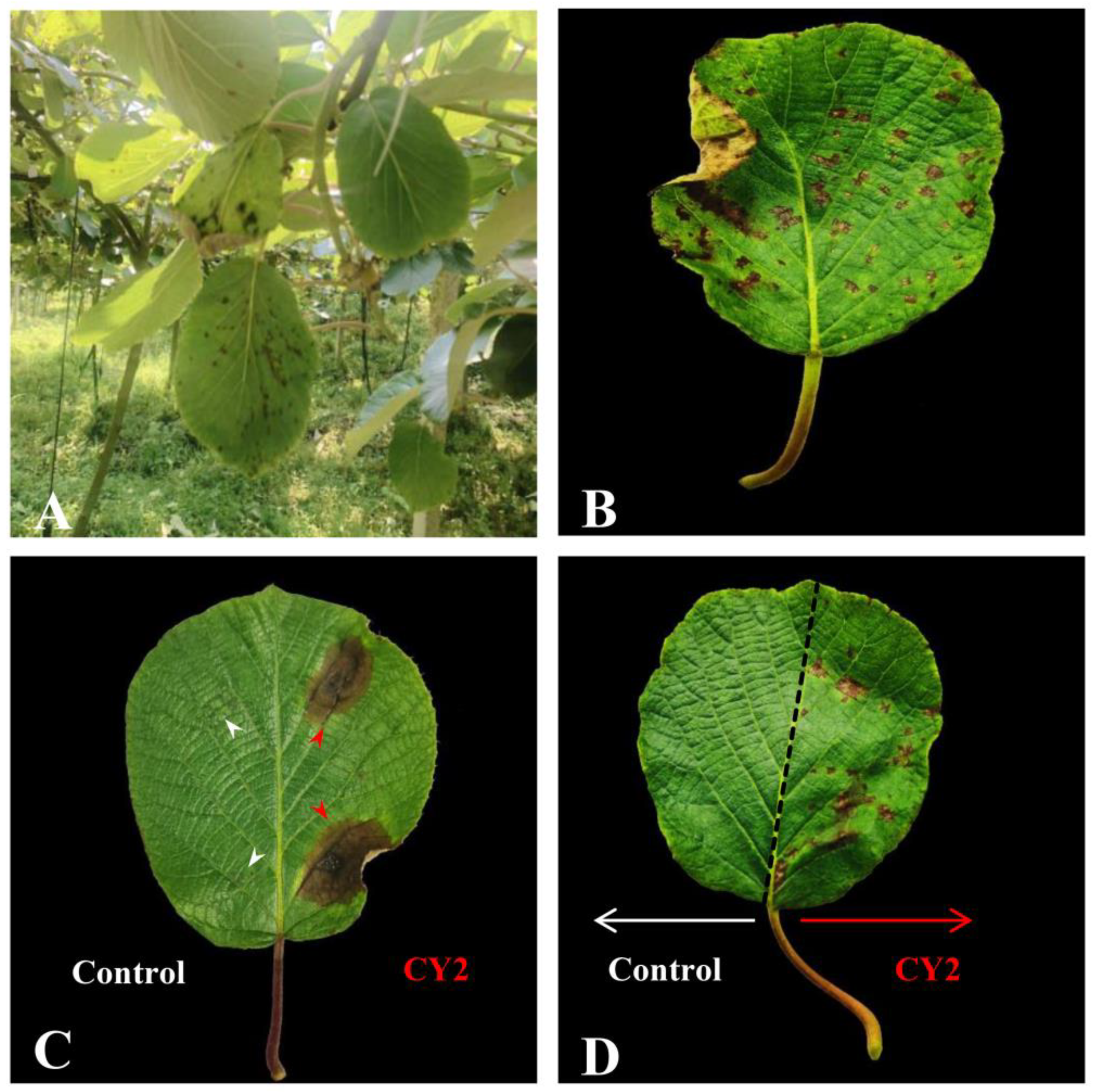
2.1. Pathogenicity Test of the Isolated Strains

Typical brown leaf spots were observed in the ‘Guichang’ kiwifruit orchards in July 2019. They appeared as a few small brown spots in the early stage and gradually expanded into larger lesions (Figure 1A,B). From the infected samples collected, a total of 37 fungal isolates were obtained. A pathogenicity test was conducted on these isolates using wounded and unwounded inoculation methods. The results showed that only isolate CY2 was pathogenic to kiwifruit. In the wounded inoculation method, the artificially inoculated leaves showed similar brown spots as those in the orchard six days after inoculation with isolate CY2 spore suspension, whereas the control inoculation did not show any brown spots (Figure 1C). In the unwounded inoculation method, the artificially inoculated leaves showed brown spots on the ninth day after inoculation with isolate CY2 spore suspension, whereas the control inoculation did not show any brown spots (Figure 1D). A morphologically similar fungus was re-isolated from the lesions of the artificially inoculated leaves. The results of the three trials were similar, which proved that the highly pathogenic isolate CY2 caused brown leaf spot disease on kiwifruit.

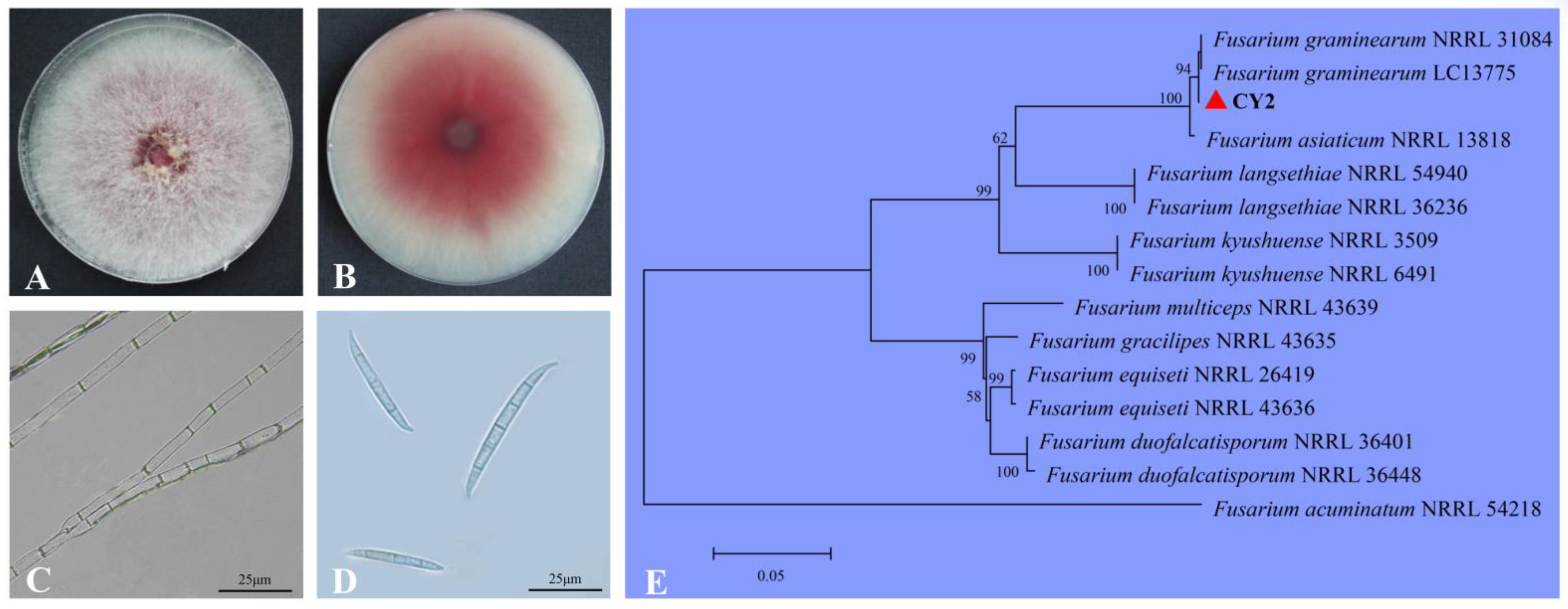
2.2. Identification of Isolate CY2

After culturing at 25 °C for six days, the colony of the highly pathogenic isolate CY2 grew rapidly on potato dextrose agar (PDA; Beijing Land Bridge Technology Co., Ltd., Beijing, China) and produced a large number of dense hyphae with red pigments (Figure 2A,B), and the mycelium had branches and septa (Figure 2C). Macroconidia of fungi cultured on carnation leaf-piece agar (CLA; 4 pieces of 5 mm long sterile carnation leaves, 10 mL of 2% water agar) had 3–5 septa, which were slender, sickle-shaped to nearly straight, with an average size of 26.4–56.7 × 3.3–7.5 μm (n = 50; Figure 2D). The isolate CY2 was identified as F. graminearum based on these morphological characteristics [10,21].
For molecular identification, different fragments of this isolate were amplified and sequences were deposited in GenBank. The sequence of the ITS region (MW871547) had 100% identity to sequences of several species stored in GenBank, including F. graminearum. The TEF-1α sequence (MW876479) was 100% homologous to that of F. graminearum (MN381089), while the RPB2 sequence (MW876480) showed 99.89% similarity to that of F. graminearum (MW233447). A phylogenetic tree was constructed using ITS, TEF1-α, and RPB2 gene sequences of isolate CY2, along with a few reference isolates obtained from GenBank (Table 1). It showed that isolate CY2 clustered together with other strains of F. graminearum obtained from GenBank. Therefore, results of the molecular analysis supported our morphological identification, which demonstrated that the highly pathogenic isolate CY2 was F. graminearum (Figure 2E).

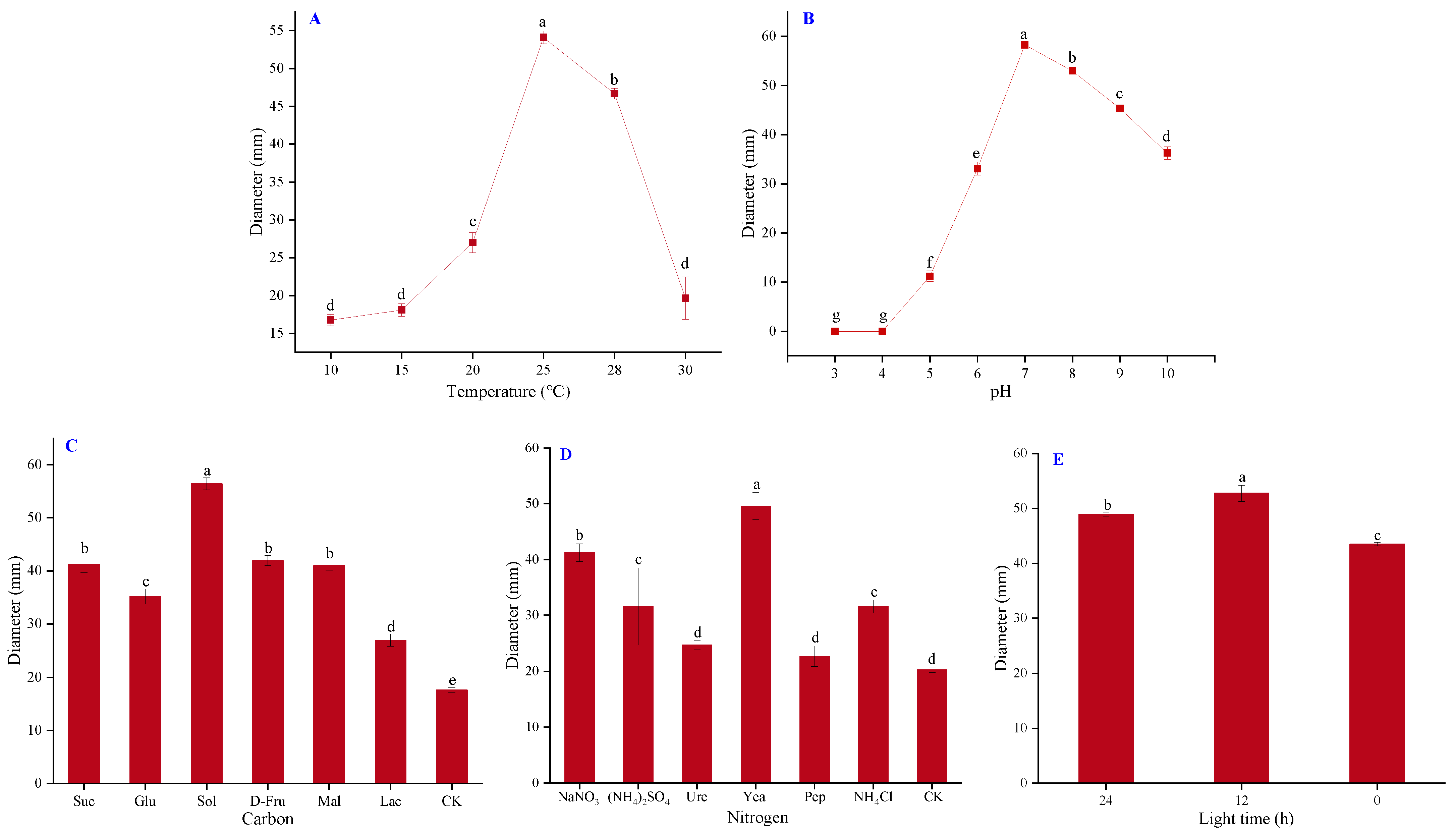
2.3. Biological Characteristics of Isolate CY2

After four days of culture at the temperature range of 10–30 °C, the colony diameter was 16.75 mm at 10 °C, 18.08 mm at 15 °C, 27 mm at 20 °C, 54.08 mm at 25 °C, 46.67 mm at 28 °C, and 19.67 mm at 30 °C (Figure 3A). The results showed that 25 °C was the optimal temperature for the growth of isolate CY2. Furthermore, the isolate grew at the range of pH 5–10, and its growth ceased at pH 3–4. The mycelial growth peak appeared at pH 7 with a colony diameter of 58.25 mm, showing that a moderate pH environment was optimal for its growth (Figure 3B). Soluble starch (Sol) was the optimal carbon source and yeast powder (Yea) was the optimal nitrogen source, as they resulted in the largest colony diameters among the treatments (Figure 3C,D). Regarding the light requirements, isolate CY2 could grow in all light conditions, but a daily 12 h photoperiod was the most beneficial for its growth (Figure 3E).

2.4. Toxicity Effects of Several Active Ingredients of Biological Fungicides against F. graminearum CY2

Screening active ingredients of biological fungicides against isolate CY2 is helpful for developing environmentally safe alternatives for the control of F. graminearum. In the present study, the sensitivity of isolate CY2 to five different active ingredients of biological fungicides, including honokiol, matrine, tetramycin, citral, and baicalein, was determined in vitro, (Figure 4 and Table 2). Positive slopes in the regression equations indicated a positive correlation between concentration and growth inhibition and R2 values close to 1 indicated that all equations were reliable. Tetramycin had the lowest EC50 value of 4.02 ± 0.05 μg mL−1, followed by honokiol and matrine, with EC50 values of 9.26 ± 0.11 and 15.2 ± 0.31 μg mL−1, respectively. Citral and baicalein showed relatively weak inhibition effects, with EC50 values of 28.6 ± 0.99 and 47.3 ± 0.12 μg mL−1, respectively. This indicated that tetramycin and honokiol had the highest antifungal activity against F. graminearum, while baicalein had the lowest.

3. Discussion

In the present study, although a total of 37 fungal isolates were obtained from the infected kiwifruit samples with symptoms of brown leaf spot, only isolate CY2 was confirmed to be a real pathogen based on Koch’s postulates [22]. Using wounded and unwounded inoculation methods, we showed that the other isolates were not pathogenic to kiwifruit, indicating they were probably saprophytic fungi on kiwifruit leaves. It is also possible that some of these fungi are pathogens, but indoor conditions were not suitable for their infection. Our morphological and molecular identification methods suggested that the pathogenic isolate CY2 was F. graminearum. The infection of kiwifruit with Fusarium species has led to concerning problems in its agricultural production [7,23,24,25,26]. However, this is the first report of the pathogen F. graminearum causing brown leaf spot on kiwifruit. High humidity is favorable to the distribution and survival of different pathogens, including F. graminearum [27], which may be an important reason why isolate CY2 was highly pathogenic to kiwifruit in the pathogenicity assays, as they were conducted under high humidity conditions. F. graminearum produces both ascospores and macroconidia, which can be transmitted by air, rain, and insects [11,28,29]. Most previous studies have used macroconidia for inoculation, since they are more readily produced than ascospores [30,31,32,33,34].
To determine the conditions suitable for the occurrence of different pathogens, it is essential to investigate their biological characteristics [35,36]. The findings of previous studies on the biological characteristics of F. graminearum were similar to ours, indicating that the optimal temperature for F. graminearum mycelial growth is 25 °C; optimal pH is 7; most suitable nitrogen source is Yea; and most suitable photoperiod is 12 h light/12 h darkness [10,37]. However, some findings of the present study differed from those of previous studies. For example, a previous study found that the most suitable carbon source for F. graminearum was glucose (Glu), while ours showed that it was Sol [10]. The differences in biological characteristics may be caused by differences in strains and regions that were evaluated in the study.
In recent years, food safety has become an important topic, and synthetic fungicides have been shown to cause serious health problems in humans [38,39,40]. Moreover, F. graminearum has developed resistance to synthetic fungicides, such as azoles and carbendazim, because of their long-term usage [41,42,43,44,45]. Therefore, it is of great significance to develop active ingredients of biological fungicides to combat this critical pathogen. In the present study, the sensitivity of F. graminearum CY2 to five active ingredients of biological fungicides was determined, and we found that among them, tetramycin and honokiol showed the highest inhibitory activities against this isolate. Tetramycin is a polyene macrolide antibiotic produced by Streptomyces ahygroscopicus var. wuzhouensis, and it has a strong antimicrobial effect and can enhance host disease resistance by inducing the activity of defense enzymes [46]. Honokiol is a polyphenolic compound obtained from Magnolia officinalis and can inhibit fungal mycelial growth by inducing apoptosis and autophagy [47]. These two active ingredients of biological fungicides displayed excellent inhibitory activities against F. graminearum isolate CY2 in vitro, suggesting that they are potential environmentally safe fungicides for treating brown leaf spot disease caused by F. graminearum in kiwifruit. However, their control efficacies against brown leaf spot disease in the field should be further investigated. Specifically, their mechanisms of antifungal action remain unknown and require further study.

4. Materials and Methods

4.1. Sampling, Isolation, and Purification

Leaf samples were collected from kiwifruit plants in the ‘Guichang’ kiwifruit orchards (Actinidia deliciosa cv. ‘Guichang’) in Xifeng County, Guizhou Province, China (27°3′15.74″ N, 106°31′13.9″ E). Infected leaf tissue was cut into small pieces (5 mm × 5 mm), and their surfaces were disinfected with 75% ethanol and washed with sterile distilled water thrice. The cleaned tissue samples were dried on sterile absorbent paper and then transferred onto PDA plates. The inoculated plates were incubated at 25 °C in a growth chamber for four days. The emerging fungal hyphae were transferred to fresh PDA plates for further culture. The obtained isolates were preserved in 20% (v/v) glycerin at −20 °C.

4.2. Pathogenicity Tests

Testing pathogenicity according to Koch’s postulates [48], the fungal isolates with various colony morphologies were cultivated on PDA (for most isolates) at 25 °C for six days. For isolate CY2, the CLA Petri plate was used, as F. graminearum could easily produce spores on this medium [21]. The conidia were detached using a sterilized glass sprayer, and the conidial concentration was adjusted to 1 × 10⁶ conidia mL−1 using a cytometer (Solarbio Science and Technology Co., Ltd., Beijing, China). Healthy kiwifruit leaves (cv. ‘Guichang’) were surface disinfected in 0.1% NaOCl, washed with water, and air dried. Two methods were used for inoculation: wounded and unwounded inoculation [23,24,25]. For wounded inoculation, kiwi leaves were pierced three times with a sterile needle, after which the pierced sites were inoculated with 5 μL of spore suspension or the same amount of sterile water (control). For unwounded inoculation, 500 μL of spore suspension was evenly sprayed on healthy kiwi leaves on the right side of the major vein, and 500 μL of sterile water was sprayed on the left side (control). All leaves with petioles were wrapped in wet cotton to retain moisture. Three replicates of each treatment were performed to assess the pathogenicity of each isolate, and the pathogenicity assay was repeated three times. The artificially inoculated leaves were cultivated in a growth chamber at 25 °C with 90% relative humidity and a light time of 12 h per day. The pathogen was re-isolated from the lesion area of inoculated leaves that developed symptoms and identified morphologically.

4.3. Morphological Characterization

The purified pathogenic fungus discs (d = 6 mm) were inoculated on PDA for mycelial growth and on CLA for macroconidium production. After culturing at 25 °C for six days, the culture characteristics of hyphae and macroconidia were observed and recorded using a binocular microscope (Leica DM500, Leica Microsystems (Shanghai) Trading Co., Ltd., Shanghai, China).

4.4. DNA Extraction, PCR Amplification, Sequencing, and Phylogenetic Analysis

After the culture at 25 °C for six days, the mycelia were collected and freeze dried in a vacuum freeze-dryer (LGJ-10E, Beijing Sihuan Scientific Instrument Factory Co., Ltd., Beijing, China). Genomic DNA was extracted using the Ezup Column Fungal Genomic DNA Extraction Kit (Sangon Bioengineering Ltd., Shanghai, China). The primers ITS1/ITS4 [49], EF1-728F/EF1-986R [50], and RPB2-5F2/fRPB2-7cR [51,52] were used to amplify the ribosomal DNA internal transcribed spacer (ITS) region, translation elongation factor 1-alpha encoding gene (TEF-1α), and the second largest subunit of RNA polymerase II encoding gene (RPB2) genes, respectively (Table 3). Polymerase chain reaction (PCR) amplification was carried out in a Bio-Rad T100TM Thermal Cycler (Bio-Rad Laboratories Co., Ltd., Shanghai, China) in a 20 μL reaction mixture comprising 10 μL of 2×Taq PCR StarMix with a loading dye (Sangon, Inc., Shanghai, China), 1 μL of the DNA template, 1 μL of each primer, and 7 μL of ddH2O. The PCR conditions were as follows: initial denaturation at 94 °C for 2 min, 35 cycles of amplification (denaturation at 94 °C for 30 s, annealing at 60 °C for 30 s, and extension at 72 °C for 60 s), and a final extension at 72 °C for 10 min. Sequencing of the PCR products was conducted by Sangon Bioengineering Ltd. (Shanghai, China), and the BLAST network service was used to compare the sequences with other sequences deposited in GenBank for similarity analysis.
Reference sequences of species closely related to the isolated pathogens were obtained from GenBank for the construction of an ITS–TEF-1αRPB2 phylogenetic tree. Multiple sequence comparison by log expectation (MUSCLE) was used to perform sequence alignments. Phylogenetic relationships were determined by MEGA v7.0 (Mega Limited Inc., Auckland, New Zealand) using the neighbor-joining method (bootstrap analysis with 1000 replicates), and Fusarium acuminatum was used as an outgroup [53,54].

4.5. Biological Characteristics of Isolate CY2

The biological characteristics of the isolate were investigated, as described in previous studies, by evaluating the effects of temperature, pH, carbon source, nitrogen source, and light on the mycelial growth of the fungal pathogen [2,10,55]. The pathogen was cultured on PDA medium in darkness at 10–30 °C to determine the optimum culture temperature and at pH 3–10 to determine the optimal pH value. Czapek Dox agar (CDA; Beijing Land Bridge Technology Co., Ltd., Beijing, China) containing equal carbon quantity of (Glu, Sol, d-fructose (D-Fru), maltose (Mal), and lactose (Lac) instead of sucrose (Suc) was used to determine the optimal carbon source, and the medium with no carbon source was used as blank control (CK); and CDA containing equal nitrogen quantity of (NH4)2SO4, urea (Ure), Yea, peptone (Pep), and NH4Cl instead of NaNO3 were used to determine the optimal nitrogen source, while the medium with no nitrogen source was used as blank control (CK). The pathogen was cultured on PDA at a photoperiod of 24 h light, 24 h darkness, or 12 h light/12 h darkness to determine the influence of light time on its growth. For each evaluation, the pathogen was cultivated on PDA Petri dishes (d = 9 cm) before transferring the mycelial discs (d = 6 mm) from fungal colonies to the center of the plates with three replicates. Except for the temperature and light treatment, all other treatments were cultured in darkness at 25 °C for four days, and colony diameter was measured in two vertical directions.

4.6. In Vitro Toxicity Tests

In vitro toxicity of several active ingredients of biological fungicides against the isolate CY2 was evaluated according to Xing’s method [56]. Active ingredients of biological fungicides were dissolved in organic solvent N,N–dimethylformamide (DMF) to prepare stock solutions, which were then diluted with water and added into the PDA medium to obtain different concentrations (Table 4). Fungicide-free PDA was used as control. Mycelial discs (d = 6 mm) from the periphery of the colonies were inoculated at the center of the plate. After incubation at 25 °C for five days, mycelial diameters were measured in two vertical directions. Three replicates were evaluated for each treatment, and one plate was randomly selected from each treatment and photographed using a digital camera (ZV-1, Sony Co., Ltd., Beijing, China). The growth inhibition rate was calculated according to the following Formula (1):
Growth inhibition rate = 100% × (Dc − Dt)/(Dc − 6)
where Dc is the diameter of the colony on the control plate, 6 mm is the diameter of inoculated mycelial discs, and Dt is the diameter of the colony grown on plates with different concentrations of active ingredients of biological fungicides. The growth inhibition rate of each treatment concentration was converted into a probability value, and the concentration was converted into a logarithmic value. The toxicity regression equation, determination coefficient (R2), and EC50 values of each treatment were calculated using the DPS data processing system [57].

4.7. Data Analysis

The data were analyzed using Microsoft Excel 2010 (Microsoft Inc., Redmond, WA, USA) and visualized using Origin v7.0 (Origin Lab Corporation, Northampton, MA, USA) [58]. One-way analysis of variance (ANOVA) was conducted with DPS v16.0 (Ruifeng Information Technology Co., Ltd., Zhejiang, China), and Duncan’s multi-range test was used to determine statistical significance at p < 0.05 [57].

5. Conclusions

In this study, we aimed to determine the causal agent of brown leaf spot disease observed in kiwifruit orchards in Guizhou. Based on Koch’s postulates, morphological identification, and molecular characteristics, F. graminearum was identified as the fungal pathogen causing this disease in kiwifruit. To the best of our knowledge, this is the first study to report that brown leaf spot on kiwifruit was caused by F. graminearum. Furthermore, the biological characteristics of F. graminearum CY2 were investigated, and the inhibitory effects of several active ingredients of biological fungicides on its growth were evaluated. Our findings are significant for the prevention and control of this important pathogen.

Author Contributions

Conceptualization, J.C. and Y.L.; Methodology, Z.Z. (Zhibo Zhao); Validation, J.S., T.C. and X.C.; Formal analysis, J.C., B.W. and Z.Z. (Zhuzhu Zhang); Investigation, J.C., L.H. and W.L.; Resources, Y.L.; Data curation, F.R.; Writing—original draft preparation, J.C. and Y.L.; Writing—review and editing, J.C. and W.W.; Visualization, J.C.; Supervision, W.L. and Y.L.; Funding acquisition, Y.L. All authors have read and agreed to the published version of the manuscript.

Funding

This study was funded by the China Agriculture Research System of MOF (296-Ministry of Agriculture Project), the Science and Technology Plan Project of Guizhou Province (grant no. QKHZC [2019]2403, [2020]1Y016, [2021]YB237), and the Talent Introducing Scientific Research Fund of Guizhou University (NO.X2021029).

Institutional Review Board Statement

Not applicable.

Informed Consent Statement

Not applicable.

Data Availability Statement

The datasets generated and/or analyzed during the study are available from the corresponding author upon reasonable request.

Conflicts of Interest

The authors declare no conflict of interest.

References

  1. Richardson, D.P.; Ansell, J.; Drummond, L.N. The nutritional and health attributes of kiwifruit: A review. Eur. J. Nutr. 2018, 57, 2659–2676. [Google Scholar] [CrossRef] [PubMed]
  2. Luo, A.; Bai, J.; Li, R.; Liu, Z.; Fang, Y.; Wang, D.; Huang, T.; Zhang, L.; Liang, J.; Kou, L. Difference of resistance to postharvest blue mold between Hongyang and Qihong kiwifruits. Food Chem. 2019, 285, 389–396. [Google Scholar] [CrossRef]
  3. Pereira, C.; Costa, P.; Pinheiro, L.; Balcão, V.M.; Almeida, A. Kiwifruit bacterial canker: An integrative view focused on biocontrol strategies. Planta 2021, 253, 49. [Google Scholar] [CrossRef] [PubMed]
  4. Jeong, I.H.; Lim, M.T.; Kim, G.H.; Han, T.W.; Koh, Y.J. Incidences of leaf spots and blights on kiwifruit in Korea. Plant Pathol. J. 2008, 24, 125–130. [Google Scholar] [CrossRef]
  5. Hawthorne, B.T.; Rees-George, J.; Samuels, G.J. Fungi associated with leaf spots and post-harvest fruit rots of kiwifruit (Actinidia chinensis) in New Zealand. N. Z. J. Bot. 1982, 20, 143–150. [Google Scholar] [CrossRef]
  6. Li, L.; Pan, H.; Chen, M.Y.; Zhang, S.J.; Zhong, C.H. First report of Nigrospora oryzae causing brown/black spot disease of kiwifruit in china. Plant Dis. 2017, 102, 243. [Google Scholar] [CrossRef]
  7. Kim, M.J.; Kwon, Y.; Kwak, Y.S. First report of kiwifruit brown leaf spot caused by Fusarium tricinctum in South Korea. J. Agric. Life Sci. 2019, 53, 135–140. [Google Scholar] [CrossRef]
  8. Dean, R.; Kan, J.; Pretorius, Z.A.; Hammond-Kosack, K.E.; Pietro, A.D.; Spanu, P.D. The top 10 fungal pathogens in molecular plant pathology. Mol. Plant Pathol. 2012, 13, 414–430. [Google Scholar] [CrossRef]
  9. Lofgren, L.A.; LeBlanc, N.R.; Certano, A.K.; Nachtigall, J.; LaBine, K.M.; Riddle, J.; Broz, K.; Dong, Y.; Bethan, B.; Kafer, C.W.; et al. Fusarium graminearum: Pathogen or endophyte of North American grasses? New Phytol. 2018, 217, 1203–1212. [Google Scholar] [CrossRef]
  10. Zhao, L.Y.; Liu, R.L.; Gao, H.Y.; Han, Y.C.; Wu, W.J.; Chen, H.J. Identification and biological characterization of dominant spoilage fungi from postharvest water bamboo shoot. J. Nucl. Agric. Sci. 2019, 33, 2354–2361. [Google Scholar] [CrossRef]
  11. Keller, M.D.; Bergstrom, G.C.; Shields, E.J. The aerobiology of Fusarium graminearum. Aerobiologia 2014, 30, 123–136. [Google Scholar] [CrossRef]
  12. Yin, J.; Hao, C.; Niu, G.; Wang, W.; Wang, G.; Xiang, P.; Xu, J.R.; Zhang, X. FgPal1 regulates morphogenesis and pathogenesis in Fusarium graminearum. Environ. Microbiol. 2020, 22, 5373–5386. [Google Scholar] [CrossRef]
  13. Audenaert, K.; Vanheule, A.; Höfte, M.; Haesaert, G. Deoxynivalenol: A major player in the multifaceted response of Fusarium to its environment. Toxins 2013, 6, 1–19. [Google Scholar] [CrossRef]
  14. Kaur, S.; Barakat, R.; Kaur, J.; Epstein, L. The effect of temperature on disease severity and growth of Fusarium oxysporum f. sp. apii races 2 and 4 in celery. Phytopathology 2022, 112, 364–372. [Google Scholar] [CrossRef]
  15. Wan, W.; Tan, J.; Wang, Y.; Qin, Y.; He, H.; Wu, H.; Zuo, W.; He, D. Responses of the rhizosphere bacterial community in acidic crop soil to pH: Changes in diversity, composition, interaction, and function. Sci. Total Environ. 2020, 700, 134418. [Google Scholar] [CrossRef]
  16. Rasiukevičiūtė, N.; Brazaitytė, A.; Vaštakaitė-Kairienė, V.; Kupčinskienė, A.; Duchovskis, P.; Samuolienė, G.; Valiuškaitė, A. The effect of monochromatic LED light wavelengths and photoperiods on Botrytis cinerea. J. Fungi 2021, 7, 970. [Google Scholar] [CrossRef]
  17. Liang, D.H.; Hu, Y. Simultaneous sulfamethoxazole biodegradation and nitrogen conversion by Achromobacter sp. JL9 using with different carbon and nitrogen sources. Bioresour. Technol. 2019, 293, 122061. [Google Scholar] [CrossRef]
  18. Tateishi, H.; Miyake, T.; Mori, M.; Kimura, R.; Sakuma, Y.; Saishoji, T. Sensitivity of Japanese Fusarium graminearum species complex isolates to metconazole. J. Pestic. Sci. 2010, 35, 419–430. [Google Scholar] [CrossRef]
  19. Yu, W.Y.; Zhang, L.G.; Qiu, J.B.; Wang, J.X.; Chen, C.J. Effect of carbendazim-8- oxyquinoline-copper, a novel chelate fungicide against Fusarium graminearum. J. Pestic. Sci. 2011, 36, 385–391. [Google Scholar] [CrossRef]
  20. Khaskheli, M.A.; Wu, L.; Chen, G.; Chen, L.; Hussain, S.; Song, D.; Liu, S.; Feng, G. Isolation and characterization of root-associated bacterial endophytes and their biocontrol potential against major fungal phytopathogens of rice (Oryza sativa L.). Pathogens 2020, 9, 172. [Google Scholar] [CrossRef]
  21. Leslie, J.F.; Summerell, B.A. The Fusarium Laboratory Manual; Blackwell Publishing: Ames, UK, 2006; pp. 179–182. [Google Scholar] [CrossRef]
  22. Saleh, A.A.; Sharafaddin, A.H.; El_Komy, M.H.; Ibrahim, Y.E.; Hamad, Y.K. Molecular and physiological characterization of Fusarium strains associated with different diseases in date palm. PLoS ONE 2021, 16, e0254170. [Google Scholar] [CrossRef] [PubMed]
  23. Zhao, Z.B.; Zhu, Q.M.; Zhi, T.H.; Li, Q.Y.; Fan, R. First report of fruit blotch on kiwifruit caused by Fusarium avenaceum in china. Plant Dis. 2020, 104, 1545. [Google Scholar] [CrossRef]
  24. Li, H.H.; Tang, W.; Liu, K.; Zhang, L.; Liu, Y.S. First report of Fusarium fujikuroi causing brown leaf spot on kiwifruit. Plant Dis. 2019, 104, 1560. [Google Scholar] [CrossRef]
  25. Chen, T.T.; Wu, X.; Dai, Y.Y.; Yin, X.H.; Zhao, Z.B.; Zhang, Z.Z.; Li, W.Z.; He, L.N.; Long, Y.H. Sensitivity testing of natural antifungal agents on Fusarium fujikuroi to investigate the potential for sustainable control of kiwifruit leaf spot disease. J. Fungi 2022, 8, 239. [Google Scholar] [CrossRef] [PubMed]
  26. Wang, C.; Ai, J.; Fan, S.; Lv, H.; Qin, H.; Yang, Y.; Liu, Y. Fusarium acuminatum: A new pathogen causing postharvest rot on stored kiwifruit in China. Plant Dis. 2015, 99, 1644. [Google Scholar] [CrossRef]
  27. Beyer, M.; Rding, S.; Ludewig, A.; Verreet, J.A. Germination and survival of Fusarium graminearum macroconidia as affected by environmental factors. J. Phytopathol. 2004, 152, 92–97. [Google Scholar] [CrossRef]
  28. Mongrain, D.; Couture, L.; Comeau, A. Natural occurrence of Fusarium graminearum on adult wheat midge and transmission to wheat spikes. Cereal Res. Commun. 2000, 28, 173–180. [Google Scholar] [CrossRef]
  29. Trail, F. For blighted waves of grain: Fusarium graminearum in the postgenomics era. Plant Physiol. 2009, 149, 103–110. [Google Scholar] [CrossRef]
  30. Arseniuk, E.; Foremska, T.E.; Goral, J.; Chelowski, J. Fusarium head blight reactions and accumulation of deoxynivalenol (DON) and some of its derivates in kernels of wheat, triticale and rye. J. Phytopathol. 1999, 147, 577–590. [Google Scholar] [CrossRef]
  31. Atanassov, Z.; Nakamura, C.; Mori, N.; Kaneda, C.; Kato, H.; Jin, Y.Z. Mycotoxin production and pathogenicity of Fusarium species and wheat resistance to fusarium head blight. Can. J. Bot. 1994, 72, 161–167. [Google Scholar] [CrossRef]
  32. Eudes, F.; Comeau, A.; Rioux, S.; Collin, J. Impact of trichothecenes on Fusarium head blight (Fusarium graminearum) development in spring wheat (Triticum aestivum). Can. J. Plant Pathol. 2001, 23, 318–322. [Google Scholar] [CrossRef]
  33. Gilbert, J.; Abramson, D.; McCallum, B.; Clear, R. Comparison of Canadian Fusarium graminearum isolates for aggressiveness, vegetative compatibility, and production of ergosterol and mycotoxins. Mycopathologia 2002, 153, 209–215. [Google Scholar] [CrossRef]
  34. Stack, R.W. A comparison of the inoculum potential of ascospores and conidia of Gibberella zea. Can. J. Plant Pathol. 1989, 11, 137–142. [Google Scholar] [CrossRef]
  35. Li, J.; Zhang, Y. Biological characteristics and isolation of Pythium ultimum causing rot of Chinese cabbage. Australas. Plant Pathol. 2020, 49, 2. [Google Scholar] [CrossRef]
  36. Yang, Y.Q.; Sun, Q.; Li, C.M.; Chen, H.F.; Lan, B. Biological characteristics and genetic diversity of Phomopsis asparagi, causal agent of asparagus stem blight. Plant Dis. 2020, 104, 2898–2904. [Google Scholar] [CrossRef]
  37. Borges Neto, C.B.; Gorgati, C.Q.; Pitelli, R.A. Influence of the photoperiod and temperature on the intensity of disease caused by Fusarium graminearum in Egeria densa and E. najas. Planta Daninha 2005, 23, 449–456. [Google Scholar] [CrossRef]
  38. Valcke, M.; Bourgault, M.H.; Rochette, L.; Normandin, L.; Samuel, O.; Belleville, D.; Blanchet, C.; Phaneuf, D. Human health risk assessment on the consumption of fruits and vegetables containing residual pesticides: A cancer and non-cancer risk/benefit perspective. Environ. Int. 2017, 108, 63–74. [Google Scholar] [CrossRef]
  39. Gonalves, D.; Ribeiro, W.R.; Gonalves, D.C.; Menini, L.; Costa, H. Recent advances and future perspective of essential oils in control Colletotrichum spp.: A sustainable alternative in postharvest treatment of fruits. Food Res. Int. 2021, 150, 110758. [Google Scholar] [CrossRef]
  40. García-Machado, F.J.; García-García, A.L.; Borges, A.A.; Jiménez-Arias, D. Root treatment with a vitamin k3 derivative: A promising alternative to synthetic fungicides against botrytis cinerea in tomato plants. Pest Manag. Sci. 2022, 78, 974–981. [Google Scholar] [CrossRef]
  41. Zhao, Y.; Chi, M.; Sun, H.; Qian, H.; Yang, J.; Huang, J. The FgCYP51B Y123H mutation confers reduced sensitivity to prochloraz and is important for conidiation and ascospore development in Fusarium graminearum. Phytopathology 2021, 111, 1420–1427. [Google Scholar] [CrossRef]
  42. Rekanović, E.; Mihajlović, M.; Potočnik, I. In vitro sensitivity of Fusarium graminearum (schwabe) to difenoconazole, prothioconazole and thiophanate-methyl. Pestic. Fitomed. 2010, 25, 325–333. [Google Scholar] [CrossRef]
  43. Sevastos, A.; Markoglou, A.; Labrou, N.E.; Flouri, F.; Malandrakis, A. Molecular characterization, fitness and mycotoxin production of Fusarium graminearum laboratory strains resistant to benzimidazoles. Pestic. Biochem. Physiol. 2016, 128, 1–9. [Google Scholar] [CrossRef]
  44. Yerkovich, N.; Cantoro, R.; Palazzini, J.M.; Torres, A.; Chulze, S.N. Fusarium head blight in Argentina: Pathogen aggressiveness, triazole tolerance and biocontrol-cultivar combined strategy to reduce disease and deoxynivalenol in wheat. Crop Prot. 2020, 137, 105300. [Google Scholar] [CrossRef]
  45. Chaves, M.; Reginatto, P.; Costa, B.; Paschoal, R.; Teixeira, M.L.; Fuentefria, A.M. Fungicide resistance in Fusarium graminearum species complex. Curr. Microbiol. 2022, 79, 62. [Google Scholar] [CrossRef]
  46. Gao, Y.; He, L.; Li, X.; Lin, J.; Mu, W.; Liu, F. Toxicity and biochemical action of the antibiotic fungicide tetramycin on Colletotrichum scovillei. Pestic. Biochem. Phys. 2018, 147, 51–58. [Google Scholar] [CrossRef]
  47. Ma, D.; Cui, X.; Zhang, Z.; Li, B.; Xu, Y.; Tian, S. Honokiol suppresses mycelial growth and reduces virulence of botrytis cinerea by inducing autophagic activities and apoptosis. Food Microbiol. 2020, 88, 103411. [Google Scholar] [CrossRef]
  48. Zhang, Y.; Chen, C.; Zhao, J.; Chen, C.; Lin, J.; Jayawardena, R.S.; Xiang, M.; Manawasinghe, I.S.; You, C. Fusarium elaeidis causes stem and root rot on Alocasia longiloba in south China. Pathogens 2021, 10, 1395. [Google Scholar] [CrossRef]
  49. Ghaderi, F.; Habibi, A. Morphological and molecular characterization of Phytophthora species associated with root and crown rot of Pomegranate in Iran. Plant Pathol. 2020, 70, 615–629. [Google Scholar] [CrossRef]
  50. Carbone, I.; Kohn, L.M.; Anderson, J.B. Patterns of descent in clonal lineages and their multilocus fingerprints are resolved with combined gene genealogies. Evolution 1999, 53, 11–21. [Google Scholar] [CrossRef]
  51. Liu, Y.J.; Whelen, S.; Hall, B.D. Phylogenetic relationships among ascomycetes: Evidence from an RNA polymerse II subunit. Mol. Biol. Evol. 1999, 16, 1799–1808. [Google Scholar] [CrossRef]
  52. Sung, G.H.; Sung, J.M.; Hywel-Jones, N.L.; Spatafora, J.W. A multi-gene phylogeny of Clavicipitaceae (Ascomycota, Fungi): Identification of localized incongruence using a combinational bootstrap approach. Mol. Phylogenet. Evol. 2007, 44, 1204–1223. [Google Scholar] [CrossRef]
  53. Kumar, S.; Glen, S.; Koichiro, T. Mega7: Molecular evolutionary genetics analysis version 7.0 for bigger datasets. Mol. Biol. Evol. 2016, 33, 1870–1874. [Google Scholar] [CrossRef]
  54. Aktaruzzaman, M.; Afroz, T.; Choi, H.W.; Lee, H.S.; Assefa, A.D.; Hahn, B.S. First report of Fusarium wilt caused by Fusarium equiseti on cabbage (Brassica oleracea var. capitate) in Korea. Plant Dis. 2020, 105, 1198. [Google Scholar] [CrossRef]
  55. Song, S.Q.; Lü, Z.; Wang, J.; Zhu, J.; Gu, M.Y.; Tang, Q.Y.; Zhang, Z.D.; Wang, W.; Zhang, L.J.; Wang, B. First report of a new potato disease caused by Galactomyces candidum F12 in china. J. Integr. Agric. 2020, 19, 2470–2476. [Google Scholar] [CrossRef]
  56. Xing, K.; Liu, Y.F.; Shen, X.Q.; Zhu, X.; Li, X.Y.; Miao, X.M.; Feng, Z.Z.; Peng, X.; Qin, S. Effect of o-chitosan nanoparticles on the development and membrane permeability of Verticillium dahliae. Carbohydr. Polym. 2017, 165, 334–343. [Google Scholar] [CrossRef]
  57. Xu, S.; Zeng, X.; Dai, S.; Wang, J.; Chen, Y.; Song, J.; Shi, Y.; Cheng, X.; Liao, S.; Zhao, Z. Turpentine derived secondary amines for sustainable crop protection: Synthesis, activity evaluation and QSAR study. J. Agric. Food Chem. 2020, 68, 11829–11838. [Google Scholar] [CrossRef]
  58. Gopal, M.; Ramappa, M.; Mohamed, A.; Dhakal, R.; Chaurasia, S. Quantitative corneal neural imaging using in vivo confocal microscopy in cases of congenital corneal anesthesia: A prospective analysis and clinical correlation. Indian J. Ophthalmol. 2021, 69, 262–266. [Google Scholar] [CrossRef]
Figure 1. (A,B) Natural field symptoms of brown leaf spot on kiwifruit. (C) In the wounded inoculation method, six days after inoculation with isolate CY2 spore suspension, the artificially inoculated leaves showed brown spots similar to those observed in the orchard, while the control inoculation did not show brown spots (white and red arrows indicate the sites of inoculation). The experiment was repeated three times with similar results. (D) On the ninth day after inoculation with isolate CY2 spore suspension, the artificially inoculated leaves showed brown spots, while no symptoms were observed on the control inoculation (the dashed line indicates the major vein, white and red arrows indicate different inoculation treatments). The experiment was repeated three times with similar results.
Pathogens 11 00673 g001
Figure 2. (A,B) Colony morphology of isolate CY2 on potato dextrose agar (PDA) after six days of cultivation. (C) Mycelia of isolate CY2 on PDA, with branches and septa. (D) Macroconidia produced on CLA with 5–6 septa. (E) The evolutionary history of the investigated Fusarium graminearum isolate CY2 (indicated by bold letters) inferred by the neighbor-joining method (1000 bootstraps for confidence level) based on the combined ITS, TEF-1α, and RPB2 genes.
Pathogens 11 00673 g002
Figure 3. Mycelial diameters under different (A) temperatures, (B) pH, (C) carbon sources, (D) nitrogen sources, and (E) light time conditions. The diameters were determined after four days of cultivation of F. graminearum isolate CY2. The error bar indicates standard deviations (SD), each value is the mean ± SD of three replicates, and different lower-case letters represent significant differences at the 5% level (p < 0.05).
Pathogens 11 00673 g003
Figure 4. (A) Mycelial growth inhibition of Fusarium graminearum isolate CY2 after the application of different active ingredients of biological fungicides under a series of concentrations, with fungicide free plates (CK) as the control. (B) EC50 value of different fungicides applied to isolate CY2. The error bar indicates standard deviations (SD), each value is the mean ± SD of three replicates, and different lower-case letters represent significant differences at the 5% level (p < 0.05).
Pathogens 11 00673 g004
Table 1. Reference isolates used in the present study and their GenBank accession numbers.
SpeciesCulture AccessionGenBank Accession
ITSTEF-1αRPB2
Fusarium graminearumNRRL 31084-MW233103MW233447
Fusarium graminearumLC 13775-MW620072MW474597
Fusarium asiaticumNRRL 13818-MW233069MW233412
Fusarium acuminatumNRRL 54218HM068326HM068316HM068336
Fusarium equisetiNRRL 26419GQ505688GQ505599GQ505777
Fusarium equisetiNRRL 43636GQ505752GQ505663GQ505841
Fusarium langsethiaeNRRL 54940-MW233138MW233482
Fusarium langsethiaeCBS 36236-MW233114MW233458
Fusarium duofalcatisporumNRRL 36401-GQ505651GQ505829
Fusarium duofalcatisporumNRRL 36448GQ505741GQ505652GQ505830
Fusarium kyushuenseNRRL 3509NR152943MW233056MW233399
Fusarium kyushuenseNRRL 6491-MW233057MW233400
Fusarium multicepsNRRL 43639GQ505755GQ505666GQ505844
Fusarium gracilipesNRRL 43635GQ505751GQ505662GQ505840
Table 2. Toxicities of different active ingredients of biological fungicides against Fusarium graminearum CY2.
Active Ingredients of Biological FungicidesRegression EquationDetermination
Coefficient (R2)
EC50 (μg mL−1)95% Confidence Interval
98% Honokiol DPY = 3.4834 + 1.5742x0.98769.26 ± 0.117.97–10.60
98% Matrine DPY = 3.1989 + 1.5251x0.983115.2 ± 0.3113.11–17.54
1.5% Tetramycin ASY = 4.3763 + 1.0306x0.98174.02 ± 0.053.10–5.23
97% Citral ASY = 2.1413 + 1.974x0.982528.6 ± 0.9923.99–32.84
98% Baicalein DPY = 2.457 + 1.5172x0.973047.3 ± 0.1239.31–57.23
Each value indicates the mean ± SD of three replicates; X and Y represent active ingredients of biological fungicides concentration and growth inhibition rate, respectively.
Table 3. Primers used in the present study.
Target
Region/Gene
DescriptionPrimerSequence 5′ → 3′Reference
ITSRegion with ribosomal RNA genes and two internal transcribed spacersITS1TCCGTAGGTGAACCTGCGG[49]
ITS4TCCTCCGCTTATTGATATGC
TEF-1αTranslation elongation factor 1-α geneEF1-728FCATCGAGAAGTTCGAGAAGG[50]
EF1-986RTACTTGAAGGAACCCTTACC
RPB2Second largest subunit of RNA polymerase IIfRPB2-7cRCCCATRGCTTGTYYRCCCAT[51]
RPB2-5F2GGGGWGAYCAGAAGAAGGC[52]
Table 4. Concentration gradient of different active ingredients of biological fungicides used for treating isolate CY2.
Active Ingredients of Biological FungicidesManufacturerConcentration Gradient (μg mL−1)
T1T2T3T4T5
98% Honokiol DPShanghai Macklin Biochemical Co., Ltd., Shanghai, China48163264
98% Matrine DPAladdin Industrial Corporation, Shanghai, China48163264
1.5% Tetramycin ASLiaoning Wkioc Bioengineering Co., Ltd., Shenyang, China48163264
97% Citral ASAladdin Industrial Corporation, Shanghai, China510204080
98% Baicalein DPShanghai Macklin Biochemical Co., Ltd., Shanghai, China10204080160
DP, dust powder; AS, aqueous solution.
Publisher’s Note: MDPI stays neutral with regard to jurisdictional claims in published maps and institutional affiliations.

