
The Effect of Pegbovigrastim Injection on Phagocytic and Oxidative Burst Activities of Peripheral Blood Granulocytes and Monocytes in Calves Challenged with Mycoplasma bovis

 , ,
, , 
Abstract
:1. Introduction
2. Results
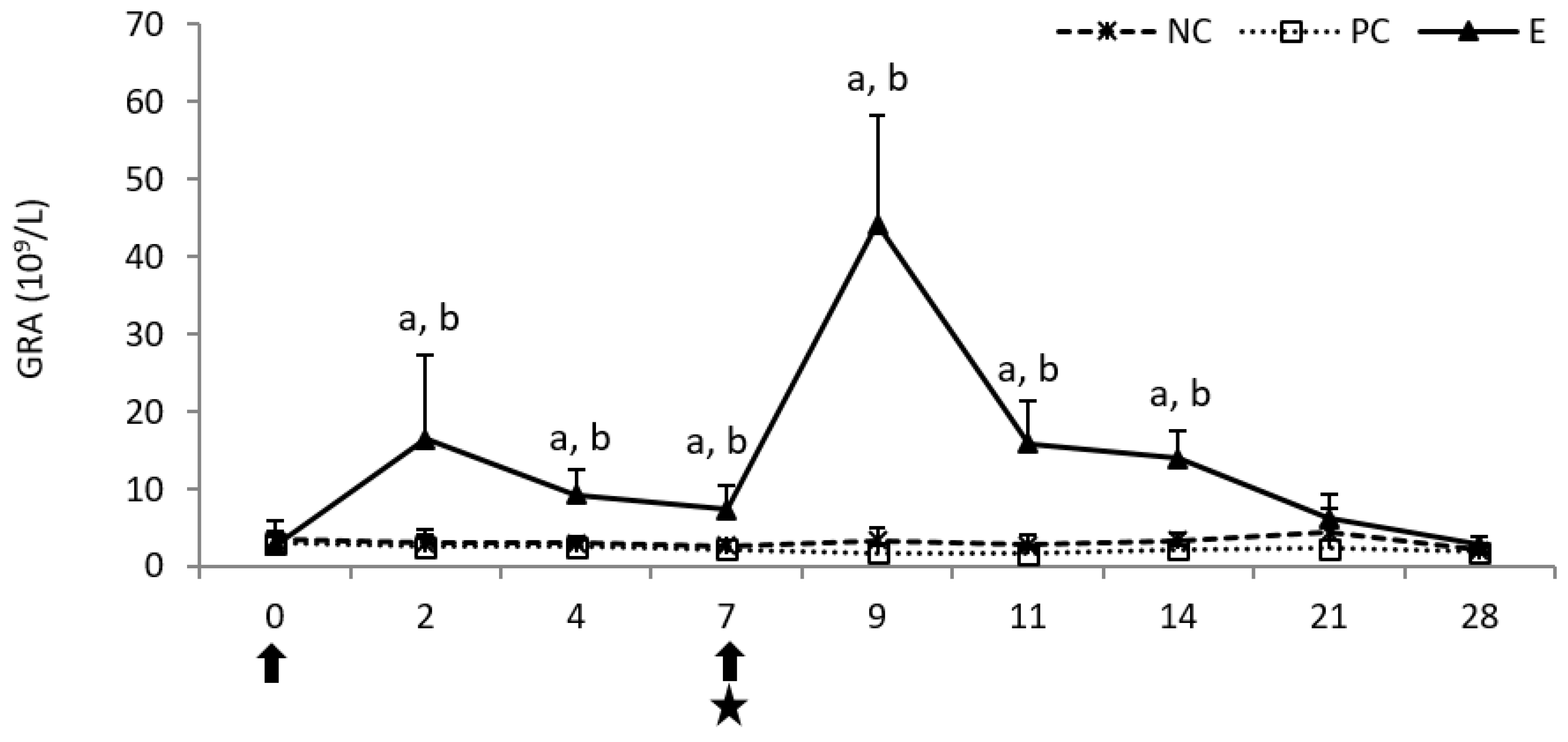
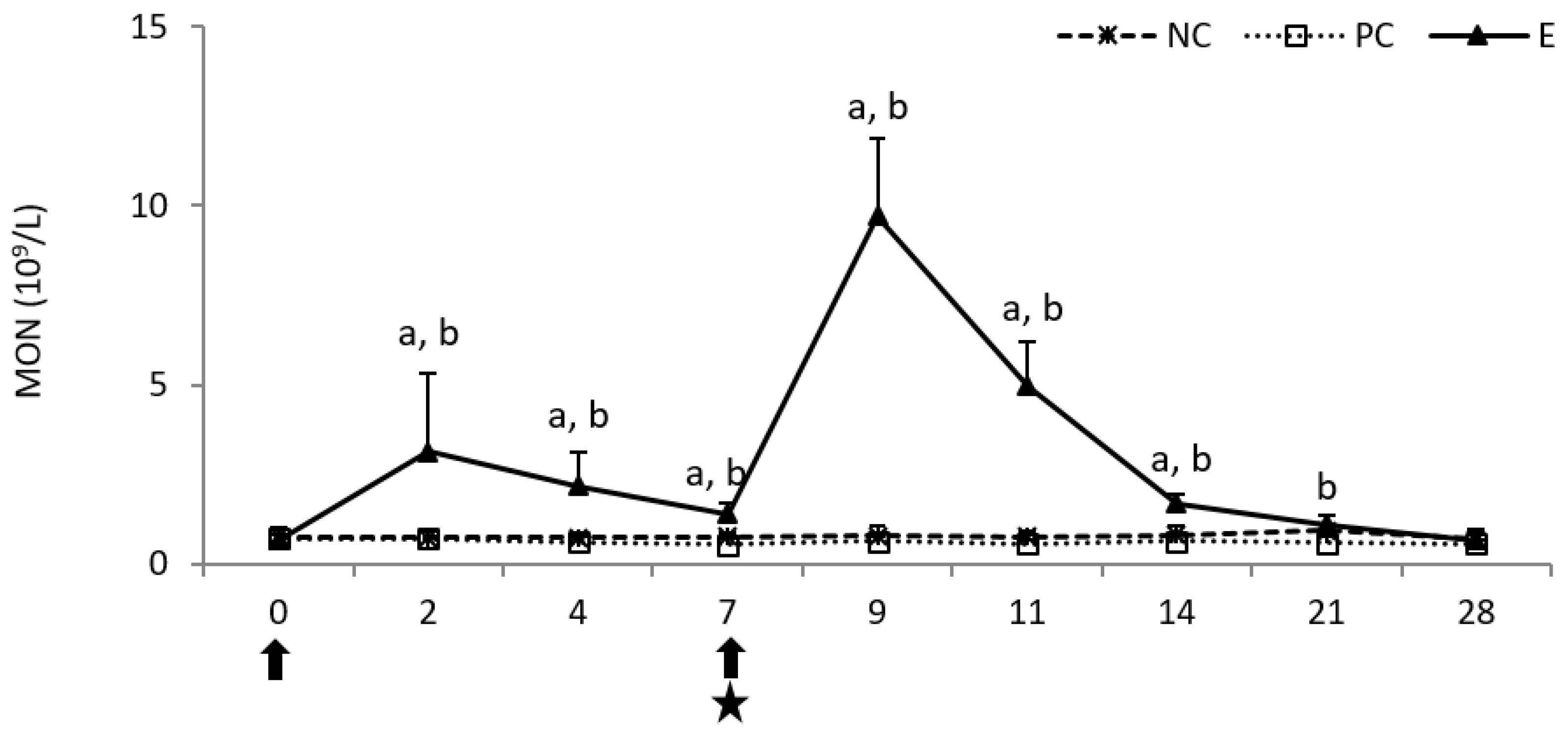
2.1. Hematology
2.2. Flow Cytometry
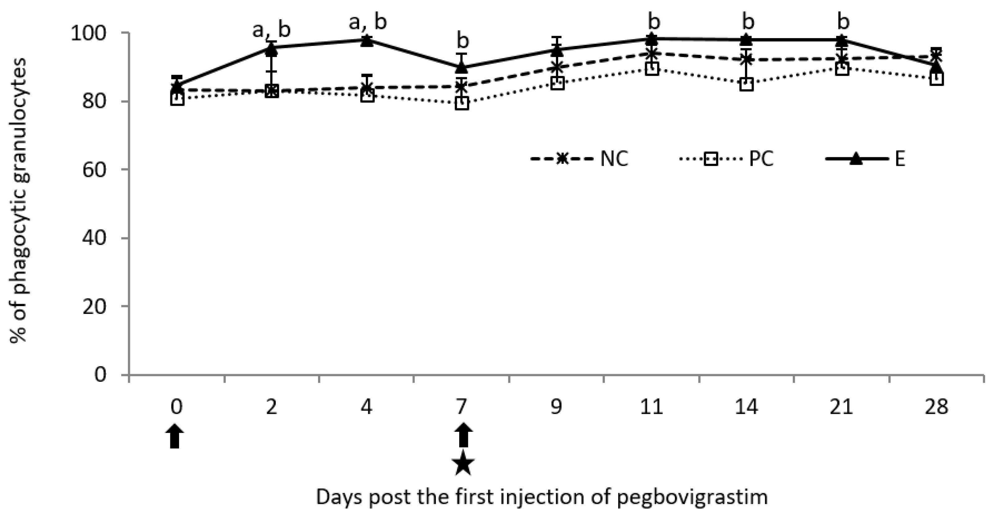
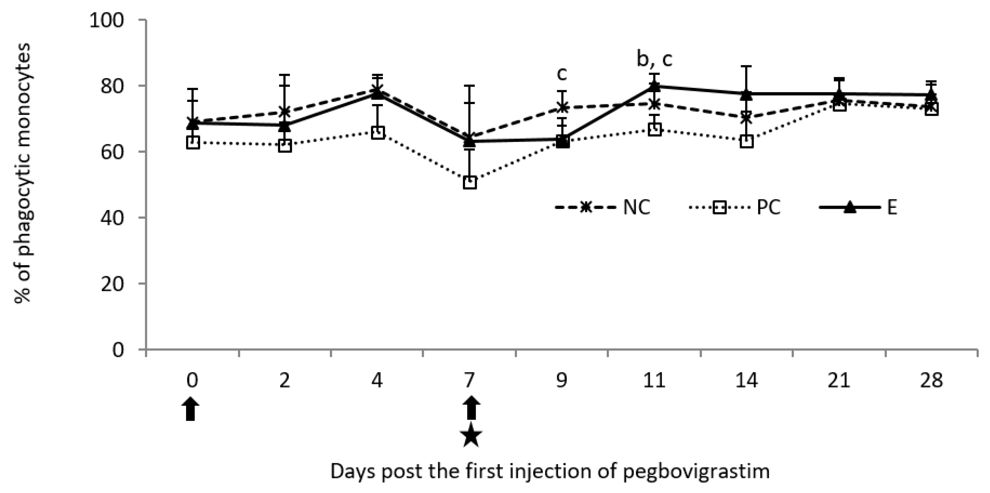
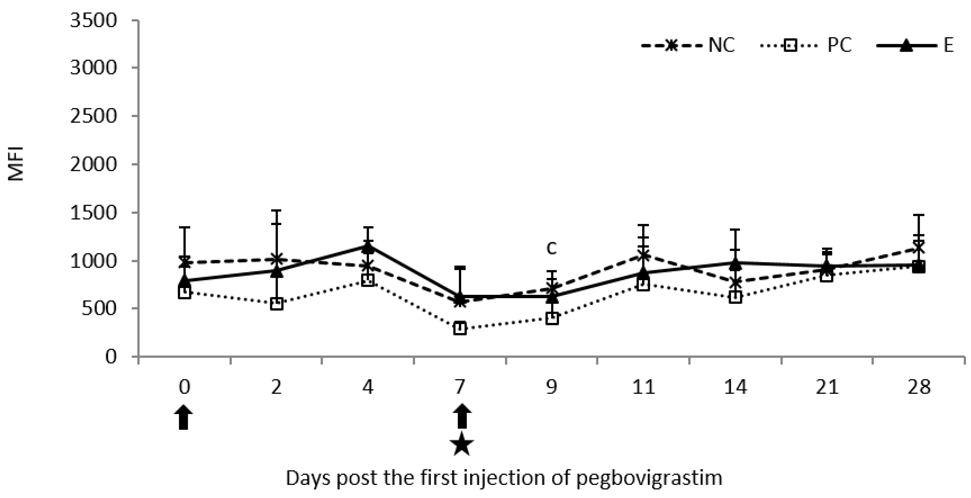
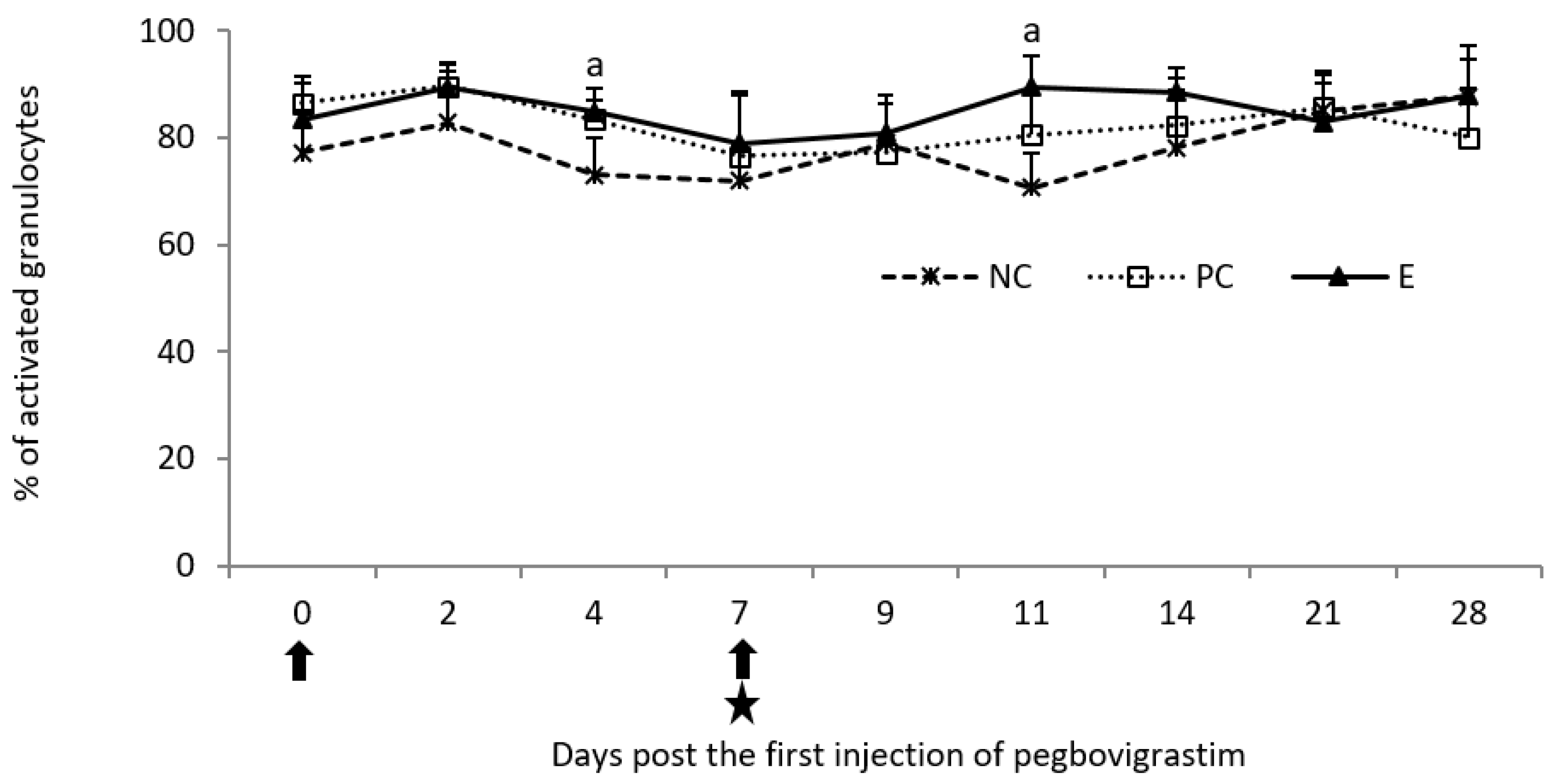
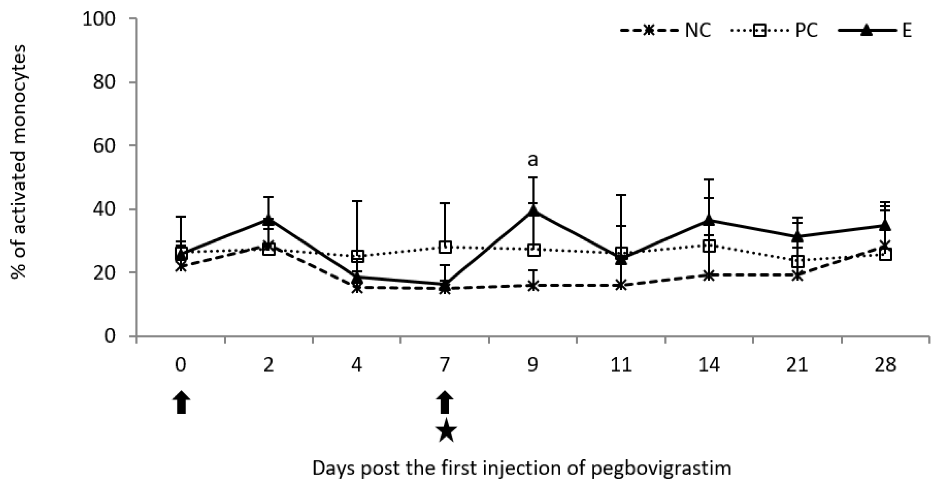
2.2.1. Phagocytic Activity
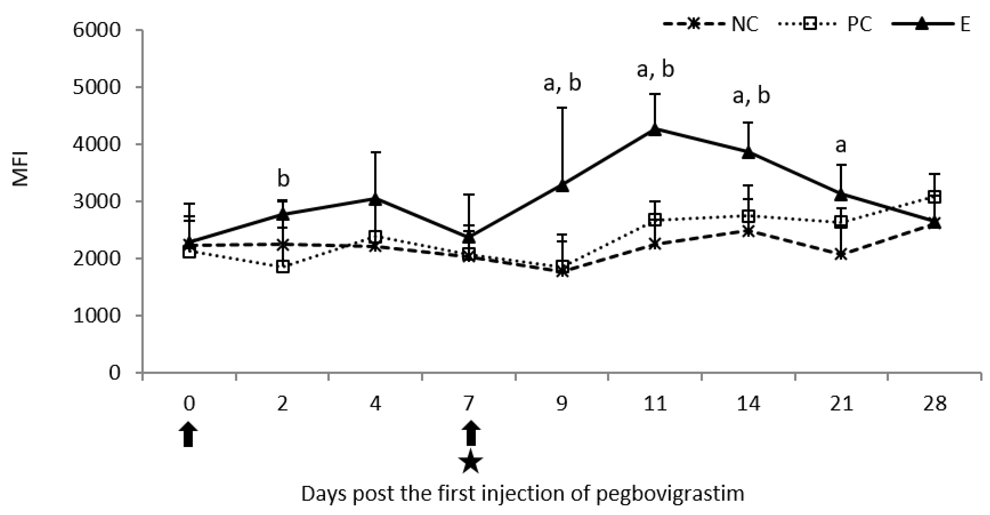
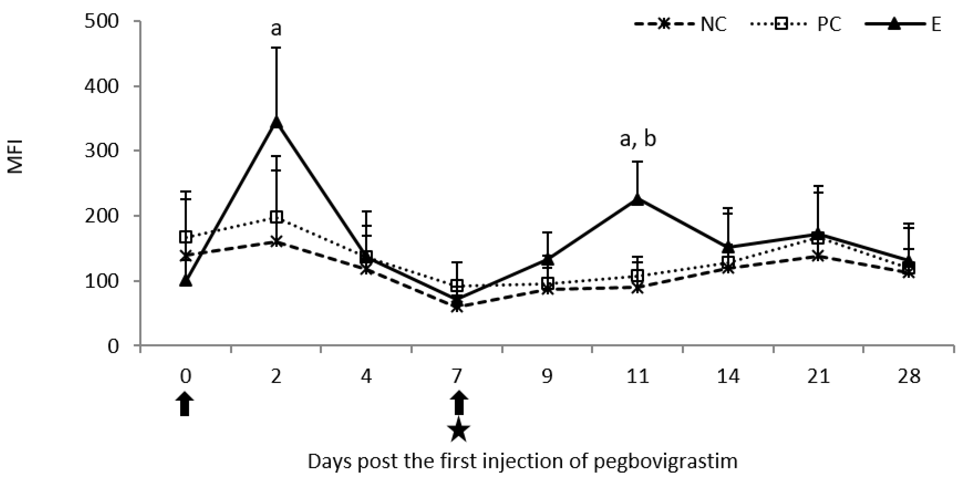
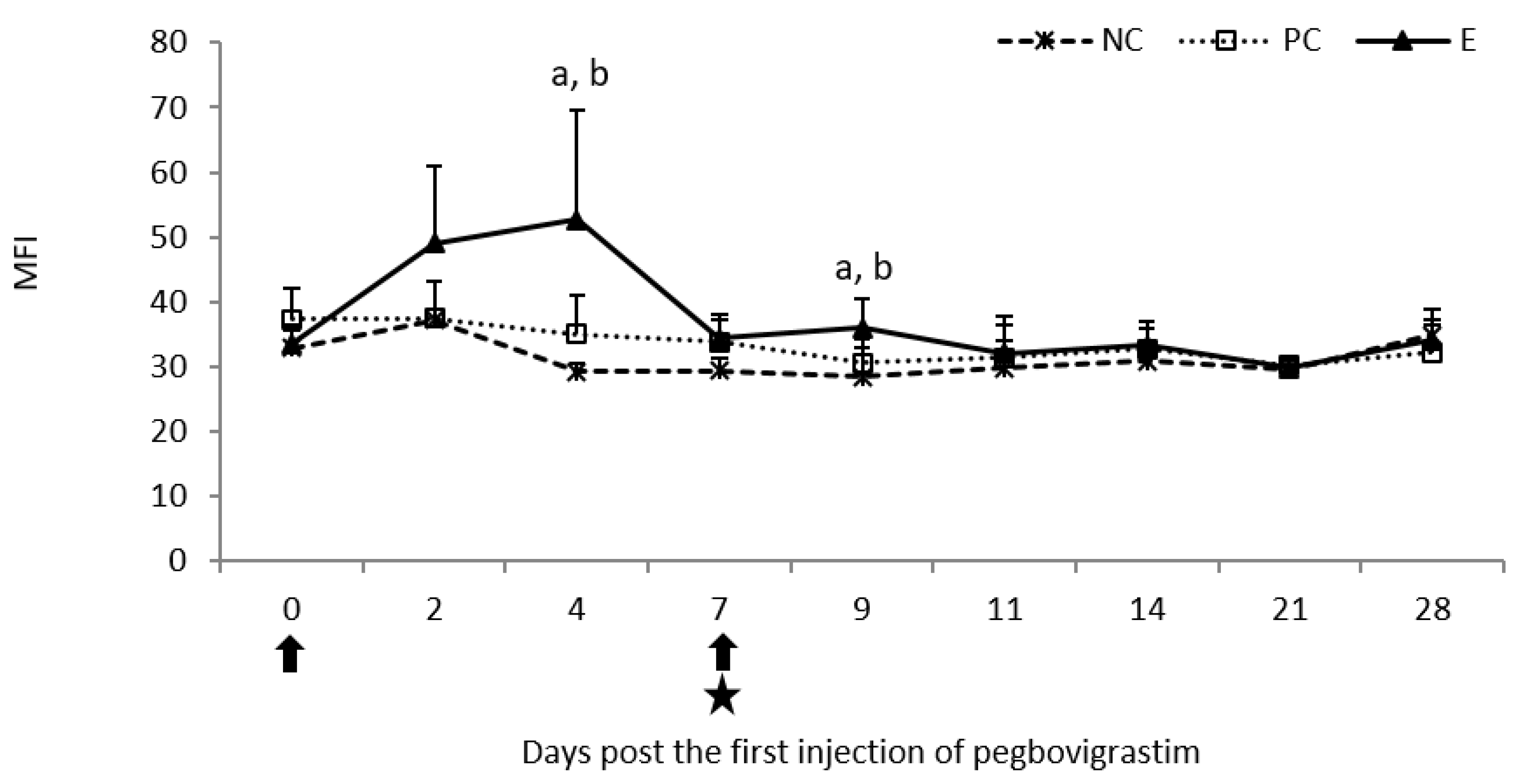
2.2.2. Oxidative Burst Activity
3. Discussion
4. Materials and Methods
4.1. Animals and Study Design
4.2. Hematological and Flow Cytometry Analyses
4.3. Statistical Analysis
Author Contributions
Funding
Institutional Review Board Statement
Informed Consent Statement
Data Availability Statement
Conflicts of Interest
References
- Dudek, K.; Nicholas, R.A.J.; Szacawa, E.; Bednarek, D. Mycoplasma bovis Infections-Occurrence, Diagnosis and Control. Pathogens 2020, 9, 640. [Google Scholar] [CrossRef] [PubMed]
- Nicholas, R.A.J.; Ayling, R.D. Mycoplasma bovis: Disease, diagnosis, and control. Res. Vet. Sci. 2003, 74, 105–112. [Google Scholar] [CrossRef]
- Nicholas, R.; Ayling, R.; McAuliffe, L. Mycoplasma Diseases of Ruminants, 1st ed.; CABI Publishing: Oxford, UK, 2008. [Google Scholar]
- Dudek, K.; Bednarek, D.; Ayling, R.D.; Szacawa, E. Immunomodulatory effect of Mycoplasma bovis in experimentally infected calves. J. Vet. Res. 2013, 57, 499–506. [Google Scholar] [CrossRef] [Green Version]
- Jimbo, S.; Suleman, M.; Maina, T.; Prysliak, T.; Mulongo, M.; Perez-Casal, J. Effect of Mycoplasma bovis on bovine neutrophils. Vet. Immunol. Immunopathol. 2017, 188, 27–33. [Google Scholar] [CrossRef] [PubMed]
- Thomas, C.B.; Van Ess, P.; Wolfgram, L.J.; Riebe, J.; Sharp, P.; Schultz, R.D. Adherence to bovine neutrophils and suppression of neutrophil chemiluminescence by Mycoplasma bovis. Vet. Immunol. and Immunopathol. 1991, 27, 365–381. [Google Scholar] [CrossRef]
- Vanden Bush, T.J.; Rosenbusch, R.F. Characterization of a lympho-inhibitory peptide produced by Mycoplasma bovis. Biochem. Biophys. Res. Commun. 2004, 315, 336–341. [Google Scholar] [CrossRef]
- Dudek, K.; Szacawa, E.; Nicholas, R.A.J. Recent Developments in Vaccines for Bovine Mycoplasmoses Caused by Mycoplasma bovis and Mycoplasma mycoides subsp. mycoides. Vaccines 2021, 9, 549. [Google Scholar] [CrossRef]
- Soehnlen, M.K.; Aydin, A.; Lengerich, E.J.; Houser, B.A.; Fenton, G.D.; Lysczek, H.R.; Burns, C.M.; Byler, L.I.; Hattel, A.L.; Wolfgang, D.R.; et al. Blinded, controlled field trial of two commercially available Mycoplasma bovis bacterin vaccines in veal calves. Vaccine 2011, 29, 5347–5354. [Google Scholar] [CrossRef]
- Nicholas, R.A.J.; Ayling, R.D.; Stipkovits, L.P. An experimental vaccine for calf pneumonia caused by Mycoplasma bovis: Clinical, cultural, serological and pathological findings. Vaccine 2002, 20, 3569–3575. [Google Scholar] [CrossRef]
- Klein, U.; de Jong, A.; Youala, M.; El Garch, F.; Stevenin, C.; Moyaert, H.; Rose, M.; Catania, S.; Gyuranecz, M.; Pridmore, A.; et al. New antimicrobial susceptibility data from monitoring of Mycoplasma bovis isolated in Europe. Vet. Microbiol. 2019, 238, 108432. [Google Scholar] [CrossRef]
- Gregory, A.D.; Hogue, L.A.; Ferkol, T.W. Regulation of systemic and local neutrophil responses by G-CSF during pulmonary Pseudomonas aeruginosa infection. Blood 2007, 109, 3235–3243. [Google Scholar] [CrossRef] [Green Version]
- Cheers, C.; Haigh, A.M.; Kelso, A.; Metcalf, D. Production of colony-stimulating factors (CSFs) during infection: Separate determinations of macrophage-, granulocyte-, granulocyte-macrophage-, and multi-CSFs. Infect. Immun. 1988, 56, 247–251. [Google Scholar] [CrossRef] [Green Version]
- Bajrami, B.; Zhu, H.; Kwak, H.-J.; Mondal, S.; Hou, Q.; Geng, G.; Karatepe, K.; Zhang, Y.C.; Nombela-Arrieta, C.; Park, S.; et al. G-CSF maintains controlled neutrophil mobilization during acute inflammation by negatively regulating CXCR2 signaling. J. Exp. Med. 2016, 213, 1999–2018. [Google Scholar] [CrossRef] [Green Version]
- Pelus, L.M.; Horowitz, D.; Cooper, S.C.; King, A.G. Peripheral blood stem cell mobilization. A role for CXC chemokines. Crit. Rev. Oncol. Hematol. 2002, 43, 257–275. [Google Scholar] [CrossRef]
- Nguyen-Jackson, H.; Panopoulos, A.D.; Zhang, H.; Li, H.S.; Watowich, S.S. STAT3 controls the neutrophil migratory response to CXCR2 ligands by direct activation of G-CSF-induced CXCR2 expression and via modulation of CXCR2 signal transduction. Blood 2010, 115, 3354–3363. [Google Scholar] [CrossRef] [Green Version]
- Nguyen-Jackson, H.T.; Li, H.S.; Zhang, H.; Ohashi, E.; Watowich, S.S. G-CSF-activated STAT3 enhances production of the chemokine MIP-2 in bone marrow neutrophils. J. Leukoc. Biol. 2012, 92, 1215–1225. [Google Scholar] [CrossRef] [Green Version]
- Theyab, A.; Algahtani, M.; Alsharif, K.F.; Hawsawi, Y.M.; Alghamdi, A.; Alghamdi, A.; Akinwale, J. New insight into the mechanism of granulocyte colony-stimulating factor (G-CSF) that induces the mobilization of neutrophils. Hematology 2021, 26, 628–636. [Google Scholar] [CrossRef]
- Trimboli, F.; Morittu, V.M.; Di Loria, A.; Minuti, A.; Spina, A.A.; Piccioli-Cappelli, F.; Trevisi, E.; Britti, D.; Lopreiato, V. Effect of Pegbovigrastim on Hematological Profile of Simmental Dairy Cows during the Transition Period. Animals 2019, 9, 841. [Google Scholar] [CrossRef] [Green Version]
- Barca, J.; Meikle, A.; Bouman, M.; Gnemmi, G.; Ruiz, R.; Schukken, Y.H. Effect of pegbovigrastim on clinical mastitis and uterine disease during a full lactation in grazing dairy cows. PLoS ONE 2021, 16, e0252418. [Google Scholar] [CrossRef]
- Cook, J.G. Effect of pegbovigrastim treatment on the incidence of post-calving antimicrobial treatments in four UK dairy herds. Vet. J. 2020, 259–260, 105479. [Google Scholar] [CrossRef]
- Kegles, F.; Madruga, O.C.; Schmoeller, E.; Bragança, L.F.; Londero, U.S.; Marins, L.; Feijó, J.O.; Corrêa, M.N.; Schmitt, E.; Del Pino, F.A.B. Hematological and biochemical parameters of dairy calves submitted to pegbovigrastim administration. J. Dairy Sci. 2019, 102, 547–556. [Google Scholar] [CrossRef] [PubMed] [Green Version]
- Crookenden, M.A.; Roche, J.R.; Heiser, A.; Kuhn-Sherlock, B.; Higham, C.D.; Phyn, C.V.C.; Turner, S.A. Effect of dose rate and timing of administration of pegbovigrastim on white blood cell responses in grazing dairy cows. J. Dairy Sci. 2021, 104, 11955–11972. [Google Scholar] [CrossRef] [PubMed]
- Dudek, K.; Bednarek, D.; Ayling, R.D.; Kycko, A.; Reichert, M. Preliminary study on the effects of enrofloxacin, flunixin meglumine and pegbovigrastim on Mycoplasma bovis pneumonia. BMC Vet. Res. 2019, 15, 371. [Google Scholar] [CrossRef] [PubMed] [Green Version]
- Chase, C.C.L. The essentials: The who, what, and where of the bovine immune system. In Bovine Immunity: Making Immunology and Vaccinology Come Alive, 1st ed.; Chase, C.C.L., Walsh, C., Casademunt, S., Viejo, J., Angás, P., Hernández, A., Gragera, J., Izaguerri, M., The team at Grupo Asís, Eds.; HIPRA, S.A.: Amer (Girona), Spain, 2022; pp. 2–29. [Google Scholar]
- Dudek, K.; Bednarek, D.; Ayling, R.D.; Kycko, A.; Szacawa, E.; Karpińska, T.A. An experimental vaccine composed of two adjuvants gives protection against Mycoplasma bovis in calves. Vaccine 2016, 34, 3051–3058. [Google Scholar] [CrossRef]
- Dudek, K.; Bednarek, D.; Szacawa, E.; Rosales, R.S.; Ayling, R.D. Flow cytometry follow-up analysis of peripheral blood leukocyte subpopulations in calves experimentally infected with field isolates of Mycoplasma bovis. Acta Vet. Hung. 2015, 63, 167–178. [Google Scholar] [CrossRef] [Green Version]
- Wojcicka-Lorenowicz, K.; Kostro, K.; Lisiecka, U.; Gąsiorek, B. Phagocytic activity and oxygen metabolism of peripheral blood granulocytes from rabbits experimentally infected with Trichophyton mentagrophytes. J. Vet. Res. 2018, 62, 43–48. [Google Scholar] [CrossRef]


Publisher’s Note: MDPI stays neutral with regard to jurisdictional claims in published maps and institutional affiliations. |
© 2022 by the authors. Licensee MDPI, Basel, Switzerland. This article is an open access article distributed under the terms and conditions of the Creative Commons Attribution (CC BY) license (https://creativecommons.org/licenses/by/4.0/).
Share and Cite
Dudek, K.; Szacawa, E.; Wasiak, M.; Bednarek, D.; Reichert, M. The Effect of Pegbovigrastim Injection on Phagocytic and Oxidative Burst Activities of Peripheral Blood Granulocytes and Monocytes in Calves Challenged with Mycoplasma bovis. Pathogens 2022, 11, 1317. https://doi.org/10.3390/pathogens11111317
Dudek K, Szacawa E, Wasiak M, Bednarek D, Reichert M. The Effect of Pegbovigrastim Injection on Phagocytic and Oxidative Burst Activities of Peripheral Blood Granulocytes and Monocytes in Calves Challenged with Mycoplasma bovis. Pathogens. 2022; 11(11):1317. https://doi.org/10.3390/pathogens11111317
Chicago/Turabian StyleDudek, Katarzyna, Ewelina Szacawa, Magdalena Wasiak, Dariusz Bednarek, and Michał Reichert. 2022. "The Effect of Pegbovigrastim Injection on Phagocytic and Oxidative Burst Activities of Peripheral Blood Granulocytes and Monocytes in Calves Challenged with Mycoplasma bovis" Pathogens 11, no. 11: 1317. https://doi.org/10.3390/pathogens11111317