Article

Genetic Characterization of the Poultry Red Mite (Dermanyssus gallinae) in Poland and a Comparison with European and Asian Isolates

by
Sylwia Koziatek-Sadłowska
* and
Rajmund Sokół
Department of Parasitology and Invasive Diseases, Faculty of Veterinary Medicine, University of Warmia and Mazury in Olsztyn, 10-719 Olsztyn, Poland
*
Author to whom correspondence should be addressed.
Pathogens 2022, 11(11), 1301; https://doi.org/10.3390/pathogens11111301
Submission received: 7 October 2022 / Revised: 28 October 2022 / Accepted: 3 November 2022 / Published: 6 November 2022
(This article belongs to the Special Issue Advances in Parasitic Diseases)

Abstract

:
(1) Background: The blood-feeding mite Dermanyssus gallinae (De Geer 1778) continues to attract wide interest from researchers and bird breeders. The aim of this study was to evaluate the genetic diversity of D. gallinae populations in five commercial laying hen farms in Poland and to determine their similarity with isolates from other countries. The study involved an analysis of a fragment of the cytochrome c oxidase subunit I gene (COI). A total of 38 isolates obtained from Polish farms and 338 sequences deposited in GenBank were analyzed. (2) Results: Haplotype No. 46 was dominant (90%) in Polish isolates and was homologous with the isolates from Great Britain, the Netherlands, Belgium, Japan, and South Korea. These results are indicative of high genetic homogeneity and common ancestry of the poultry red mite and point to a common source of infestation in the examined farms. (3) Conclusions: The genetic diversity of D. gallinae should be further studied to promote a better understanding of how this parasite is disseminated within and between countries.

1. Introduction

The poultry red mite (Dermanyssus gallinae) (De Geer 1778) (Mesostigmata: Dermanissidae) is a broadly distributed species found on all continents except Antarctica. The poultry red mite colonizes various bird-rearing systems, regardless of production type or flock size [1]. Severe mite infestations cause a parasitic disease known as dermanyssosis. The disease compromises the well-being and health status of birds, increases mortality, and decreases laying performance and egg quality [1,2,3,4,5,6,7,8]. Its treatment is expensive, and mite infestations can generate serious economic losses in poultry farming. The losses associated with D. gallinae infestations in European layer farms were estimated at EUR 130 million in 2004 [9] and EUR 231 million in 2017 [10]. These data clearly indicate that red poultry mite infestations pose a serious problem in the poultry sector.
The genus Dermanyssus presently comprises 25 species of hematophagous mites which have been classified into two subgenera—Dermanyssus (hirustus group and dermanyssus group) and Microdermanyssus [11,12,13]. Species belonging to the first subgenus share many morphological traits and host specificity patterns. In turn, species of the gallinae group are difficult to distinguish based on their morphological characteristics, and colonize various bird species [12,14]. In order to identify intraspecific diversity in the genus Dermanyssus, the COI gene was found to be the most informative marker [12,15]. A comparative analysis of the nucleotide sequence of the COI gene revealed intraspecies variations between geographically distant populations of D. gallinae. The gene of 16S rRNA was informative as well as the COI gene. Research has shown that fragments of the internal transcribed spacer (ITS) were least useful marker to identify intraspecific variations within populations of D. gallinae [16,17,18].
It appears that genetic variation in D. gallinae can contribute to the plasticity in host choice and can increase the tolerance to adverse environmental conditions and selection pressures (mutations, genetic drift, natural selection and geographical isolation) [12,15]. In poultry red mites, genetic variation can result from the acquisition of resistance to a prolonged and excessive use of chemical agents [7,12,15,19].
Research into the genetic diversity of D. gallinae can promote a better understanding of the parasite’s population dynamics and dissemination within and between countries [20]. The resulting knowledge can be used to identify genetic markers of resistance to acaricides and to develop alternative strategies and methods to eradicate poultry red mite infestations [12,15,17]. The phylogenetic relatedness of D. gallinae populations in Polish poultry farms has not been fully elucidated to date. Therefore, the aim of this study was to evaluate the genetic diversity of D. gallinae populations in selected commercial poultry farms in Poland and to determine their similarity with isolates deposited in GenBank.

2. Materials and Methods

2.1. Dermanyssus gallinae

Female D. gallinae for genetic analysis were obtained from five commercial layer farms in the Polish regions of Warmia and Mazury (farms A and B), Mazovia (farms C and D), and Kuyavia-Pomerania (farm E). The mites were collected with a system of traps developed by Sokół [21]. The acquired samples were transported to a laboratory and stored at a temperature of −20 °C until analysis.

2.2. Isolation of Genomic DNA

Adult D. gallinae females were randomly selected. The mites were identified to species level based on the morphological characteristics described by Di Palma et al. [22] under a binocular stereo zoom microscope (Olympus SZ, 40× magnification). Individual mites were placed in Eppendorf tubes (1.5 mL), 300 µL of ultrapure water was added, and the contents were homogenized with the use of the Tissue Grinder Mixy Professional (NIPPON Genetics Europe, Düren, Germany). Genomic DNA was isolated with the Sherlock AX Kit (A&A Biotechnology, Gdańsk, Poland) according to the manufacturer’s protocol. The extracted DNA was suspended in 50 µL of ultrapure water. The resulting DNA was stored at a temperature of −20 °C until further analysis. The purity and quantity of the isolated DNA were checked with a spectrophotometer at a wavelength of A260/A280 and A230/A260.

2.3. PCR Assay

A PCR assay was conducted with the use of the StartWarm HS-PCR Mix (A&A Biotechnology, Gdańsk, Poland; catalogue No. 2017-100). The reaction mixture was composed of Taq DNA polymerase (0.1 U/µL), PCR buffer, magnesium chloride (2.5 mM), dNTPs (0.5 mM each), specific primers—forward FCOIDG (5′-CATTAATATTAACTGCACCTGACA TG-3′) and reverse RCOIDG (5′-CCCGTGGAGTGTTGAAATTCA TGA-3′) [16] or forward CO1Fyuw114 (5′-AGATCTTTAATTGAAGGGGG-3′) and reverse CO1Ryuw114 (5′-AAGATCAAAGAATCGGTGG-3′) [17] (0.5 µM each), and DNA (30–60 ng). The reaction mix had a final volume of 25 µL (12.5 µL of StartWarm HS-PCR; 1 µL of each primer, 4–5 µL of DNA, 8–14 µL of ultrapure water) or 50 µL (25 µL of StartWarm HS-PCR; 1–2 µL of each primer, 10–15 µL of DNA, 6.5–8 µL of ultrapure water). The DNA fragments amplified with the use of FCOIDG + RCOIDG and CO1Fyuw114 + CO1Ryuw114 primers had a length of 737 base pairs (bp) and 681 bp, respectively. Ultrapure nuclease-free water was added to the reaction mix in the negative control reaction. The PCR cycling conditions were as follows: initial denaturation at 95 °C for 10 min, followed by 35–40 cycles of denaturation at 95 °C for 20 s, primer annealing at 52–54 °C for 30 s, elongation at 72 °C for 90 s, and final elongation at 72 °C for 10 min. The reactions were carried out with the use of the Light Cycler Nano thermocycler (Roche, Basel, Switzerland). The PCR products were separated by electrophoresis on a 2% agarose gel with Midori Green Advance DNA Stain (NIPPON Genetics Europe, Düren, Germany). The results of the PCR assay were visualized in the Gel Doc EZ imaging system (Bio-Rad, Hercules, CA, USA) with a 100 bp molecular weight marker (Generuler 100BP DNA Ladder, Life Technologies, Carlsbad, CA, USA; catalogue No. SM0243). The products with the expected size were cut out from the agarose gel, purified with the Gel-out reagent kit (A&A Biotechnology, Gdańsk, Poland) according to the manufacturer’s protocol, and sequenced.

2.4. Sequencing

The obtained amplicons were sequenced by Genomed SA (Warsaw, Poland) with the use of the BigDye® Terminator v3.1 Cycle Sequencing Kit (Applied Biosystems, Life Technologies).

2.5. Phylogenetic Analysis

2.5.1. Consensus Sequences

The obtained sequences were edited in the BioEdit Sequence Alignment Editor. The quality of the resulting data was checked by analyzing the chromatograms, and ambiguous and noisy sites at the ends were removed. Low-quality reads were rejected. Antisense sequences were transcribed to sense strands using the reverse complement function. Pairs of sequences were aligned for every sample with ClustalW (to derive consensus sequences). The chromatograms corresponding to specific samples were checked to correct any sequencing errors. Non-overlapping end reads were removed. All consensus sequences were assembled with ClustalW and were trimmed to equal length. A total of 38 nucleotide sequences were obtained. The sequences were saved in a text file in FASTA format and used in further analyses.

2.5.2. Homologous Sequences

Homologous sequences were obtained from the NCBI database with the use of the BLAST tool (search parameters: Dermanyssus gallinae; algorithm parameters; general parameters; max. target sequences: 1000). A total of 463 nucleotide sequences were exported (as of 7 August 2020) and saved in a FASTA file.

2.5.3. Sequence Alignment Analysis

The generated sequences and homologous sequences were aligned with the use of ClustalW in the BioEdit environment. Sequences that were significantly shorter (less than 75% of the length of the generated sequences) were removed. All sequences were trimmed to the shortest sequence. The aligned sequences were saved in a FASTA file. A total of 376 sequences with a length of 552 bp were used in the analyses. The analyzed fragment corresponds to the nucleotide sequence from 28,218 to 28,768 of the D. gallinae genome (GenBank reference No. QVRM01004456.1).

2.5.4. Haplotype Analysis

The sequences were collapsed to haplotypes using FaBox (https://users-birc.au.dk/palle/php/fabox/index.php (accessed on 15 August 2020)). Haplotypes are sequences that differ by at least one nucleotide or an insertion/deletion (indel) in one position. The sequences representing the studied haplotypes (one sequence per haplotype) were saved in a FASTA file and subjected to a phylogenetic analysis.

2.5.5. Generation of a Phylogenetic Tree
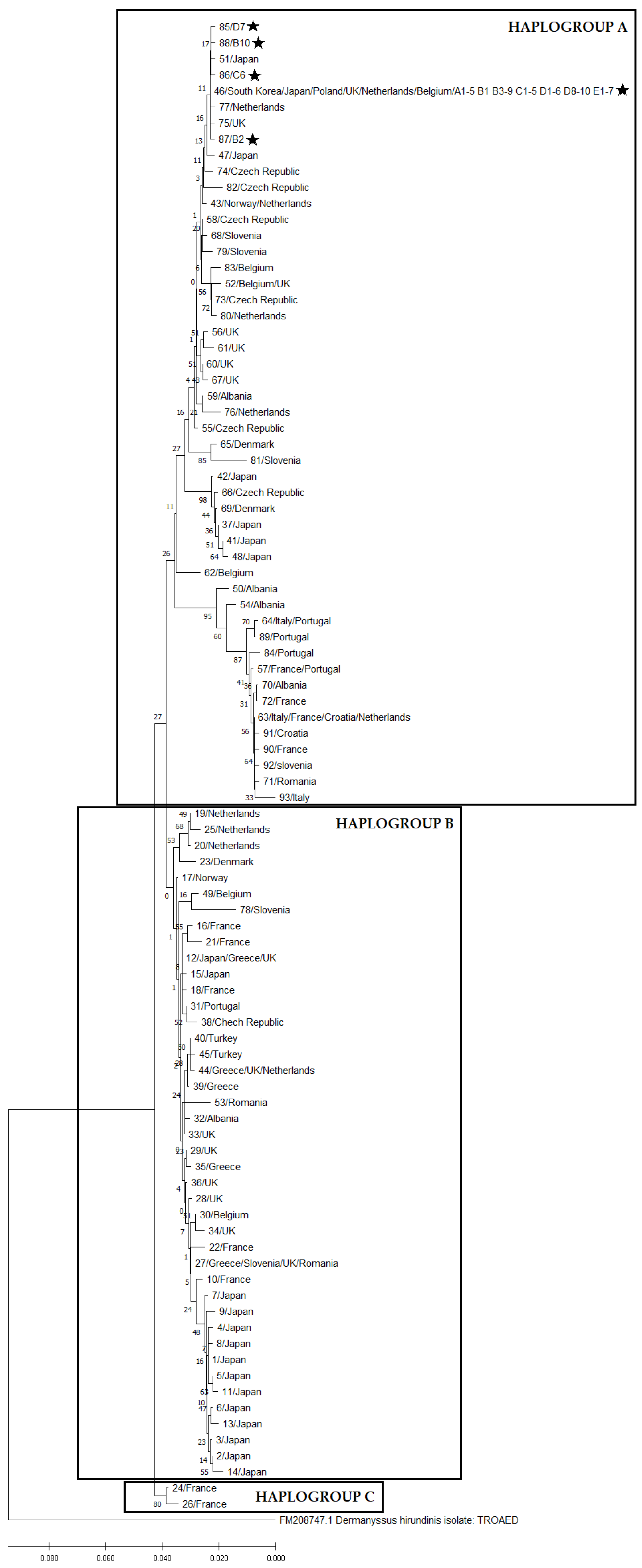
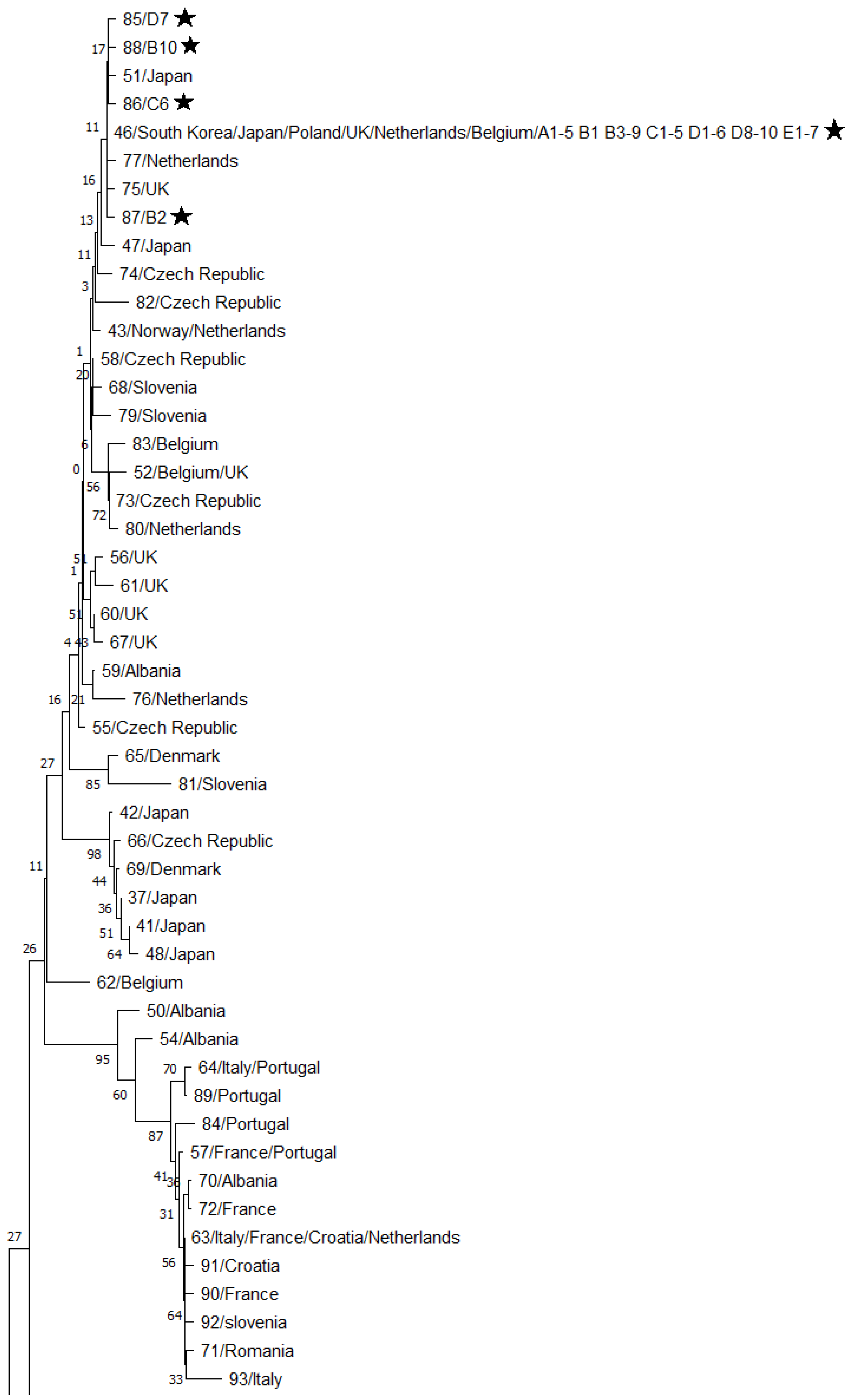
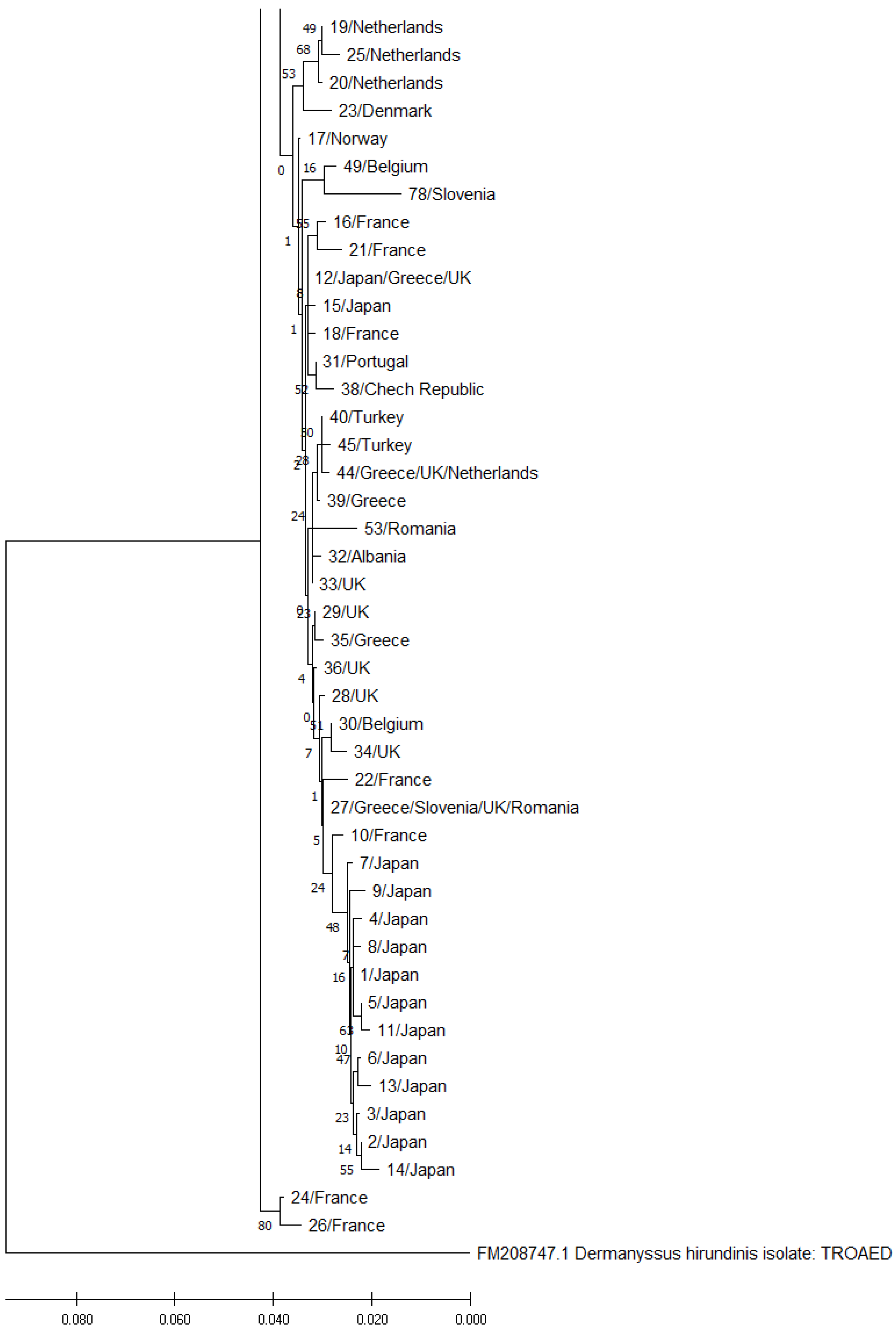
A phylogenetic tree was generated by the neighbor-joining method (1000 bootstrap) with the use of MEGA X software. The phylogenetic tree was rooted by incorporating a homologous sequence from an external source—a fragment of the cytochrome oxidase gene of Dermanyssus hirundinis (FM208747.1). Evolutionary distances were estimated with the Kimura two-parameter (K2P) model and were expressed by the number of base substitutions per site. Codon positions 1, 2, 3 sequences were considered. All ambiguous positions were removed in each sequence pair (pairwise deletion option).

3. Results

A total of 93 haplotypes were identified in 376 nucleotide sequences of the 552-bp fragment of the COI gene in D. gallinae (including 38 sequences obtained in the study and 338 sequences obtained from the GenBank database). These haplotypes were randomly assigned numbers from 1 to 93. Haplotype numbers, GenBank reference numbers of the corresponding sequences, and countries of origin are presented in Table 1. Based on the structure of the phylogenetic tree, we divided the identified haplotypes into three haplogroups: haplogroup A (49 haplotypes), haplogroup B (42 haplotypes), and haplogroup C (2 haplotypes), which is consistent with the findings of Øines and Brännström [16] for D. gallinae in northern Europe. The phylogenetic tree with the haplogroups is presented in Figure 1. The haplogroups A and B are enlarged in Figure 2 and Figure 3.
The isolates from Polish poultry farms, including 5 isolates from farm A, 10 isolates from farm B, 6 isolates from farm C, 10 isolates from farm D, and 7 isolates from farm E, were assigned to 5 haplotypes belonging to the haplogroup A. These isolates were numbered 46, 85, 86, 87, and 88. Haplotype 46 contained 34 isolates (approx. 90% of all isolates). Haplotypes 85–88 were identified in single cases. The percentages of the identified haplotypes in the studied farms are presented in Table 2. Dermanyssus gallinae strains belonging to the haplogroups B and C were not identified in any of the samples we sequenced.
An optimal phylogenetic tree with a sum of branch lengths = 0.48525737 is presented in Figure 1.
In Polish isolates, single nucleotide substitutions were observed relative to the nucleotide sequence of the most prevalent haplotype 46. A T-to-C transition at position 550 was noted in haplotype 85. A A-to-T transversion was observed at position 552 in haplotype 86. A A-to T-transversion was noted at position 353 in haplotype 87. A G-to-A transition was observed at position 501 in haplotype 88. Insertion/deletion polymorphisms were not detected. The transversion in haplotype 87 led to the replacement of D with V in the polypeptide chain. The remaining mutations did not change the sequence of the coded polypeptide chain.

4. Discussion

All GenBank sequences that were homologous with the sequences of Polish isolates were considered in our genetic analysis. The reference sequences originated from various countries around the world, mostly Europe (Table 1) but also Japan and South Korea. Two lines of D. gallinae, classified as haplogroup A and haplogroup B, were identified as the dominant lines in the world. A third line, referred to as haplogroup C, was also identified; to date, it has been detected only in France [12]. The haplogroup C is genetically distant from haplogroups A and B, which is why haplogroup C strains are regarded as cryptic species of D. gallinae [16]. The haplotypes from groups A and B are ubiquitous around the world, which indicates that the parasite is transmitted between countries and continents [16,17,23]. The geographic distribution of the haplotypes in different countries suggests that haplogroup B strains have a preference for regions with a temperate climate, whereas haplogroup A strains occur in regions with a more severe climate.
The study demonstrated that D. gallinae haplotype 46 belonging to the haplogroup A was the most prevalent in the analyzed poultry farms (Table 2). Haplotype 46 was previously identified in Great Britain, the Netherlands, Belgium, Japan, and South Korea [16,24,23]. This haplotype was also detected by Roy et al. [12] who analyzed isolates from southern Poland. Haplotypes 85–88 were identified in individual samples, and the analysis revealed that these haplotypes probably emerged as a mutation of haplotype 46. Haplotypes 85–88, which we found in the present study, have not been identified in other countries. These observations confirm the high genetic homogeneity of D. gallinae populations in the analyzed Polish regions. However, based on the presented results, it cannot be concluded with sufficient certainty whether the currently occurring D. gallinae is a native species or whether it migrated to Poland from other European countries.
Gaweł et al. [25] reported higher levels of genetic diversity in D. gallinae isolates from the Polish regions of Wielkopolska, Opole, Łódź, Lubusz, and Lower Silesia. The cited authors identified four subtypes corresponding to haplogroup A and haplogroup B in the present study. The isolates from the regions of Łódź and Lubusz (10%) belonged to subtype A and were homologous to the isolates from France, the Netherlands, Denmark, and Australia. Isolates from the Wielkopolska region (83%) were homologous to the isolates from France and Poland. Two isolates from the regions of Opole and Wielkopolska were homologous to French isolates. According to the cited authors, the Polish population of D. gallinae is not genetically different from the populations in other European countries, which was confirmed in the current study. These data and the present findings suggest that the number of parasite transmission routes is higher in south-western than in northern Poland.
Similarly to Poland, a low genetic diversity of D. gallinae was also reported in Romania, Japan, and Turkey [16,23]. In contrast, D. gallinae populations in Greece, Great Britain, and Belgium are highly genetically diverse, and around 30 different haplotypes have been identified in these countries. Øines and Brännström [16] identified 32 haplotypes in an analysis of D. gallinae isolates from Sweden and Norway. Only single haplotypes were detected in most farms. These observations point to a high number of parasite transmission routes to poultry farms in Sweden and Norway. However, no haplotypes were shared between Norway and Sweden, which points to little or no exchange of D. gallinae strains between these countries.
The fact that females of D. gallinae are heterozygotic may influence the number of haplotypes. It implies that diversity may be underestimated and haplotypes cannot be unambiguously assigned in heterozygous individuals.
The results of this study indicate that D. gallinae populations in the examined Polish layer farms are highly genetically homogeneous and have a common ancestry, which suggests that parasitic infestations have a common source. The genetic diversity of D. gallinae should be studied to promote a better understanding of how this parasite is disseminated within and between countries. Understanding the spread routes of different populations of D. gallinae is important to advance our knowledge of their epidemiology and develop combat strategies, as different populations of D. gallinae may display differences in resistance to acaricides, pathogenicity and vectorial capacity [13].

Author Contributions

Conceptualization, S.K.-S.; Formal analysis, S.K.-S.; Investigation, S.K.-S.; Methodology, S.K.-S.; Resources, S.K.-S. and R.S.; Software, S.K.-S.; Supervision, S.K.-S. and R.S.; Validation, S.K.-S.; Visualization, S.K.-S.; Writing—original draft, S.K.-S.; Writing—review & editing, R.S. All authors have read and agreed to the published version of the manuscript.

Funding

This research received no external funding.

Institutional Review Board Statement

Not applicable.

Data Availability Statement

The nucleotide sequences obtained and analyzed during the current study are available in the GenBank database (accession numbers: OL547403–OL547440).

Acknowledgments

Project financially co-supported by the Minister of Science and Higher Education within the program entitled “Regional Initiative of Excellence” for the years 2019–2023, Project No. 010/RID/2018/19, amount of funding 12.000.000 PLN.

Conflicts of Interest

The authors declare no conflict of interest.

References

  1. Sparagano, O.A.E.; Pavlicevic, A.; Murano, T.; Camarda, A.; Sahibi, H.; Kilpinen, O.; Mul, M.; Van Emous, R.; Le Bouquin, S.; Hoel, K.; et al. Prevalence and key figures for the poultry red mite Dermanyssus gallinae infections in poultry farm systems. Exp. Appl. Acarol. 2009, 48, 3–10. [Google Scholar] [CrossRef] [PubMed]
  2. Hoglund, J.; Nordenfors, H.; Uggla, A. Prevalence of the poultry red mite, Dermanyssus gallinae, in different types of production systems for egg layers in Sweden. Poult. Sci. 1995, 74, 1793–1798. [Google Scholar] [CrossRef] [PubMed]
  3. Cencek, T.; Ziomko, I.; Topór, W. Inwazja Dermanyssus gallinae przyczyną masowych padnięć kacząt brojlerów. Med. Wet. 2002, 58, 353–355. [Google Scholar]
  4. Guy, J.H.; Khajavi, M.; Hlalel, M.M.; Sparagano, O.A.E. Red mite (Dermanyssus gallinae) prevalence in laying units in northern England. Br. Poult. Sci. 2004, 45, 5–6. [Google Scholar]
  5. Fiddes, M.D.; Le Gresley, S.; Parsons, D.G.; Epe, C.; Coles, G.C.; Stafford, K.A. Prevalence of the poultry red mite (Dermanyssus gallinae) in England. Vet. Rec. 2005, 157, 233–235. [Google Scholar] [CrossRef]
  6. Sparagano, O.A.E.; George, D.R.; Harrington, D.W.; Giangaspero, A. Significance and control of the poultry red mite, Dermanyssus gallinae. Annu. Rev. Entomol. 2014, 59, 447–466. [Google Scholar] [CrossRef] [Green Version]
  7. Sokół, R.; Koziatek-Sadłowska, S.; Michalczyk, M. The influence of Dermanyssus gallinae and different lighting regimens on selected blood proteins, corticosterone levels and egg production in layer hens. Vet. Res. Commun. 2019, 43, 31–36. [Google Scholar] [CrossRef] [Green Version]
  8. Koziatek-Sadłowska, S.; Sokół, R. Changes in the percentages of B- and T-lymphocytes and antibody titres in laying hens infested with Dermanyssus gallinae—A preliminary study. Animals 2020, 10, 987. [Google Scholar] [CrossRef]
  9. van Emous, R.A. Wage war against the red mite! Poult. Int. 2005, 44, 26–33. [Google Scholar]
  10. Flochlay, A.S.; Thomas, E.; Sparagano, O.A.E. Poultry red mite (Dermanyssus gallinae) infestation: A broad impact parasitological disease that still remains a significant challenge for the egg-laying industry in Europe. Parasites Vectors 2017, 10, 357. [Google Scholar] [CrossRef]
  11. Moss, W.W. The mite genus Dermanyssus: A survey, with description of Dermanyssus trochilinis, n. sp., and a revised key to the species (Acari: Mesostigmata: Dermanyssidae). J. Med. Entomol. 1978, 14, 627–640. [Google Scholar] [CrossRef]
  12. Roy, L.; Dowling, A.P.G.; Chauve, C.; Buronfosse, T. Delimiting species boundaries within Dermanyssus Dugès, 1834 (Acari: Dermanyssidae) using a total evidence approach. Mol. Phylogenet. Evol. 2009, 50, 446–470. [Google Scholar] [CrossRef] [PubMed]
  13. Roy, L.; Buronfosse, T. Using mitochondrial and nuclear sequence data for disentangling population structure in complex pest species: A case study with Dermanyssus gallinae. PLoS ONE 2011, 6, e22305. [Google Scholar] [CrossRef] [PubMed]
  14. Roy, L.; Chauve, C.M.; Buronfosse, T. Contrasted ecological repartition of the northern fowl mite Ornithonyssus sylviarum (Mesostigmata: Macronyssidae) and the chicken red mite Dermanyssus gallinae (Mesostigmata: Dermanyssidae). Acarologia 2010, 50, 207–219. [Google Scholar] [CrossRef] [Green Version]
  15. Marangi, M.; Cafiero, M.A.; Capelli, G.; Camarda, A.; Sparagano, O.A.E.; Giangaspero, A. Evaluation of the poultry red mite, Dermanyssus gallinae (Acari: Dermanyssidae), susceptibility to some acaricides in field populations from Italy. Exp. Appl. Acarol. 2009, 48, 11–18. [Google Scholar] [CrossRef]
  16. Øines, Ø.; Brännström, S. Molecular investigations of cytochrome c oxidase subunit I (COI) and the internal transcribed spacer (ITS) in the poultry red mite, Dermanyssus gallinae, in northern Europe and implications for its transmission between laying poultry farms. Med. Vet. Entomol. 2011, 25, 402–412. [Google Scholar] [CrossRef]
  17. Chu, T.T.; Murano, T.; Uno, Y.; Usui, T.; Yamaguchi, T. Molecular epidemiological characterization of poultry red mite, Dermanyssus gallinae, in Japan. J. Vet. Med. Sci. 2015, 77, 1397–1403. [Google Scholar] [CrossRef]
  18. Potenza, L.; Cafiero, M.A.; Camarda, A.; La Salandra, G.; Cucchiarini, L.; Dachà, M. Characterization of Dermanyssus gallinae (Acarina: Dermanissydae) by sequence analysis of the ribosomal internal transcribed spacer regions. Vet. Res. Commun. 2009, 33, 611–618. [Google Scholar] [CrossRef]
  19. Chauve, C. The poultry red mite Dermanyssus gallinae: Current situation and future prospects. Vet. Parasitol. 1998, 8, 364–376. [Google Scholar]
  20. Marangi, M.; Cantacessi, C.; Sparagano, O.A.; Camarda, A.; Giangaspero, A. Molecular characterization and phylogenetic inferences of Dermanyssus gallinae isolates in Italy within an European framework. Med. Vet. Entomol. 2014, 28, 447–452. [Google Scholar] [CrossRef]
  21. Sokół, R.; Romaniuk, K. Przebieg i dynamika inwazji Dermanyssus gallinae w fermie kur niosek. Med. Wet. 2007, 63, 484–486. [Google Scholar]
  22. Di Palma, A.; Giangaspero, A.; Cafiero, M.A.; Germinara, G.S. A gallery of the key characters to ease identification of Dermanyssus gallinae (Acari: Gamasida: Dermanyssidae) and allow differentiation from Ornithonyssus sylviarum (Acari: Gamasida: Macronyssidae). Parasites Vectors 2012, 5, 104. [Google Scholar] [CrossRef] [PubMed] [Green Version]
  23. Karp-Tatham, E.; Küster, T.; Angelou, A.; Papadopoulos, E.; Nisbet, A.J.; Xia, D.; Tomley, F.M.; Blake, D.P. Phylogenetic inference using cytochrome c oxidase subunit I (COI) in the poultry red mite, Dermanyssus gallinae in the United Kingdom relative to a European framework. Front. Vet. Sci. 2020, 7, 553. [Google Scholar] [CrossRef]
  24. Oh, S.; Noh, G.; Yi, S.; Do, Y.; Kim, E.; Yoo, J. Molecular epidemiological characterization of poultry red mite (Dermanyssus gallinae) collected from Korea. Korean J. Vet. Serv. 2019, 42, 161–167. [Google Scholar]
  25. Gaweł, Ł. Analiza genetyczna populacji ptaszyńca w Polsce. In Proceedings of the III Międzynarodowa Konferencja Techniczna Eimeriana Avia—Aktualne i Przyszłe Wyzwania w Strategiach Kontroli Kokcydiozy i Innych Chorób Inwazyjnych Drobiu—Aspekty Prawne i Terapeutyczne, Wroclaw, Poland, 20–22 February 2020; p. 102. [Google Scholar]
Figure 1. A phylogenetic tree generated by the neighbor-joining method, including haplotype numbers and countries of origin (the percentage of replicated trees, where related taxa were grouped in the bootstrap test (1000 replications), are presented next to the branches. The tree was drawn to scale, and branch lengths are given in the same units as the evolutionary distances used to infer the phylogenetic tree). (Asterisks indicate the sequences obtained in this study).
Pathogens 11 01301 g001
Figure 2. A fragment of the phylogenetic tree presenting the haplogroup A, including haplotype numbers and countries of origin. (Asterisks indicate the sequences obtained in this study).
Pathogens 11 01301 g002
Figure 3. A fragment of the phylogenetic tree presenting the haplogroups B and C, including haplotype numbers and countries of origin.
Pathogens 11 01301 g003
Table 1. Haplotypes considered in the genetic analysis, with the number of haplotypes and the country of the origin (the sequences obtained in this study are underlined).
No.Number of HaplotypesSequence (GenBank ID)CountryRef.
134MK599418Japan
LC029558, LC029544, LC029537, LC029535, LC029534, LC029533, LC029531, LC029528, LC029524, LC029523, LC029522, LC029521, LC029515, LC029513, LC029512, LC029511, LC029505, LC029494, LC029492, LC029491, LC029488, LC029483, LC029481, LC029480, LC029479, LC029477, LC029476, LC029473, LC029472, LC029469, LC029468, LC029464, LC029460,Japan[17]
214LC029542, LC029536, LC029504, LC029501, LC029499, LC029493, LC029489, LC029487, LC029482, LC029474, LC029471, LC029470, LC029466, LC029463,Japan[17]
31LC029529Japan[17]
41LC029467Japan[17]
52LC029540, LC029503Japan[17]
62LC029520, LC029462Japan[17]
72LC029507, LC029478Japan[17]
81LC029500Japan[17]
91LC029458Japan[17]
101FM208732France[12]
111LC029553Japan[17]
1213LC029551, LC029550, LC029549, LC029548, LC029547, LC029526, LC029519, LC029518, LC029517, LC029497, LC029496,Japan[17]
LR812399Greece[23]
LR812378Great Britain[23]
131LC029502Japan[17]
141LC029465Japan[17]
151LC029525Japan[17]
161AM921857France[12]
171AM921853Norway[12]
181FM208722France[12]
191FM207497Netherlands[12]
201FM207495Netherlands[12]
211AM921858France[12]
221FM208739France[12]
231AM921856Denmark[12]
241AM921863France[12]
251FM207498Netherlands[12]
261AM921865France[12]
2750LR812452, LR812451, LR812450, LR812449, LR812448, LR812447, LR812433, LR812432, LR812431, LR812430, LR812429, LR812428, LR812427, LR812418, LR812417, LR812416, LR812415, LR812414, LR812413, LR812412, LR812411, LR812410, LR812409, LR812408, LR812407, LR812405, LR812404, LR812403, LR812402, LR812398, LR812397, LR812396, LR812395, LR812394, LR812393, LR812392, LR812391,Greece[23]
LR812406Slovenia[23]
LR812390, LR812373, LR812372, LR812371, LR812370, LR812369,Great Britain[23]
LR812346, LR812345, LR812343, LR812342, LR812341, LR812340,Romania[23]
LR812406, LR812406, LR812406Slovenia[23]
281LR812383Great Britain[23]
291LR812375Great Britain[23]
303LR812339, LR812333, LR812332,Belgium[23]
313LR812312, LR812311, LR812310Portugal[23]
322LR812290, LR812289Albania[23]
333LR812477, LR812377, LR812376.1Great Britain[23]
345LR812476, LR812475, LR812474, LR81247, LR812472Great Britain[23]
351LR812434Greece[23]
361LR812374Great Britain[23]
371LC029552Japan[17]
381LR812317Czechia[23]
391LR812420Greece[23]
405LR812351, LR812350, LR812349, LR812348, LR812347Turkey[23]
4119LC029557, LC029556, LC029554, LC029546, LC029545, LC029541, LC029532, LC029527, LC029514, LC029510, LC029509, LC029506, LC029498, LC029490, LC029486, LC029485, LC029475, LC029459, LC029457,Japan[17]
421LC029484Japan[17]
432AM921852Norway[12]
LR812361Netherlands[23]
4427LR812446, LR812445, LR812444, LR812443, LR812442, LR812441, LR812440, LR812439, LR812438, LR812437, LR812436, LR812435, LR812426, LR812425, LR812424, LR812423, LR812422, LR812421, LR812419, LR812401, LR812400Netherlands[23]
LR812385, LR812384, LR812381, LR812380
LR812379
Great Britain[23]
LR812367Netherlands[23]
451LR812352Turkey[23]
4655MN249083, MN249082, MN249080, MN249079, MN249078, MN249077, MN249076, MN249075, MN249074, MN249073, MN249072South Korea[24]
LC029538, LC029516, LC029508, LC029495Japan[17]
AM921854Poland[12]
LR812388Great Britain[23]
LR812363, LR812362Netherlands[23]
LR812337, LR812336Belgium[23]
A1-A5, B1, B3-9, C1-5, D 1-6, D8-10, E1-7 (OL547403-436)Poland
472LC029543, LC029461Japan[17]
481LC029539Japan[17]
491LR812334Belgium[23]
501MT232060Albania
512LC029555, LC029530Japan[17]
522FM208717Belgium[12]
LR812470Great Britain[23]
531LR812344Romania[23]
541MT232059Albania
551LR812321Czechia[23]
562LR812468
LR812467
Great Britain[23]
574AM921864France[12]
LR812307Portugal[23]
LR812458, LR812455Italy[23]
582LR812319, LR812318Czechia[23]
591LR812288Albania[23]
608LR812469, LR812466, LR812465, LR812464, LR812463, LR812462, LR812387, LR812386Great Britain[23]
611LR812389Great Britain[23]
621LR812335Belgium[23]
6322MT230034, MT230032, KY025552Italy
FM208725, FM208718, FM208719, FM208733France[12]
LR812301, LR812300, LR812299, LR812298,France[23]
LR812296, LR812294, LR812293Croatia[23]
LR812460.1, LR812459, LR812457, LR812456, LR812454, LR812453Italy[23]
LR812366, LR812365Netherlands[23]
644MT230033Italy
LR812305, LR812304, LR812303Portugal[23]
654LR812325, LR812324, LR812323, LR812326Denmark[23]
663LR812315, LR812314, LR812313Czechia[23]
671LR812382Great Britain[23]
681LR812357Slovenia[23]
695LR812331, LR812330, LR812329, LR812328, LR812327Denmark[23]
708MT232061, LR812292, LR812291, LR812287, LR812286, LR812285, LR812284, LR812140Albania[23]
712KX984130, KX984129Romania
721FM208737France[12]
731LR812320Czechia[23]
741LR812316Czechia[23]
751LR812471Great Britain[23]
761LR812364Netherlands[23]
771LR812360Netherlands[23]
781LR812359Slovenia[23]
793LR812358, LR812355, LR812353Slovenia[23]
801LR812368Netherlands[23]
811LR812356Slovenia[23]
821LR812322Czechia[23]
831LR812338Belgium[23]
841LR812308Portugal[23]
851D7 (OL547437)Poland
861C6 (OL547438)Poland
871B2 (OL547439)Poland
881B10 (OL547440)Poland
891LR812306Portugal[23]
902LR812302, LR812297France[23]
911LR812295Croatia[23]
921LR812354Slovenia[23]
931LR812461Italy[23]
Table 2. Percentages of the identified haplotypes in farms A–E.
Sampling Site (Farm)Haplotype Number
4685868788
A100%----
B80%--10%10%
C83%-17%--
D90%10%---
E100%----
Publisher’s Note: MDPI stays neutral with regard to jurisdictional claims in published maps and institutional affiliations.

