Article

Evaluation of Lactiplantibacillus plantarum KAU007 against Low-Pathogenic Avian Influenza Virus (H9N2)

1
Department of Biological Sciences, Faculty of Science, King Abdulaziz University, Jeddah 21589, Saudi Arabia
2
Center of Excellence in Bionanoscience Research, King Abdulaziz University, Jeddah 21589, Saudi Arabia
3
Department of Biotechnology, Yeungnam University, Gyeongsansi 38541, Gyeongsanbuk-do, Korea
*
Authors to whom correspondence should be addressed.
Pathogens 2022, 11(11), 1246; https://doi.org/10.3390/pathogens11111246
Submission received: 13 October 2022 / Revised: 16 October 2022 / Accepted: 24 October 2022 / Published: 27 October 2022
(This article belongs to the Special Issue 10th Anniversary of Pathogens—Feature Papers)

Abstract

:
Avian influenza A viruses (AIVs) pose a persistent threat to humans owing to their reassortment and antigenic drift properties. Among them is H9N2, a low-pathogenic avian influenza virus first discovered in the non-human host and later found infective to humans with huge pandemic potential. In recent years, antiviral resistance has become an increasing threat to public health. Additionally, vaccination against AIVs is becoming increasingly challenging with little success due to antigenic drift. This has resulted in a growing demand for products that can replace the presently in-use medications and the development of innovative antiviral therapies. In this study, we systematically investigate the antiviral potential of lactic acid bacteria against H9N2. Bacteria that produce lactic acid are commonly used in food processing. In addition, these bacteria are considered more affordable, effective, and safe “nutraceuticals” than other alternative medicines. We tested Lactiplantibacillus plantarum KAU007 against the low-pathogenic avian influenza virus (H9N2). As confirmed by the hemagglutination assay, KAU007 showed potent antiviral activity against H9N2 and vigorous antioxidant activity. The CFCS showed a dose-dependent reduction in the levels of IL-6 and IFN-γ. Thus, KAU007 might be considered a potential H9N2 target-based probiotic.

1. Introduction

Viral diseases with pandemic potential have significantly miffed the world population and economy for centuries. A recent study has shown that climate change will force new animal encounters that could drive the emergence of more newly viral diseases [1]. In humans, zoonotic transmission of viral pathogens has been a critical route for newly emerging viruses that have afflicted them for decades. Avian influenza A viruses (AIVs) have become a major threat to humans and livestock across the globe. Recent outbreaks due to newly emerged AIVs in poultry sectors have been the most devastating factors and human concern [2,3]. AIVs belong to the Orthomyxoviridae family and are enveloped pleomorphic with an eight-segmented single-stranded RNA genome [4]. Based on their hemagglutinin (HA) and neuraminidase (NA) antigenic properties, AIVs are grouped into diverse subtypes, viz., HA (H1-H18) and NA (N1-N11) [5]. Owing to their antigenic drift and shift features, the resurgence of AIVs may pose a future pandemic and zoonotic threat to any country, necessitating sporadic monitoring and developing of efficient antiviral therapy. Based on the pathotyping, they have been classified into high and low pathogenic AIVs and are becoming endemic to Asian and African regions. The fast and constant evolution of AIVs makes surveillance and control extremely difficult. Several entirely AIVs have broken the species barrier in recent years, causing human illness and even death. AIV subtypes such as H5N1, H6N1, H7N9, H9N2, H10N8, and H5N6 are widely circulating and posing a threat to humans across the globe [6]. Among AIVs, H9N2 has caused significant economic losses, especially when co-infected with other respiratory illnesses [7,8,9]. In 1966, the H9N2 virus was isolated from turkeys in the US state of Wisconsin and now, it is known that it can infect broad hosts such as chickens, quail, ducks, pigs, geese, and humans [10,11]. Phylogenetic analysis of H9N2 viruses has revealed several clades belonging to the G1-like lineage and the Y280-like lineages [12]. Based on pathotyping and molecular characterization, H9N2 subtype viruses are classified as low-pathogenicity avian viruses (LPAIV) with the ability to infect broad hosts such as chickens, turkeys, quail, ducks, pigs, geese, and humans. H9N2 has been a major threat to the poultry industry and humans, owing to its pandemic potential and high rates of zoonotic infections [13]. In 1998, China reported the first human infection with the H9N2 virus; since then, human infections have been documented in a number of nations, suggesting a severe concern in the future [11,14]. From 1998 and early 2021, 74 H9N2 human infections were reported, mostly in youngsters who had previously been exposed to poultry which further highlights its zoonotic potential [10,11,12,13,14]. Existing genetically diverse H9N2 viruses are considered a moderate pandemic risk due to their global dispersion, extensive host range, and reassortment capabilities [15]. Furthermore, it has been observed that the H9N2 strain of low pathogenic avian influenza virus (LPAIV) can easily undergo genetic reassortment and transfer internal gene segments to highly pathogenic avian influenza viruses (HPAIV) H5 and H7 [16,17]. For example, constant internal gene segment transfer of H9N2 leads to new human-infecting H5N1, H5N6, H7N9, and H10N8 influenza subtypes, highlighting the virus’s potential pandemic threat.
Vaccination is a cost-effective and efficient way to prevent and control virus outbreaks. Previous studies have revealed that immunizing chickens against AIVs such as HPAI H7N77 and H5N18 lower viral transmission rates [18]. As a result, inactivated vaccinations have been employed to prevent H9N2 epidemics in the poultry sector. Although several countries used inactivated vaccines to prevent disease, the virus continues to spread in vaccinated chicken farms, probably due to antigenic drift [19]. Although the antigenic drift may have been the primary cause of H9N2 virus failure, other factors such as insufficient vaccination coverage, inefficient vaccination use, or a low dose may also have contributed to vaccination failure [20]. As a result, finding new antiviral remedies for H9N2 is critical from both a human and a poultry perspective. In this regard, harnessing the potential of probiotic bacteria against AIVs such as H9N2 is a promising approach owing to its multiple beneficial traits to their hosts and protection. There have been reports of probiotics effectively treating viral infections of the intestine, respiratory system, and urogenital system [21]. Previous studies have shown that lactic acid bacteria (LAB) and their metabolites show solid antiviral activity against viral pathogens [22,23,24]. The suppression of virus replication appears to be one of the most essential antiviral mechanisms discovered so far using LAB [25]. However, LAB and their metabolites, such as bacteriocins, can also trigger the host immune response providing resistance to viral infections [22,23]. LAB strains have been effectively utilized to treat dental, gastrointestinal, and vaginal infections for many years, and some can even lower serum cholesterol [21]. Despite scientific and technological advances, the AIVs continue to threaten human health and the global economy. Hence, finding efficient antiviral agents and a better understanding of viral infections with the potential to cause pandemics can thus aid in preventing future pandemics. In this regard, LAB have a lot of potential for “virus warfare”, notably by reducing the use of toxic virucidal chemicals in several industries while simultaneously promoting growth. A recent study demonstrated that L. plantarum was an effective method of delivering antigens from pathogens to immunize against infections caused by these pathogens [26]. Therefore, this study aims to evaluate the antiviral effect of KAU007 isolated from camel milk against LPAIV.

2. Materials and Methods

2.1. Bacteria Culture and CFCS Preparation

L. plantarum KAU007 strain was previously isolated from camel milk [27]. To prepare the cell-free culture supernatant (CFCS), the isolate was cultured on de Man Rogosa (MRS) broth at 37 °C for 24 h, followed by centrifugation at 10,000 rpm at 4 °C for 10 min. The supernatant was transferred into a fresh sterile tube and syringe filtered using a 0.22 µm pore size syringe. The sterile CFCS of L. plantarum KAU007 was used in different concentrations viz., 2.5, 5, and 10 mg/mL.

2.2. Cell Culture, Cytotoxicity, and Antiviral Activity of CFCS of L. plantarum

The most frequently utilized cell line for the isolation and propagation of human influenza viruses (HIVs) is Madin–Darby canine kidney (MDCK) cells. In this study, the MDCK cell line was regularly cultivated in the lab using the same procedure as previously described [27,28,29,30]. Before evaluating the antiviral effect of L. plantarum CFCS on H9N2 grown in MDCK cell lines, we first examined the cytotoxic effect of L. plantarum CFCS on MDCK cells following the procedure [27]. H9N2 influenza virus (A/Korea/01/2009) was previously acquired from KCDA, Korea, and grown on MDCK cell lines for 72 h with 4% CO2 at 37 °C. CFCS of L. plantarum KAU007 was used to treat the H9N2 virus for 1 h at 37 °C in doses of 2.5, 5, and 10 mg/mL under 5% CO2. The CFCS-treated H9N2 was then injected into MDCK cell lines which were subsequently grown for 48 h at 37 °C in a humid environment with 5 percent CO2. After two days, the plates were checked for cytopathic effects (CPEs).

2.3. Hemagglutination Assay

In this study, dual inoculation of H9N2 (106.5 EID50/0.1 mL) and CFCS of L. plantarum (2.5, 5, and 10 mg/mL) were inoculated into 11 days embryonated eggs using microinjection syringe. The eggs were then incubated for 4 to 5 days in egg incubator with 70% humidity at 37 °C. Similarly, control eggs were inoculated with PBS. The survival rate of the embryonated eggs was then assessed. For hemagglutination test, the allantoic fluid from eggs chilled at 4 °C for 2 h was harvested into sterilize tubes. A two-fold dilution of treated samples was made in 50 µL using PBS in V shaped 96-well microplate. Finally, equal volume of 1% SPF chicken RBC was added to diluted samples and allowed to hemagglutination for 30 min [30].

2.4. Estimation of Cytokines and Antioxidant Activity after CFCS L. plantarum KAU007

In this work, the impact of CFCS on cytokine profile was assessed using three different CFCS dosages (2.5, 5, and 10 mg/mL). Briefly, cells were treated with CFCS, and IFN-γ and IL-6 levels were determined using quantitative sandwich ELISA kits (R&D Systems, Inc., Minneapolis, MN, USA), per the manufacturer’s recommendations. The plates were read at 450 nm. The results were given in mg of protein per plate.
The antioxidant activity of KAU007 was determined by estimating the nitrate scavenging ability [31], DPPH radical scavenging ability [32], and SOD-like activity by following the method previously described [32,33,34,35,36].

2.5. Statistical Analysis

Statistical significance was determined by comparing untreated control with treated groups using two-way ANOVA. The graphs were made using GraphPad Prism (v 9.4.1GraphPad Software, San Diego, CA, USA). Each experiment was conducted in triplicate, and data obtained were presented as means ± standard errors. A p-value ≥ 0.05 was considered significant.

3. Results and Discussion

Lactobacillus plantarum stains have been found to be a viable alternative for treating bacterial and viral infections owing to their potent antibacterial and antiviral properties [27,37]. In this study, we examined the antiviral activity of L. plantarum KAU007 (GenBank accession No. OM442911) against the low-pathogenic avian influenza virus (H9N2). H9N2 has been a major threat to the poultry industry and humans due to its pandemic potential and high rates of zoonotic infections. Therefore, developing a successful antiviral medication against H9N2 is crucial from a human and poultry perspective.

3.1. To Evaluate the Antiviral Effect of CFCS of L. plantarum against H9N2 in MDCK Cells

Previously, we evaluated the cytotoxicity of CFCS by measuring the viability of MDCK cells that had been treated with different concentrations of CFCS using the MTT test [27]. The results showed no appreciable effects of different concentrations of CFCS on the MDCK cells after 24 h compared to the untreated control cells, demonstrating that CFCS of L. plantarum has no cytotoxic effects on mammalian cells. Previous studies have also revealed that CFCS has no effect on normal human cells but is highly cytotoxic to cancer cells [38,39]. Further, we systematically examined the antiviral activity of L. plantarum CFCS against H9N2. Firstly, we evaluate the H9N2-induced CPE in MDCK cells using a viral dose (106.5 EID50/0.1 mL). Our results show a significant reduction of viable MDCK cells (p < 0.001) compared to control or uninfected MDCK cells. However, pretreatment of MDCK cells with L. plantarum CFCS (5 and 10 mg/mL) showed no H9N2-induced CPE after 72 h of post-viral infection compared to non-CFCS treated and infected cells (Figure 1). Interestingly, even after treating H9N2-infected cells with 2.5 mg/mL CFCS; (not significantly different from infected cells, p = 0.09); differences in the viability of the cells were still seen. These results are consistent with previous reports on the antiviral activity of CFCS of L. plantarum against various viral pathogens [29]. This adds to the growing body of evidence that has shown that probiotic bacteria and their metabolites are the most promising strategies for treating viral illnesses.

3.2. Evaluation of Antiviral Activity of L. plantarum CFCS against H9N2 Using Embryonated Eggs and Hemagglutination Assay

In this study, dual inoculation of H9N2 (106.5 EID50/0.1 mL) and CFCS of L. plantarum (2.5, 5, and 10 mg/mL) were inoculated into embryonated eggs. The survival rate of the embryonated eggs was then assessed. Our findings showed that the most significant (p < 0.001) survival rate for embryonated eggs was enhanced by 10 mg/mL CFCS (Table 1). These findings add to the growing body of evidence supporting L. plantarum’s potential antiviral activity against H9N2 and suggest that it may represent a good candidate for antiviral treatment in the future. Alternatively, we investigated the hemagglutination assay (HA) to identify hemagglutinating substances in egg culture and amniotic fluid collected from embryonated eggs against H9N2. Our results showed that CFCS inhibited hemagglutination with all the three tested concentrations of 2.5 mg/mL, 5 mg/mL, and 10 mg/mL (Table 2). Previous studies have also revealed that L. plantarum AA09a can efficiently decrease viral infectivity [40].

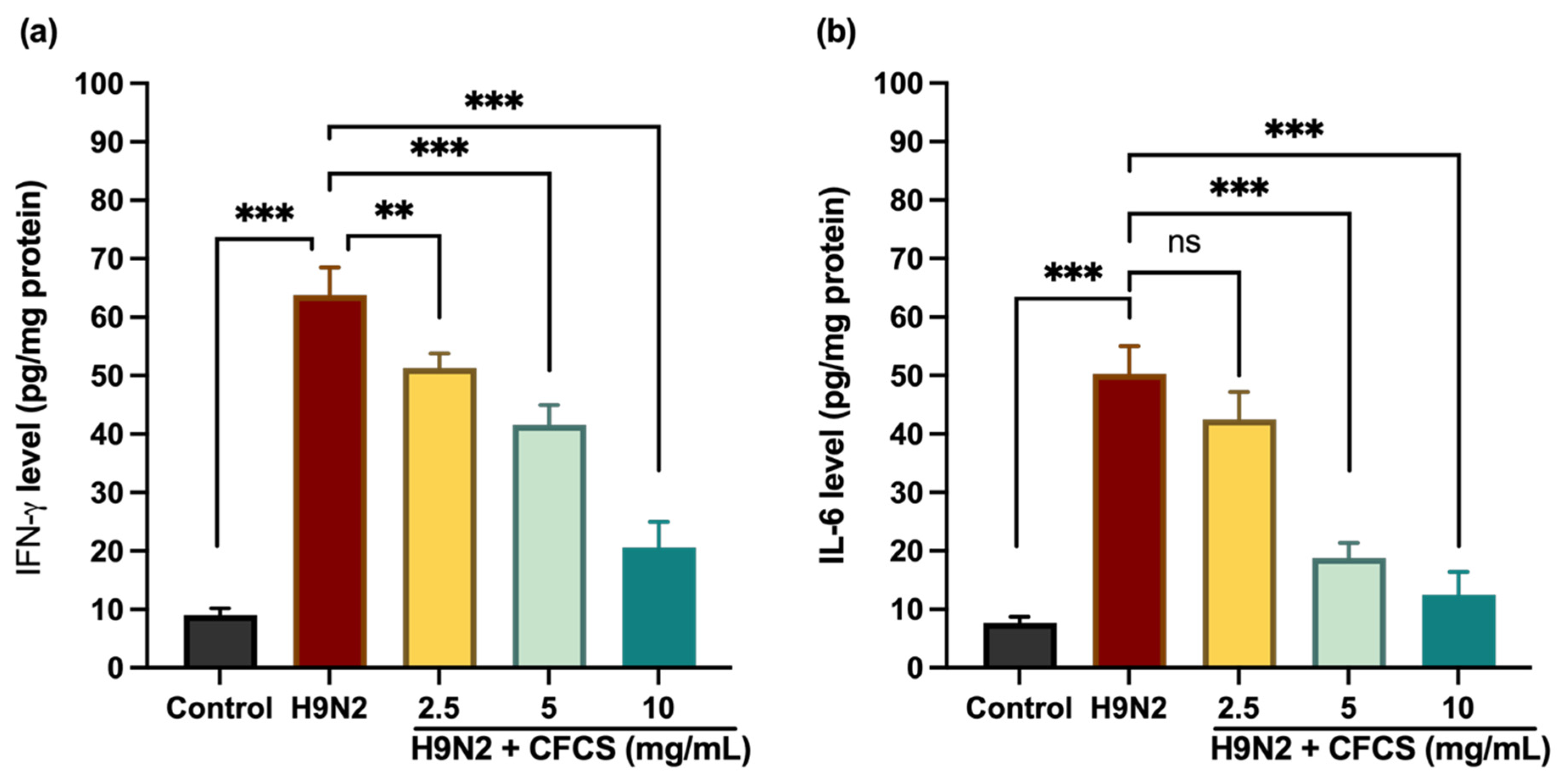
3.3. Effect of L. plantarum CFCS on Proinflammatory Cytokines

Virus infections trigger the expression of cytokines and chemokines as part of proinflammatory response. In this study, we examine the effect of L. plantarum CFCS on proinflammatory cytokines by monitoring the expression of two proinflammatory signature cytokines, IFN-γ and IL-6. Based on our findings, the expression levels of IFN-γ and IL-6 significantly decreased after CFCS treatment. In contrast, H9N2 infected cells showed dramatic increase in IFN-γ levels (62.80 ± 4.7 pg/mL) compared to uninfected or control cells (9 ± 1.2 pg/mL) (p < 0.001). Figure 2 shows that CFCS significantly reduced the level of IFN-γ in a dose-dependent manner at concentrations of 5 and 10 mg/mL, with p values of <0.001. However, there was no significant difference between H9N2-infected cells and CFCS (2.5 mg) treated cells in terms of IL-6 expression levels (p = 0.1). The expression levels of IL-6 were significantly higher in H9N2-infected cells than in non-infected cells. However, pretreatment of L. plantarum CFCS (5, and 10 mg/mL) significantly decreases the levels of IL-6 when compared to H9N2 infected cells (p < 0.001). Previous studies have shown that L. plantarum reduces proinflammatory cytokines such as TNF, IL-6, and IL-8 [40].

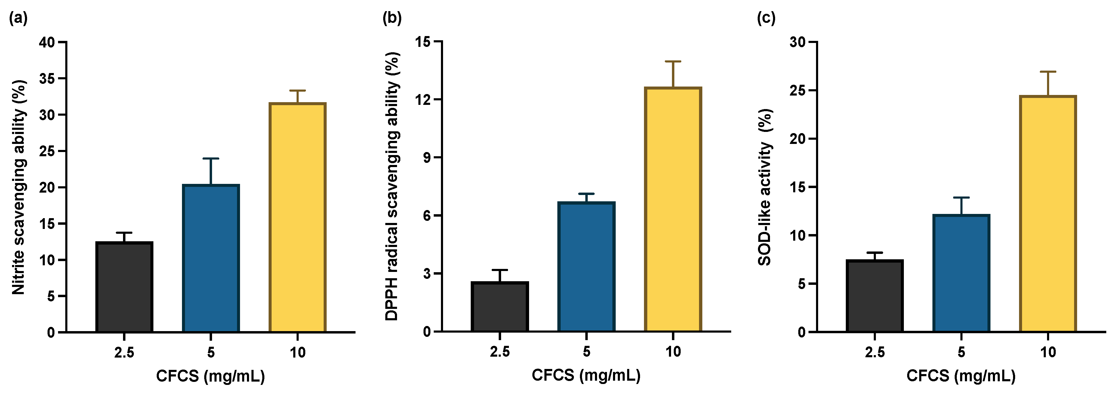
3.4. Effect of L. plantarum CFCS on Antioxidant System

Reactive nitrogen species (RNS) and reactive oxygen species (ROS) play a vital role in immunoreaction against microbial pathogens [41]. However, excess RNS/ROS production results in oxidative stress, which in turn causes DNA damage, lipid peroxidation, and protein oxidation [42]. L. plantarum bacteria strains induce antioxidant enzymatic activity, which protects cells from oxidative stress. In this study, we systematically investigate how L. plantarum KAU007 CFCS affects the activity of antioxidants, viz., nitrate radical scavenging activity, DPPH radical scavenging activity, and superoxide dismutase. According to our findings, CFCS (2.5, 5, and 10 mg/mL) significantly boosts the activity of all three antioxidants, but CFCS (10 mg/mL) exhibited the highest antioxidant activity as shown in Figure 3. The ability of L. plantarum KAU007 CFCS to scavenge free radicals was dosage-dependent, as shown in Figure 3. Previous investigations have also emphasized the significance of the L. plantarum strains in triggering antioxidant activity [43,44].
The use of probiotics in fermented foods and dairy products has a long evolutionary history of benefiting the host [34,36,45,46,47,48,49,50,51]. As dietary supplements, probiotics have become increasingly popular in recent decades. The effectiveness of probiotics has also been confirmed in thousands of scientific studies, demonstrating their effectiveness against various ailments, including bacterial, viral, and fungal infections [22,27,28,29,30,40,52,53,54,55,56,57]. Recent years have seen an increase in the popularity of LAB because of its antibacterial and probiotic properties. A growing body of research suggests that it may represent a promising alternative to synthetic drugs [58,59,60,61,62,63,64,65,66].
This study tested the effectiveness of camel milk isolate, L. plantarum KAU007 against the LPIV virus. In higher concentrations, KAU007 CFCS is more effective at nearly eradicating the virus. The isolated CFCS from KAU007 showed no cytotoxicity to MDCK cells [27]. Based on these results, the KAU007 could be considered a safe candidate that may not cause host cell damage. The in vitro antiviral activity assay conducted on L. plantarum KAU007 against the LPAI virus shows high antiviral activity. To combat LPAI, the most effective dose of CFCS was 10 mg/mL from KAU007.
We observed similar results in SPF embryonated eggs, with maximum antiviral activity at a concentration of 10 mg/mL of CFCS. Therefore, the study underpins that L. plantarum KAU007 is an anti-influenza prophylactic probiotic found in camel milk. Further, the results reinforce the study of the mechanism of action of KAU007 against LPAI, as well as the mechanisms of action against other influenza strains, in order to develop effective therapeutic candidates to prevent influenza virus-induced respiratory disease.

4. Conclusions

Using probiotic antagonist microbes and their metabolic products could be a promising therapeutic strategy against viral infections. In this study, we investigate the role of L. plantarum KAU007 against H9N2 using different approaches. Our results revealed that L. plantarum KAU007 has potent antiviral activity against H9N2 and could be a promising candidate for future antiviral therapy. In the future, multi-omics and other high throughput immunotechnological approaches will be needed to identify the metabolites in L. plantarum KAU007 responsible for its antiviral capabilities, which will open up new possibilities for the mechanisms underlying its antiviral capabilities in antiviral development.

Author Contributions

Conceptualization, I.A.R.; methodology, I.A.R., M.R.K. and J.S.M.S.; software, I.A.R.; validation, I.A.R.; formal analysis, I.A.R.; investigation, I.A.R. and S.A.; resources, I.A.R.; data curation, M.R.K.; writing—original draft preparation, I.A.R. and S.A.; writing—review and editing, I.A.R.; visualization, S.A., M.R.K. and J.S.M.S.; supervision, I.A.R.; project administration, I.A.R.; funding acquisition, I.A.R. All authors have read and agreed to the published version of the manuscript.

Funding

This Project was funded by the Deanship of Scientific Research (DSR) at King Abdulaziz University, Jeddah, under grant No. (G: 796-130-1441). The authors, therefore, acknowledge with thanks DSR for technical and financial support.

Institutional Review Board Statement

Not applicable.

Informed Consent Statement

Not applicable.

Data Availability Statement

The data generated are cited in the manuscript.

Conflicts of Interest

The authors declare no conflict of interest. The funders had no role in the design of the study; in the collection, analyses, or interpretation of data; in the writing of the manuscript; or in the decision to publish the results.

References

  1. Carlson, C.J.; Albery, G.F.; Merow, C.; Trisos, C.H.; Zipfel, C.M.; Eskew, E.A.; Olival, K.J.; Ross, N.; Bansal, S. Climate change increases cross-species viral transmission risk. Nature 2022, 607, 555–562. [Google Scholar] [CrossRef] [PubMed]
  2. Su, S.; Bi, Y.; Wong, G.; Gray, G.C.; Gao, G.F.; Li, S. Epidemiology, Evolution, and Recent Outbreaks of Avian Influenza Virus in China. J. Virol. 2015, 89, 8671–8676. [Google Scholar] [CrossRef] [PubMed] [Green Version]
  3. Arafat, N.; Eladl, A.H.; Marghani, B.H.; Saif, M.A.; El-Shafei, R. Enhanced infection of avian influenza virus H9N2 with infectious laryngeotracheitis vaccination in chickens. Vet. Microbiol. 2018, 219, 8–16. [Google Scholar] [CrossRef] [PubMed]
  4. Barberis, A.; Boudaoud, A.; Gorrill, A.; Loupias, J.; Ghram, A.; Lachheb, J.; Alloui, N.; Ducatez, M.F. Full-length genome sequences of the first H9N2 avian influenza viruses isolated in the Northeast of Algeria. Virol. J. 2020, 17, 108. [Google Scholar] [CrossRef] [PubMed]
  5. Fouchier, R.A.M.; Munster, V.; Wallensten, A.; Bestebroer, T.M.; Herfst, S.; Smith, D.; Rimmelzwaan, G.F.; Olsen, B.; Osterhaus, A.D.M.E. Characterization of a Novel Influenza A Virus Hemagglutinin Subtype (H16) Obtained from Black-Headed Gulls. J. Virol. 2005, 79, 2814–2822. [Google Scholar] [CrossRef] [Green Version]
  6. Bailey, E.S.; Choi, J.Y.; Fieldhouse, J.K.; Borkenhagen, L.K.; Zemke, J.; Zhang, D.; Gray, G.C. The continual threat of influenza virus infections at the human–animal interface. Evol. Med. Public Health 2018, 2018, 192–198. [Google Scholar] [CrossRef] [Green Version]
  7. Sun, Y.; Liu, J. H9N2 influenza virus in China: A cause of concern. Protein Cell 2015, 6, 18–25. [Google Scholar] [CrossRef] [Green Version]
  8. Wang, S.; Jiang, N.; Shi, W.; Yin, H.; Chi, X.; Xie, Y.; Hu, J.; Zhang, Y.; Li, H.; Chen, J.-L. Co-infection of H9N2 Influenza A Virus and Escherichia coli in a BALB/c Mouse Model Aggravates Lung Injury by Synergistic Effects. Front. Microbiol. 2021, 12, 670688. [Google Scholar] [CrossRef]
  9. Pan, Q.; Liu, A.; Zhang, F.; Ling, Y.; Ou, C.; Hou, N.; He, C. Co-infection of broilers with Ornithobacterium rhinotracheale and H9N2 avian influenza virus. BMC Veter. Res. 2012, 8, 104. [Google Scholar] [CrossRef] [Green Version]
  10. Carnaccini, S.; Perez, D.R. H9 Influenza Viruses: An Emerging Challenge. Cold Spring Harb. Perspect. Med. 2020, 10, a038588. [Google Scholar] [CrossRef] [Green Version]
  11. Peiris, M.; Yuen, K.; Leung, C.; Chan, K.; Ip, P.; Lai, R.; Orr, W.; Shortridge, K. Human infection with influenza H9N2. Lancet 1999, 354, 916–917. [Google Scholar] [CrossRef]
  12. Shanmuganatham, K.; Feeroz, M.M.; Jones-Engel, L.; Walker, D.; Alam, S.M.R.; Hasan, M.K.; McKenzie, P.; Krauss, S.; Webby, R.J.; Webster, R.G. Genesis of Avian Influenza H9N2 in Bangladesh. Emerg. Microbes Infect. 2014, 3, 1–17. [Google Scholar] [CrossRef] [PubMed]
  13. Lin, Y.P.; Shaw, M.; Gregory, V.; Cameron, K.; Lim, W.; Klimov, A.; Subbarao, K.; Guan, Y.; Krauss, S.; Shortridge, K.; et al. Avian-to-human transmission of H9N2 subtype influenza A viruses: Relationship between H9N2 and H5N1 human isolates. Proc. Natl. Acad. Sci. USA 2000, 97, 9654–9658. [Google Scholar] [CrossRef] [Green Version]
  14. World Health Organization Influenza at the Human-Animal Interface Summary and Assessment. Available online: https://shorturl.hk/bUFDbU (accessed on 21 January 2022).
  15. The SJCEIRS H9 Working Group; Schultz-Cherry, S.; Thomas, P. Assessing the fitness of distinct clades of influenza A (H9N2) viruses. Emerg. Microbes Infect. 2013, 2, e75. [Google Scholar] [CrossRef]
  16. Nagy, A.; Mettenleiter, T.C.; Abdelwhab, E.M. A brief summary of the epidemiology and genetic relatedness of avian influenza H9N2 virus in birds and mammals in the Middle East and North Africa. Epidemiol. Infect. 2017, 145, 3320–3333. [Google Scholar] [CrossRef] [Green Version]
  17. Gu, M.; Xu, L.; Wang, X.; Liu, X. Current situation of H9N2 subtype avian influenza in China. Vet. Res. 2017, 48, 49. [Google Scholar] [CrossRef] [Green Version]
  18. Sitaras, I.; Rousou, X.; Kalthoff, D.; Beer, M.; Peeters, B.; de Jong, M.C.M. Role of vaccination-induced immunity and antigenic distance in the transmission dynamics of highly pathogenic avian influenza H5N1. J. R. Soc. Interface 2016, 13, 20150976. [Google Scholar] [CrossRef] [PubMed] [Green Version]
  19. Pu, J.; Wang, S.; Yin, Y.; Zhang, G.; Carter, R.A.; Wang, J.; Xu, G.; Sun, H.; Wang, M.; Wen, C.; et al. Evolution of the H9N2 influenza genotype that facilitated the genesis of the novel H7N9 virus. Proc. Natl. Acad. Sci. USA 2015, 112, 548–553. [Google Scholar] [CrossRef] [Green Version]
  20. Poland, G.A. Influenza Vaccine Failure: Failure to Protect or Failure to Understand? Expert Rev. Vaccines 2018, 17, 495–502. [Google Scholar] [CrossRef]
  21. Tiwari, S.K.; Dicks, L.M.T.; Popov, I.V.; Karaseva, A.; Ermakov, A.M.; Suvorov, A.; Tagg, J.R.; Weeks, R.; Chikindas, M.L. Probiotics at War Against Viruses: What Is Missing from the Picture? Front. Microbiol. 2020, 11, 1877. [Google Scholar] [CrossRef]
  22. AL Kassaa, I.; Hober, D.; Hamze, M.; Chihib, N.E.; Drider, D. Antiviral Potential of Lactic Acid Bacteria and Their Bacteriocins. Probiotics Antimicrob. Proteins 2014, 6, 177–185. [Google Scholar] [CrossRef] [PubMed]
  23. Drider, D.; Bendali, F.; Naghmouchi, K.; Chikindas, M.L. Bacteriocins: Not Only Antibacterial Agents. Probiotics Antimicrob. Proteins 2016, 8, 177–182. [Google Scholar] [CrossRef] [PubMed]
  24. Chikindas, M.L.; Weeks, R.; Drider, D.; Chistyakov, V.A.; Dicks, L.M. Functions and emerging applications of bacteriocins. Curr. Opin. Biotechnol. 2018, 49, 23–28. [Google Scholar] [CrossRef] [PubMed]
  25. Kanmani, P.; Albarracin, L.; Kobayashi, H.; Hebert, E.M.; Saavedra, L.; Komatsu, R.; Gatica, B.; Miyazaki, A.; Ikeda-Ohtsubo, W.; Suda, Y.; et al. Genomic Characterization of Lactobacillus delbrueckii TUA4408L and Evaluation of the Antiviral Activities of its Extracellular Polysaccharides in Porcine Intestinal Epithelial Cells. Front. Immunol. 2018, 9, 2178. [Google Scholar] [CrossRef] [PubMed]
  26. del Rio, B.; Dattwyler, R.J.; Aroso, M.; Neves, V.; Meirelles, L.; Seegers, J.F.M.L.; Gomes-Solecki, M. Oral Immunization with Recombinant Lactobacillus plantarum Induces a Protective Immune Response in Mice with Lyme Disease. Clin. Vaccine Immunol. 2008, 15, 1429–1435. [Google Scholar] [CrossRef] [Green Version]
  27. Rather, I.A.; Kamli, M.R.; Sabir, J.S.M.; Paray, B.A. Potential Antiviral Activity of Lactiplantibacillus plantarum KAU007 against Influenza Virus H1N1. Vaccines 2022, 10, 456. [Google Scholar] [CrossRef] [PubMed]
  28. Majumder, R.; Rather, I.A.; Bajpai, V.K.; Park, Y.-H. In vitro antiviral activity of Lactobacillus plantarum using SPF embryonated eggs and hemagglutination assay. Bangladesh J. Pharmacol. 2015, 10, 688–691. [Google Scholar] [CrossRef] [Green Version]
  29. Rather, I.A.; Choi, K.-H.; Bajpai, V.K.; Park, Y.-H. Antiviral mode of action of Lactobacillus plantarum YML009 on Influenza virus H1N1. Bangladesh J. Pharmacol. 2015, 10, 475–482. [Google Scholar] [CrossRef] [Green Version]
  30. Seo, B.; Rather, I.; Kumar, V.; Choi, U.; Moon, M.; Lim, J.; Park, Y. Evaluation of Leuconostoc mesenteroides YML003 as a probiotic against low-pathogenic avian influenza (H9N2) virus in chickens. J. Appl. Microbiol. 2012, 113, 163–171. [Google Scholar] [CrossRef]
  31. Choi, D.; Cho, K.-A.; Na, M.-S.; Choi, H.-S.; Kim, Y.-O.; Lim, D.-H.; Cho, S.J.; Cho, H. Effect of bamboo oil on antioxidative activity and nitrite scavenging activity. J. Ind. Eng. Chem. 2008, 14, 765–770. [Google Scholar] [CrossRef]
  32. Bajpai, V.K.; Rather, I.A.; Park, Y.-H. Partially Purified Exo-Polysaccharide from Lactobacillus Sakei Probio 65 with Antioxidant, α-Glucosidase and Tyrosinase Inhibitory Potential. J. Food Biochem. 2016, 40, 264–274. [Google Scholar] [CrossRef]
  33. Bajpai, V.K.; Alam, M.B.; Quan, K.T.; Kwon, K.-R.; Ju, M.-K.; Choi, H.-J.; Lee, J.S.; Yoon, J.-I.; Majumder, R.; Rather, I.A.; et al. Antioxidant efficacy and the upregulation of Nrf2-mediated HO-1 expression by (+)-lariciresinol, a lignan isolated from Rubia philippinensis, through the activation of p38. Sci. Rep. 2017, 7, 46035. [Google Scholar] [CrossRef] [PubMed] [Green Version]
  34. Oh, N.; Kwon, H.; Lee, H.; Joung, J.; Lee, J.; Lee, K.; Shin, Y.; Baick, S.; Park, M.; Kim, Y.; et al. Preventive effect of fermented Maillard reaction products from milk proteins in cardiovascular health. J. Dairy Sci. 2014, 97, 3300–3313. [Google Scholar] [CrossRef] [PubMed] [Green Version]
  35. Rather, I.A.; Sabir, J.S.M.; Asseri, A.H.; Ali, S. Antifungal Activity of Human Cathelicidin LL-37, a Membrane Disrupting Peptide, by Triggering Oxidative Stress and Cell Cycle Arrest in Candida auris. J. Fungi 2022, 8, 204. [Google Scholar] [CrossRef]
  36. Al Malki, A.; Yoon, S.-H.; Firoz, A.; Ali, H.M.; Park, Y.-H.; Hor, Y.-Y.; Rather, I.A. Characterization of New Probiotic Isolates from Fermented Ajwa Dates of Madinah and Their Anti-Inflammatory Potential. Appl. Sci. 2022, 12, 5082. [Google Scholar] [CrossRef]
  37. Ooi, M.F.; Foo, H.L.; Loh, T.C.; Mohamad, R.; Rahim, R.A.; Ariff, A. A refined medium to enhance the antimicrobial activity of postbiotic produced by Lactiplantibacillus plantarum RS5. Sci. Rep. 2021, 11, 7617. [Google Scholar] [CrossRef]
  38. Motevaseli, E.; Shirzad, M.; Akrami, S.M.; Mousavi, A.-S.; Mirsalehian, A.; Modarressi, M.H. Normal and tumour cervical cells respond differently to vaginal lactobacilli, independent of pH and lactate. J. Med. Microbiol. 2013, 62, 1065–1072. [Google Scholar] [CrossRef]
  39. Chuah, L.-O.; Foo, H.L.; Loh, T.C.; Alitheen, N.B.M.; Yeap, S.K.; Mutalib, N.E.A.; Rahim, R.A.; Yusoff, K. Postbiotic metabolites produced by Lactobacillus plantarum strains exert selective cytotoxicity effects on cancer cells. BMC Complement. Altern. Med. 2019, 19, 114. [Google Scholar] [CrossRef] [Green Version]
  40. Sunmola, A.A.; Ogbole, O.O.; Faleye, T.O.C.; Adetoye, A.; Adeniji, J.A.; Ayeni, F.A. Antiviral potentials of Lactobacillus plantarum, Lactobacillus amylovorus, and Enterococcus hirae against selected Enterovirus. Folia Microbiol. 2019, 64, 257–264. [Google Scholar] [CrossRef]
  41. Kozlov, E.; Ivanova, E.; Grechko, A.; Wu, W.-K.; Starodubova, A.; Orekhov, A. Involvement of Oxidative Stress and the Innate Immune System in SARS-CoV-2 Infection. Diseases 2021, 9, 17. [Google Scholar] [CrossRef]
  42. Jeon, H.-J.; Choi, H.-S.; Lee, O.-H.; Jeon, Y.-J.; Lee, B.-Y. Inhibition of Reactive Oxygen Species (ROS) and Nitric Oxide (NO) by Gelidium elegans Using Alternative Drying and Extraction Conditions in 3T3-L1 and RAW 264.7 Cells. Prev. Nutr. Food Sci. 2012, 17, 122–128. [Google Scholar] [CrossRef] [PubMed] [Green Version]
  43. Chang, H.M.; Foo, H.L.; Loh, T.C.; Lim, E.T.C.; Mutalib, N.E.A. Comparative Studies of Inhibitory and Antioxidant Activities, and Organic Acids Compositions of Postbiotics Produced by Probiotic Lactiplantibacillus plantarum Strains Isolated from Malaysian Foods. Front. Vet. Sci. 2021, 7, 602280. [Google Scholar] [CrossRef] [PubMed]
  44. Tian, Y.; Yang, W.; Song, J.; Wu, Y.; Ni, B. Hepatitis B Virus X Protein-Induced Aberrant Epigenetic Modifications Contributing to Human Hepatocellular Carcinoma Pathogenesis. Mol. Cell. Biol. 2013, 33, 2810–2816. [Google Scholar] [CrossRef] [PubMed] [Green Version]
  45. Park, C.W.; Youn, M.; Jung, Y.-M.; Kim, H.; Jeong, Y.; Lee, H.-K.; Kim, H.O.; Lee, I.; Lee, S.W.; Kang, K.H.; et al. New Functional Probiotic Lactobacillus sakei Probio 65 Alleviates Atopic Symptoms in the Mouse. J. Med. Food 2008, 11, 405–412. [Google Scholar] [CrossRef] [PubMed]
  46. Parvez, S.; Malik, K.A.; Ah Kang, S.; Kim, H.-Y. Probiotics and their fermented food products are beneficial for health. J. Appl. Microbiol. 2006, 100, 1171–1185. [Google Scholar] [CrossRef]
  47. Drouault, S. Health Effects of Lactic Acid Bacteria Ingested in Fermented Milk. Vet. Res. 2001, 32, 101–117. [Google Scholar] [CrossRef] [Green Version]
  48. Apás, A.L.; Arena, M.E.; Elias, A.; González, F.; González, S.N. Beneficial Effects of Fermented Sugarcane Residue with Goat Probiotic on Gut Health. Int. J. Agric. Innov. Res. 2015, 3, 1–8. [Google Scholar]
  49. Adedokun, E.O.; Rather, I.A.; Bajpai, V.K.; Choi, K.-H.; Park, Y.-H. Isolation and Characterization of Lactic Acid Bacteria from Nigerian Fermented Foods and Their Antimicrobial Activity. J. Pure Appl. Microbiol. 2014, 8, 3411–3420. [Google Scholar]
  50. Nagao, F.; Nakayama, M.; Muto, T.; Okumura, K. Effects of a Fermented Milk Drink ContainingLactobacillus caseiStrain Shirota on the Immune System in Healthy Human Subjects. Biosci. Biotechnol. Biochem. 2000, 64, 2706–2708. [Google Scholar] [CrossRef]
  51. Makino, S.; Ikegami, S.; Kume, A.; Horiuchi, H.; Sasaki, H.; Orii, N. Reducing the risk of infection in the elderly by dietary intake of yoghurt fermented with Lactobacillus delbrueckii ssp. bulgaricus OLL1073R-1. Br. J. Nutr. 2010, 104, 998–1006. [Google Scholar] [CrossRef] [Green Version]
  52. Bajpai, V.K.; Rather, I.A.; Majumder, R.; Alshammari, F.H.; Nam, G.-J.; Park, Y.-H. Characterization and Antibacterial Mode of Action of Lactic Acid BacteriumLeuconostoc mesenteroidesHJ69 from Kimchi. J. Food Biochem. 2017, 41, e12290. [Google Scholar] [CrossRef]
  53. Bajpai, V.K.; Han, J.-H.; Rather, I.A.; Park, C.; Lim, J.; Paek, W.K.; Lee, J.S.; Yoon, J.-I.; Park, Y.-H. Characterization and Antibacterial Potential of Lactic Acid Bacterium Pediococcus pentosaceus 4I1 Isolated from Freshwater Fish Zacco koreanus. Front. Microbiol. 2016, 7, 2037. [Google Scholar] [CrossRef] [PubMed]
  54. Rather, I.A.; Seo, B.J.; Kumar, V.J.R.; Choi, U.-H.; Choi, K.-H.; Lim, J.; Park, Y.-H. Biopreservative potential of Lactobacillus plantarum YML007 and efficacy as a replacement for chemical preservatives in animal feed. Food Sci. Biotechnol. 2014, 23, 195–200. [Google Scholar] [CrossRef]
  55. Ahmad Rather, I.; Seo, B.J.; Rejish Kumar, V.J.; Choi, U.-H.; Choi, K.-H.; Lim, J.H.; Park, Y.-H. Isolation and characterization of a proteinaceous antifungal compound from Lactobacillus plantarum YML007 and its application as a food preservative. Lett. Appl. Microbiol. 2013, 57, 69–76. [Google Scholar] [CrossRef] [PubMed]
  56. Anwar, F.; Altayb, H.N.; Al-Abbasi, F.A.; Al-Malki, A.L.; Kamal, M.A.; Kumar, V. Antiviral effects of probiotic metabolites on COVID-19. J. Biomol. Struct. Dyn. 2021, 39, 4175–4184. [Google Scholar] [CrossRef]
  57. Youn, H.-N.; Lee, D.-H.; Lee, Y.-N.; Park, J.-K.; Yuk, S.-S.; Yang, S.-Y.; Lee, H.-J.; Woo, S.-H.; Kim, H.-M.; Lee, J.-B.; et al. Intranasal administration of live Lactobacillus species facilitates protection against influenza virus infection in mice. Antivir. Res. 2012, 93, 138–143. [Google Scholar] [CrossRef]
  58. Tomasik, P.; Tomasik, P. Probiotics, Non-Dairy Prebiotics and Postbiotics in Nutrition. Appl. Sci. 2020, 10, 1470. [Google Scholar] [CrossRef] [Green Version]
  59. Sharifi-Rad, J.; Rodrigues, C.; Stojanović-Radić, Z.; Dimitrijević, M.; Aleksić, A.; Neffe-Skocińska, K.; Zielińska, D.; Kołożyn-Krajewska, D.; Salehi, B.; Prabu, S.M.; et al. Probiotics: Versatile Bioactive Components in Promoting Human Health. Medicina 2020, 56, 433. [Google Scholar] [CrossRef]
  60. Rather, I.A.; Majumder, R.; Alshammari, F.H.; Park, J.G.; Bajpai, V.K. Review-Ulcerative colitis and probiotics: An overview. Pak. J. Pharm. Sci. 2016, 29, 1877–1880. [Google Scholar]
  61. Santosa, S.; Farnworth, E.; Jones, P.J.H. Probiotics and Their Potential Health Claims. Nutr. Rev. 2006, 64, 265–274. [Google Scholar] [CrossRef]
  62. Rather, I.A.; Bajpai, V.K.; Kumar, S.; Lim, J.; Paek, W.K.; Park, Y.-H. Probiotics and Atopic Dermatitis: An Overview. Front. Microbiol. 2016, 7, 507. [Google Scholar] [CrossRef] [PubMed] [Green Version]
  63. Reid, G.; Jass, J.; Sebulsky, M.T.; McCormick, J.K. Potential Uses of Probiotics in Clinical Practice. Clin. Microbiol. Rev. 2003, 16, 658–672. [Google Scholar] [CrossRef] [PubMed] [Green Version]
  64. Suez, J.; Zmora, N.; Segal, E.; Elinav, E. The pros, cons, and many unknowns of probiotics. Nat. Med. 2019, 25, 716–729. [Google Scholar] [CrossRef] [PubMed]
  65. Mangrolia, U.; Osborne, J.W. Probiotics in Counteracting the Role of Neutrophils in Cancer Metastasis. Vaccines 2021, 9, 1306. [Google Scholar] [CrossRef] [PubMed]
  66. Bajpai, V.K.; Chandra, V.; Kim, N.-H.; Rai, R.; Kumar, P.; Kim, K.; Aeron, A.; Kang, S.C.; Maheshwari, D.K.; Na, M.; et al. Ghost probiotics with a combined regimen: A novel therapeutic approach against the Zika virus, an emerging world threat. Crit. Rev. Biotechnol. 2018, 38, 438–454. [Google Scholar] [CrossRef]
Figure 1. Cytopathic effect of the H9N2 virus. MTT assays were used to measure cell viability. Each experiment was performed three times, and the results are presented as a mean + standard error. To determine statistical significance, treatment groups were compared with control (PBS only) and H9N2 (no treatment). p-values: *** < 0.001, ** < 0.01. ns = non-significant.
Pathogens 11 01246 g001
Figure 2. H9N2 virus-challenged mammalian kidney cells treated with CFCS show a reduction in interleukin levels. (a) IFN-γ and (b) IL-6. Results were presented as means + SE for the three experiments conducted in triplicate. p-values: ** < 0.01, *** < 0.001, ns = non-significant.
Pathogens 11 01246 g002
Figure 3. Antioxidant activity of CFCS isolated from L. plantarum KAU007 (a) nitrate radical scavenging activity of CFCS. (b) DPPH radical scavenging activity of CFCS. (c) Superoxide dismutase (SOD) such as activity of CFCS. Data are expressed as mean ± SD (n = 3).
Pathogens 11 01246 g003
Table 1. An evaluation of the effects of CFCS on the survival rate of embryonated eggs infected with H9N2.
GroupsTreatmentSurvival/TotalSurvival Percentage
ConPBS 2/2100%
H9N2107.5 EID50/0.1 mL5/955%
H9N2 + KAU007—ICFCS (2.5 mg/mL)3/475%
H9N2 + KAU007—IICFCS (5 mg/mL)5/683%
H9N2 + KAU007—IIICFCS (10 mg/mL)8/988.88%
Viral titer: 106.5 EID50/0.1 mL; Con: PBS only.
Table 2. Inhibition of hemagglutination by CFCS of Lactiplantibacillus plantarum KAU007.
Treatment
Hemagglutinating activityConKAU007 CFCS (2.5 mg/mL)KAU007 CFCS (5 mg/mL)KAU007 CFCS (10 mg/mL)
01:41:81:8
Viral titer: 106.5 EID50/Suppp0.1 mL; Con: PBS only.
Publisher’s Note: MDPI stays neutral with regard to jurisdictional claims in published maps and institutional affiliations.

