Article

Diversity of Rickettsiales in Rhipicephalus microplus Ticks Collected in Domestic Ruminants in Guizhou Province, China

1
National Institute for Communicable Disease Control and Prevention, Chinese Center for Disease Control and Prevention, Changping District, Beijing 102206, China
2
College of Life Sciences, Shandong First Medical University & Shandong Academy of Medical Sciences, Taian 271016, China
3
Tongzhou Center for Disease Control and Prevention, Tongzhou District, Beijing 101100, China
4
Guizhou Center for Disease Control and Prevention, Guiyang 550004, China
5
Liuzhi Center for Disease Control and Prevention, Liupanshui 553400, China
6
Tianjin Key Laboratory of Food and Biotechnology, Tianjin University of Commerce, Beichen District, Tianjin 300134, China
*
Author to whom correspondence should be addressed.
These authors contributed equally to this work.
Pathogens 2022, 11(10), 1108; https://doi.org/10.3390/pathogens11101108
Submission received: 1 August 2022 / Revised: 19 September 2022 / Accepted: 23 September 2022 / Published: 27 September 2022
(This article belongs to the Section Ticks)

Abstract

:
Rhipicephalus microplus ticks are vectors for multiple pathogens infecting animals and humans. Although the medical importance of R. microplus has been well-recognized and studied in most areas of China, the occurrence of tick-borne Rickettsiales has seldom been investigated in Guizhou Province, Southwest China. In this study, we collected 276 R. microplus ticks from cattle (209 ticks) and goats (67 ticks) in three locations of Guizhou Province. The Rickettsia, Anaplasma, and Ehrlichia were detected by targeting the 16S rRNA gene and were further characterized by amplifying the key genes. One Rickettsia (Ca. Rickettsia jingxinensis), three Ehrlichia (E. canis, E. minasensis, Ehrlichia sp.), and four Anaplasma (A. capra, A. ovis, A. marginale, Ca. Anaplasma boleense) species were detected, and their gltA and groEL genes were recovered. Candidatus Rickettsia jingxinensis, a spotted fever group of Rickettsia, was detected in a high proportion of the tested ticks (88.89%, 100%, and 100% in ticks from the three locations, respectively), suggesting the possibility that animals may be exposed to this type of Rickettsia. All the 16S, gltA, groEL, and ompA sequences of these strains are 100% identical to strains reported in Ngawa, Sichuan Province. E. minasensis, A. marginale, and Candidatus Anaplasma boleense are known to infect livestock such as cattle. The potential effects on local husbandry should be considered. Notably, E. canis, A. ovis, and A. capra have been reported to infect humans. The relatively high positive rates in Qianxinan (20.99%, 9.88%, and 4.94%, respectively) may indicate the potential risk to local populations. Furthermore, the genetic analysis indicated that the E. minasensis strains in this study may represent a variant or recombinant. Our results indicated the extensive diversity of Rickettsiales in R. microplus ticks from Guizhou Province. The possible occurrence of rickettsiosis, ehrlichiosis, and anaplasmosis in humans and domestic animals in this area should be further considered and investigated.

1. Introduction

Ticks (Acari: Ixodida) are a group of hematophagous ectoparasites that feed on terrestrial vertebrates. They are globally distributed and divided into three major families, namely the Ixodidae, Argasidae, and Nuttalliellidae. Ticks are believed to be second only to mosquitoes as vectors for human pathogens and are the primary vectors for pathogens of wild and domestic animals [1]. They play an important role in the ecology of numerous human and animal pathogens [2,3,4,5]. Annually, tick-borne pathogens are responsible for over 100,000 cases of illness in humans worldwide [5]. Tick-borne zoonotic pathogens infecting humans include tick-borne encephalitis virus (TBEV), severe fever with thrombocytopenia syndrome virus (SFTSV), Crimean–Congo hemorrhagic fever virus (CCHFV), Rickettsia rickettsia (the agent of Rocky Mountain spotted fever), R. sibirica (the agent of Siberian tick typhus), Anaplasma phagocytophilum (the agent of human granulocytic anaplasmosis), A. capra, Borrelia burgdorferi sensu lato (the agent of Lyme disease), B. persica (the agent of relapsing fever), and Babesia microti (the agent of human babesiosis) [2,3,4].
Rhipicephalus microplus is distributed in Asia, Latin America, the Middle East, and East and South Africa [6]. It is considered to be the most important tick infesting various domestic and wild animals, and results in huge economic losses throughout tropical and subtropical regions. The preferred hosts of R. microplus include cattle, deer, sheep, goats, horses, and buffalo [7]. Occasionally, it also bites humans [7]. As an important vector of pathogens, R. microplus harbors a large variety of animal (including human) pathogens such as spotted fever group Rickettsia (SFGR, agents of human spotted fever), Anaplasma phagocytophilum, A. capra, A. marginale (the agent of bovine anaplasmosis), Ehrlichia chaffeensis, E. canis (the agent of canine ehrlichiosis), E. minasensis (the agent of bovine ehrlichiosis), Babesia bigemina, B. bovis (the agent of bovine babesiosis), Borrelia spp. (agents of Lyme disease and relapsing fever), and severe fever with thrombocytopenia syndrome virus (SFTSV) [8,9,10,11], thus resulting in infections in both humans and domestic animals.
As one of the most prevalent tick species in China, R. microplus has been recorded in at least 188 counties. Furthermore, it was predicted to be distributed in 678 counties of China and to potentially affect 350 million people [11]. Extensive studies have been performed on R. microplus-vectored pathogens in China. From 1950 to 2018, multiple human pathogens including viruses (SFTSV, Jingmen tick virus, Tacheng tick virus, and Bocavirus), bacteria (SFGR, E. chaffeensis, A. phagocytophilum, and A. capra), and protozoans (B. bigemina and B. microti) have been detected in R. microplus from China [11]. Although R. microplus only occasionally infests humans, it may play an important role in the natural circulation of these pathogens, thus increasing the exposure to humans and domestic animals.
Rickettsia, Anaplasma, and Ehrlichia, belonging to the order Rickettsiales, are important vector-borne pathogens that are mainly vectored by ticks. They infect a wide variety of mammals, including humans, and are of great veterinary and medical importance. Although there have been many studies on the molecular detection of these tick-borne pathogens in China, reports on these pathogens are remarkably scarce in Guizhou Province [12,13,14], a subtropical mountainous province with an area of 176,167 km2 in southwest China. To investigate the geographic distribution and species composition of Rickettsia, Anaplasma, and Ehrlichia pathogens in R. microplus ticks from this area, we collected ticks from three locations in Guizhou Province and detected these pathogens in them.

2. Results

2.1. Tick Samples

All 276 ticks were morphologically identified to be R. microplus by observing the capitula, legs, anal groove, and caudal appendage. All 276 ticks were fully or partially engorged. All the obtained COI sequences had >99% identity to R. microplus, confirming the morphological identification of these ticks. Phylogenetic analysis of the COI genes showed that the ticks formed various clades in the phylogenetic tree, indicating the polymorphism of this species in Guizhou Province (Figure 1).

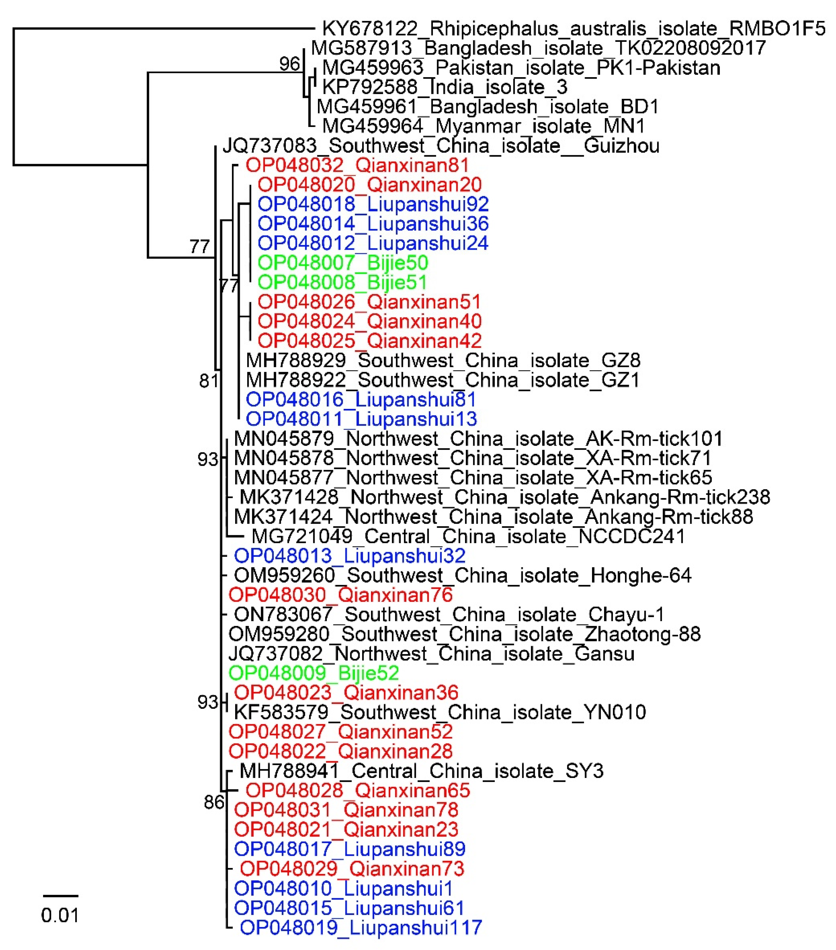
2.2. Detection and Analysis of Rickettsia

The PCR results showed that R. microplus ticks from all three locations had high positive rates for Rickettsia. Ticks from Qianxinan, Liupanshui, and Bijie had positive rates as high as 88.89% (72/81), 100% (126/126), and 100% (69/69), respectively (Table 1). All the Rickettsia 16S, gltA, and ompA sequences were 100% identical to strains of Candidatus Rickettsia jingxinensis, a spotted fever group Rickettsia. For the groEL gene, they were 100% identical to the uncultured Rickettsia sp. clone tick 28 we previously identified in Ngawa in Sichuan Province, which also represents a Candidatus Rickettsia jingxinensis strain [15] (Figure 2). In the phylogenetic tree based on concatenated sequences (Supplementary Material Figure S1), all the strains were closely related to Candidatus Rickettsia jingxinensis strain. These results clearly indicated that Candidatus Rickettsia jingxinensis is widespread and highly prevalent in Guizhou Province.

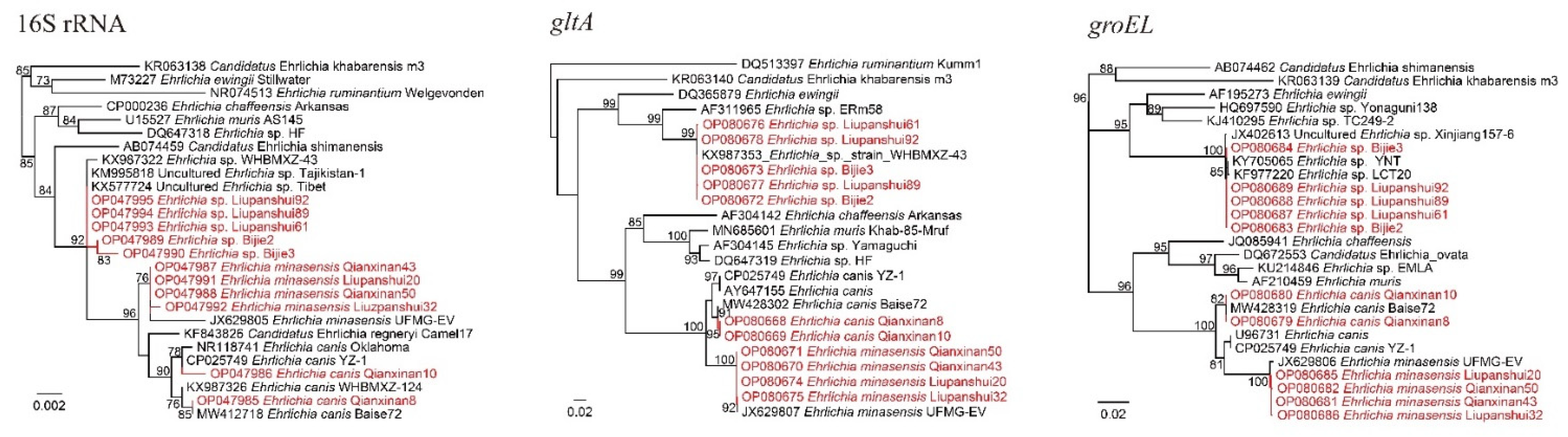
2.3. Detection and Analysis of Ehrlichia

Three Ehrlichia species were detected and characterized, namely E. canis, E. minasensis, and Ehrlichia sp. Ehrlichia canis was detected only in Qianxinan, with a positive rate of 20.99% (17/81) (Table 1). The 16S rRNA sequences of the randomly selected strains (E. canis Qianxinan8 and E. canis Qianxinan10) had three different nucleotides and they were divided into different clades in the phylogenetic tree. However, their gltA and groEL genes were identical to each other, and they both showed the highest identity to E. canis strains we previously identified in Guangxi Province of Southwest China, which is adjacent to Guizhou Province [10]. Ehrlichia minasensis was detected in both Qianxinan and Liupanshui, with positive rates of 2.47% (2/81) and 5.56% (7/126) (Table 1). Although their 16S rRNA gene sequences had the highest (99.85–100%) similarities to the uncultured Ehrlichia sp. clone Honghe-42 and only 99.42–99.59% similarity to E. minasensis strains, the gltA and groEL sequences were both closely related to E. minasensis strain UFMG-EV (99.89–100% for gltA, 99.64–99.73% for groEL). In the phylogenetic tree based on concatenated sequences of these genes, all these strains were in the same clade as E. minasensis strain UFMG-EV (Supplementary Material Figure S1). These data support the proposal that these strains may represent an E. minasensis variant or recombinant.
Except for E. canis and E. minasensis, an uncultured Ehrlichia species was detected in Bijie and Liupanshui, with positive rates of 10.14% (7/69) and 3.97% (5/126), respectively (Table 1). Genetic analysis indicated that the 16S gene sequences of the strains from Bijie have 99.76–99.92% identity to an uncultured Ehrlichia sp. clone from Tibet, the uncultured Ehrlichia sp. clone Dehong-18, and Ehrlichia sp. strain WHBMXZ-41, while all the strains from Liupanshui were 100% identical to these strains. Meanwhile, the gltA and groEL sequences were both closely related to Ehrlichia sp. strains detected in R. microplus from Wuhan city, Hubei Province (Ehrlichia sp. strain WHBMXZ-43, strain WHBMXZ-40, strain WHBMXZ-41, etc.), with similarities of 99.79–100%. In the phylogenetic trees, all the sequences from these strains were closely clustered (Figure 3).

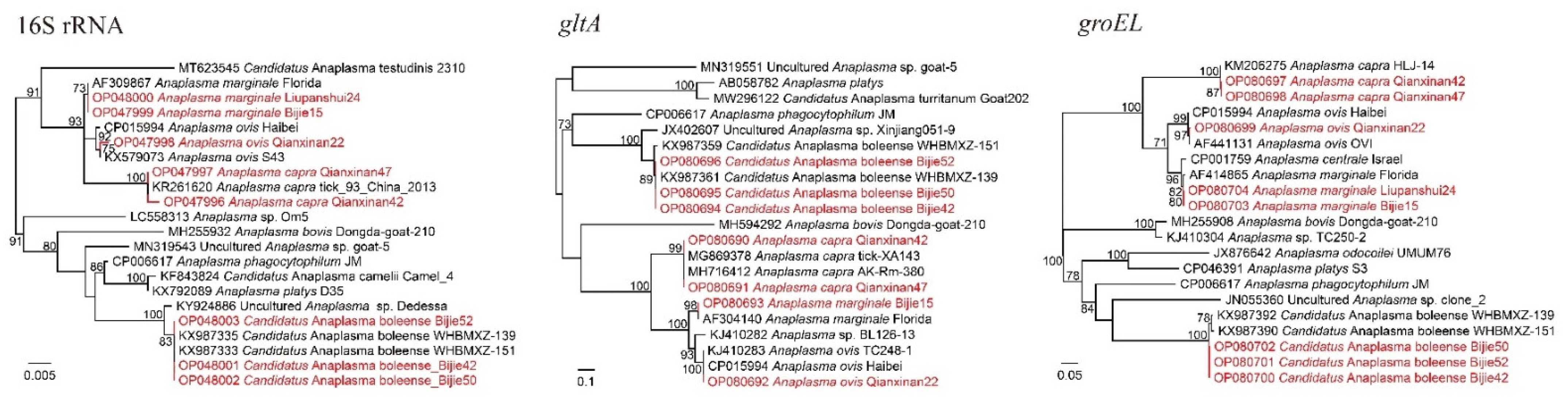
2.4. Detection and Analysis of Anaplasma

Four Anaplasma species were identified in these samples: A. marginale, A. ovis, A. capra, and Candidatus Anaplasma boleense. Anaplasma marginale was detected in both Bijie (43.48%, 30/69) and Liupanshui (1.59%, 2/126) (Table 1). The representative strains Bijie15 and Liupanshui24 were both closely related to A. marginale strains, with the similarities of key genes varying from 99.78% to 100%. Alongside for A. marginale, Candidatus Anaplasma boleense was identified in ticks from Bijie (8.70%, 6/69). The BLASTn results showed that these strains were mostly related to strains reported in Wuhan City, Hubei Province. However, some variations were observed. Although the 16S genes had as high as 99.91–100% homology to Candidatus Anaplasma boleense strain WHBMXZ-151, strain WHANSA-29, and strain WHANSP-48, the recovered gltA sequences were 99.86% identical to strain WHBMXZ-139 but had similarities of lower than 97.75% to any other Candidatus Anaplasma boleense strains. As for the groEL genes, all the strains were only 99.10% identical to strain WHBMXZ-139, strain WHBMXZ-151, and strain WHBMXZ-45. In the phylogenetic tree based on groEL, the strains in this study formed a distinct cluster, indicating that this species has evolved for a long time in this area.
In ticks from Qianxinan, A. ovis (9.88%, 8/81) and A. capra (4.94%, 4/81) were detected (Table 1), both of which are human pathogens. The 16S, gltA, and groEL sequences were all closely related to the A. capra strains reported elsewhere in China, with similarities of 99.69–99.92%, 99.88–100%, and 99.70% to other strains, respectively. All these strains clustered with other A. capra strains and formed a clade in the phylogenetic trees (Figure 4).
All the obtained sequences have been submitted to the GenBank Database (the accession numbers are shown in Supplementary Table S1).

3. Discussion

In China, R. microplus has been recorded in 188 counties [11]. In a previous study, we identified various Rickettsia, Anaplasma, and Ehrlichia species in R. microplus ticks from Hainan, Yunnan, Guangxi, Sichuan, and Hubei Provinces in China [10,16]. In this study, one Rickettsia, four Anaplasma, and three Ehrlichia species in total were detected and characterized in Guizhou Province.
All ticks from the three locations had high positive rates of Candidatus Rickettsia jingxinensis. Candidatus Rickettsia jingxinensis is a spotted fever group Rickettsia widely distributed in China and other countries. After its first report in Jilin Province, Northeast China [17], this Rickettsia species has been reported in Shaanxi, Sichuan, Guangxi, and Yunnan Provinces in China [10,12,18], as well as some neighboring countries, including Korea, Thailand, and India (GenBank Accession No. MN463681.1–MN463706.1) [19,20]. In previous studies, remarkably high positive rates of this Rickettsia (as high as 69.7% in H. longicornis from Shaanxi Province) in some areas have been observed [18]. In this study, the high positive rates in all three locations suggest the possibility that this Rickettsia may be a symbiont of this ixodid. Furthermore, it has been indicated that different endosymbiotic Rickettsia species may be unable to co-infect the same organ of the same tick, which is called “interference” [21]. In this study, only high positive rates of Candidatus Rickettsia jingxinensis were observed, while no other Rickettsia species were detected. It would be interesting to know whether this has resulted from the interference effect of Candidatus Rickettsia jingxinensis. Although this Rickettsia has only been detected in ticks up until now and there is no solid evidence proving its ability to infect animals or humans, it is phylogenetically close to some Rickettsia species pathogenic to humans such as R. japonica and R. heilongjiangensis. Furthermore, a gltA sequence (KU853023) obtained from a patient in China with 99.91% nucleotide identity to Ca. R. jingxinensis has been submitted to the GenBank database, suggesting its possible human pathogenicity. For the reasons above, more attention should be paid to its pathogenicity and further studies are needed.
Ehrlichia canis is a tick-borne pathogen mainly vectored and transmitted by Rhipicephalus sanguineus. It has also been reported in some other tick species such as R. microplus, R. linnaei, R. bursa, etc. [10,22,23]. As the causative agent of canine monocytic ehrlichiosis (CME), E. canis is one of the most prevalent tick-borne pathogens infecting dogs worldwide. Furthermore, it also infects other animals such as goats, sheep, deer, and red foxes [24,25,26]. As early as 1989, E. canis was reported to infect humans and cause a series of symptoms in the United States [27]. In recent years, more cases of infection have been reported in other countries such as Costa Rica [28]. In this study, E. canis was observed in ticks collected from goats in Qianxinan, suggesting that a tick–goat–tick cycle may exist in this area. Genetic and phylogenetic analysis indicated that these E. canis strains were closely related to those previously reported in Baise City in Guangxi Province [10], and they represented a variant that is different from most E. canis strains.
Ehrlichia minasensis, an Ehrlichia species closely related to E. canis, has been reported in China, Canada, Brazil, Malaysia, Ethiopia, and South Africa, suggesting its worldwide distribution [29]. It has long been considered the etiologic agent of tick-borne bovine ehrlichiosis [29]. This is the first report that E. minasensis exists in Guizhou Province. Notably, the 16S sequences of all the E. minasensis strains in this study were more genetically related to those of other Ehrlichia species, indicating that some genetic recombination may have occurred.
In total, four Anaplasma species were detected in these samples, namely A. capra, A. ovis, A. marginale, and Candidatus Anaplasma boleense. In previous studies, R. microplus has been recognized as the vector of A. marginale, the agent of bovine anaplasmosis [30]. In this study, A. marginale had a high positive rate (30/69, 43.48%) in ticks collected from Bijie City, indicating that A. marginale is circulating in this area. Its potential effects on local husbandry should be considered. Of note, two zoonotic pathogens, A. capra and A. ovis, were detected, which occasionally infect humans and cause a series of symptoms [31,32]. In previous studies, the prevalence of A. capra and A. ovis was reported in other locations in Guizhou Province. Our results confirmed the existence of these pathogens and further proved their wide distribution in Guizhou Province, despite the relatively low positive rates and the low anthropophily of R. microplus.
In summary, this study contributed to our knowledge on the species’ abundance and the genetic diversity of Rickettsiales bacteria in Guizhou Province. Notably, some of these bacteria have been reported to infect humans. Considering the spatial proximity and close contact between humans and the ticks’ hosts, these results may indicate the potential risk of human exposure.

4. Methods

4.1. Sample Collection and DNA Extraction

From August to October 2021, 276 ticks were collected from 50 domestic animals (26 cattle and 24 goats) in Bijie City, Liupanshui City, and Qianxinan Bouyei-and-Miao Autonomous Prefecture (Supplementary Material Figure S2). In August 2021, 14 ticks from 3 cattle (Bos taurus) and 67 ticks from 24 goats (Capra hircus) were collected in Puan County of Qianxinan Bouyei-and-Miao Autonomous Prefecture (25.78° N 104.95° E). In September, 126 ticks were collected from 17 cattle in Liuzhi Special District of Liupanshui City (26.20° N 105.48° E,). In October 2021, 69 ticks were collected from six cattle in Qianxi County of Bijie City (27.01° N, 106.03° W). The ticks were carefully removed from the body surfaces of the animals using tweezers. All the ticks were morphologically identified and then individually screened for tick-borne pathogen species [33]. In addition, in order to confirm the identification of the tick species, a randomly chosen subsample was confirmed by amplifying and sequencing the COI gene [10]. In total, 26 ticks (26/276, 9.42%) were randomly selected from the three locations (Bijie: 3 ticks; Liupanshui: 10 ticks; Qianxinan: 13 ticks) for molecular confirmation. Before DNA extraction, each tick was washed three times using phosphate buffered saline (PBS) to remove environmental contamination and then thoroughly ground in a mortar with PBS (100 uL). The homogenates were manually placed in 1.5 mL Eppendorf tubes, and the DNA was extracted using Omega Mollusc DNA extraction kits (Omega Bio-Tek, Norcross, GA, USA). The DNA was eluted in 60 μL of an elution buffer and then kept in a freezer at −80 °C before molecular detection.

4.2. Molecular Detection of Rickettsiale

All the DNA samples were screened for the existence of Rickettsiales by amplifying conserved regions of the 16S rRNA gene by nested or semi-nested PCR. Rickettsia was detected using the protocol previously shown in [10], generating approximately 900 bp of PCR product. The Anaplasmataceae bacteria including Ehrlichia, and Anaplasma were detected using the primers as shown [15], generating approximately 400 bp of PCR product. Here, ddH2O was set as the negative control for each PCR. All the PCR products were electrophoresed in 1.0% agarose gels, and the PCR products that met the expected length were subjected to Sanger sequencing. The 16S sequences thus obtained were aligned with reference sequences in the GenBank database using BLASTn to initially determine the bacterial species or genus.

4.3. Amplification and Analysis of Key Genes

To exactly determine the bacterial species and further characterize the detected strains, representative strains were selected from the positive samples, and longer 16S fragments (1184 bp for Rickettsia, 1119–1250 bp for Ehrlichia, and 1099–1299 bp for Anaplasma) were amplified from these using primers [10,18]. The citrate synthase gene (gltA) and 60 kDa chaperonin (groEL) genes were also amplified [10,18,34] for all the representative Rickettsia, Ehrlichia, and Anaplasma strains. For the Rickettsia strains, the outer membrane protein A (ompA) sequences were additionally obtained using primers as shown in [18]. All the primers are shown in Supplementary Material Table S2.

4.4. Sequence and Phylogenetic Analysis

All the obtained sequences were assembled and edited using BioEdit software (North Carolina State University) and then aligned with sequences in the GenBank database using BLASTn to determine the nucleotides’ similarity. For phylogenetic analysis, all the recovered sequences were aligned with the reference sequences using the ClustalW method with the MEGA program, version 5.2 [35]. Representative strains were selected from most, if not all, validated Rickettsia, Ehrlichia, and Anaplasma species as reference sequences. Due to the limited quantity of validated Ehrlichia and Anaplasma species, some unvalidated species were also included. A substitution model test was performed to determine the phylogenetic model with the best fit. Maximum likelihood (ML) trees based on the aligned sequences were constructed by the GTR+I+G model using PhyML v3.2 [36]. All the trees were rooted at the mid-point.

Supplementary Materials

The following supporting information can be downloaded at: https://www.mdpi.com/article/10.3390/pathogens11101108/s1. Table S1. GenBank numbers of the Rickettsia, Anaplasma, and Ehrlichia sequences obtained in this study. Table S2. The primers used for amplification of the 16S, gltA, groEL, and ompA genes from Rickettsia, Anaplasma, and Ehrlichia by nested PCR or hemi-nested PCR. Figure S1. Phylogenetic trees constructed by PhyML 3.0 software (GTR model) based on concatenated sequences of the Rickettsia, Ehrlichia, and Anaplasma strains. Figure S2. Map showing the locations where the samples were collected: Puan County of Qianxinan Bouyei-and-Miao Autonomous Prefecture; Liuzhi Special District of Liupanshui City; and Qianxi County of Bijie City.

Author Contributions

Conceptualization, M.L. (Miao Lu). and K.L.; Formal analysis, W.W. and K.L.; Funding acquisition, X.G.; Investigation, M.L. (Miao Lu), C.M., Y.S., Y.L., M.L. (Mengyao Li) and G.Z.; Methodology, M.L. (Miao Lu), C.M. and Y.S.; Project administration, K.L.; Resources, X.G., J.Z. and G.T.; Supervision, C.M.; Writing—original draft, K.L.; Writing—review & editing, X.G. and K.L. All authors have read and agreed to the published version of the manuscript.

Funding

This work was funded by the Key Supporting Scientific Research Projects of Beijing Road Medical Sector, General Hospital of Xinjiang Military Region (2022jzbjl16); the National Natural Science Foundation of China (Grant No. 82102390); and the State Key Laboratory for Infectious Disease Prevention and Control (Grant No. 2021SKLID507, 2021SKLID510), Science & Technology Development Foundation of the University of Tianjin Municipal City (Grant No: 20140614).

Institutional Review Board Statement

Not applicable.

Informed Consent Statement

Not applicable.

Data Availability Statement

All the sequence files are available from the NCBI database (the accession numbers are shown in Table S1).

Acknowledgments

We sincerely thank Xiaojing Jin and Qi Li for their generous help.

Conflicts of Interest

The authors declare no conflict of interest.

References

  1. Mansfield, K.L.; Jizhou, L.; Phipps, L.P.; Johnson, N. Emerging ttick-borne viruses in the twenty-first century. Front. Cell. Infect. Microbiol. 2017, 7, 298. [Google Scholar] [CrossRef] [PubMed]
  2. Madison-Antenucci, S.; Kramer, L.D.; Gebhardt, L.L.; Kauffman, E. Emerging tick-borne diseases. Clin. Microbiol. Rev. 2020, 33, e00083-18. [Google Scholar] [CrossRef] [PubMed]
  3. Mediannikov, O.; Fenollar, F. Looking in ticks for human bacterial pathogens. Microb. Pathog. 2014, 77, 142–148. [Google Scholar] [CrossRef] [PubMed]
  4. Rafinejad, J.; Choubdar, N.; Oshaghi, M.; Piazak, N.; Satvat, T.; Mohtarami, F.; Barmaki, A. Detection of Borrelia persica Infection in Ornithodoros tholozani using PCR targeting rrs gene and xenodiagnosis. Iran. J. Public Health 2011, 40, 138–145. [Google Scholar] [PubMed]
  5. de la Fuente, J.; Estrada-Pena, A.; Venzal, J.M.; Kocan, K.M.; Sonenshine, D.E. Overview: Ticks as vectors of pathogens that cause disease in humans and animals. Front. Biosci. 2008, 13, 6938–6946. [Google Scholar] [CrossRef]
  6. Kamani, J.; Apanaskevich, D.A.; Gutiérrez, R.; Nachum-Biala, Y.; Baneth, G.; Harrus, S. Morphological and molecular identification of Rhipicephalus (Boophilus) microplus in Nigeria, West Africa: A threat to livestock health. Exp. Appl. Acarol. 2017, 73, 283–296. [Google Scholar] [CrossRef] [PubMed]
  7. Ali, A.; Khan, M.A.; Zahid, H.; Yaseen, P.M.; Qayash Khan, M.; Nawab, J.; Ur Rehman, Z.; Ateeq, M.; Khan, S.; Ibrahim, M. Seasonal dynamics, record of ticks infesting humans, wild and domestic animals and molecular phylogeny of Rhipicephalus microplus in Khyber Pakhtunkhwa Pakistan. Front. Physiol. 2019, 10, 793. [Google Scholar] [CrossRef]
  8. Cordeiro, M.D.; de Azevedo Baêta, B.; Cepeda, P.B.; Teixeira, R.C.; Ribeiro, C.C.D.U.; de Almeida Valim, J.R.; Pinter, A.; da Fonseca, A.H. Experimental infection of Rickettsia parkeri in the Rhipicephalus microplus tick. Ticks Tick-Borne Dis. 2018, 9, 93–96. [Google Scholar] [CrossRef] [PubMed]
  9. Zhang, X.L.; Deng, Y.P.; Yang, T.; Li, L.Y.; Cheng, T.Y.; Liu, G.H.; Duan, D.Y. Metagenomics of the midgut microbiome of Rhipicephalus microplus from China. Parasites Vectors 2022, 15, 48. [Google Scholar] [CrossRef] [PubMed]
  10. Lu, M.; Tian, J.; Pan, X.; Qin, X.; Wang, W.; Chen, J.; Guo, W.; Li, K. Identification of Rickettsia spp., Anaplasma spp., and an Ehrlichia canis-like agent in Rhipicephalus microplus from Southwest and South-Central China. Ticks Tick-Borne Dis. 2022, 13, 101884. [Google Scholar] [CrossRef] [PubMed]
  11. Zhao, G.P.; Wang, Y.X.; Fan, Z.W.; Ji, Y.; Liu, M.J.; Zhang, W.H.; Li, X.L.; Zhou, S.X.; Li, H.; Liang, S.; et al. Mapping ticks and tick-borne pathogens in China. Nat. Commun. 2021, 12, 1075. [Google Scholar] [CrossRef] [PubMed]
  12. Peng, Y.; Wang, K.; Zhao, S.; Yan, Y.; Wang, H.; Jing, J.; Jian, F.; Wang, R.; Zhang, L.; Ning, C. Detection and phylogenetic characterization of Anaplasma capra: An emerging pathogen in sheep and goats in China. Front. Cell. Infect. Microbiol. 2018, 8, 283. [Google Scholar] [CrossRef] [PubMed]
  13. Wang, Q.; Guo, W.B.; Pan, Y.S.; Jiang, B.G.; Du, C.H.; Que, T.C.; Zhan, L.; Wu, J.H.; Yu, M.H.; Cui, X.M.; et al. Detection of novel spotted fever group Rickettsiae (Rickettsiales: Rickettsiaceae) in ticks (Acari: Ixodidae) in Southwestern China. J. Med. Entomol. 2021, 58, 1363–1369. [Google Scholar] [CrossRef] [PubMed]
  14. Liu, Z.; Ma, M.; Wang, Z.; Wang, J.; Peng, Y.; Li, Y.; Guan, G.; Luo, J.; Yin, H. Molecular survey and genetic identification of Anaplasma species in goats from central and southern China. Appl. Environ. Microbiol. 2012, 78, 464–470. [Google Scholar] [CrossRef]
  15. Lu, M.; Tian, J.; Zhao, H.; Jiang, H.; Qin, X.; Wang, W.; Li, K. Molecular Survey of Vector-Borne Pathogens in Ticks, Sheep Keds, and Domestic Animals from Ngawa, Southwest China. Pathogens 2022, 11, 606. [Google Scholar] [CrossRef]
  16. Lu, M.; Tang, G.P.; Bai, X.S.; Qin, X.C.; Wang, W.; Guo, W.P.; Li, K. Molecular Detection of tick-borne pathogens in ticks collected from Hainan Island, China. Biomed. Environ. Sci. 2021, 34, 581–586. [Google Scholar] [CrossRef]
  17. Liu, H.; Li, Q.; Zhang, X.; Li, Z.; Wang, Z.; Song, M.; Wei, F.; Wang, S.; Liu, Q. Characterization of rickettsiae in ticks in northeastern China. Parasites Vectors 2016, 9, 498. [Google Scholar] [CrossRef]
  18. Guo, W.P.; Wang, Y.H.; Lu, Q.; Xu, G.; Luo, Y.; Ni, X.; Zhou, E.M. Molecular detection of spotted fever group rickettsiae in hard ticks, northern China. Transbound. Emerg. Dis. 2019, 66, 1587–1596. [Google Scholar] [CrossRef]
  19. Park, H.J.; Kim, J.; Choi, Y.J.; Kim, H.C.; Klein, T.A.; Chong, S.T.; Jiang, J.; Richards, A.L.; Jang, W.J. Tick-borne rickettsiae in Midwestern region of Republic of Korea. Acta Trop. 2021, 215, 105794. [Google Scholar] [CrossRef]
  20. Takhampunya, R.; Korkusol, A.; Pongpichit, C.; Yodin, K.; Rungrojn, A.; Chanarat, N.; Promsathaporn, S.; Monkanna, T.; Thaloengsok, S.; Tippayachai, B.; et al. Metagenomic Approach to Characterizing Disease Epidemiology in a Disease-Endemic Environment in Northern Thailand. Front. Microbiol. 2019, 10, 319. [Google Scholar] [CrossRef] [Green Version]
  21. Gurfield, N.; Grewal, S.; Cua, L.S.; Torres, P.J.; Kelley, S.T. Endosymbiont interference and microbial diversity of the Pacific coast tick, Dermacentor occidentalis, in San Diego County, California. PeerJ 2017, 5, e3202. [Google Scholar] [CrossRef] [PubMed]
  22. Kaewmongkol, G.; Lukkana, N.; Yangtara, S.; Kaewmongkol, S.; Thengchaisri, N.; Sirinarumitr, T.; Jittapalapong, S.; Fenwick, S.G. Association of Ehrlichia canis, hemotropic Mycoplasma spp. and Anaplasma platys and severe anemia in dogs in Thailand. Vet. Microbiol. 2017, 201, 195–200. [Google Scholar] [CrossRef]
  23. Neave, M.J.; Mileto, P.; Joseph, A.; Reid, T.J.; Scott, A.; Williams, D.T.; Keyburn, A.L. Comparative genomic analysis of the first Ehrlichia canis detections in Australia. Ticks Tick-Borne Dis. 2022, 13, 101909. [Google Scholar] [CrossRef] [PubMed]
  24. Chisu, V.; Loi, F.; Mura, L.; Tanda, A.; Chessa, G.; Masala, G. Molecular detection of Theileria sergentii/orientalis/buffeli and Ehrlichia canis from aborted ovine and caprine products in Sardinia, Italy. Vet. Med. Sci. 2021, 7, 1762–1768. [Google Scholar] [CrossRef]
  25. Bezerra-Santos, M.A.; Nguyen, V.L.; Iatta, R.; Manoj, R.R.S.; Latrofa, M.S.; Hodžić, A.; Dantas-Torres, F.; Mendoza-Roldan, J.A.; Otranto, D. Genetic variability of Ehrlichia canis TRP36 in ticks, dogs, and red foxes from Eurasia. Vet. Microbiol. 2021, 255, 109037. [Google Scholar] [CrossRef]
  26. Li, Y.; Chen, Z.; Liu, Z.; Liu, J.; Yang, J.; Li, Q.; Li, Y.; Luo, J.; Yin, H. Molecular survey of Anaplasma and Ehrlichia of red deer and Sika deer in Gansu, China in 2013. Transbound. Emerg. Dis. 2016, 63, e228–e236. [Google Scholar] [CrossRef]
  27. Conrad, M.E. Ehrlichia canis: A tick-borne rickettsial-like infection in humans living in the southeastern United States. Am. J. Med. Sci. 1989, 297, 35–37. [Google Scholar] [CrossRef]
  28. Bouza-Mora, L.; Dolz, G.; Solórzano-Morales, A.; Romero-Zuñiga, J.J.; Salazar-Sánchez, L.; Labruna, M.B.; Aguiar, D.M. Novel genotype of Ehrlichia canis detected in samples of human blood bank donors in Costa Rica. Ticks Tick-Borne Dis. 2017, 8, 36–40. [Google Scholar] [CrossRef]
  29. Moura de Aguiar, D.; Pessoa Araújo Junior, J.; Nakazato, L.; Bard, E.; Aguilar-Bultet, L.; Vorimore, F.; Leonidovich Popov, V.; Moleta Colodel, E.; Cabezas-Cruz, A. Isolation and characterization of a novel pathogenic strain of Ehrlichia minasensis. Microorganisms 2019, 7, 528. [Google Scholar] [CrossRef]
  30. Ribeiro, M.F.; Lima, J.D. Morphology and development of Anaplasma marginale in midgut of engorged female ticks of Boophilus microplus. Vet. Parasitol. 1996, 61, 31–39. [Google Scholar] [CrossRef]
  31. Li, H.; Zheng, Y.C.; Ma, L.; Jia, N.; Jiang, B.G.; Jiang, R.R.; Huo, Q.B.; Wang, Y.W.; Liu, H.B.; Chu, Y.L.; et al. Human infection with a novel tick-borne Anaplasma species in China: A surveillance study. Lancet Infect. Dis. 2015, 15, 663–670. [Google Scholar] [CrossRef]
  32. Chochlakis, D.; Ioannou, I.; Tselentis, Y.; Psaroulaki, A. Human anaplasmosis and Anaplasma ovis variant. Emerg. Infect. Dis. 2010, 16, 1031–1032. [Google Scholar] [CrossRef] [PubMed]
  33. Namgyal, J.; Lysyk, T.J.; Couloigner, I.; Checkley, S.; Gurung, R.B.; Tenzin, T.; Dorjee, S.; Cork, S.C. Identification, Distribution, and Habitat Suitability Models of Ixodid Tick Species in Cattle in Eastern Bhutan. Trop. Med. Infect. Dis. 2021, 6, 27. [Google Scholar] [CrossRef] [PubMed]
  34. Guo, W.P.; Tian, J.H.; Lin, X.D.; Ni, X.B.; Chen, X.P.; Liao, Y.; Yang, S.Y.; Dumler, J.S.; Holmes, E.C.; Zhang, Y.Z. Extensive genetic diversity of Rickettsiales bacteria in multiple mosquito species. Sci. Rep. 2016, 6, 38770. [Google Scholar] [CrossRef]
  35. Kumar, S.; Stecher, G.; Tamura, K. MEGA7: Molecular Evolutionary Genetics Analysis Version 7.0 for Bigger Datasets. Mol. Biol. Evol. 2016, 33, 1870–1874. [Google Scholar] [CrossRef]
  36. Guindon, S.; Delsuc, F.; Dufayard, J.F.; Gascuel, O. Estimating maximum likelihood phylogenies with PhyML. Methods Mol. Biol. 2009, 537, 113–137. [Google Scholar] [CrossRef] [Green Version]
Figure 1. Phylogenetic trees constructed by PhyML 3.0 software (GTR model) (Sourced by Stéphane Guindon in Montpellier, France) based on the COI sequences (633 bp) of Rhipicephalus microplus ticks from three locations in Guizhou Province (3 ticks from Bijie, 10 ticks from Liupanshui, and 13 ticks from Qianxinan). Green, Bijie City; Blue, Liupanshui City; Red, Qianxinan Prefecture.
Pathogens 11 01108 g001
Figure 2. Phylogenetic trees constructed by PhyML 3.0 software (GTR model) (Sourced by Stéphane Guindon in Montpellier, France) based on the nucleotide sequences of 16S rRNA (1184 bp) and the groEL (1042 bp), gltA (985 bp), and ompA (706 bp) genes of Rickettsia strains. Red: Sequences obtained in this study.
Pathogens 11 01108 g002
Figure 3. Phylogenetic trees constructed by PhyML 3.0 software (Sourced by Stéphane Guindon in Montpellier, France) (GTR model) based on the nucleotide sequences of 16S rRNA (1119–1250 bp) and the gltA (920–933 bp) and groEL (1113 bp) genes of Ehrlichia strains. Red: Sequences obtained in this study.
Pathogens 11 01108 g003
Figure 4. Phylogenetic trees constructed by PhyML 3.0 software (Sourced by Stéphane Guindon in Montpellier, France) (GTR model) based on the nucleotide sequences of 16S rRNA (1099–1299 bp) and the gltA (536–911 bp) and groEL (779–1338 bp) genes of Anaplasma strains. Red: Sequences obtained in this study.
Pathogens 11 01108 g004
Table 1. Positive rates of Rickettsiales in 276 Rhipicephalus microplus ticks in three locations (Qianxinan, Liupanshui, and Bijie) of Guizhou Province, 2021.
Rickettsiales SpeciesQianxinanLiupanshuiBijieTotal
RickettsiaCandidatus Rickettsia jingxinensis72/81 (88.89%)126/126 (100.00%)69/69 (100.00%)267/276 (96.74%)
EhrlichiaEhrlichia canis17/81 (20.99%)0/126 (0.00%)0/69 (0.00%)16/276 (6.16%)
Ehrlichia minasensis2/81 (2.47%)7/126 (5.56%)0/69 (0.00%)9/276 (3.26%)
Ehrlichia sp.0/81 (0.00%)5/126 (3.97%)7/69 (10.14%)12/276 (4.35%)
AnaplasmaAnaplasma capra4/81 (4.94%)0/126 (0.00%)0/69 (0.00%)4/276 (1.45%)
Anaplasma marginale0/81 (0.00%)2/126 (1.59%)30/69 (43.48%)32/276 (11.59%)
Anaplasma ovis8/81 (9.88%)0/126 (0.00%)0/69 (0.00%)8/276 (2.90%)
Candidatus Anaplasma boleense0/81 (0.00%)0/126 (0.00%)6/69 (8.70%)6/276 (2.17%)
Publisher’s Note: MDPI stays neutral with regard to jurisdictional claims in published maps and institutional affiliations.

