
A Systematic Review of Zoonotic Enteric Parasites Carried by Flies, Cockroaches, and Dung Beetles


Abstract
:1. Introduction
2. Methods
2.1. Search Strategy
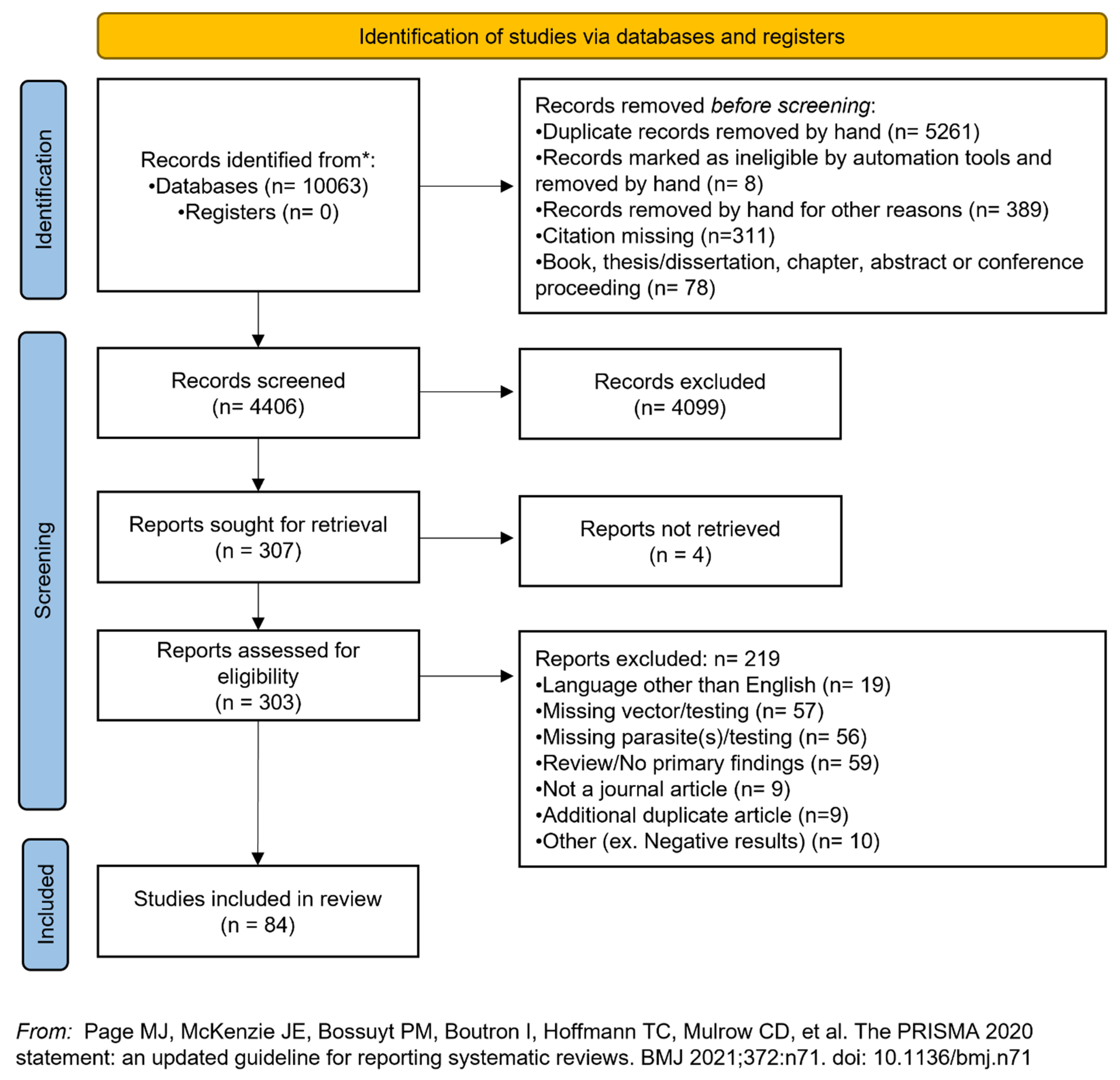
2.2. Screening Process and Study Selection
2.3. Data Extraction
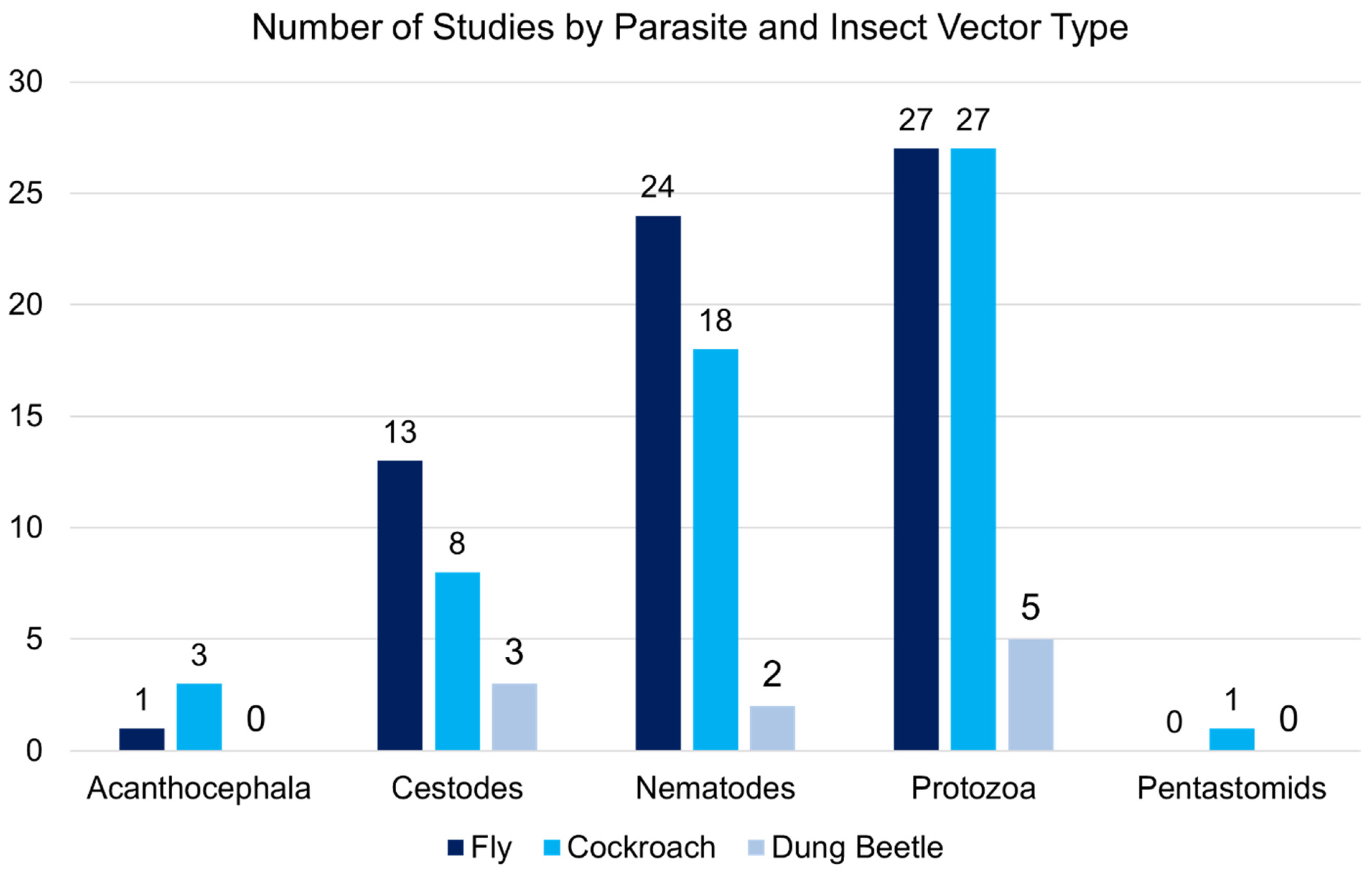
3. Results
Zoonotic Enteric Parasites and Vectors Included in Review
4. Discussion
4.1. Protozoa
4.2. Cestodes
4.3. Nematodes
4.4. Acanthocephala and Pentastomida
4.5. Parasites of Potential Zoonotic Concern
4.6. Non-Pathogenic and Non-Zoonotic Organisms
4.7. Sampling Locations and Risk Factors for Exposure
4.8. Recommendations
4.9. Limitations
5. Conclusions
Supplementary Materials
Author Contributions
Funding
Institutional Review Board Statement
Informed Consent Statement
Data Availability Statement
Acknowledgments
Conflicts of Interest
References
- Graczyk, T.K.; Knight, R.; Tamang, L. Mechanical transmission of human protozoan parasites by insects. Clin. Microbiol. Rev. 2005, 18, 128–132. [Google Scholar] [CrossRef] [Green Version]
- Hadi, A.M. Study of Flyborne Parasites (Brachycera): A Review. Plant Arch. 2020, 20, 2419–2429. [Google Scholar]
- Hayati, R.Z.; Susanna, D. The Human Pathogens Carried by the Cockroaches in the Food-Related Environment Potentially Causing a Foodborne Diseases: A Systematic Review. Malays. J. Public Health Med. 2020, 20, 159–170. [Google Scholar] [CrossRef]
- Nichols, E.; Gomez, A. Dung beetles and fecal helminth transmission: Patterns, mechanisms and questions. Parasitology 2014, 141, 614–623. [Google Scholar] [CrossRef]
- Khamesipour, F.; Lankarani, K.B.; Honarvar, B.; Kwenti, T.E. A systematic review of human pathogens carried by the housefly (Musca domestica L.). BMC Public Health 2018, 18, 1049. [Google Scholar] [CrossRef]
- World Health Organization. The Housefly: Training and Information Guide; No. WHO/VBC/90.987; World Health Organization: Geneva, Switzerland, 1991. [Google Scholar]
- Rozendaal, J.A. Vector Control: Methods for Use by Individuals and Communities; World Health Organization: Geneva, Switzerland, 1997.
- Olsen, A.R. Regulatory action criteria for filth and other extraneous materials: III. Review of flies and foodborne enteric disease. Regul. Toxicol. Pharmacol. 1998, 28, 199–211. [Google Scholar] [CrossRef] [Green Version]
- Graczyk, T.K.; Knight, R.; Gilman, R.H.; Cranfield, M.R. The role of non-biting flies in the epidemiology of human infectious diseases. Microbes Infect. 2001, 3, 231–235. [Google Scholar] [CrossRef]
- Atiokeng Tatang, R.; Tsila, H.; Wabo Poné, J. Medically important parasites carried by cockroaches in Melong Subdivision, Littoral, Cameroon. J. Parasitol. Res. 2017, 2017, 7967325. [Google Scholar] [CrossRef] [Green Version]
- Zahraei-Ramazani, A.R.; Saghafipour, A.; Vatandoost, H. Control of American cockroach (Periplaneta americana) in municipal sewage disposal system, Central Iran. J. Arthropod-Borne Dis. 2018, 12, 172. [Google Scholar] [CrossRef]
- Donkor, E.S. Cockroaches and food-borne pathogens. Environ. Health Insights 2020, 14, 1178630220913365. [Google Scholar] [CrossRef]
- Doi, H.; Gałęcki, R.; Mulia, R.N. The merits of entomophagy in the post COVID-19 world. Trends Food Sci. Technol. 2021, 110, 849–854. [Google Scholar] [CrossRef]
- Ramírez-Restrepo, L.; Halffter, G. Copro-necrophagous beetles (Coleoptera: Scarabaeinae) in urban areas: A global review. Urban Ecosyst. 2016, 19, 1179–1195. [Google Scholar] [CrossRef]
- Nichols, E.; Alarcón, V.; Forgie, S.; Gomez-Puerta, L.A.; Jones, M.S. Coprophagous insects and the ecology of infectious diseases of wildlife. ILAR J. 2017, 58, 336–342. [Google Scholar] [CrossRef]
- Barnes, A.N.; Davaasuren, A.; Baasandagva, U.; Gray, G.C. A systematic review of zoonotic enteric parasitic diseases among nomadic and pastoral people. PLoS ONE 2017, 12, e0188809. [Google Scholar] [CrossRef] [Green Version]
- Page, M.J.; McKenzie, J.E.; Bossuyt, P.M.; Boutron, I.; Hoffmann, T.C.; Mulrow, C.D.; Shamseer, L.; Tetzlaff, J.M.; Akl, E.A.; Brennan, S.E. The PRISMA 2020 statement: An updated guideline for reporting systematic reviews. BMJ 2021, 372, n71. [Google Scholar] [CrossRef]
- Chandler, A.C. Some factors affecting the propagation of hookworm infections in the Asansol mining settlement, with special reference to the part played by cockroaches in mines. Indian Med. Gaz. 1926, 61, 209. [Google Scholar]
- Roberts, E.W. The part played by the faeces and vomit-drop in the transmission of Entamoeba histolytica by Musca domestica. Ann. Trop. Med. Parasitol. 1947, 41, 129–142. [Google Scholar] [CrossRef]
- Schiller, E.L. Studies on the helminth fauna of Alaska. XIX. An experimental study on blowflow (Phormia regina) transmission of hydatid disease. Exp. Parasitol. 1954, 3, 161–166. [Google Scholar] [CrossRef]
- Heinz, H.; Brauns, W. The Ability of Plies to transmit Ova of Echinococcus granulosus to Human Foods. S. Afr. J. Med. Sci. 1955, 20, 131–132. [Google Scholar]
- Laarman, J. Transmission of experimental toxoplasmosis by Stomoxys calcitrans. Doc. Med. Geogr. Trop. 1956, 8, 293–298. [Google Scholar]
- Miller, A.; Chi-Rodriguez, E.; Nichols, R.L. The fate of helminth eggs and protozoan cysts in human feces infested by dung beetles (Coleoptera: Scarabaeidae). Am. J. Trop. Med. Hyg. 1961, 10, 748–754. [Google Scholar] [CrossRef]
- Paim, G.; Queiroz, J. The Ability of Musca domestica to carry Toxoplasma gondii. Arch. Fac. Hig. Saude Publ. Univ. S. Paulo 1963, 28, 213–216. [Google Scholar]
- Pegg, E.J. Infection of dogs by Toxocara canis carried by flies. Parasitology 1971, 62, 409–414. [Google Scholar] [CrossRef]
- Wallace, G.D. Experimental transmission of Toxoplasma gondii by filth-flies. Am. J. Trop. Med. Hyg. 1971, 20, 411–413. [Google Scholar] [CrossRef]
- Nadzhafov, I.G. Role of flies in the epidemiology of human helminthiases in the Azerbaijan SSR. Med. Parazitol. 1972, 41, 168–172. [Google Scholar]
- Wallace, G.D. Experimental transmission of Toxoplasma gondii by cockroaches. J. Infect. Dis. 1972, 126, 545–547. [Google Scholar] [CrossRef]
- Young, P. Investigations on experimental transmission of Trichinella spiralis between rodents and cockroaches. Proc. La. Acad. Sci. 1974, 37, 54–59. [Google Scholar]
- Young, P.L. Studies on the transmission of helminth ova by cockroaches. Proc. Okla. Acad. Sci. 1975, 55, 169–174. [Google Scholar]
- Chinchilla, M.; Ruiz, A. Cockroaches as possible transport hosts of Toxoplasma gondii in Costa Rica. J. Parasitol. 1976, 62, 140–142. [Google Scholar] [CrossRef]
- Gonzalez, J.; Mishra, G. Spontaneous parasitism of cockroach (insecta dictyoptera) in Tunis. Their part as an intermediate host of Mastophorus muris (Gmelin 1970) and Moniliformis moniliformis (Bremser 1811). Arch. L’institut Pasteur Tunis 1976, 53, 211–238. [Google Scholar]
- Khan, A.; Huq, F. Disease agents carried by flies in Dacca city. Bangladesh Med. Res. Counc. Bull. 1978, 4, 86–93. [Google Scholar] [PubMed]
- Smith, D.D.; Frenkel, J. Cockroaches as vectors of Sarcocystis muris and of other coccidia in the laboratory. J. Parasitol. 1978, 64, 315–319. [Google Scholar] [CrossRef]
- Lonc, E. The possible role of the soil fauna in the epizootiology of cysticercosis in cattle. II. Dung beetles—A biotic factor in the transmission of Taenia saginata eggs. Angew. Parasitol. 1980, 21, 139–144. [Google Scholar]
- Markus, M.B. Flies as natural transport hosts of Sarcocystis and other coccidia. J. Parasitol. 1980, 66, 361–362. [Google Scholar] [CrossRef] [PubMed]
- Kasprzak, W.; Majewska, A. Transmission of Giardia cysts. I. The role of flies and cockroaches. Wiad. Parazytol. 1981, 27, 555–563. [Google Scholar]
- Dipeolu, O. Laboratory investigations into the role of Musca vicina and Musca domestica in the transmission of parasitic helminth eggs and larvae. Int. J. Zoonoses 1982, 9, 57–61. [Google Scholar]
- Lawson, J.; Gemmell, M. The potential role of blowflies in the transmission of taeniid tapeworm eggs. Parasitology 1985, 91, 129–143. [Google Scholar] [CrossRef]
- Sulaiman, S.; Sohadi, A.R.; Yunus, H.; Iberahim, R. The role of some cyclorrhaphan flies as carriers of human helminths in Malaysia. Med. Vet. Entomol. 1988, 2, 1–6. [Google Scholar] [CrossRef]
- Sulaiman, S.; Sohadi, A.R.; Jeffery, J. Human helminth parasite burdens on cyclorrhaphan flies (Diptera) trapped at an aboriginal settlement in Malaysia. Bull. Entomol. Res. 1989, 79, 625–629. [Google Scholar] [CrossRef]
- Umeche, N.; Mandah, L. Musca domestica as a carrier of intestinal helminths in Calabar, Nigeria. East Afr. Med. J. 1989, 66, 349–352. [Google Scholar]
- Saitoh, Y.; Itagaki, H. Dung Beetles, Onthophagus spp., as Potential Transport Hosts of Feline Coccidia. Jpn. J. Vet. Sci. 1990, 52, 293–297. [Google Scholar] [CrossRef] [PubMed] [Green Version]
- Monzon, R.; Sanchez, A.; Tadiaman, B.; Najos, O.; Valencia, E.; De Rueda, R.; Ventura, J. A comparison of the role of Musca domestica (Linnaeus) and Chrysomya megacephala (Fabricius) as mechanical vectors of helminthic parasites in a typical slum area of Metropolitan Manila. Southeast Asian J. Trop. Med. Public Health 1991, 22, 222–228. [Google Scholar] [PubMed]
- Stoddart, R.; Crompton, D.; Walters, D. Influence of host strain and helminth isolate on the first phase of the relationship between rats and Moniliformis moniliformis (Acanthocephala). J. Parasitol. 1991, 77, 372–377. [Google Scholar] [CrossRef]
- Chinchilla, M.; Guerrero, O.; Castro, A.; Sabah, J. Cockroaches as transport hosts of the protozoan Toxoplasma gondii. Rev. Biol. Trop. 1994, 42, 329–331. [Google Scholar] [PubMed]
- Zerpa, R.; Huicho, L. Childhood Cryptosporidial Diarrhea Associated with Identification of Cryptosporidium sp. in the Cockroach periplaneta Americana. Pediatr. Infect. Dis. J. 1994, 13, 546–548. [Google Scholar] [CrossRef]
- Juris, P.; Világiová, I.; Plachý, P. The importance of flies (Diptera-Brachycera) in the dissemination of helminth eggs from sewage treatment plants. Vet. Med. 1995, 40, 289–292. [Google Scholar]
- Graczyk, T.K.; Cranfield, M.R.; Fayer, R.; Bixler, H. House flies (Musca domestica) as transport hosts of Cryptosporidium parvum. Am. J. Trop. Med. Hyg. 1999, 61, 500–504. [Google Scholar] [CrossRef] [PubMed] [Green Version]
- Mathison, B.A.; Ditrich, O. The fate of Cryptosporidium parvum oocysts ingested by dung beetles and their possible role in the dissemination of cryptosporidiosis. J. Parasitol. 1999, 85, 678–681. [Google Scholar] [CrossRef] [PubMed]
- Doiz, O.; Clavel, A.; Morales, S.; Varea, M.; Castillo, F.J.; Rubio, C.; Gómez-Lus, R. House fly (Musca domestica) as a transport vector of Giardia lamblia. Folia Parasitol. 2000, 47, 330–333. [Google Scholar] [CrossRef] [Green Version]
- Graczyk, T.K.; Fayer, R.; Knight, R.; Mhangami-Ruwende, B.; Trout, J.M.; Da Silva, A.J.; Pieniazek, N.J. Mechanical transport and transmission of Cryptosporidium parvum oocysts by wild filth flies. Am. J. Trop. Med. Hyg. 2000, 63, 178–183. [Google Scholar] [CrossRef] [Green Version]
- Oliveira, V.C.D.; Mello, R.P.D.; d’Almeida, J.M. Muscoid dipterans as helminth eggs mechanical vectors at the zoological garden, Brazil. Rev. Saude Publica 2002, 36, 614–620. [Google Scholar] [CrossRef] [Green Version]
- Graczyk, T.K.; Grimes, B.H.; Knight, R.; Da Silva, A.J.; Pieniazek, N.J.; Veal, D.A. Detection of Cryptosporidium parvum and Giardia lamblia carried by synanthropic flies by combined fluorescent in situ hybridization and a monoclonal antibody. Am. J. Trop. Med. Hyg. 2003, 68, 228–232. [Google Scholar] [CrossRef]
- Pai, H.-H.; Ko, Y.; Chen, E. Cockroaches (Periplaneta americana and Blattella germanica) as potential mechanical disseminators of Entamoeba histolytica. Acta Trop. 2003, 87, 355–359. [Google Scholar] [CrossRef]
- Chan, O.; Lee, E.K.; Hardman, J.M.; Navin, J.J. The cockroach as a host for Trichinella and Enterobius vermicularis: Implications for public health. Hawaii Med. J. 2004, 63, 74–77. [Google Scholar]
- Graczyk, T.K.; Grimes, B.H.; Knight, R.; Szostakowska, B.; Kruminis-Lozowska, W.; Racewicz, M.; Tamang, L.; Dasilva, A.J.; Myjak, P. Mechanical transmission of Cryptosporidium parvum oocysts by flies. Wiadomości Parazytol. 2004, 50, 243–247. [Google Scholar]
- Szostakowska, B.; Kruminis-Lozowska, W.; Racewicz, M.; Knight, R.; Tamang, L.; Myjak, P.; Graczyk, T.K. Cryptosporidium parvum and Giardia lamblia recovered from flies on a cattle farm and in a landfill. Appl. Environ. Microbiol. 2004, 70, 3742–3744. [Google Scholar] [CrossRef] [Green Version]
- Tatfeng, Y.; Usuanlele, M.; Orukpe, A.; Digban, A.; Okodua, M.; Oviasogie, F.; Turay, A. Mechanical transmission of pathogenic organisms: The role of cockroaches. J. Vector Borne Dis. 2005, 42, 129. [Google Scholar]
- Conn, D.B.; Weaver, J.; Tamang, L.; Graczyk, T.K. Synanthropic flies as vectors of Cryptosporidium and Giardia among livestock and wildlife in a multispecies agricultural complex. Vector-Borne Zoonotic Dis. 2007, 7, 643–652. [Google Scholar] [CrossRef]
- Getachew, S.; Gebre-Michael, T.; Erko, B.; Balkew, M.; Medhin, G. Non-biting cyclorrhaphan flies (Diptera) as carriers of intestinal human parasites in slum areas of Addis Ababa, Ethiopia. Acta Trop. 2007, 103, 186–194. [Google Scholar] [CrossRef]
- Kinfu, A.; Erko, B. Cockroaches as carriers of human intestinal parasites in two localities in Ethiopia. Trans. R. Soc. Trop. Med. Hyg. 2008, 102, 1143–1147. [Google Scholar] [CrossRef]
- Sasmal, N.; Pahari, T.; Laha, R. Experimental infection of the cockroach Periplaneta americana with Toxocara canis and the establishment of patent infections in pups. J. Helminthol. 2008, 82, 97–100. [Google Scholar] [CrossRef]
- Fetene, T.; Worku, N. Public health importance of non-biting cyclorrhaphan flies. Trans. R. Soc. Trop. Med. Hyg. 2009, 103, 187–191. [Google Scholar] [CrossRef]
- Förster, M.; Klimpel, S.; Sievert, K. The house fly (Musca domestica) as a potential vector of metazoan parasites caught in a pig-pen in Germany. Vet. Parasitol. 2009, 160, 163–167. [Google Scholar] [CrossRef]
- Mowlavi, G.; Mikaeili, E.; Mobedi, I.; Kia, E.; Masoomi, L.; Vatandoost, H. A survey of dung beetles infected with larval nematodes with particular note on Copris lunaris beetles as a vector for Gongylonema sp. in Iran. Korean J. Parasitol. 2009, 47, 13. [Google Scholar] [CrossRef]
- Racewicz, M.; Kruminis-Łozowska, W.; Gabre, R.M.; Stańczak, J. The occurrence of Cryptosporidium spp. in synanthropic flies in urban and rural environments. Wiad. Parazytol. 2009, 55, 231–236. [Google Scholar] [PubMed]
- Chamavit, P.; Sahaisook, P.; Niamnuy, N. The majority of cockroaches from the Samutprakarn province of Thailand are carriers of parasitic organisms. EXCLI J. 2011, 10, 218. [Google Scholar] [PubMed]
- Fetene, T.; Worku, N.; Huruy, K.; Kebede, N. Cryptosporidium recovered from Musca domestica, Musca sorbens and mango juice accessed by synanthropic flies in Bahirdar, Ethiopia. Zoonoses Public Health 2011, 58, 69–75. [Google Scholar] [CrossRef] [PubMed]
- Ribeiro, M.J.R.; Dias, S.M.F.; Teshima, E.; Barboni, A.R. Unhealthy environment and social aspects associated with intestinal pathogens isolated of dipteral. Eng. Sanit. Ambient. 2011, 16, 83–90. [Google Scholar] [CrossRef]
- Ryan, U.; Yang, R.; Gordon, C.; Doube, B. Effect of dung burial by the dung beetle Bubas bison on numbers and viability of Cryptosporidium oocysts in cattle dung. Exp. Parasitol. 2011, 129, 1–4. [Google Scholar] [CrossRef] [Green Version]
- El-Sherbini, G.T.; Gneidy, M.R. Cockroaches and flies in mechanical transmission of medical important parasites in Khaldyia Village, El-Fayoum, Governorate, Egypt. J. Egypt. Soc. Parasitol. 2012, 240, 165–174. [Google Scholar] [CrossRef]
- Adenusi, A.A.; Adewoga, T.O.S. Human intestinal parasites in non-biting synanthropic flies in Ogun State, Nigeria. Travel Med. Infect. Dis. 2013, 11, 181–189. [Google Scholar] [CrossRef] [PubMed]
- Adenusi, A.A.; Adewoga, T.O. Studies on the potential and public health importance of non-biting synanthropic flies in the mechanical transmission of human enterohelminths. Trans. R. Soc. Trop. Med. Hyg. 2013, 107, 812–818. [Google Scholar] [CrossRef]
- Lalander, C.; Diener, S.; Magri, M.E.; Zurbrügg, C.; Lindström, A.; Vinnerås, B. Faecal sludge management with the larvae of the black soldier fly (Hermetia illucens)—From a hygiene aspect. Sci. Total Environ. 2013, 458, 312–318. [Google Scholar] [CrossRef] [PubMed]
- Tetteh-Quarcoo, P.B.; Donkor, E.S.; Attah, S.K.; Duedu, K.O.; Afutu, E.; Boamah, I.; Olu-Taiwo, M.; Anim-Baidoo, I.; Ayeh-Kumi, P.F. Microbial carriage of cockroaches at a tertiary care hospital in Ghana. Environ. Health Insights 2013, 7, EHI-S12820. [Google Scholar] [CrossRef] [PubMed] [Green Version]
- Bunchu, N.; Silaram, M.; Sukontason, K.; Sukontason, K.L.; Chaiwong, T. Isolation of Toxocara eggs from flies in Northeast Thailand. J. Med. Assoc. Thail. 2014, 97, S25–S28. [Google Scholar]
- Lima, M.S.C.S.; Soares, M.R.A.; Pederassi, J.; Aguiar, B.C.G.; Pereira, C.A.S. The housefly Musca domestica L. (Diptera: Muscidae) as a potential paratenic host in the city of Bom Jesus-Piauí, Brazil. Comun. Sci. 2014, 5, 349–355. [Google Scholar]
- Gomez-Puerta, L.A.; Lopez-Urbina, M.T.; Garcia, H.H.; Gonzalez, A.E. Longevity and viability of Taenia solium eggs in the digestive system of the beetle Ammophorus rubripes. Rev. Bras. Parasitol. Veter. 2014, 23, 94–97. [Google Scholar] [CrossRef] [Green Version]
- Hamu, H.; Debalke, S.; Zemene, E.; Birlie, B.; Mekonnen, Z.; Yewhalaw, D. Isolation of intestinal parasites of public health importance from cockroaches (Blattella germanica) in Jimma Town, Southwestern Ethiopia. J. Parasitol. Res. 2014, 2014, 186240. [Google Scholar] [CrossRef] [Green Version]
- Isaac, C.; Orue, P.O.; Iyamu, M.I.; Ehiaghe, J.I.; Isaac, O. Comparative analysis of pathogenic organisms in cockroaches from different community settings in Edo State, Nigeria. Korean J. Parasitol. 2014, 52, 177. [Google Scholar] [CrossRef]
- Zhao, Z.; Dong, H.; Wang, R.; Zhao, W.; Chen, G.; Li, S.; Qi, M.; Zhang, S.; Jian, F.; Zhao, J. Genotyping and subtyping Cryptosporidium parvum and Giardia duodenalis carried by flies on dairy farms in Henan, China. Parasites Vectors 2014, 7, 190. [Google Scholar] [CrossRef] [Green Version]
- Cazorla Perfetti, D.; Morales, P.; Navas, P. Isolation of intestinal parasites from American cockroach (Periplaneta americana) in Coro, Falcon state, Venezuela. Boletín Malariol. Salud Ambient. 2015, 55, 184–193. [Google Scholar]
- Muñoz, D.J. Bacterial and Parasite Agents in Adult Housefly Musca domestica Collected in El Peñón, Sucre State, Venezuela. Rev. Cient. Fac. Cienc. Vet. 2015, 25, 159–166. [Google Scholar]
- Farah Haziqah, M.; Asyiqin, N.; Mohd Khalid, M.; Suresh, K.; Rajamanikam, A.; Chandrawathani, P.; Mohd Zain, S. Short Communication Current status of Blastocystis in cockroaches. Trop. Biomed. 2017, 34, 741–745. [Google Scholar]
- González-García, T.; Muñoz-Guzmán, M.; Sánchez-Arroyo, H.; Prado-Ochoa, M.; Cuéllar-Ordaz, J.; Alba-Hurtado, F. Experimental transmission of Toxocara canis from Blattella germanica and Periplaneta americana cockroaches to a paratenic host. Vet. Parasitol. 2017, 246, 5–10. [Google Scholar] [CrossRef] [PubMed]
- Martínez-Girón, R.; Martínez-Torre, C.; van Woerden, H.C. The prevalence of protozoa in the gut of German cockroaches (Blattella germanica) with special reference to Lophomonas blattarum. Parasitol. Res. 2017, 116, 3205–3210. [Google Scholar] [CrossRef]
- Oguz, B.; Ozdal, N.; Orunc Kilinc, O.; Deger, M.S. First investigation on vectorial potential of Blattella germanica in Turkey. Ank. Univ. Vet. Fak. Derg. 2017, 64, 141–144. [Google Scholar]
- Adenusi, A.A.; Akinyemi, M.I.; Akinsanya, D. Domiciliary cockroaches as carriers of human intestinal parasites in Lagos Metropolis, Southwest Nigeria: Implications for public health. J. Arthropod-Borne Dis. 2018, 12, 141. [Google Scholar] [CrossRef]
- Hemmati, S.; Afshar, A.A.; Mohammadi, M.A.; Afgar, A.; Nasibi, S.; Harandi, M.F. Experimental and field investigation of non-biting flies as potential mechanical vectors of Echinococcus granulosus eggs. Exp. Parasitol. 2018, 189, 43–48. [Google Scholar] [CrossRef]
- Paliy, A.; Sumakova, N.; Mashkey, A.; Petrov, R.; Ishchenko, K. Contamination of animal-keeping premises with eggs of parasitic worms. Biosyst. Divers. 2018, 26, 327–333. [Google Scholar] [CrossRef] [Green Version]
- Paliy, A.; Sumakova, N.; Ishchenko, K. Biological control of house fly. Ukr. J. Ecol. 2018, 8, 230–234. [Google Scholar]
- Valles, L.T.; Alejos, M.; Antonini, M.; Escobar, C.; Perez, M.; Perez, F.; Ramirez, D.; Tovar, A.; Najul, M. Enteroparasitary contamination of flies captured in the Palavecino municipality, Lara state, Venezuela, 2017. Rev. Med. Urug. 2018, 34, 217–221. [Google Scholar]
- Vargas-Calla, A.; Gomez-Puerta, L.A.; Pajuelo, M.J.; Garcia, H.H.; Gonzalez, A.E.; Cysticercosis Working Group in Peru. Molecular detection of taeniid eggs in beetles collected in an area endemic for Taenia solium. Am. J. Trop. Med. Hyg. 2018, 99, 1198. [Google Scholar] [CrossRef]
- Gałęcki, R.; Sokół, R. A parasitological evaluation of edible insects and their role in the transmission of parasitic diseases to humans and animals. PLoS ONE 2019, 14, e0219303. [Google Scholar] [CrossRef] [Green Version]
- Muller, A.; Wiedmer, S.; Kurth, M. Risk evaluation of passive transmission of animal parasites by feeding of black soldier fly (Hermetia illucens) larvae and prepupae. J. Food Prot. 2019, 82, 948–954. [Google Scholar] [CrossRef]
- Dokmaikaw, A.; Suntaravitun, P. Prevalence of parasitic contamination of cockroaches collected from fresh markets in Chachoengsao province, Thailand. Kobe J. Med. Sci. 2019, 65, E118. [Google Scholar]
- Ma, L.; Zhang, Y.; Qiao, H.; Li, S.; Wang, H.; Zhang, N.; Zhang, X. Cockroach as a Vector of Blastocystis sp. is Risk for Golden Monkeys in Zoo. Korean J. Parasitol. 2020, 58, 583. [Google Scholar] [CrossRef] [PubMed]
- van Woerden, H.C.; Martínez-Girón, R.; Martínez-Torre, C. Protozoan cysts in faecal pellets of German cockroaches (Blattella germanica), with particular emphasis on Lophomonas blattarum. Acta Parasitol. 2020, 65, 831–836. [Google Scholar] [CrossRef] [PubMed]
- Barnes, A.N.; Davaasuren, A.; Baasandavga, U.; Lantos, P.M.; Gonchigoo, B.; Gray, G.C. Zoonotic enteric parasites in Mongolian people, animals, and the environment: Using One Health to address shared pathogens. PLoS Negl. Trop. Dis. 2021, 15, e0009543. [Google Scholar] [CrossRef] [PubMed]
- Motevalli-Haghi, S.F.; Shemshadian, A.; Nakhaei, M.; Faridnia, R.; Dehghan, O.; Shafaroudi, M.M.; Kelarijani, M.N.; Nikookar, S.H.; Kalani, H.; Fakhar, M. First report of Lophomonoas spp. in German Cockroaches (Blatella germanica) Trapped in Hospitals, Northern Iran. J. Parasit. Dis. 2021, 45, 937–943. [Google Scholar] [CrossRef] [PubMed]
- Sands, B.; Wall, R. Dung beetles reduce livestock gastrointestinal parasite availability on pasture. J. Appl. Ecol. 2017, 54, 1180–1189. [Google Scholar] [CrossRef] [Green Version]
- Fincher, G.T. Dung beetles as biological control agents for gastrointestinal parasites of livestock. J. Parasitol. 1973, 59, 396–399. [Google Scholar] [CrossRef] [PubMed]
- Yoshikawa, H.; Wu, Z.; Howe, J.; Hashimoto, T.; Geok-Choo, N.; Tan, K.S. Ultrastructural and phylogenetic studies on Blastocystis isolates from cockroaches. J. Eukaryot. Microbiol. 2007, 54, 33–37. [Google Scholar] [CrossRef] [PubMed]
- Zaman, V.; Ng, G.; Suresh, K.; Yap, E.; Singh, M. Isolation of Blastocystis from the cockroach (Dictyoptera: Blattidae). Parasitol. Res. 1993, 79, 73–74. [Google Scholar] [CrossRef]
- Bauerfeind, R.; Von Graevenitz, A.; Kimmig, P.; Schiefer, H.G.; Schwarz, T.; Slenczka, W.; Zahner, H. Zoonoses: Infectious Diseases Transmissible from Animals to Humans; John Wiley & Sons: Hoboken, NJ, USA, 2015. [Google Scholar]
- Dehghani, R.; Kassiri, H. A brief review on the possible role of houseflies and cockroaches in the mechanical transmission of coronavirus disease 2019 (COVID-19). Arch. Clin. Infect. Dis. 2020, 15, e102863. [Google Scholar] [CrossRef] [Green Version]


| Zoonotic Enteric Parasite(s) † | Vector(s) | Country of Study | Sample Source | Type of Iinfection | Citation | |
|---|---|---|---|---|---|---|
| Class and Family | Genus an/or Species and Natural Prevalence (%), When Provided | |||||
| Chromadorea, Ascarididae | Ascaris spp. | Cockroach | India | Village area | Experimental | Chandler 1926 [18] |
| Chromadorea Ancylostomatidae | Hookworm (Unspecified) | |||||
| Enoplea, Trichuridae | Trichuris spp. | |||||
| Lobosa, Entamoebidae | Entamoeba histolytica | Fly | England | Laboratory | Experimental | Roberts 1947 [19] |
| Cestoda, Taeniidae | Echinococcus spp. | Fly | United States | Laboratory | Experimental | Schiller 1954 [20] |
| Cestoda, Taeniidae | Echinococcus granulosus | Fly | South Africa | Laboratory | Experimental | Heinz and Brauns 1955 [21] |
| Conoidasida, Sarcocystidae | Toxoplasma gondii | Fly | Netherlands | Laboratory | Experimental | Laarman 1956 [22] |
| Chromadorea, Ascarididae | Ascaris lumbricoides | Dung beetle | United States | Farm/Field | Experimental | Miller et al. 1961 [23] |
| Zoomastigophora, Hexamitidae | Giardia lamblia | |||||
| Chromadorea Ancylostomatidae | Hookworm (Necator americanus) | |||||
| Enoplea, Trichuridae | Trichuris trichiura | |||||
| Conoidasida, Sarcocystidae | T. gondii | Fly | Brazil | Laboratory | Experimental | Paim and Queiroz 1963 * [24] |
| Chromadorea, Toxocaridae | Toxocara canis | Fly | England | Laboratory | Experimental | Pegg 1971 [25] |
| Conoidasida, Sarcocystidae | T. gondii | Fly | United States | Laboratory | Experimental | Wallace 1971 [26] |
| Chromadorea, Ascarididae | Ascaris spp. | Fly | Azerbaijan | Laboratory Village area | Mixed | Nadzhafov 1972 * [27] |
| Chromadorea Ancylostomatidae | Hookworm (Unspecified) | |||||
| Cestoda, Hymenolepididae | Hymenolepis nana | |||||
| Enoplea, Trichuridae | Trichuris spp. | |||||
| Conoidasida, Sarcocystidae | T. gondii | Cockroach | United States | Laboratory | Experimental | Wallace 1972 [28] |
| Enoplea, Trichinellidae | Trichinella spiralis | Cockroach | United States | Laboratory | Experimental | Young and Babero 1974 [29] |
| Chromadorea, Ascarididae | Ascaris columnaris (Baylisascaris procyonis) | Cockroach | United States | Laboratory | Experimental | Young 1975 [30] |
| Ascaris suum | ||||||
| Cestoda, Dipylidiidae | Dipylidium caninum | |||||
| Cestoda, Hymenolepididae | Hymenolepis dimenuta | |||||
| H. nana | ||||||
| Cestoda, Mesocestoididae | Mesocestoides lineatus | |||||
| Chromadorea, Physalopteridae | Physaloptera turgida§ | |||||
| Chromadorea, Setariidae | Setaria equina§ | |||||
| Chromadorea, Oxyuridae | Syphacia obvelata | |||||
| Chromadorea, Toxocaridae | Toxascaris leonine§ | |||||
| T. canis | ||||||
| Toxocara cati | ||||||
| Conoidasida, Sarcocystidae | T. gondii | Cockroach | Costa Rica | Unspecified | Experimental | Chinchilla and Ruiz 1976 [31] |
| Lobosa, Entamoebidae | Entamoeba spp. | Cockroach | Tunisia | Urban area | Natural | Gonzalez and Mishra 1976 * [32] |
| Archiacanthocephala, Moniliformidae | Moniliformis moniliformis | |||||
| Chromadorea, Ascarididae | A. lumbricoides (18.8–62.5%) | Fly | Bangladesh | Abattoir/Butchery/Slaughterhouse/ Food market Hospital Household Open defecation area Public transportation Waste disposal area | Natural | Khan and Huq 1978 [33] |
| Zoomastigophora, Hexamitidae | Giardia spp. (6.2%) | |||||
| Chromadorea Ancylostomatidae | Hookworm (Unspecified; 15.6%) | |||||
| Enoplea, Trichuridae | Trichuris trichiura (46.9%) | |||||
| Conoidasida, Sarcocystidae | Sarcocystis muris | Cockroach | United States | Laboratory | Experimental | Smith and Frenkel 1978 [34] |
| T. gondii | ||||||
| Cestoda, Taeniidae | Taenia saginata | Dung beetle | Poland | Unspecified | Experimental | Lonc 1980 [35] |
| Conoidasida, Sarcocystidae | Sarcocystis spp. | Fly | England | Dog Kennel | Natural | Markus 1980 [36] |
| Zoomastigophora, Hexamitidae | Giardia intestinalis | Cockroach Fly | Poland | Open defecation area Waste disposal area | Mixed | Kasprzak and Majewska 1981 * [37] |
| Chromadorea, Ascarididae | A. lumbricoides | Fly | Nigeria | School/University | Experimental | Dipeolu 1982 [38] |
| Chromadorea Ancylostomatidae | Hookworm (Unspecified) | |||||
| Cestoda, Taeniidae | Taenia hydatigena§ | Fly | New Zealand | Farm/Field Laboratory | Mixed | Lawson and Gemmell 1985 [39] |
| Chromadorea, Ascarididae | A. lumbricoides | Fly | Malaysia | Farm/Field Household Waste disposal area | Natural | Sulaiman et al. 1988 [40] |
| Chromadorea Ancylostomatidae | Hookworm (Necator americanus) | |||||
| Enoplea, Trichuridae | T. trichiura | |||||
| Chromadorea, Ascarididae | A. lumbricoides | Fly | Malaysia | Household | Natural | Sulaiman et al. 1989 [41] |
| Chromadorea Ancylostomatidae | Hookworm (Necator americanus and/or Ancylostoma duodenale §) | |||||
| Enoplea, Trichuridae | T. trichiura | |||||
| Chromadorea, Ascarididae | A. lumbricoides (0.20–0.81%) | Fly | Nigeria | Food market Household | Natural | Umeche and Mandah 1989 [42] |
| Chromadorea, Strongyloididae | Strongyloides stercoralis (0.40–1.80%) | |||||
| Chromadorea, Toxocaridae | T. canis (2.40–2.11%) | |||||
| Conoidasida, Sarcocystidae | T. gondii | Dung beetle | Japan | Laboratory School/University | Mixed | Saitoh and Itagaki 1990 [43] |
| Chromadorea, Ascarididae | Ascaris spp. (72.8–82.4%) | Fly | Philippines | Urban area | Natural | Monzon et al. 1991 [44] |
| Enoplea, Capillariidae | Capillaria hepatica (0.0–0.005%) | |||||
| Chromadorea Ancylostomatidae | Hookworm (Unspecified; (0.02–13.1%) | |||||
| Cestoda, Taeniidae | Taenia spp. (0.005–0.02%) | |||||
| Chromadorea, Toxocaridae | Toxocara spp. (0.005–0.04%) | |||||
| Enoplea, Trichuridae | T. trichiura (18.8–60.1%) | |||||
| Archiacanthocephala, Moniliformidae | M. moniliformis | Cockroach | Scotland | Laboratory | Experimental | Stoddart et al. 1991 [45] |
| Conoidasida, Sarcocystidae | T. gondii | Cockroach | Costa Rica | Laboratory | Experimental | Chinchilla et al. 1994 [46] |
| Conoidasida, Cryptosporidiidae | Cryptosporidium spp. | Cockroach | Peru | Garden Household | Natural | Zerpa and Huicho 1994 [47] |
| Chromadorea, Ascarididae | Ascaris spp. | Fly | Slovakia | Wastewater treatment area | Natural | Juris et al. 1995 * [48] |
| Enoplea, Capillariidae | Capillaria spp. | |||||
| Cestoda, Hymenolepididae | Hymenolepis spp. | |||||
| Cestoda, Taeniidae | Taenia spp. | |||||
| Chromadorea, Toxocaridae | Toxocara spp. | |||||
| Enoplea, Trichuridae | Trichuris spp. | |||||
| Conoidasida, Cryptosporidiidae | Cryptosporidium parvum | Fly | United States | Laboratory | Experimental | Graczyk et al. 1999 [49] |
| Conoidasida, Cryptosporidiidae | C. parvum | Dung beetle | Czech Republic | Farm/Field Forest | Experimental | Mathison and Ditrich 1999 [50] |
| Zoomastigophora, Hexamitidae | G. lamblia (22%) | Fly | Spain | Farm/Field | Natural | Doiz et al. 2000 [51] |
| Conoidasida, Cryptosporidiidae | C. parvum | Fly | United States | Farm/Field | Mixed | Graczyk et al. 2000 [52] |
| Palaeacanthocephala, Unspecified | Acanthocephala spp. | Fly | Brazil | Waste disposal area Zoo | Natural | de Oliveira et al. 2002 * [53] |
| Chromadorea, Ascarididae. | Ascaris spp. | |||||
| Enoplea, Capillariidae | Capillaria spp. | |||||
| Chromadorea, Ascarididae. | Toxascaris spp. § | |||||
| Chromadorea, Toxocaridae | Toxocara spp. | |||||
| Chromadorea, Trichostrongylidae | Trichostrongylidae spp. | |||||
| Enoplea, Trichuridae | Trichuris spp. | |||||
| Chromadorea, Oxyuridae | Unspecified oxyuridae spp. | |||||
| Conoidasida, Cryptosporidiidae | C. parvum | Fly | United States | Farm/Field Food market Waste disposal area | Natural | Graczyk et al. 2003 [54] |
| Zoomastigophora, Hexamitidae | G. lamblia | |||||
| Lobosa, Entamoebidae | Entamoeba histolytica/dispar (10.3–25.4%) | Cockroach | Taiwan | Kitchen area Laboratory School/University | Mixed | Pai et al. 2003 [55] |
| Chromadorea, Oxyuridae | Enterobius vermicularis (3%) | Cockroach | United States | Hospital School/University | Natural | Chan et al. 2004 [56] |
| Enoplea, Trichinellidae | Trichinella spp. (1.0%) | |||||
| Conoidasida, Cryptosporidiidae | C. parvum | Fly | United States Poland | Farm/Field Laboratory Waste disposal area | Mixed | Graczyk et al. 2004 [57] |
| Conoidasida, Cryptosporidiidae | C. parvum | Fly | Poland | Farm/Field Waste disposal area | Natural | Szostakowska et al. 2004 [58] |
| Zoomastigophora, Hexamitidae | G. lamblia | |||||
| Chromadorea, Ascarididae | A. lumbricoides | Cockroach | Nigeria | Household | Natural | Tatfeng et al. 2005 [59] |
| Litostomatea, Balantiididae | Balantidium coli | |||||
| Conoidasida, Cryptosporidiidae | C. parvum | |||||
| Lobosa, Entamoebidae | E. histolytica | |||||
| Chromadorea, Oxyuridae | E. vermicularis | |||||
| Chromadorea, Strongyloididae | S. stercoralis | |||||
| Enoplea, Trichuridae | T. trichiura | |||||
| Conoidasida, Cryptosporidiidae | Cryptosporidium spp. (55.56%) | Fly | United States | Farm/Field Garden School/University | Natural | Conn et al. 2007 [60] |
| Zoomastigophora, Hexamitidae | Giardia spp. (7.94%) | |||||
| Chromadorea, Ascarididae | A. lumbricoides | Fly | Ethiopia | Abattoir/Butchery/Slaughterhouse/ Food market Open defecation area Waste disposal area | Natural | Getachew et al. 2007 [61] |
| Conoidasida, Cryptosporidiidae | Cryptosporidium spp. | |||||
| Lobosa, Entamoebidae | E. histolytica/dispar | |||||
| Zoomastigophora, Hexamitidae | G. lamblia | |||||
| Chromadorea Ancylostomatidae | Hookworm (Unspecified) | |||||
| Cestoda, Hymenolepididae | H. nana | |||||
| Chromadorea, Strongyloididae | S. stercoralis | |||||
| Cestoda, Taeniidae | Taenia spp. | |||||
| Enoplea, Trichuridae | T. trichiura | |||||
| Chromadorea, Ascarididae | A. lumbricoides | Cockroach | Ethiopia | Household | Natural | Kinfu and Erko 2008 [62] |
| Lobosa, Entamoebidae | E. histolytica/dispar | |||||
| Chromadorea, Oxyuridae | E. vermicularis | |||||
| Cestoda, Taeniidae | Taenia spp. | |||||
| Enoplea, Trichuridae | T. trichiura | |||||
| Chromadorea, Toxocaridae | T. canis | Cockroach | India | Kitchen area | Experimental | Sasmal et al. 2008 [63] |
| Chromadorea, Ascarididae | A. lumbricoides (36.9%) | Fly | Ethiopia | Abattoir/Butchery/Slaughterhouse/ Food market Waste disposal area | Natural | Fetene and Worku 2009 [64] |
| Conoidasida, Cryptosporidiidae | Cryptosporidium spp. (16.7%) | |||||
| Lobosa, Entamoebidae | E. histolytica/dispar (48.1%) | |||||
| Zoomastigophora, Hexamitidae | G. lamblia (10.4%) | |||||
| Chromadorea Ancylostomatidae | Hookworm (Unspecified; 13.0%) | |||||
| Cestoda, Hymenolepididae | H. nana (0.6%) | |||||
| Chromadorea, Strongyloididae | S. stercoralis (1.7%) | |||||
| Cestoda, Taeniidae | Taenia spp. (8.4%) | |||||
| Enoplea, Trichuridae | T. trichiura (38.8%) | |||||
| Chromadorea, Ascarididae | A. suum | Fly | Germany | Farm/Field Laboratory | Mixed | Förster et al. 2009 [65] |
| Chromadorea, Metastrongylidae | Metastrongylus spp. § | |||||
| Chromadorea, Strongyloididae | Strongyloides ransomi§ | |||||
| Enoplea, Trichuridae | Trichuris suis§ | |||||
| Chromadorea, Gongylomatidae | Gongylonema spp. (17.7%) | |||||
| Chromadorea, Rhabditidae | Rhabditis spp. (2.2%) | |||||
| Chromadorea, Gongylomatidae | Gongylonema spp. (17.7%) | Dung beetle | Iran | Farm/Field | Natural | Mowlavi et al. 2009 [66] |
| Chromadorea, Rhabditidae | Rhabditis spp. (2.2%) | |||||
| Conoidasida, Cryptosporidiidae | Cryptosporidium spp. (18.9%) | Fly | Poland | Farm/Field Waste disposal area | Natural | Racewicz et al. 2009 * [67] |
| Chromadorea, Ascarididae | A. lumbricoides (0.3%) | Cockroach | Thailand | Food market | Natural | Chamavit et al. 2011 [68] |
| Litostomatea, Balantiididae | B. coli (5.8%) | |||||
| Bigyra, Blastocystidae | Blastocystis hominis (1.2%) | |||||
| Conoidasida, Cryptosporidiidae | Cryptosporidium spp. (28.1%) | |||||
| Conoidasida, Eimeriidae | Cyclospora spp. (1.3%) § | |||||
| Lobosa, Entamoebidae | E. histolytica/dispar (4.6%) | |||||
| Chromadorea, Strongyloididae | S. stercoralis (0.8%) | |||||
| Cestoda, Taeniidae | Taenia spp. (0.1%) | |||||
| Enoplea, Trichuridae | T. trichiura (0.3%) | |||||
| Conoidasida, Cryptosporidiidae | Cryptosporidium spp. | Fly | Ethiopia | Abattoir/Butchery/Slaughterhouse/ Farm/Field Food market Open defecation area | Mixed | Fetene et al. 2011 [69] |
| Unspecified | Unspecified helminths and protozoa | Fly | Brazil | Waste disposal area | Natural | Ribeiro et al. 2011 * [70] |
| Conoidasida, Cryptosporidiidae | Cryptosporidium spp. | Dung beetle | Australia | Unspecified | Experimental | Ryan et al. 2011 [71] |
| Chromadorea, Ascarididae | Flies Ascaris spp. | Fly Cockroach | Egypt | Household Open defecation area | Natural | El-Sherbini and Gneidy 2012 [72] |
| Chromadorea, Oxyuridae | E. vermicularis | |||||
| Chromadorea Ancylostomatidae | Hookworm (Unspecified) | |||||
| Cestoda, Hymenolepididae | H. nana | |||||
| Enoplea, Trichuridae | T. trichiura | |||||
| Cockroaches Unspecified parasitic agents | ||||||
| Chromadorea, Ascarididae | A. lumbricoides (34.08%) | Fly | Nigeria | Abattoir/Butchery/Slaughterhouse/ Food market Open defecation area Waste disposal area | Natural | Adenusi and Adewoga 2013 [73] |
| Conoidasida, Cryptosporidiidae | Cryptosporidium spp. (1.81%) | |||||
| Lobosa, Entamoebidae | E. histolytica/dispar (27.26%) | |||||
| Zoomastigophora, Hexamitidae | G. lamblia (3.34%) | |||||
| Chromadorea Ancylostomatidae | Hookworm (Unspecified; 20.45%) | |||||
| Cestoda, Hymenolepididae | H. nana (1.11%) | |||||
| Chromadorea, Strongyloididae | S. stercoralis (3.89%) | |||||
| Cestoda, Taeniidae | Taenia spp. (2.36%) | |||||
| Enoplea, Trichuridae | T. trichiura (25.87%) | |||||
| Chromadorea, Ascarididae | A. lumbricoides (52.2%) | Fly | Nigeria | Open defecation area Waste disposal area | Natural | Adenusi and Adewoga 2013 [74] |
| Cestoda, Taeniidae | Taenia spp. (1.0%) | |||||
| Enoplea, Trichuridae | T. trichiura (47.2%) | |||||
| Chromadorea, Ascarididae | A. suum | Fly | Sweden | Laboratory | Experimental | Lalander et al. 2013 [75] |
| Chromadorea Ancylostomatidae | Hookworm (Ancylostoma duodenale) (4.9%) § | Cockroach | Ghana | Hospital | Natural | Tetteh-Quarcoo et al. 2013 [76] |
| Cestoda, Hymenolepididae | H. nana (1.6%) | |||||
| Cestoda, Taeniidae | Taenia spp. (1.6%) | |||||
| Chromadorea, Toxocaridae | Toxocara spp. | Fly | Thailand | Farm/Field Food market School/University Waste disposal area | Natural | Bunchu et al. 2014 [77] |
| Chromadorea, Ascarididae | Ascaris spp. | Fly | Brazil | Farm/Field School/University | Natural | Cruz Souza Lima et al. 2014 [78] |
| Lobosa, Entamoebidae | Entamoeba spp. | |||||
| Chromadorea, Oxyuridae | E. vermicularis | |||||
| Zoomastigophora, Hexamitidae | Giardia spp. | |||||
| Cestoda, Hymenolepididae | H. nana | |||||
| Cestoda, Taeniidae | Taenia spp. | |||||
| Enoplea, Trichuridae | Trichuris spp. | |||||
| Cestoda, Taeniidae | Taenia solium | Dung beetle | Peru | Farm/Field | Experimental | Gomez-Puerta et al. 2014 [79] |
| Chromadorea, Ascarididae | A. lumbricoides | Cockroach | Ethiopia | Household | Natural | Hamu et al. 2014 [80] |
| Litostomatea, Balantiididae | B. coli | |||||
| Lobosa, Entamoebidae | Entamoeba spp. | |||||
| Zoomastigophora, Hexamitidae | G. duodenalis | |||||
| Cestoda, Taeniidae | Taenia spp. | |||||
| Enoplea, Trichuridae | T. trichiura | |||||
| Unspecified Strongyloides-like nematodes | ||||||
| Chromadorea, Ascarididae | A. lumbricoides (2.9–13.2%) | Cockroach | Nigeria | Household | Natural | Isaac et al. 2014 [81] |
| Litostomatea, Balantiididae | B. coli (1.1–1.2%) | |||||
| Lobosa, Entamoebidae | E. histolytica (1.2–2.2%) | |||||
| Enoplea, Trichuridae | T. trichiura (4.4–4.7%) | |||||
| Unspecified coccidia spp. (3.3%) § | ||||||
| Conoidasida, Cryptosporidiidae | Cryptosporidium spp. | Fly | China | Farm/Field | Natural | Zhao et al. 2014 [82] |
| Zoomastigophora, Hexamitidae | Giardia spp. | |||||
| Chromadorea, Ascarididae | Ascaris spp. | Cockroach | Venezuela | Food market Hospital School/University | Natural | Cazorla Perfetti et al. 2015 * [83] |
| Bigyra, Blastocystidae | Blastocystis spp. (82.9%) | |||||
| Conoidasida, Eimeriidae | Cyclospora spp. § | |||||
| Chromadorea, Oxyuridae | E. vermicularis | |||||
| Bigyra, Blastocystidae | Blastocystis spp. | Fly | Venezuela | Waste disposal area | Natural | Muñoz 2015 * [84] |
| Conoidasida, Eimeriidae | Cyclospora cayetanensis§ | |||||
| Lobosa, Entamoebidae | E. histolytica | |||||
| Zoomastigophora, Hexamitidae | G. intestinalis | |||||
| Chromadorea, Toxocaridae | Toxocara spp. | |||||
| Chromadorea, Ascarididae | Ascaris spp. (33.76%) | Cockroach | Cameroon | Household | Natural | Atiokeng Tatang et al. 2017 [10] |
| Enoplea, Capillariidae | Capillaria spp. (6.16%) | |||||
| Chromadorea Ancylostomatidae | Hookworm (Unspecified; 4.86%) | |||||
| Chromadorea, Toxocaridae | Toxocara spp. (4.86%) | |||||
| Enoplea, Trichuridae | T. trichiura (11.97%) | |||||
| Bigyra, Blastocystidae | Blastocystis spp. (40.4%) | Cockroach | Malaysia | Food market Household Waste disposal area | Natural | Farah et al. 2017 [85] |
| Chromadorea, Toxocaridae | T. canis | Cockroach | Mexico | Laboratory | Experimental | González-García et al. 2017 [86] |
| Unspecified | Amoeba spp. (25.4%) | Cockroach | Spain | Hospital Kitchen area School/University | Natural | Martínez-Girón et al. 2017 [87] |
| Chromadorea, Ascarididae | A. lumbricoides (3%) | Cockroach | Turkey | Household | Natural | Oğuz et al. 2017 [88] |
| Bigyra, Blastocystidae | B. hominis (41%) | |||||
| Lobosa, Entamoebidae | E. histolytica/dispar (16.7%) | |||||
| Zoomastigophora, Hexamitidae | Giardia spp. (13.6%) | |||||
| Chromadorea, Toxocaridae | Toxocara spp. (3%) | |||||
| Chromadorea, Trichostrongylidae | Trichostrongylus spp. (1.5%) | |||||
| Enoplea, Trichuridae | T. trichiura (1.5%) | |||||
| Unspecified unsporulated coccidial oocyst (7.6%) § | ||||||
| Chromadorea, Ascarididae | A. lumbricoides (61.3%) | Cockroach | Nigeria | Household Kitchen area | Natural | Adenusi et al. 2018 [89] |
| Conoidasida, Cryptosporidiidae | Cryptosporidium spp. (13.85) | |||||
| Lobosa, Entamoebidae | E. histolytica/dispar (44.1%) | |||||
| Chromadorea, Oxyuridae | E. vermicularis (17.2%) | |||||
| Zoomastigophora, Hexamitidae | G. lamblia (18.7%) | |||||
| Chromadorea Ancylostomatidae | Hookworm (Unspecified; 11.6%) | |||||
| Cestoda, Hymenolepididae | H. nana (11.6%) | |||||
| Chromadorea, Strongyloididae | S. stercoralis (11.7%) | |||||
| Cestoda, Taeniidae | Taenia spp./Echinococcus spp. (10.5%) | |||||
| Enoplea, Trichuridae | T. trichiura (55.8%) | |||||
| Cestoda, Taeniidae | E. granulosus | Fly | Iran | Abattoir/Butchery/Slaughterhouse/ Farm/Field | Mixed | Hemmati et al. 2018 [90] |
| Chromadorea, Toxocaridae | T. canis | Fly | Ukraine | Dog kennel | Natural | Paliy et al. 2018 [91] |
| Enoplea, Trichuridae | Trichuris vulpis | |||||
| Chromadorea, Ascarididae | A. suum | Fly | Ukraine | Farm/Field | Natural | Paliy et al. 2018 [92] |
| Chromadorea, Chabertiidae | Oesophagostomum dentatum§ | |||||
| Enoplea, Trichuridae | T. suis§ | |||||
| Bigyra, Blastocystidae | Blastocystis spp. | Fly | Venezuela | Unspecified | Natural | Valles et al. 2018 * [93] |
| Lobosa, Entamoebidae | E. histolytica/dispar | |||||
| Cestoda, Taeniidae | T. hydatigena§ | Dung beetle | Peru | Farm/Field Village area | Natural | Vargas-Calla et al. 2018 [94] |
| T. solium | ||||||
| Palaeacanthocephala, Unspecified | Acanthocephala spp. (0.67%) | Cockroach | Various | Farm/Field Pet store | Natural | Gałęcki and Sokół 2019 [95] |
| Litostomatea, Balantiididae | Balantidium spp. (4.67%) | |||||
| Conoidasida, Cryptosporidiidae | Cryptosporidium spp. (11.87%) | |||||
| Lobosa, Entamoebidae | Entamoeba spp. (4.53%) | |||||
| Chromadorea, Physalopteridae | Physaloptera spp. (3.07%) | |||||
| Cestoda, Unspecified | Unspecified cysticercoids (0.53%) | |||||
| Maxillopoda, Unspecified | Unspecified pentastomida spp. (0.67%) | |||||
| Chromadorea, Spiruridae | Unspecified spiruroidea spp. (1.87%) | |||||
| Chromadorea, Ascarididae | A. suum | Fly | Germany | Laboratory | Experimental | Muller et al. 2019 [96] |
| Chromadorea, Ascarididae | A. lumbricoides (5.9%) | Cockroach | Thailand | Food market | Natural | Dokmaikaw and Suntaravitun 2020 [97] |
| Litostomatea, Balantiididae | B. coli (1.1%) | |||||
| Bigyra, Blastocystidae | B. hominis (6.6%) | |||||
| Conoidasida, Cryptosporidiidae | Cryptosporidium spp. (15.4%) | |||||
| Conoidasida, Eimeriidae | Cyclospora spp. (7.0%) § | |||||
| Lobosa, Entamoebidae | E. histolytica/dispar (8.5%) | |||||
| Chromadorea Ancylostomatidae | Hookworm (Unspecified; 2.2%) | |||||
| Chromadorea, Strongyloididae | S. stercoralis (4.4%) | |||||
| Cestoda, Taeniidae | Taenia spp. (5.1%) | |||||
| Chromadorea, Toxocaridae | Toxocara spp. (8.5%) | |||||
| Enoplea, Trichuridae | T. trichiura (6.3%) | |||||
| Bigyra, Blastocystidae | Blastocystis spp. (82.8%) | Cockroach | China | Zoo | Natural | Ma et al. 2020 [98] |
| Litostomatea, Balantiididae | B. coli (2.1%) | Cockroach | Spain | Household | Natural | van Woerden et al. 2020 [99] |
| Conoidasida, Cryptosporidiidae | Cryptosporidium spp. (9%) | |||||
| Lobosa, Entamoebidae | Entamoeba spp. (12.7%) | |||||
| Unspecified coccidia spp. (8.4%) § | ||||||
| Conoidasida, Cryptosporidiidae | Cryptosporidium spp. (0.9%) | Fly | Mongolia | Household Kitchen area | Natural | Barnes et al. 2021 [100] |
| Zoomastigophora, Hexamitidae | Giardia spp. (14.8%) | |||||
| Bigyra, Blastocystidae | Blastocystis spp. (2.1%) | Cockroach | Iran | Hospital | Natural | Motevalli-Haghi et al. 2021 [101] |
| Risk Factor | Citations |
|---|---|
| Inadequate water and sanitation services or infrastructure at household or community level | [10,18,19,21,23,33,37,38,40,41,42,44,52,54,59,61,64,70,72,73,74,75,78,81,82,89,100] |
| Open defecation site near human or animal activities | [10,18,23,40,43,44,61,64,69,72,74,81] |
| Unmanaged animal waste near human or animal activities | [10,26,28,31,36,40,43,44,46,50,60,66,74,81,94,100] |
| Poor environmental hygiene, overcrowding, open slaughter, and/or a lack of garbage removal and processing services | [35,38,40,41,42,44,47,52,54,55,58,64,67,70,72,73,74,76,78,81,82,84,88,89,91,100] |
| Seasonality and environmental conditions for insect vector proliferation | [18,28,29,38,39,40,41,62,69,71,82,90,91,97] |
| Unsafe food preparation, storage, sale, and/or service | [19,20,21,26,29,33,38,40,46,54,55,57,62,63,68,69,72,73,81,85,90,101] |
| Insect vector feeding behaviors and preferences, movement patterns, and living habitat predilection | [19,29,30,31,34,35,39,43,46,49,52,54,55,57,59,60,61,64,71,72,73,76,79,85,86,88,90,97,101] |
| Animal contact, husbandry, and proximity to living spaces | [10,18,20,21,26,28,42,44,46,47,50,52,53,57,60,65,66,90,91,94,98,100] |
| Purposeful or accidental ingestion of contaminated insect vector by animals or humans | [25,30,34,56,66,72,75,95,96] |
Publisher’s Note: MDPI stays neutral with regard to jurisdictional claims in published maps and institutional affiliations. |
© 2022 by the authors. Licensee MDPI, Basel, Switzerland. This article is an open access article distributed under the terms and conditions of the Creative Commons Attribution (CC BY) license (https://creativecommons.org/licenses/by/4.0/).
Share and Cite
Patel, A.; Jenkins, M.; Rhoden, K.; Barnes, A.N. A Systematic Review of Zoonotic Enteric Parasites Carried by Flies, Cockroaches, and Dung Beetles. Pathogens 2022, 11, 90. https://doi.org/10.3390/pathogens11010090
Patel A, Jenkins M, Rhoden K, Barnes AN. A Systematic Review of Zoonotic Enteric Parasites Carried by Flies, Cockroaches, and Dung Beetles. Pathogens. 2022; 11(1):90. https://doi.org/10.3390/pathogens11010090
Chicago/Turabian StylePatel, Avi, Meg Jenkins, Kelly Rhoden, and Amber N. Barnes. 2022. "A Systematic Review of Zoonotic Enteric Parasites Carried by Flies, Cockroaches, and Dung Beetles" Pathogens 11, no. 1: 90. https://doi.org/10.3390/pathogens11010090