Article

Development of a Multiplex PCR and Magnetic DNA Capture Assay for Detecting Six Species Pathogens of the Genera Anaplasma and Ehrlichia in Canine, Bovine, Caprine and Ovine Blood Samples from Grenada, West Indies

1
Department of Pathobiology, School of Veterinary Medicine, St. George’s University, St. George, West Indies, Grenada
2
Center of Excellence of Vector Borne Diseases, Department of Diagnostic Medicine/Pathobiology, College of Veterinary Medicine, Kansas State University, Manhattan, KS 66506, USA
3
Department of Small Animal Medicine & Surgery, School of Veterinary Medicine, St Georges University, St. George, West Indies, Grenada
4
Department of Large Animal Medicine & Surgery, School of Veterinary Medicine, St. George’s University, St. George, West Indies, Grenada
*
Author to whom correspondence should be addressed.
Current address: Office of Field Operations, Food Safety and Inspection Service, United States Department of Agriculture, Edinburg, VA 22824, USA.
Pathogens 2021, 10(2), 192; https://doi.org/10.3390/pathogens10020192
Submission received: 30 December 2020 / Revised: 3 February 2021 / Accepted: 7 February 2021 / Published: 10 February 2021
(This article belongs to the Collection Advances in Tick Research)

Abstract

:
Infections with tick-borne pathogens belonging to Anaplasma/Ehrlichia in various vertebrate hosts are a persistent problem resulting in nonspecific clinical signs during early infection. Diagnosis of single and multi-infections with these pathogens, causing diseases in companion/agricultural animals and people, remains a challenge. Traditional methods of diagnosis, such as microscopy and serology, have low sensitivity and specificity. Polymerase chain reaction (PCR) assays are widely used to detect early-phase infections, since these have high sensitivity and specificity. We report the development and validation of an assay involving PCR followed by magnetic capture method using species-specific oligonucleotides to detect six Anaplasma/Ehrlichia species pathogens in canine, bovine, caprine, and ovine blood samples. Overall, the assay application to 455 samples detected 30.1% (137/455) positives for one or more out of six screened pathogens. Single-pathogen infections were observed in 94.9% (130/137) of the positive samples, while co-infections were detected in 5.1% (7/137). Anaplasma marginale infection in cattle had the highest detection rate (34.4%), followed by canines positive for Anaplasma platys (16.4%) and Ehrlichia canis (13.9%). The assay aided in documenting the first molecular evidence for A. marginale in cattle and small ruminants and Ehrlichia chaffeensis and Ehrlichia ewingii in dogs in the Caribbean island of Grenada.

1. Introduction

For over three decades, Anaplasma and Ehrlichia species pathogens have been known to cause diseases in humans, while in pets and livestock these infections have been well-documented for many decades [1,2,3]. In the United States, human infections with Anaplasma and Ehrlichia species are identified as the second leading cause of tick-borne diseases after Lyme disease [4]. Clinical outcomes of ehrlichiosis and anaplasmosis vary from asymptomatic infections to severe, potentially fatal illness in animals and humans. Two or more tick-borne infections are also common in vertebrate hosts [5,6,7,8,9,10,11,12,13,14,15,16,17]. Multi-infections with tick-borne pathogens may enhance the disease severity and complicate the clinical presentation in a host [18,19,20]. Gaunt et al. [21] reported a greater pathophysiological response in dogs experimentally co-infected with Ehrlichia canis and Anaplasma platys, than when infected with either one of the pathogens. Multiple-pathogen infections can also persist for months to years and complicate a patient’s clinical presentation, substantially influencing the progression of the diseases, while also creating challenges for laboratory diagnosis [13,22,23].
Early detection of these infections, when antibiotic treatment is most effective, is often very challenging. This is because early signs and symptoms of these illnesses are nonspecific, making clinical diagnosis difficult [24]. Assays that can rapidly confirm and discriminate between tick-borne rickettsial pathogens are limited and not readily available at an affordable cost. The Indirect immunofluorescence antibody (IFA) assays performed on paired acute and convalescent sera are considered the gold-standard for serologic confirmation of rickettsial infections [25,26]. However, IFA assays are insensitive during the acute phase of rickettsial infection [24,27,28,29,30], because during the early infection stage pathogen-specific antibodies are yet to develop. For tick-borne rickettsial infections, seroconversion usually occurs within two to four weeks, at which time pathogen-specific antibodies can be detected. Therefore, the Centers for Disease Prevention and Control, USA, recommends performing an IgG IFA assay on acute and convalescent-phase samples (sampled two to four weeks apart) in tandem, a four-fold or greater increase in the antibody titer is evidence of seroconversion and reflects current infection [30]. For the detection of Anaplasma marginale in particular, the World Organization for Animal Health recommends performing microscopic examination of the freshly prepared blood smears as well as polymerase chain reaction (PCR) assays [31]. PCR assays are based on the principle of artificial amplification of species-specific DNA and are widely used for rapid, sensitive, and specific detection of Anaplasma/Ehrlichia species, in the whole blood specimens collected during the acute stages of illness. These molecular methods include both conventional and real-time quantitative PCR assays targeting mostly 16S rRNA or 16S rDNA [6,32,33,34,35,36,37]. Other PCR assays use primers targeting genes such as dsb [38], groEL [39,40], msp1a, and msp4 of A. marginale [41,42], major outer membrane protein genes of Ehrlichia species such as p28-p30/MAP1 [43,44], and citrate synthase gene gltA [45]. However, most of these assays only detect a limited number of Anaplasma/Ehrlichia species. Recently, new technologies have been developed for molecular diagnosis of tick-borne rickettsial infections and identifying the infecting agent. For example, Michelet et al., [46] utilized a microfluidics system to perform parallel real-time PCRs to test ticks for the presence of 25 bacterial and 12 parasitic species simultaneously.
In Grenada, one of the Windward Islands of the Caribbean, Anaplasma and Ehrlichia infections are highly prevalent in dogs, and are considered endemic in cattle infections with A. marginale as judged by serological analysis [47,48,49,50,51,52,53], and have recently been reported in small ruminants [54]. In dogs, these infections are primarily transmitted by the vector Rhipicephalus sanguineus (brown dog tick). Reports on PCR and serology-based assays have identified co-infections in dogs of Grenada to both E. canis and A. platys [48,49,50]. Molecular evidence of Anaplasma and Ehrlichia infections in ruminants in Grenada, however, is limited, and infections by multiple pathogens are also not well documented [47]. Moreover, Ehrlichia ewingii, Ehrlichia chaffeensis, and Ehrlichia ruminantium infections have not previously been reported from Grenada, although they have been documented from some of the islands of the Caribbean [47,48,49,50,54,55]. Grenada experiences both human and animal international movements throughout the year from the Americas, Africa, Asia, and Europe since it is an educational hub and a popular tourist destination. With the population influx, the possibility of the introduction of exotic ticks and tick-borne rickettsial pathogens in Grenada is high. The currently available assays in Grenada (point-of-care enzyme-linked immunosorbent assay and conventional PCR) only focus on E. canis and A. platys due to the endemic status of these two pathogens. Due to globalization and Grenada’s unique status as a tourist and educational destination, this is no longer sufficient for the detection and control of tick-borne rickettsial infection.
Therefore, in this study, we report the development and validation of a new multiplex PCR coupled with oligonucleotide probe based multi-analyte profiling (xMAP) bead assay for the simultaneous detection of six different Anaplasma/Ehrlichia species pathogens in animal-blood samples. The assay uses the basic principles of a 16S rRNA-based real-time quantitative PCR assay, as previously described [37], combined with the Luminex xMAP hybridization technology. This technology utilizes advanced “solution-phase kinetics” in combination with optics and digital signaling to allow a high degree of multiplexing (up to 50 analytes). Other benefits of xMAP technology are the reduced sample volume requirements and fast results. The assay involves in vitro amplification of a 100 bp 16S rDNA gene fragment targeting six different species of Anaplasma and Ehrlichia and is followed by the capture and detection of species-specific amplicons using pathogen-specific complementary oligonucleotide probes attached to magnetic beads. The xMAP hybridization assay offers a distinct advantage similar to several previously reported similar methods for detecting human and veterinary pathogens [56,57,58,59].

2. Results

2.1. Optimization of the xMAP Assay

To develop an xMAP assay with high analytical sensitivity and specificity, optimization was performed by varying primer-annealing temperatures, amplification cycles, and varying MgCl2 concentrations. For the xMAP hybridization step, we optimized the probe concentration, the amount of PCR product used, hybridization time and temperature, and the ‘washed’ versus ‘no-wash’ protocols. While we used our previously reported species-specific probes for five pathogens [37], the A. marginale-specific probe required designing a new probe and optimization. The newly designed probe gave higher median fluorescence intensity (MFI) value for the xMAP assay when tested with the positive control plasmid. The final optimized xMAP protocol for all experiments was as follows: PCR was conducted for 35 cycles with an annealing at 50 °C for 30 s, extension at 72 °C for 30 s, and 2.5 mM of MgCl2. For xMAP analyses, the no-wash protocol was used with 0.1 nmols each of the probes and 5 µL of the PCR product. The probe hybridization temperature and time to achieve an optimal balance between the sensitivity and specificity were 55 °C and 15 min, respectively, for the xMAP analysis.

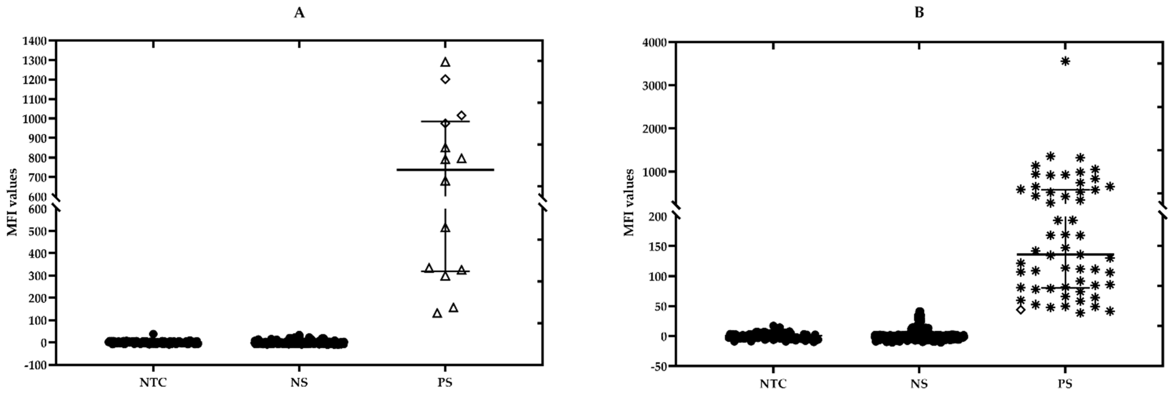
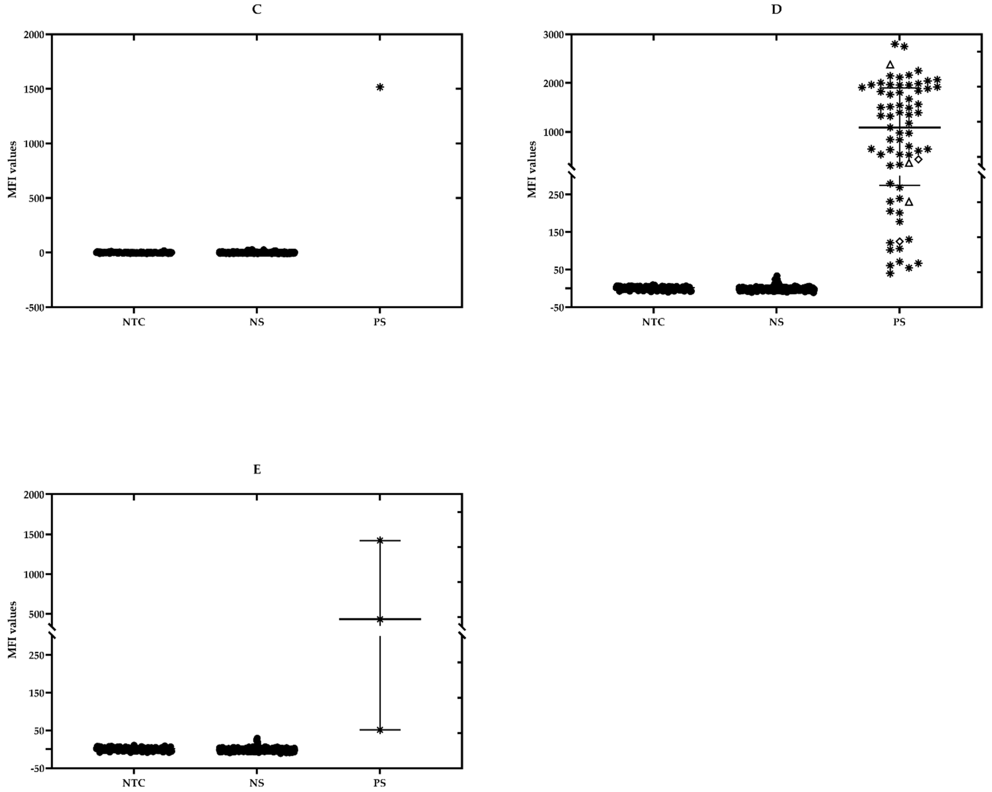
2.2. Analytical Specificity

The MFI data from all the samples in each assay was corrected for background (F − F0, where F is the MFI value of a sample, and F0 is the average background MFI value of the no-template controls (NTCs)). The multiplex analysis performed with different dilutions of positive control plasmid DNAs revealed that each species was correctly detected by its respective probe-bead set without cross-reactions with any other probe-bead sets. No positive MFI signal above the cut-off value was observed for any probe-bead set for which the corresponding specific plasmid DNA was not present. The six-plex xMAP assay had the highest (100%) analytical specificity, as no hybridization signals were observed for DNA templates from known negative animals (MFI values of negative animal samples did not differ from no-template controls) (Table 1 and Table 2). A decrease in the MFI values was observed when two plasmid combinations were present in the hybridization mix as compared to the MFI of a single species. This MFI reduction was more evident in mixtures where differences in concentrations were above one order of magnitude. For example, when 10,000 copies of E. canis were present singly, an average MFI value of 2088.6 was recorded (Table 1) but, when mixed with 100 copies of E. chaffeensis, the MFI value for E. canis decreased to 1500.5 (Table 2). It was also observed that some two plasmid combinations, at nonequivalent concentrations at a ratio greater than 100-fold, would only result in a positive MFI for the plasmid DNA having the higher concentration. For example, a combination of 10,000 copies of E. chaffeensis and 100 copies of E. canis gave average MFI values of 2231.6 and 28.2, respectively, without the background correction (Table 2). However, when the MFI values were corrected for background (F − F0), an average MFI of 2212.1, (2231.6 − 19.5) was calculated for 10,000 copies E. chaffeensis while 100 copies of E. canis resulted in an average MFI of 7.9 (28.2 − 20.3), a value below the cut-off (21.8) for E. canis probe-bead set (Table 2).

2.3. The Limit of Detection and Analytical Sensitivity

The detection limit was 10 copies/µL for E. canis, E. chaffeensis, and A. platys and 100 copies/µL for A. marginale, E. ewingii, and E. ruminantium. For analytical sensitivity, the MFIs differed by at least two times the MFI signal between copy numbers 10, 100, 1000, and 10,000 for E. canis, E. chaffeensis, and A. platys species; however, for A. marginale, E. ewingii, and E. ruminantium the analytical sensitivity of the MFI signals was less and not able to distinguish between 10 and 100 copies of the template.

2.4. Repeatability

Assessment of intra-assay and inter-assay variability was determined by the percentage of coefficient of variation (%CV) of replicates run either within the plate (intra-assay) or between the plates (inter-assay). Each probe-bead set gave a different value for the intra-assay and inter-assay %CV. Therefore, the intra-assay %CV ranged between 2% to 9%, and the inter-assay %CV ranged between 4% to 19%. These values are within the acceptable range; according to Luminex [60], the values for intra-assay %CV and inter-assay %CV should be below 10 and 20, respectively.

2.5. Testing of the Field Samples

A total of 455 blood samples collected from the six parishes in Grenada were analyzed by performing PCR and xMAP assays (Table 3) (Figure 1). The geographic location with most positive samples was concentrated in the southern half of the island, in St. George and St. David’s parishes (Figure 1). Positive and negative controls were included as part of the analysis. Figure 2 represents the distribution of the MFIs for each detected bacterial species in the six-plex xMAP assay for all samples. Anaplasma marginale was primarily detected in cattle and small ruminant blood samples, whereas E. canis and A. platys were detected predominantly in dog blood samples. The highest MFI values detected among A. marginale and A. platys positives were 1290.3 and 2799.8, respectively. Similarly, the highest MFI values for E. canis, and E. chaffeensis positive samples were 3561.3, and 1422.8, respectively. In contrast, the lowest MFI values detected for positives were as follows: A. marginale, 131.8; A. platys, 40.2; E. canis, 38.7; and E. chaffeensis, 51.8. Sample from a dog reacted with the E. ewingii probe and had an MFI of 1515.8 (Figure 2). None of the samples were positive for E. ruminantium.
Of the 455 samples analyzed, 137 (30.1%) tested positive for one or more pathogens. Of all the positive samples, 130 (94.9%) tested positive for a single pathogen infection, and seven samples (5.1%) tested positive for infections with two different pathogens. Single-pathogen infections were the highest in cattle for A. marginale (11/32; 34.3%), followed by dogs for E. canis, A. platys, and E. chaffeensis (110/358; 30.7%). Unique findings included finding E. canis in one goat, and A. platys in the blood of five ruminants (three bovine and two small ruminants).
Co-infection was not detected in any cattle and small ruminant samples, whereas 1.9% (7/358) of the dog samples tested positive for co-infection with two rickettsial pathogens; E. canis and E. chaffeensis in two dogs, E. canis and A. platys in four dogs, and A. platys and E. ewingii in one dog (Table 4).

2.6. Confirmatory PCR Assays

In order to confirm and validate the results obtained from this newly developed xMAP assay, conventional PCR assays and direct sequencing were performed on the extracted genomic DNA for a subset of the field samples. The target gene for PCR assays and sequencing were msp1a for A. marginale and 16S rRNA for E. canis, E. chaffeensis, and E. ewingii. The sequences shown in Table 5 had at least 94% identity with the reference sequences. All these sequences have been deposited in the GenBank (Accession numbers; MW474807-15, MW486117, and MW486118). xMAP results for A. platys positives were not confirmed via the conventional PCR method.

3. Discussion

In this study, we described the development and application of an xMAP six-plex PCR and oligonucleotide bead-based assay having high analytical specificity and sensitivity. The three-step assay involves: 1) PCR amplification from a sample DNA targeting a 100 bp 16S rRNA gene segment common to the six rickettsial pathogens; 2) PCR product hybridization with species-specific probes captured on magnetic beads and 3) detection of the hybrids by xMAP suspension array technology on a 96-well plate format. This assay has a quick turnaround time (3.5 h) and tests for six different pathogen DNAs simultaneously, which can be expanded to detect DNA targets from many other hemoparasite infections in a diverse host species. In particular, MagPix analyzers have the capability to test up to 84 different samples in addition to 12 controls in a 96-well plate format. Therefore, the assay has a broader applicability than a conventional PCR assay.
Anaplasma and Ehrlichia genera-specific primer sets targeting the 16S rRNA gene fragment described previously by Sirigireddy and Ganta [37] enabled the amplification of all six selected rickettsial pathogen-specific DNAs in a single step. Within the amplicon includes variable region sequences specific for each species allowing the design of magnetic capture probes, which permitted the identification of pathogen-specific detections simultaneously on the xMAP platform. One major advantage of this assay is that it can test DNA samples from different sources as demonstrated in the present study through the application of canine, bovine, ovine, and caprine blood samples. To validate the performance characteristics of this six-plex assay, we performed experiments to define the analytical specificity and sensitivity, detection limit, and repeatability. We achieved a 100% analytical specificity for the assay after limiting the PCR cycles to 35 and optimizing the hybridization step at 55 °C. Any deviation from these two parameters resulted in either cross-reactions amongst the probe-bead sets or a decrease in the MFI signals. Although there was a decrease in the MFI signals when two different species-specific positive control plasmid DNAs were mixed at nonequivalent concentrations above 10-fold, this did not preclude the detection of the plasmids as positives. The decrease in MFI when the assay included two different DNA targets was attributed to increased competition between the amplicons during the PCR step rather than xMAP assay detection, as reported previously [61]. We observed that some two plasmid combinations, at nonequivalent concentrations at a ratio greater than 100-fold, would result in a positive MFI detection only for the pathogen DNA present at the higher concentration. This could be a potential limitation of the assay in situations where clinical samples are co-infected with two or more pathogens differing in bacteremia by greater than 100-fold. However, this issue is not likely to be clinically significant because the antibiotic treatment regime for all these pathogens is the same. The detection limit for each of the analytes was between 10 and 100 copies/µL, with good analytical sensitivity between log fold concentrations of copy numbers that were above the limit of detection. The high analytical specificity and sensitivity of this assay was not affected by spiking the samples with pathogen-negative DNA from dogs or cattle. The results from these experiments illustrate that the assay is both sensitive and specific for detecting the target species even when genomic DNA from the host species is present during PCR and xMAP analysis.
The application of the xMAP six-plex PCR assay to 455 field samples detected 30.1% (137/455) of positives, where amongst these positive samples we found 34.3% (11/32) cattle for A. marginale, 16.4% (59/358) dogs for A. platys, and 13.9% (50/358) dogs for E. canis. These results are consistent with prior published reports from other endemic regions for bovine anaplasmosis and canine rickettsial pathogens of the world, including the Caribbean region [62,63,64,65,66]. Co-infection with E. canis and A. platys in the Grenadian dog population was 1.1% (4/358) and this observation is also similar to previous reports from the Caribbean islands of St. Kitts and Republic of Haiti [67,68]. In 2006, the reported prevalence of Grenadian dogs based on conventional PCR for E. canis and A. platys was 24.7% and 19.2%, respectively, with 5.5% of dual infections [50]. While the previously published data is consistent with the data reported here, the current study had a lower prevalence in E. canis and A. platys, which may reflect natural fluctuations in the pathogen distribution rather than the sensitivity differences in the assays. Although statistical comparisons were not performed between the prior published data and the current data, the reported variations in results may be due to differences in sample selection sites. Landscape and climatic differences among the various parishes in Grenada may have contributed to some variation in prevalence of the pathogens as noted in the current study compared to the previous reports. However, a more extensive study of the island is necessary. Anaplasma marginale has been known to infect bovine species worldwide, particularly in tropical, subtropical, and temperate regions [69,70,71,72,73]. However, in the Caribbean region, molecular detection of A. marginale infections in bovine species (e.g., cattle and buffalo) have only been reported from Cuba and Puerto Rico [74,75,76]. The present study augments those previous findings, and it is the first report on the molecular detection of A. marginale in cattle and small ruminants from Grenada. Previously, only serological data had been reported demonstrating exposure to A. marginale in the livestock animals in Grenada [47,53] and the current data validates the existence of the predicted cattle infections with the pathogen.
The xMAP assay analysis performed on various field specimens also revealed novel data. For example, our study is the second in reporting the presence of E. canis in goat blood [54]. Additional investigations are warranted to define the significance of E. canis infections in goats in causing disease in this host. The xMAP analysis of samples from domestic ruminants also resulted in the identification of A. platys DNA in five samples tested from domestic ruminants (cattle, goats, and sheep). This is the first study to report A. platys in these animal species. Additional investigations are necessary to determine the significance of A. platys infection to the ruminant population health. This study is also the first to report co-infections in dogs with E. chaffeensis and E. canis and with E. ewingii and A. platys. The sample cohort of dogs investigated in this study included some dogs having a travel history from the USA where E. chaffeensis and E. ewingii infections are more widespread. Therefore, the presence of E. chaffeensis and E. ewingii in dogs residing in Grenada may represent dogs originating from the USA. Since Grenada is a popular tourist and education destination with frequent movement of both humans and pet animals from other countries, including the USA and Canada, the introduction of new bacterial infections and tick species by means of importation of animals to the island cannot be ruled out.

4. Materials and Methods

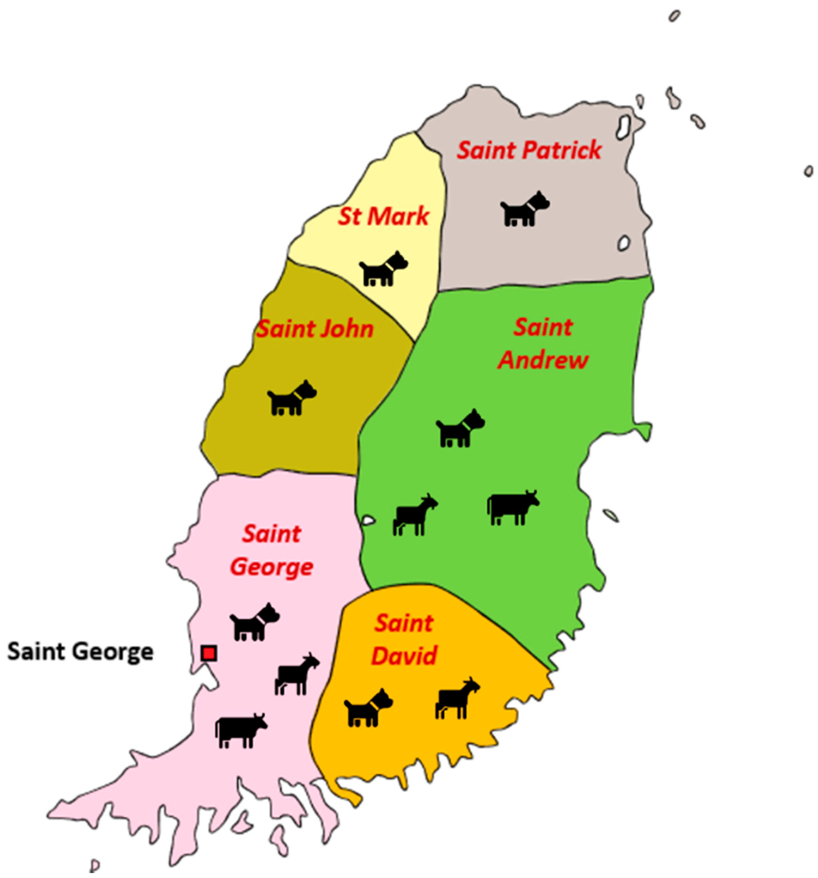
4.1. Collection of the Field Specimens

A total of 455 samples were collected between the years 2014 and 2018. These samples included whole blood collected from canines (n = 358), caprine and ovine (n = 65), and bovine (n = 32). (Table 3). The sample cohort for dogs was comprised of community-owned (mostly free roaming) dogs that were presented to the Small Animal Clinic (as part of diagnostic service) and to the Junior Surgery Laboratory (for blood sampling prior to spay and neuter surgeries) of the School of Veterinary Medicine (SVM) at St. George’s University (SGU). Cattle blood-samples were collected at farms in several parishes of Grenada (Figure 1) and from animals brought to the Large Animal Medicine and Surgery clinic of SVM, SGU. Small ruminants were sampled at various farms located in the parishes of Grenada (Figure 1). Bleeding and sample collections for all the animals were performed as per the approved Institutional Animal Care and Use Committee protocols of SGU. Blood samples were collected in EDTA-anticoagulant tubes and stored at 4 °C until processing, which typically occurred within 24 h. Table 3 represents the Grenada parish location of the blood samples collected from each animal species.

4.2. DNA Extraction

Genomic DNA (gDNA) from the blood samples was isolated from 100 µL of blood using the DNeasy Blood and Tissue kit (Qiagen, Hilden, Germany) as per the manufacturer’s instruction. Yield and purity of DNAs were determined using a Nanodrop™ 2000 Spectrophotometer (Thermo Scientific, Waltham, Massachusetts, US) and then all DNAs were stored at −20 °C until subsequent analyses.

4.3. Optimization of the PCR and xMAP Hybridization Assay Conditions

To achieve a balance between the analytical sensitivity and specificity of the assay developed in the present study, PCR and xMAP assay conditions were optimized, as shown in Table 6. Previous reports that used the PCR-based xMAP technology have reported the use of PCR cycles from 35 [77,78] up to 45 [79,80] depending on the target-genes. In the present study, 16S rRNA gene fragment was used for the PCR protocol, and the cycling conditions were carefully optimized after testing 30, 35, and 40 cycles in combination with different primer annealing temperatures (50 °C and 52 °C) and MgCl2 concentrations (1.5 mM and 2.5 mM).
For the xMAP hybridization step, different concentrations of the probes (0.1 and 0.2 nmol/µL) were tested in combination with different hybridization temperatures (50, 52, 55 and 60 °C), and incubation times (10, 15, and 20 min). A washed versus a no-wash protocol was also tested [55]. The probe for A. marginale was redesigned since the original probe [37] performed sub-optimally with various test conditions.

4.4. DNA Amplification for xMAP Assay

PCRs were performed to amplify the Anaplasma/Ehrlichia common 100 bp fragment corresponding to the 16S rRNA gene segment, as described previously [37] using forward primer EHRANA-F (5′-CTCAGAACGAACGCTGG-3′) and reverse primer EHRANA-R2bio (5′/5Biosg/GCATTACTCACCCGTCTGC-3′) (Integrated DNA Technologies, Coralville, Iowa, US). The reverse primer was 5′-biotinylated to allow conjugation of streptavidin phycoerythrin (SAPE) for detection via xMAP assay by Luminex (Austin, Texas, US). All amplification reactions contained 12.5 µL of 2X Platinum Hot Start Master Mix (1.5 mM MgCl2, 200 µM of each dNTP, and 1 U of Taq Platinum Polymerase (Invitrogen, California, US), an additional 0.5 µL of 1mM MgCl2 to increase the concentration to 2.5 mM, 1.25 µL of each of primers at 0.5 µM, 1 µL of gDNA template (10–20 ng/µL), and 8.5 µL of nuclease-free water to make the final volume to 25 µL. For the positive controls, six recombinant plasmid DNAs containing inserts corresponding to a 100 bp 16S rRNA gene-segments of A. marginale, A. platys, E. canis, E. chaffeensis, E. ewingii, and E. ruminantium, as described previously [37], were diluted to the copy numbers 100, 500, 1000, and 10,000. These plasmid controls were used in the PCR assays to serve as serial dilution positive controls. Ten reactions were included at each assay to serve as NTCs where gDNA solution was replaced with nuclease-free water. PCR thermal cycler conditions consisted of an initial denaturation step of 4 min at 94 °C, followed by 35 cycles of denaturation for 30 s at 94 °C, annealing of 30 s at 50 °C and an extension of 30 s at 72 °C. Subsequently, a final extension step was set at 72 °C for 5 min and the samples were stored at 4 °C.

4.5. Oligonucleotide xMAP Assay

4.5.1. Oligonucleotide Probe Design

Species-specific oligonucleotide probes (size range between 21 and 32 bp) corresponding to the variable regions located within the amplicons were prepared as described previously [37]. Anaplasma marginale-specific probe was designed in the current study and listed in Table 7, as the previously designed probe was found to be suboptimal for the xMAP analysis. All probes were manufactured with the inclusion of a six-carbon amino linker attached to the 5′ end (Integrated DNA Technologies, Coralville, IA, USA).

4.5.2. Oligonucleotide Probe Coupling to xMAP Beads

Species-specific oligonucleotide probes were conjugated to six unique sets of fluorescent-dyed magnetic carboxylated MagPlex® Microspheres (beads) (Luminex, Austin, TX, USA) by a chemical reaction attaching the carboxy groups on the beads to the amine group of the 5′ end probe liners, as per the manufacturer’s protocol [60]. Six different bead-set stocks (represented as R20, R25, R34, R38, R43, R48, and R53) were then resuspended by being vortexed at 20 rpm for 1 to 2 min and sonicated for 1 min. Five million beads from each bead-set stock were coupled to amine-linked species-specific oligonucleotide probes protocol as per manufacturer’s protocol [60]. Coupling efficiency was evaluated by hybridization of the coupled beads with two-fold dilutions of femtomolar concentrations of biotinylated oligonucleotide sequences that were complementary to the probes coupled to the bead-sets. The degree of hybridization was evaluated as outlined below.

4.5.3. Direct Hybridization of Blood-Derived DNA Samples to Six Oligonucleotide Probe-Coupled xMAP Beads

For these experiments, a no-wash protocol was followed [60]. Five micro liters of biotinylated PCR products were mixed with 33 µL of the six species-specific oligonucleotide bead mixtures and the volumes were raised to 50 µL with the addition of Tris-ethylene-diamine-tetraacetic acid (TE) buffer. The probe-bead mixture was calculated to contain about 23 beads/µL in 1.5× tetramethyl ammonium chloride (TMAC) hybridization buffer (4.5 M TMAC, 0.15% Sarkosyl, 75 mM Tris HCl, 6 mM EDTA pH 8.0). Biotin-labeled PCR products made from six recombinant plasmids and 10 NTCs were used as positive and negative controls, respectively. The NTCs served to calculate background MFI for each xMAP assay. The hybridization reaction was performed in Bio-Rad Hard-shell 96-well thin wall PCR plates (Hercules, CA, USA) at 55 °C for 15 min in Eppendorf Mastercycler® pro (Hamburg, Germany). Twenty-five microliters of SAPE (New England Biolabs, Ipswich, MA, USA) in 1× TMAC buffer at a final concentration of 4 µg/µL was added to each reaction well and was incubated at 55 °C for a further 5 min. Each bead was analyzed by a red-light emitting diode (LED), which identified unique fluorescent dyes coating the bead region for each probe coupled bead-set and a green LED, which detects the SAPE signal of the hybridization between the amplified biotinylated-product and with complementary oligonucleotide probe(s). All analyses were performed on a MAGPIX® instrument (Luminex, Austin, TX, USA) using xPONENT version 4.2 software (Luminex Corporation, Austin, TX, USA). The analysis was performed at 55 °C with an average of ~750 beads present for each of the six bead regions representing 750 replicate measurements for each bead region. An internal wash-step for each sample was carried out during the analysis to ensure removal of unbound SAPE reporter in the supernatant from interfering with the imaging chamber before reading the microspheres. The MFI data from all the samples in each assay was corrected for background (F − F0, where F is the MFI value of a sample, and F0 is the average background MFI value of the NTCs). For each probe hybridization, positive and negative cut-off values were calculated as the arithmetic mean of MFI values for the NTCs replicates included in each assay plus three standard deviations (SD) from the mean.

4.5.4. Determination of the Analytical Specificity of the Luminex Assay

The analytical specificity of all probe-bead sets was tested against recombinant plasmids (i) to identify a single DNA species when all six probe-bead sets are present and (ii) to identify combinations of two different positive control plasmids when added to the six-oligo bead sets. Table 8 illustrates the experimental set-up for different plasmid combinations tested to determine the analytical specificity of the Luminex assay. Every analysis was performed with 5 µL of PCR amplicon containing positive control plasmid DNA as described in the above section. To simulate natural infection, plasmids were spiked with known negative genomic DNAs (3–5 ng/µL) recovered from dog and cattle blood. The spiked DNAs from known negatives were run separately as additional controls along with the NTCs.

4.5.5. Determination of Limit of Detection and Analytical Sensitivity

Detection limit is defined as the lowest concentration of an analyte detected as positive by an assay [81]. To determine the detection limit of the assay, serial dilutions of plasmid controls of each species were used in PCRs. Six 10-fold dilutions created 10,000 to 0 copies/µL of each plasmid control [82]. To determine the ability of the assay to detect differences between MFIs of plasmid copy numbers or analytical sensitivity, the MFI of the plasmid controls were compared between each dilution or copy number. To simulate co-infections, a combination of two different control plasmids was added into wells containing the six different oligonucleotide coupled beads. All the plasmids at different concentrations were spiked with known negative dog or cattle DNA for PCR and xMAP experiments so that each reaction well also contained gDNA from negative field samples.

4.5.6. Intra-Assay and Inter-Assay Variability

The intra-assay variability (repeatability or precision within a plate or run) was calculated by testing plasmid controls at concentrations of 100 and 10,000 copies/µL, in four replicates each, on a single plate. The inter-assay repeatability or precision between plates and runs was calculated by running the six plasmid controls at various dilutions (100, 500, 1000, and 10,000 copies/µL) in four different plates each run on four different days. The percent of coefficient of variation (%CV) for the intra-assay and inter-assay variability was determined by dividing the standard deviation of the replicates by the mean, then multiplied by 100.

4.6. Confirmation of the Results by PCR and Sequencing

Results for a subset of xMAP positive samples were confirmed using conventional PCR assays followed by sequencing. The primers targeted different genes or regions than those used for the xMAP assay (Table 9) to confirm the pathogen-DNA in the field samples. Amplicons were extracted and sent for direct sequencing to the sequencing facility of Molecular Cloning Laboratories (South San Francisco, CA, USA). The sequencing histograms were cleaned and compared to the sequence-database present in GenBank® using the Nucleotide Basic Local Alignment Search Tool of the National Center for Biotechnology Information.

5. Conclusions

In conclusion, this novel six-plex oligonucleotide PCR-based bead assay is highly specific, sensitive, and repeatable for the simultaneous detection of six Anaplasma/Ehrlichia species frequently observed in vertebrate hosts and tick vectors. The assay identified multi-infections in dogs with two Anaplasma/Ehrlichia species, which is consistent with prior reports in Grenada using conventional PCR. Thus, it may contribute to our understanding of the expansion of vertebrate hosts and vectors for these pathogens, their prevalence and geographic spread, and to assess possible zoonotic concerns.

Author Contributions

Conceptualization, B.S., R.R.G., D.S., A.A., K.G. and M.J.W.; Data curation, B.S., R.R.G., D.S., A.A., M.L.-P., I.K., E.C., C.M.B. and M.J.W.; Funding acquisition, R.R.G. and M.J.W.; Investigation, B.S., R.R.G., A.A., M.L.-P., V.M.B., I.K., E.C., C.M.B. and M.J.W.; Project administration, B.S. and M.J.W.; Supervision, R.R.G., D.S. and M.J.W.; Validation, B.S., R.R.G. and M.J.W.; Writing—original draft, B.S.; Writing—review & editing, B.S., R.R.G., D.S., A.A., M.L.-P., V.M.B., I.K., E.C., C.M.B., K.G. and M.J.W. All authors have read and agreed to the published version of the manuscript.

Funding

This research was funded by One Health Research Initiative OHRI-10-26-8 grant awarded by WINDREF at SGU, Grenada, West Indies to M.J.W., and the PHS grant # AI070908 to R.R.G. from the National Institute of Allergy and Infectious Diseases, National Institutes of Health, USA.

Institutional Review Board Statement

The study was conducted according to the guidelines approved by the Institutional Animal Care and Use Committee of ST. GEORGE’S UNIVERSITY (protocol #17006-R dated 4 July 2017).

Informed Consent Statement

Informed consent was obtained from all the owners of the animal-subjects involved in the study.

Data Availability Statement

The new nucleic acid sequences have been deposited in the database of GenBank. Accession numbers provided by GenBank have been included in the manuscript under the Table 5.

Acknowledgments

The authors gratefully acknowledge St. George’s University, Grenada, West Indies for funding the project under One Health Research Initiative (OHRI-10-26-8), awarded to Melinda J. Wilkerson. This work was also supported by the PHS grant # AI070908 to Roman R. Ganta from the National Institute of Allergy and Infectious Diseases, National Institutes of Health, USA. We are grateful to all the students and clinicians at the Small Animal Clinic, Department of Large Animal Medicine and Surgery, and Department of Small Animal Medicine and Surgery, SVM, SGU for their assistance in sample collection. The skilled technical assistance provided by Elizabeth Peach is greatly appreciated.

Conflicts of Interest

The authors declare no conflict of interest. The funders had no role in the design of the study; in the collection, analyses, or interpretation of data; in the writing of the manuscript, or in the decision to publish the results.

References

  1. Dumler, J.S.; Barbet, A.F.; Bekker, C.P.; Dasch, G.A.; Palmer, G.H.; Ray, S.C.; Rikihisa, Y.; Rurangirwa, F.R. Reorganization of genera in the families Rickettsiaceae and Anaplasmataceae in the order Rickettsiales: Unification of some species of Ehrlichia with Anaplasma, Cowdria with Ehrlichia and Ehrlichia with Neorickettsia, descriptions of six new species combinations and designation of Ehrlichia equi and ‘HGE agent’ as subjective synonyms of Ehrlichia phagocytophila. Int. J. Syst. Evol. Microbiol. 2001, 51, 2145–2165. [Google Scholar] [CrossRef] [PubMed] [Green Version]
  2. Rymaszewska, A.; Grenda, S. Bacteria of the genus Anaplasma–characteristics of Anaplasma and their vectors: A review. Vet. Med. 2008, 53, 573–584. [Google Scholar] [CrossRef] [Green Version]
  3. Rar, V.; Golovljova, I. Anaplasma, Ehrlichia, and “Candidatus Neoehrlichia” bacteria: Pathogenicity, biodiversity, and molecular genetic characteristics, a review. Infect. Genet. Evol. 2011, 11, 1842–1861. [Google Scholar] [CrossRef]
  4. Centers for Disease Control and Prevention. Tickborne Disease Surveillance Data Summary. Available online: https://www.cdc.gov/ticks/data-summary/index.html (accessed on 22 August 2020).
  5. Alhassan, A.; Pumidonming, W.; Okamura, M.; Hirata, H.; Battsetseg, B.; Fujisaki, K.; Yokoyama, N.; Igarashi, I. Development of a single-round and multiplex PCR method for the simultaneous detection of Babesia caballi and Babesia equi in horse blood. Vet. Parasitol. 2005, 129, 43–49. [Google Scholar] [CrossRef]
  6. Breitschwerdt, E.B.; Hegarty, B.C.; Hancock, S.I. Sequential evaluation of dogs naturally infected with Ehrlichia canis, Ehrlichia chaffeensis, Ehrlichia equi, Ehrlichia ewingii, or Bartonella vinsonii. J. Clin. Microbiol. 1998, 36, 2645–2651. [Google Scholar] [CrossRef] [Green Version]
  7. Chang, Y.F.; Novosel, V.; Chang, C.F.; Kim, J.B.; Shin, S.J.; Lein, D.H. Detection of human granulocytic ehrlichiosis agent and Borrelia burgdorferi in ticks by polymerase chain reaction. J. Vet. Diagn. Investig. 1998, 10, 56–59. [Google Scholar] [CrossRef] [Green Version]
  8. Cui, Y.; Zhang, Y.; Jian, F.; Zhang, L.; Wang, R.; Cao, S.; Wang, X.; Yan, Y.; Ning, C. Development of duplex PCR for simultaneous detection of Theileria spp. and Anaplasma spp. in sheep and goats. Exp. Parasitol. 2017, 176, 1–7. [Google Scholar] [CrossRef]
  9. Hoskins, J.D.; Breitschwerdt, E.B.; Gaunt, S.D.; French, T.W.; Burgdorfer, W. Antibodies to Ehrlichia canis, Ehrlichia platys, and spotted fever group rickettsiae in Louisiana dogs. J. Vet. Intern. Med. 1988, 2, 55–59. [Google Scholar] [CrossRef] [PubMed]
  10. Hua, P.; Yuhai, M.; Shide, T.; Yang, S.; Bohai, W.; Xiangrui, C. Canine ehrlichiosis caused simultaneously by Ehrlichia canis and Ehrlichia platys. Microbiol. Immunol. 2000, 44, 737–739. [Google Scholar] [CrossRef] [PubMed]
  11. Kordick, S.K.; Breitschwerdt, E.B.; Hegarty, B.C.; Southwick, K.L.; Colitz, C.M.; Hancock, S.I.; Bradley, J.M.; Rumbough, R.; McPherson, J.T.; MacCormack, J.N. Coinfection with multiple tick-borne pathogens in a Walker Hound kennel in North Carolina. J. Clin. Microbiol. 1999, 37, 2631–2638. [Google Scholar] [CrossRef] [Green Version]
  12. Lorusso, V.; Wijnveld, M.; Majekodunmi, A.O.; Dongkum, C.; Fajinmi, A.; Dogo, A.G.; Thrusfield, M.; Mugenyi, A.; Vaumourin, E.; Igweh, A.C.; et al. Tick-borne pathogens of zoonotic and veterinary importance in Nigerian cattle. Parasites Vectors 2016, 9, 217. [Google Scholar] [CrossRef] [Green Version]
  13. Maggi, R.G.; Mascarelli, P.E.; Havenga, L.N.; Naidoo, V.; Breitschwerdt, E.B. Co-infection with Anaplasma platys, Bartonella henselae and Candidatus Mycoplasma haematoparvum in a veterinarian. Parasit Vectors 2013, 6, 103. [Google Scholar] [CrossRef] [PubMed] [Green Version]
  14. Meinkoth, J.H.; Ewing, S.A.; Cowell, R.L.; Dawson, J.E.; Warner, C.K.; Mathew, J.S.; Bowles, M.; Thiessen, A.E.; Panciera, R.J.; Fox, C. Morphologic and molecular evidence of a dual species ehrlichial infection in a dog presenting with inflammatory central nervous system disease. J. Vet. Intern. Med. 1998, 12, 389–393. [Google Scholar] [CrossRef]
  15. Njiiri, N.E.; Bronsvoort, B.M.; Collins, N.E.; Steyn, H.C.; Troskie, M.; Vorster, I.; Thumbi, S.M.; Sibeko, K.P.; Jennings, A.; van Wyk, I.C.; et al. The epidemiology of tick-borne haemoparasites as determined by the reverse line blot hybridization assay in an intensively studied cohort of calves in western Kenya. Vet. Parasitol. 2015, 210, 69–76. [Google Scholar] [CrossRef] [Green Version]
  16. Rajput, Z.I.; Hu, S.H.; Arijo, A.G.; Habib, M.; Khalid, M. Comparative study of Anaplasma parasites in tick carrying buffaloes and cattle. J. Zhejiang Univ. Sci. B 2005, 6, 1057–1062. [Google Scholar] [CrossRef] [PubMed] [Green Version]
  17. Ringo, A.E.; Adjou Moumouni, P.F.; Taioe, M.; Jirapattharasate, C.; Liu, M.; Wang, G.; Gao, Y.; Guo, H.; Lee, S.H.; Zheng, W.; et al. Molecular analysis of tick-borne protozoan and rickettsial pathogens in small ruminants from two South African provinces. Parasitol. Int. 2018, 67, 144–149. [Google Scholar] [CrossRef]
  18. De Tommasi, A.S.; Otranto, D.; Dantas-Torres, F.; Capelli, G.; Breitschwerdt, E.B.; de Caprariis, D. Are vector-borne pathogen co-infections complicating the clinical presentation in dogs? Parasites Vectors 2013, 6, 97. [Google Scholar] [CrossRef] [PubMed] [Green Version]
  19. Mylonakis, M.E.; Koutinas, A.F.; Baneth, G.; Polizopoulou, Z.; Fytianou, A. Mixed Ehrlichia canis, Hepatozoon canis, and presumptive Anaplasma phagocytophilum infection in a dog. Vet. Clin. Pathol. 2004, 33, 249–251. [Google Scholar] [CrossRef] [PubMed]
  20. Tuttle, A.D.; Birkenheuer, A.J.; Juopperi, T.; Levy, M.G.; Breitschwerdt, E.B. Concurrent bartonellosis and babesiosis in a dog with persistent thrombocytopenia. J. Am. Vet. Med. Assoc. 2003, 223, 1306–1310. [Google Scholar] [CrossRef]
  21. Gaunt, S.; Beall, M.; Stillman, B.; Lorentzen, L.; Diniz, P.; Chandrashekar, R.; Breitschwerdt, E. Experimental infection and co-infection of dogs with Anaplasma platys and Ehrlichia canis: Hematologic, serologic and molecular findings. Parasit Vectors 2010, 3, 33. [Google Scholar] [CrossRef] [Green Version]
  22. Gal, A.; Harrus, S.; Arcoh, I.; Lavy, E.; Aizenberg, I.; Mekuzas-Yisaschar, Y.; Baneth, G. Coinfection with multiple tick-borne and intestinal parasites in a 6-week-old dog. Can. Vet. J. 2007, 48, 619–622. [Google Scholar] [PubMed]
  23. Otranto, D.; Dantas-Torres, F.; Breitschwerdt, E.B. Managing canine vector-borne diseases of zoonotic concern: Part two. Trends Parasitol. 2009, 25, 228–235. [Google Scholar] [CrossRef] [PubMed]
  24. Chapman, A.S.; Bakken, J.S.; Folk, S.M.; Paddock, C.D.; Bloch, K.C.; Krusell, A.; Sexton, D.J.; Buckingham, S.C.; Marshall, G.S.; Storch, G.A.; et al. Diagnosis and management of tickborne rickettsial diseases: Rocky Mountain spotted fever, ehrlichioses, and anaplasmosis--United States: A practical guide for physicians and other health-care and public health professionals. MMWR Recomm. Rep. 2006, 55, 1–27. [Google Scholar] [PubMed]
  25. Reller, M.E.; Dumler, J.S. Ehrlichia, Anaplasma, and related intracellular bacteria. Man. Clin. Microbiol. 2015, 1135–1149. [Google Scholar] [CrossRef]
  26. Walker, D.H.; Bouyer, D.H. Rickettsia and orientia. In Manual of Clinical Microbiology, 11th ed.; Jorgensen, J.H., Carroll, K.C., Funke, G., Pfaller, M.A., Landry, M.L., Richter, S.S., Warnock, D.W., Carroll, K.C., Funke, G., Bernard, K.A., et al., Eds.; American Society of Microbiology Press: Washington, DC, USA, 2015; pp. 1122–1134. [Google Scholar]
  27. Chandrashekar, R.; Mainville, C.A.; Beall, M.J.; O’Connor, T.; Eberts, M.D.; Alleman, A.R.; Gaunt, S.D.; Breitschwerdt, E.B. Performance of a commercially available in-clinic ELISA for the detection of antibodies against Anaplasma phagocytophilum, Ehrlichia canis, and Borrelia burgdorferi and Dirofilaria immitis antigen in dogs. Am. J. Vet. Res. 2010, 71, 1443–1450. [Google Scholar] [CrossRef]
  28. Malheiros, J.; Costa, M.M.; do Amaral, R.B.; de Sousa, K.C.M.; André, M.R.; Machado, R.Z.; Vieira, M.I.B. Identification of vector-borne pathogens in dogs and cats from Southern Brazil. Ticks Tick Borne Dis. 2016, 7, 893–900. [Google Scholar] [CrossRef] [Green Version]
  29. O’Connor, T.P.; Hanscom, J.L.; Hegarty, B.C.; Groat, R.G.; Breitschwerdt, E.B. Comparison of an indirect immunofluorescence assay, western blot analysis, and a commercially available ELISA for detection of Ehrlichia canis antibodies in canine sera. Am. J. Vet. Res. 2006, 67, 206–210. [Google Scholar] [CrossRef] [PubMed]
  30. Biggs, H.M.; Behravesh, C.B.; Bradley, K.K.; Dahlgren, F.S.; Drexler, N.A.; Dumler, J.S.; Folk, S.M.; Kato, C.Y.; Lash, R.R.; Levin, M.L.; et al. Diagnosis and Management of Tickborne Rickettsial Diseases: Rocky Mountain Spotted Fever and Other Spotted Fever Group Rickettsioses, Ehrlichioses, and Anaplasmosis—United States. MMWR Recomm. Rep. 2016, 65, 1–44. [Google Scholar] [CrossRef]
  31. World Organization for Animal Health. Manual of Diagnostic Tests and Vaccines for Terrestrial Animals, Chapter 3.4.1. Bovine Anaplasmosis. Available online: https://www.oie.int/en/standard-setting/terrestrial-manual/access-online/ (accessed on 9 February 2021).
  32. Chen, S.M.; Dumler, J.S.; Bakken, J.S.; Walker, D.H. Identification of a granulocytotropic Ehrlichia species as the etiologic agent of human disease. J. Clin. Microbiol. 1994, 32, 589–595. [Google Scholar] [CrossRef] [PubMed] [Green Version]
  33. Courtney, J.W.; Kostelnik, L.M.; Zeidner, N.S.; Massung, R.F. Multiplex real-time PCR for detection of anaplasma phagocytophilum and Borrelia burgdorferi. J. Clin. Microbiol. 2004, 42, 3164–3168. [Google Scholar] [CrossRef] [Green Version]
  34. Dawson, J.E.; Biggie, K.L.; Warner, C.K.; Cookson, K.; Jenkins, S.; Levine, J.F.; Olson, J.G. Polymerase chain reaction evidence of Ehrlichia chaffeensis, an etiologic agent of human ehrlichiosis, in dogs from southeast Virginia. Am. J. Vet. Res. 1996, 57, 1175–1179. [Google Scholar]
  35. Eddlestone, S.M.; Gaunt, S.D.; Neer, T.M.; Boudreaux, C.M.; Gill, A.; Haschke, E.; Corstvet, R.E. PCR detection of Anaplasma platys in blood and tissue of dogs during acute phase of experimental infection. Exp. Parasitol. 2007, 115, 205–210. [Google Scholar] [CrossRef]
  36. Hulínská, D.; Langrová, K.; Pejcoch, M.; Pavlásek, I. Detection of Anaplasma phagocytophilum in animals by real-time polymerase chain reaction. Apmis 2004, 112, 239–247. [Google Scholar] [CrossRef]
  37. Sirigireddy, K.R.; Ganta, R.R. Multiplex detection of Ehrlichia and Anaplasma species pathogens in peripheral blood by real-time reverse transcriptase-polymerase chain reaction. J. Mol. Diagn. 2005, 7, 308–316. [Google Scholar] [CrossRef] [Green Version]
  38. Doyle, C.K.; Labruna, M.B.; Breitschwerdt, E.B.; Tang, Y.W.; Corstvet, R.E.; Hegarty, B.C.; Bloch, K.C.; Li, P.; Walker, D.H.; McBride, J.W. Detection of medically important Ehrlichia by quantitative multicolor TaqMan real-time polymerase chain reaction of the dsb gene. J. Mol. Diagn. 2005, 7, 504–510. [Google Scholar] [CrossRef] [Green Version]
  39. Benevenute, J.L.; Dumler, J.S.; Ogrzewalska, M.; Roque, A.L.R.; Mello, V.V.C.; de Sousa, K.C.M.; Gonçalves, L.R.; D’Andrea, P.S.; de Sampaio Lemos, E.R.; Machado, R.Z.; et al. Assessment of a quantitative 5’ nuclease real-time polymerase chain reaction using groEL gene for Ehrlichia and Anaplasma species in rodents in Brazil. Ticks Tick Borne Dis. 2017, 8, 646–656. [Google Scholar] [CrossRef] [PubMed] [Green Version]
  40. Lew, A.E.; Gale, K.R.; Minchin, C.M.; Shkap, V.; de Waal, D.T. Phylogenetic analysis of the erythrocytic Anaplasma species based on 16S rDNA and GroEL (HSP60) sequences of A. marginale, A. centrale, and A. ovis and the specific detection of A. centrale vaccine strain. Vet. Microbiol. 2003, 92, 145–160. [Google Scholar] [CrossRef]
  41. Lew, A.E.; Bock, R.E.; Minchin, C.M.; Masaka, S. A msp1alpha polymerase chain reaction assay for specific detection and differentiation of Anaplasma marginale isolates. Vet. Microbiol. 2002, 86, 325–335. [Google Scholar] [CrossRef]
  42. Vidotto, M.C.; Kano, S.F.; Gregori, F.; Headley, S.A.; Vidotto, O. Phylogenetic analysis of Anaplasma marginale strains from Paraná State, Brazil, using the msp1alpha and msp4 genes. J. Vet. Med. B Infect. Dis. Vet. Public Health 2006, 53, 404–411. [Google Scholar] [CrossRef]
  43. Gusa, A.A.; Buller, R.S.; Storch, G.A.; Huycke, M.M.; Machado, L.J.; Slater, L.N.; Stockham, S.L.; Massung, R.F. Identification of a p28 gene in Ehrlichia ewingii: Evaluation of gene for use as a target for a species-specific PCR diagnostic assay. J. Clin. Microbiol. 2001, 39, 3871–3876. [Google Scholar] [CrossRef] [PubMed] [Green Version]
  44. Zhang, C.; Xiong, Q.; Kikuchi, T.; Rikihisa, Y. Identification of 19 polymorphic major outer membrane protein genes and their immunogenic peptides in Ehrlichia ewingii for use in a serodiagnostic assay. Clin. Vaccine Immunol. 2008, 15, 402–411. [Google Scholar] [CrossRef] [PubMed] [Green Version]
  45. Inokuma, H.; Brouqui, P.; Drancourt, M.; Raoult, D. Citrate synthase gene sequence: A new tool for phylogenetic analysis and identification of Ehrlichia. J. Clin. Microbiol. 2001, 39, 3031–3039. [Google Scholar] [CrossRef] [PubMed] [Green Version]
  46. Michelet, L.; Delannoy, S.; Devillers, E.; Umhang, G.; Aspan, A.; Juremalm, M.; Chirico, J.; van der Wal, F.J.; Sprong, H.; Boye Pihl, T.P.; et al. High-throughput screening of tick-borne pathogens in Europe. Front. Cell. Infect. Microbiol. 2014, 4, 103. [Google Scholar] [CrossRef]
  47. Gibson, K.; Fitzpatrick, D.; Stone, D.; Noel, T.; MacPherson, C. Vector-borne diseases in the Caribbean: History and current status. Cab. Rev. 2016, 11, 1–28. [Google Scholar] [CrossRef]
  48. Lanza-Perea, M.; Zieger, U.; Qurollo, B.A.; Hegarty, B.C.; Pultorak, E.L.; Kumthekar, S.; Bruhl-Day, R.; Breitschwerdt, E.B. Intraoperative bleeding in dogs from Grenada seroreactive to Anaplasma platys and Ehrlichia canis. J. Vet. Intern. Med. 2014, 28, 1702–1707. [Google Scholar] [CrossRef]
  49. Wilkerson, M.J.; Black, K.E.; Lanza-Perea, M.; Sharma, B.; Gibson, K.; Stone, D.M.; George, A.; Nair, A.D.; Ganta, R.R. Initial development and preliminary evaluation of a multiplex bead assay to detect antibodies to Ehrlichia canis, Anaplasma platys, and Ehrlichia chaffeensis outer membrane peptides in naturally infected dogs from Grenada, West Indies. J. Vet. Diagn. Investig. 2017, 29, 109–114. [Google Scholar] [CrossRef] [Green Version]
  50. Yabsley, M.J.; McKibben, J.; Macpherson, C.N.; Cattan, P.F.; Cherry, N.A.; Hegarty, B.C.; Breitschwerdt, E.B.; O’Connor, T.; Chandrashekar, R.; Paterson, T.; et al. Prevalence of Ehrlichia canis, Anaplasma platys, Babesia canis vogeli, Hepatozoon canis, Bartonella vinsonii berkhoffii, and Rickettsia spp. in dogs from Grenada. Vet. Parasitol. 2008, 151, 279–285. [Google Scholar] [CrossRef]
  51. Camus, E.; Barré, N. Vector situation of tick-borne diseases in the Caribbean islands. Vet. Parasitol. 1995, 57, 167–176. [Google Scholar] [CrossRef]
  52. Camus, E.; Maran, M.; Montenegro-James, S.; Accipe, A. Sero-Epidemiological Survey on Bovine Tick-Borne Diseases in the Lesser Antilles. In Proceedings of the Final Research Co-Ordination Meetings of FAO/IAEA/SIDA Co-Ordinated Research Projects, Guadeloupe, Lesser Antilles, France, 13–17 June 1994; International Atomic Energy Agency: Vienna, Austria, 1998; pp. 241–245. [Google Scholar]
  53. Camus, E.; Montenegro-James, S. Bovine anaplasmosis and babesiosis in the Lesser Antilles: Risk assessment of an unstable epidemiologic situation. Vet. Res. 1994, 25, 313–317. [Google Scholar]
  54. Zhang, J.; Kelly, P.; Guo, W.; Xu, C.; Wei, L.; Jongejan, F.; Loftis, A.; Wang, C. Development of a generic Ehrlichia FRET-qPCR and investigation of ehrlichioses in domestic ruminants on five Caribbean islands. Parasit. Vectors 2015, 8, 506. [Google Scholar] [CrossRef] [PubMed] [Green Version]
  55. Gondard, M.; Cabezas-Cruz, A.; Charles, R.A.; Vayssier-Taussat, M.; Albina, E.; Moutailler, S. Ticks and Tick-Borne Pathogens of the Caribbean: Current Understanding and Future Directions for More Comprehensive Surveillance. Front. Cell. Infect. Microbiol. 2017, 7, 490. [Google Scholar] [CrossRef] [PubMed]
  56. Christopher-Hennings, J.; Araujo, K.P.; Souza, C.J.; Fang, Y.; Lawson, S.; Nelson, E.A.; Clement, T.; Dunn, M.; Lunney, J.K. Opportunities for bead-based multiplex assays in veterinary diagnostic laboratories. J. Vet. Diagn. Investig. 2013, 25, 671–691. [Google Scholar] [CrossRef] [PubMed] [Green Version]
  57. Livengood, J.; Hutchinson, M.L.; Thirumalapura, N.; Tewari, D. Detection of Babesia, Borrelia, Anaplasma, and Rickettsia spp. in Adult Black-Legged Ticks (Ixodes scapularis) from Pennsylvania, United States, with a Luminex Multiplex Bead Assay. Vector Borne Zoonotic Dis. 2020, 20, 406–411. [Google Scholar] [CrossRef]
  58. Reslova, N.; Huvarova, V.; Hrdy, J.; Kasny, M.; Kralik, P. A novel perspective on MOL-PCR optimization and MAGPIX analysis of in-house multiplex foodborne pathogens detection assay. Sci. Rep. 2019, 9, 2719. [Google Scholar] [CrossRef]
  59. Reslova, N.; Michna, V.; Kasny, M.; Mikel, P.; Kralik, P. xMAP Technology: Applications in Detection of Pathogens. Front. Microbiol. 2017, 8, 55. [Google Scholar] [CrossRef]
  60. Angeloni, S.D.S.; Dunbar, S.; Stone, V.; Swift, S. xMAP® Cookbook. A Collection of Methods and Protocols for Developing Multiplex Assays with xMAP Technology. Available online: https://cdn2.hubspot.net/hubfs/128032/Cookbook/BR76862.xMAPCookbook.Ed4.WR.pdf (accessed on 9 February 2021).
  61. Ros-García, A.; Juste, R.A.; Hurtado, A. A highly sensitive DNA bead-based suspension array for the detection and species identification of bovine piroplasms. Int. J. Parasitol. 2012, 42, 207–214. [Google Scholar] [CrossRef] [PubMed]
  62. Abanda, B.; Paguem, A.; Abdoulmoumini, M.; Kingsley, M.T.; Renz, A.; Eisenbarth, A. Molecular identification and prevalence of tick-borne pathogens in zebu and taurine cattle in North Cameroon. Parasit. Vectors 2019, 12, 448. [Google Scholar] [CrossRef] [PubMed]
  63. Kelly, P.J.; Xu, C.; Lucas, H.; Loftis, A.; Abete, J.; Zeoli, F.; Stevens, A.; Jaegersen, K.; Ackerson, K.; Gessner, A.; et al. Ehrlichiosis, babesiosis, anaplasmosis and hepatozoonosis in dogs from St. Kitts, West Indies. PLoS ONE 2013, 8, e53450. [Google Scholar] [CrossRef] [Green Version]
  64. Lara, B.; Conan, A.; Thrall, M.A.; Ketzis, J.K.; Branford, G.C.; Rajeev, S. Serologic and Molecular Diagnosis of Anaplasma platys and Ehrlichia canis Infection in Dogs in an Endemic Region. Pathogens 2020, 9, 488. [Google Scholar] [CrossRef]
  65. Peter, S.G.; Aboge, G.O.; Kariuki, H.W.; Kanduma, E.G.; Gakuya, D.W.; Maingi, N.; Mulei, C.M.; Mainga, A.O. Molecular prevalence of emerging Anaplasma and Ehrlichia pathogens in apparently healthy dairy cattle in peri-urban Nairobi, Kenya. BMC Vet. Res. 2020, 16, 364. [Google Scholar] [CrossRef]
  66. Tana-Hernández, L.; Navarrete-Arroyo, K.; Ron-Román, J.; Reyna-Bello, A.; Chávez-Larrea, M.A. PCR-diagnosis of Anaplasma marginale in cattle populations of Ecuador and its molecular identification through sequencing of ribosomal 16S fragments. BMC Vet. Res. 2017, 13, 392. [Google Scholar] [CrossRef] [PubMed] [Green Version]
  67. Loftis, A.D.; Kelly, P.J.; Freeman, M.D.; Fitzharris, S.; Beeler-Marfisi, J.; Wang, C. Tick-borne pathogens and disease in dogs on St. Kitts, West Indies. Vet. Parasitol. 2013, 196, 44–49. [Google Scholar] [CrossRef] [PubMed] [Green Version]
  68. Starkey, L.A.; Newton, K.; Brunker, J.; Crowdis, K.; Edourad, E.J.P.; Meneus, P.; Little, S.E. Prevalence of vector-borne pathogens in dogs from Haiti. Vet. Parasitol. 2016, 224, 7–12. [Google Scholar] [CrossRef]
  69. Carelli, G.; Decaro, N.; Lorusso, A.; Elia, G.; Lorusso, E.; Mari, V.; Ceci, L.; Buonavoglia, C. Detection and quantification of Anaplasma marginale DNA in blood samples of cattle by real-time PCR. Vet. Microbiol. 2007, 124, 107–114. [Google Scholar] [CrossRef] [PubMed]
  70. Cossío-Bayúgar, R.; Rodríguez, S.D.; García-Ortiz, M.A.; García-Tapia, D.; Aboytes-Torres, R. Bovine anaplasmosis prevalence in northern Veracruz state, Mexico. Prev. Vet. Med. 1997, 32, 165–170. [Google Scholar] [CrossRef]
  71. da Silva, N.B.; Taus, N.S.; Johnson, W.C.; Mira, A.; Schnittger, L.; Valente, J.D.M.; Vidotto, O.; Masterson, H.E.; Vieira, T.; Ueti, M.W.; et al. First report of Anaplasma marginale infection in goats, Brazil. PLoS ONE 2018, 13, e0202140. [Google Scholar] [CrossRef] [Green Version]
  72. Torioni de Echaide, S.; Knowles, D.P.; McGuire, T.C.; Palmer, G.H.; Suarez, C.E.; McElwain, T.F. Detection of cattle naturally infected with Anaplasma marginale in a region of endemicity by nested PCR and a competitive enzyme-linked immunosorbent assay using recombinant major surface protein 5. J. Clin. Microbiol. 1998, 36, 777–782. [Google Scholar] [CrossRef] [Green Version]
  73. Yousefi, A.; Rahbari, S.; Shayan, P.; Sadeghi-dehkordi, Z.; Bahonar, A. Molecular detection of Anaplasma marginale and Anaplasma ovis in sheep and goat in west highland pasture of Iran. Asian Pac. J. Trop. Biomed. 2017, 7, 455–459. [Google Scholar] [CrossRef]
  74. Fosgate, G.T.; Urdaz-Rodríguez, J.H.; Dunbar, M.D.; Rae, D.O.; Donovan, G.A.; Melendez, P.; Dobek, G.L.; Alleman, A.R. Diagnostic accuracy of methods for detecting Anaplasma marginale infection in lactating dairy cattle of Puerto Rico. J. Vet. Diagn. Investig. 2010, 22, 192–199. [Google Scholar] [CrossRef] [Green Version]
  75. Díaz-Sánchez, A.A.; Meli, M.L.; Obregón Álvarez, D.; Fonseca-Rodríguez, O.; Cabezas-Cruz, A.; Hofmann-Lehmann, R.; Corona-González, B. Development and application of a multiplex TaqMan® real-time qPCR assay for the simultaneous detection of Anaplasma marginale and Theileria annulata and molecular characterization of Anaplasma marginale from cattle in Western Cuba. Ticks Tick Borne Dis. 2020, 11, 101356. [Google Scholar] [CrossRef] [PubMed]
  76. Obregón, D.; Cabezas-Cruz, A.; Armas, Y.; Silva, J.B.; Fonseca, A.H.; André, M.R.; Alfonso, P.; Oliveira, M.C.S.; Machado, R.Z.; Corona-González, B. High co-infection rates of Babesia bovis, Babesia bigemina, and Anaplasma marginale in water buffalo in Western Cuba. Parasitol. Res. 2019, 118, 955–967. [Google Scholar] [CrossRef] [PubMed]
  77. Bergval, I.; Sengstake, S.; Brankova, N.; Levterova, V.; Abadía, E.; Tadumaze, N.; Bablishvili, N.; Akhalaia, M.; Tuin, K.; Schuitema, A.; et al. Combined species identification, genotyping, and drug resistance detection of Mycobacterium tuberculosis cultures by MLPA on a bead-based array. PLoS ONE 2012, 7, e43240. [Google Scholar] [CrossRef] [PubMed] [Green Version]
  78. Wuyts, V.; Roosens, N.H.; Bertrand, S.; Marchal, K.; De Keersmaecker, S.C. Guidelines for optimisation of a multiplex oligonucleotide ligation-PCR for characterisation of microbial pathogens in a microsphere suspension array. BioMed Res. Int. 2015, 2015, 790170. [Google Scholar] [CrossRef] [PubMed]
  79. Deshpande, A.; Gans, J.; Graves, S.W.; Green, L.; Taylor, L.; Kim, H.B.; Kunde, Y.A.; Leonard, P.M.; Li, P.E.; Mark, J.; et al. A rapid multiplex assay for nucleic acid-based diagnostics. J. Microbiol. Methods 2010, 80, 155–163. [Google Scholar] [CrossRef]
  80. Thierry, S.; Hamidjaja, R.A.; Girault, G.; Löfström, C.; Ruuls, R.; Sylviane, D. A multiplex bead-based suspension array assay for interrogation of phylogenetically informative single nucleotide polymorphisms for Bacillus anthracis. J. Microbiol. Methods 2013, 95, 357–365. [Google Scholar] [CrossRef]
  81. Armbruster, D.A.; Pry, T. Limit of blank, limit of detection and limit of quantitation. Clin. Biochem. Rev. 2008, 29 (Suppl. 1), S49–S52. [Google Scholar]
  82. Sharma, B. Development of a PCR Based Direct DNA Hybridization Oligonucleotide Microbead Assay for Detection of Ehrlichia and Anaplasma Species in Animals and Ticks from Grenada, West Indies. Ph.D. Thesis, St. George’s University, West Indies, Grenada, 2020. [Google Scholar]
  83. Chang, W.L.; Pan, M.J. Specific amplification of Ehrlichia platys DNA from blood specimens by two-step PCR. J. Clin. Microbiol. 1996, 34, 3142–3146. [Google Scholar] [CrossRef] [Green Version]
  84. Anderson, B.E.; Sumner, J.W.; Dawson, J.E.; Tzianabos, T.; Greene, C.R.; Olson, J.G.; Fishbein, D.B.; Olsen-Rasmussen, M.; Holloway, B.P.; George, E.H.; et al. Detection of the etiologic agent of human ehrlichiosis by polymerase chain reaction. J. Clin. Microbiol. 1992, 30, 775–780. [Google Scholar] [CrossRef] [Green Version]
  85. Anderson, B.E.; Greene, C.E.; Jones, D.C.; Dawson, J.E. Ehrlichia ewingii sp. nov., the etiologic agent of canine granulocytic ehrlichiosis. Int. J. Syst. Bacteriol. 1992, 42, 299–302. [Google Scholar] [CrossRef] [Green Version]
Figure 1. Map of Grenada showing six different parishes and the type of samples collected from each parish.
Pathogens 10 00192 g001
Figure 2. Scatter plot illustrating the distribution of corrected median fluorescent intensity values for the 455 samples obtained by the hybridization with different probes within the six-plex xMAP assay. PS: positive samples; NS: negative samples and NTC: No Template Controls (n = 92) from nine different assays and used to calculate the cut-offs. (A) A. marginale; (B) E. canis; (C) E. ewingii; (D) A. platys; (E) E. chaffeensis. ‘△’ blood from cattle; ‘◇’ blood from small ruminants; ‘✴’ blood from dogs. Whiskers in each plot represent the interquartile range (Median—middle line and lower and upper lines mean 25 and 75 percentiles of the distribution).
Pathogens 10 00192 g002aPathogens 10 00192 g002b
Table 1. Median fluorescence intensity (MFI) values of each probe-bead set shown by a single plasmid present in the hybridization mix.
DNA TargetsMFI for Hybridization of Species-Specific Oligonucleotides to DNA Targets
A. marginaleE. canisE. ewingiiA. platysE. chaffeensisE. ruminantium
NTCs c23.5 ± 11.1 a
(>34.6)
24 ± 14.1 a
(>38.5)
23.1 ± 11.4 a
(>35)
24.6 ± 10.8 a
(>35.8)
23.1 ± 10.2 a
(>33.5)
25.5 ± 10.2 a
(>36)
A. marginale #670.5b ± 118.621.6 ± 3.724 ± 2.622 ± 2.621.6 ± 4.624 ± 3.4
E. canis #21.6 ± 1.12088.6b ± 92.521.6 ± 222 ± 222 ± 124 ± 1.7
E. ewingii #18.3 ± 1.520.3 ± 0.51605.3b ± 14720.6 ± 2.519.3 ± 1.121 ± 2
A. platys #21 ± 2.622.8 ± 3.121.6 ± 1.11870.2b ± 27021.3 b ± 3.224.6 ± 3
E. chaffeensis #23.5 ± 2.325.3 ± 224 ± 1.723.6 ± 1.12715.8b ± 321.427 ± 1.7
E. ruminantium #20.3 ± 1.522.3 ± 2.321.6 ± 1.521 ± 1.719.3 ± 2828b ± 83.9
The MFI values shown in the table are the average MFI values of each sample run in three independent assays ± standard deviations (SD) (without background correction). a Cut-off values defined as mean ± 3SD of the no-template controls (NTCs) for each probe-bead set obtained with replicates of each sample run in three independent assays. Values in parenthesis indicates the cut off incorporating mean + 3SD. b In bold are the values considered as positive (based on mean ± 3SD). c No Template Controls (PCR grade water). # Plasmid present at 10,000 copies/µL.
Table 2. MFI values of each probe-bead set shown by two plasmid combinations present in the hybridization mix.
DNA TargetsMFI for Hybridization of Species-Specific Oligonucleotides to DNA Targets
A. marginaleE. canisE. ewingiiA. platysE. chaffeensisE. ruminantium
NTC c19.2 ± 4.5 a
(>23.7)
20.3 ± 1.5 a (>21.8)20.3 ± 1.5 a
(>21.8)
21.1 ± 4.2 a
(>25.3)
19.5 ± 1.5 a
(>21)
22 ± 2.4 a
(>24.4)
Neg D d17.8 ± 0.519 ± 018 ± 0.819.2 ± 0.518 ± 0.820.5 ± 1
Neg C e19.5 ± 0.519.8 ± 0.919.2 ± 0.920 ± 0.819.2 ± 0.521.5 ± 2
EC-AP f20 ± 01344.2b ± 16.220.5 ± 0.5331.1b ± 7.320.8 ± 1.723.6 ± 1.3
AP-EC g18.9 ± 0.236b ± 2.119.8 ± 1.21242.6b ± 39.320.2 ± 2.321.6 ± 1.7
EC-ECH h21.2 ± 0.91500.5b ± 19.222.8b ± 1.822 ± 0.855b ± 024.5b ± 1.2
ECH-EC i18.8 ± 1.728.2b ± 2.618.5 ± 218.9 ± 1.62231.6b ± 80.920.4 ± 1.7
AM-ECH j1077.6b ± 26.820 ± 0.825.2b ± 0.919.8 ± 0.5107.5b ± 2.621.8 ± 0.5
ECH-AM k25.8b ± 1.726.2b ± 1.223.5b ± 0.524.9 ± 13997b ± 120.827b ± 2
The MFI values shown in the table are the average MFI values of each sample run in four replicates within the assays ± SD (without background correction). a Cut-off values defined as mean ± 3SD of the NTCs for each probe-bead set obtained with four-replicates of each sample run on the same plate. Values in parenthesis indicates the cut off incorporating mean + 3SD. b In bold are the values considered as positive (based on mean ± 3SD). c No Template Control (PCR grade water). d Spike-DNA sample from known negative dog. e Spike-DNA sample from known negative cattle. f Ehrlichia canis at 10,000 copies/µL mixed with A. platys 100 copies/µL and spiked with negative dog DNA. g Anaplasma platys at 10,000 copies/µL mixed with E. canis 100 copies/µL and spiked with negative dog DNA. h Ehrlichia canis at 10,000 copies/µL mixed E. chaffeensis 100 copies/µL and spiked with negative dog DNA. i Ehrlichia chaffeensis at 10,000 copies/µL mixed with E. canis 100 copies/µL and spiked with negative dog DNA. j Anaplasma marginale at 10,000 copies/µL mixed E. chaffeensis 100 copies/µL and spiked with negative cattle DNA. k Ehrlichia chaffeensis at 10,000 copies/µL mixed A. marginale 100 copies/µL and spiked with negative cattle DNA.
Table 3. The number and parish location of the animals sampled.
Species/# Sampled# of SamplesParish (# Sampled)Year of Collection
SGSASMSPSDSJUk
Canine/353358 *18548124501582014–2018
Caprine and Ovine/65651825--22--2017–2018
Bovine/3232257-----2017
Total (%)455
228
(50.1)
80
(17.6)
12
(2.6)
4
(0.8)
72
(15.8)
1
(0.2)
58
(12.7)
* Blood samples from five dogs were collected twice (one week apart). SG: Saint George parish; SA: Saint Andrew parish; SM: Saint Mark parish; SP: Saint Patrick parish; SD: Saint David parish; SJ: Saint John parish; Uk: Unknown (location not recorded).
Table 4. Number and percentage of animals that tested positive for single or multiple bacterial species by the Luminex assay.
Bacterial sp.CanineBovineCaprine and OvineTotal
Animal sp.
Single speciesinfectionsAM-11/32 (34.3)3/65 (4.6)14
EC50/358 (13.9)-1/65 (1.5)51
AP59/358 (16.4)3/32 (9.3)2/65 (3)64
ECH1/358 (0.2)--1
Total110/358 (30.7)14/32 (43.7)6/65 (9.2)130
Co-infectionsEC-ECH2/358 (0.5)--2
EC-AP4/358 (1.1)--4
EE-AP1/358 (0.2)--1
Total7/358 (1.9)0/36 (0)0/65 (0)7
Grand Total117/358 (32.6)14/32 (43.7)6/65 (9.2)137
Unique findings are in bold. AM: A marginale; EC: E. canis; EE: E. ewingii; AP: A. platys; ECH: E. chaffeensis; and ER: E. ruminantium. Hyphenated abbreviations indicate co-infections of two different pathogens.
Table 5. Homology between deposited sequences and reference sequences in GenBank.
SpeciesTarget Gene# of Samples Tested# of Samples SequencedDeposited
Sequence
GenBank #s
Length (bp)Percentage of Identity (%)Reference
Sequence
A. marginalemsp1a82MW486117
MW486118
568
326
94.00
94.00
NC_012026
NC_012026
E. canis16S rRNA62MW474807
MW474808
335
335
99.40
99.40
NR_118741
NR_118741
E. chaffeensis16S rRNA106MW474809
MW474810
MW474811
MW474812
MW474813
MW474814
300
334
318
334
333
334
100.00
100.00
100.00
100.00
100.00
100.00
NR_074500
NR_074500
NR_074500
NR_074500
NR_074500
NR_074500
E. ewingii16S rRNA61MW474815308100.00NR_074500
Table 6. xMAP assay optimization at different levels.
Optimization ConditionsTest Conditions
PCR optimizationPrimer Annealing temperature (°C)50, 52
MgCl2 concentration (mM)1.5, 2.5
PCR cycle numbers30, 35, 40
xMAP optimizationHybridization Temperature (°C)50, 52, 55, 60
Hybridization time (min.)10, 15, 20
PCR product volume (µL)5, 10
xMAP protocolWashed, no-wash
Concentration of the probes (nmol/µL)0.1, 0.2
Table 7. Sequences of the oligonucleotide probes that were covalently linked to the carboxylated microspheres used in the development of xMAP assay for the detection of species identification of Ehrlichia/Anaplasma in animals.
ProbesBacterial SpeciesSequences (5′-3′)xMAP COOH-Microsphere Regions for Probe BindingReference
RG270EcanE. canisTATAGCCTCTGGCTATAGGAAATTGTTAGR25[37]
RG266EchafE. chaffeensisCTTATAACCTTTTGGTTATAAATAATTGTTAGR43[37]
RG268EewinE. ewingiiCTAAATAGTCTCTGATTTAGATAGTTGTTAGR34[37]
RG260ErumE. ruminantiumGTTATTTATAGCTTCGGCTATR48[37]
RG272AplatA. platysCGGATTTTTGTCGTAGCTTGCTATGATR38[37]
RG262AmargA. marginaleCGTATACGCAGCTTGCTGCGTR20This study
R20, R25, R34, R38, R43, and R48 represent bead-set stocks with different spectral properties.
Table 8. Combinations of plasmid DNA mixes analyzed to determine analytical specificity.
Species Mix
1 2 3 4 5 6 7 and 8 9 and 10 11 and 12
E. canis
E. chaffeensis
E. ewingii
E. ruminantium
A. platys
A. marginale
✓ Shows the presence of the corresponding bacterial plasmid DNA in the mix. ✗ Indicates the absence of the corresponding bacterial plasmid DNA in the mix. Positive control plasmid mixtures 1 to 6 contain single bacterial species (indicated by ✓) at 10,000 copies/µL in each mixture. Positive control plasmid mixtures 7 and 8, 9 and 10, and 11 and 12 contain two bacterial species (indicated by ✓) at 10,000 and 100 copies/µL.
Table 9. Primers used for confirmatory PCR assays for various species.
SpeciesTarget GenePrimer NameSequence (5′→3′)Amplicon Size (bp)Reference
A. marginalemsp1aMSP1aF1
MSP1aRN
GCATTACAACGCAACGCTTGAG
CAGGAGCACCACCAAACATCATCACA
1638This study
A. platys16S rRNAEP2
EP3
GAAGATAATGACGGTACCC
CGTTTTGTCTCTGTGTTG
385[83]
E. canis16S rRNAECA
HE3
CAATTATTTATAGCCTCTGGCTATAGG
TATAGGTACCGTCATTATCTTCCCTAT
385[34]
E. chaffeensis16S rRNAHE1
HE3
CAATTGCTTATAACCTTTTGGTTATAAAT
TATAGGTACCGTCATTATCTTCCCTAT
385[84]
E. ewingii16S rRNAEE72
HE3
CAATTCCTAAATAGTCTCTGACTATT
TATAGGTACCGTCATTATCTTCCCTAT
385[85]
Publisher’s Note: MDPI stays neutral with regard to jurisdictional claims in published maps and institutional affiliations.

