Article

Using Aerosol OT in Hexane Solution to Synthesize Calcium Nitrate Self-Healing Refined Microcapsules for Construction Applications

1
Department of Civil Engineering, Munib and Angela Masri Faculty of Engineering, Aqaba University of Technology, Aqaba P.O. Box 11947, Jordan
2
Department of Construction Management, University of Houston, Houston, TX 77204, USA
3
Department of Civil and Architectural Engineering, Qatar University, Doha P.O. Box 2713, Qatar
*
Author to whom correspondence should be addressed.
Buildings 2022, 12(6), 751; https://doi.org/10.3390/buildings12060751
Submission received: 21 April 2022 / Revised: 27 May 2022 / Accepted: 30 May 2022 / Published: 31 May 2022
(This article belongs to the Collection Sustainable and Green Construction Materials)

Abstract

:
The micro-encapsulation procedure of calcium nitrate in urea-formaldehyde shell is well known. The most recent developed method for the synthesis of the calcium nitrate self-healing micro-capsules was based on the in-situ polymerization using water-in-oil emulsion. Although the microcapsules’ yield was significantly improved using this approach, incorporating the micro-capsules into concrete mixes has been found to reduce strength. One potential strength reduction cause might be the presence of sulfonic acid as a component in the continuous (oil) phase. As the anionic surfactant, Aerosol OT (AOT) has been widely used to prepare water-in-oil emulsions and to form aggregates in non-polar solvents; submicron calcium nitrate refined microcapsules were synthesized using AOT in hexane solution. While the aqueous phase in the original encapsulation procedure has not been altered, the continuous organic phase was prepared by dissolving AOT in hexane. The prepared microcapsules were characterized using Scanning Electron Microscopy (SEM). The preliminary assessment of the effect of incorporating of the refined microcapsules into cementitious materials has been carried out by preparing mortar mixes using 75% capsules’ concentration (by weight of cement). The reported yield values, average shell thickness, and average diameter of the prepared microcapsules were found satisfactory. Moreover, the mortar samples containing calcium nitrate refined microcapsules that were prepared using the proposed method did not experience significant reduction in their mechanical properties. Hence, such encapsulation procedure may be adopted for further investigation of the self-healing efficiency in cementitious materials of the microcapsules prepared using the proposed procedure. Future work shall be directed towards this end.

1. Introduction

Cracking is a major problem that severely affects the durability of concrete structures. Normally, surface cracking is repaired using mortar and other repair techniques that are costly and time-consuming. Hence, research has been recently directed towards the utilization of smart materials to minimize concrete’s cracks, increase its durability, and prevent its structural damage. The incorporation of self-healing microcapsules into cementitious mixes is currently considered as a promising alternative to traditional crack prevention and repair of concrete structures.
The cracks associated with excessive stresses are conceptually used to stimulate the self-healing mechanism in concrete elements containing self-healing microcapsules. The microcapsule’s structure consists of a shell containing a healing agent, which will be released when a crack tip reaches the microcapsule and ruptures its surrounding shell. The interaction between the released healing agent and catalyst will form calcium-silicate hydrate gels (C-S-H) that fill and prevent the cracks from propagating.
Various healing agent materials, such as sodium silicate and polyurethane, have been investigated in the literature [1,2,3]. However, their healing potentials have been found satisfactory these materials had limited use due to their high costs. As an alternative, low-cost healing agent, calcium nitrate has been encapsulated inside a urea-formaldehyde shell by Hassan, et al. [4]. The produced microcapsules had a satisfactory healing efficiency. However, they have adverse effects on concrete compressive and flexural strengths [5]. Accordingly, a modification to the original calcium nitrate micro-encapsulation procedure was proposed to mitigate the associated strength reductions [6]. In their work, the authors have concluded that the sulfonic acid catalyst used in the original encapsulation procedure was a potential cause for the strength reduction. They have reported that the sulfuric acid may act as retarder and reduces the hydration rate of Portland cement. Hence, they have modified the original micro-encapsulation method by altering the composition of the continuous phase while keeping the aqueous phase unchanged. The modification consisted of using low Hydrophilic–Lipophilic (HLB) emulsifier; Span 85 (Sorbitane trioleate), as a partial replacement of the sulfonic acid catalyst [6]. The continuous phase has been designed to include 9.1% of HLB emulsifier and 0.18% of sulfonic acid (by weight of water in the aqueous phase) dissolved in hexane as an organic solvent. The authors have also suggested that the incorporation of microcapsules may be limited to 0.75% (by weight of cement) to keep the strength reductions below 10%. However, this microcapsule percentage has resulted in about 40% reduction of the elastic modulus. Moreover, the SEM images of the modified microcapsules showed that the average diameter of the modified microcapsules (70 µm) was larger than that of the microcapsules prepared using the original procedure (51 µm).
In 2019, Milla, et al. [7] proposed another modification to the microencapsulation methodology that consists of reducing the amount of the sulfonic acid catalyst to 0.1 g and adding 0.8 g of Sorbitan monostearate (Span 60) surfactant to stabilize the emulsion instead of Span 85 proposed by Al-Ansari, et al. [6]. The agitation rates of 800 rpm and 1500 rpm have produced microcapsules with averages diameters of 50 µm and 22 µm, respectively. The authors have then used the produced microcapsules in steel fiber reinforced concrete mixes and compared their healing efficiency with those prepared using the original procedure with 0.6 g sulfonic acid [4]. The results have shown that all specimens containing microcapsules had significantly higher healing efficiencies than the control ones (without microcapsules). However, the authors concluded that the specimens containing microcapsules prepared using the original procedure had better healing efficiency than those produced using the modified procedure (with Span 60). The addition of Span 60 emulsifier has resulted in the formation of hydrophobic shell coating which increased the hydrophobic attraction between the particles. This has increased the agglomeration and has decreased the uniformity of the dispersion of capsules throughout the concrete matrix. He and Shi [8] conducted a study to evaluate the feasibility of a self-healing system consists of calcium nitrate (Ca(NO3)2)/urea-formaldehyde (UF) microcapsules and polyvinyl alcohol (PVA) microfibers to improve the early-age durability of cement mortars. The reported results demonstrated that incorporating a mixture of Ca(NO3)2)/(UF) microcapsules and PVA microfibers together eliminated 25% of the total shrinkage during 35 days curing period. Moreover, the admixed self-healing system could decrease more than 75% of the gas permeability of the tested mortar samples. However, reductions in the compressive strength and chloride migration coefficient could be witnessed due to the incorporation of such self-healing system into the mortar mixes.
Previous studies have demonstrated the significant and critical effect of microcapsule diameters on the healing process in cementitious mixtures [9]. Many of the studies have proved that the optimum self-healing efficiency is achieved by incorporating higher percentages of smaller size microcapsules. It has been reported that the optimum healing efficiency may be associated with the integration of microcapsules with diameters less than 30 µm [6]. Accordingly, other modifications may be proposed to the calcium nitrate encapsulation procedure to refine the physical and chemical properties of the produced microcapsules. Producing refined and smaller sized self-healing microcapsules will allow for increasing healing efficiency in composites with smaller gap spacings [10]. The smaller sized capsules will be more uniformly distributed within the matrix and will be able to fill the macro and microcracks as well, leading to enhanced healing efficiency. Recently, Farshi Azhar, et al. [11] utilized storing epoxy and calcium nitrate as healing agents to be encapsulated in the urea-formaldehyde shell. They could produce microcapsules with perfect thermal stability (up to 260 °C), outer rough surface, 1–100 μm diameter and shell thicknesses in the range of 0.2–0.6 µm. The authors demonstrated that the prepared microcapsules improved the durability of cementitious materials and provided high performance microcracks’ sealing.
The anionic surfactant Dioctyl Sodium Sulfosuccinate (Aerosol-OT, AOT) is extensively used as a stabilizer to prepare oil and water emulsions [12,13,14,15,16]. It is also used for pharmaceutical formulations due to its biocompatibility [12]. AOT is soluble in aliphatic compounds and can form aggregates in non-polar solvents without the addition of any co-surfactant. Such advantages over other surfactants make the AOT suitable for the preparation of water-in-oil emulsions [17,18]. Accordingly, researchers had successful attempts to synthesize different nano particles using AOT in hexane solutions [17,19,20,21]. For example, AOT dissolved in n-Hexane was used to synthesize pullulan nanoparticles with sizes less than 50 nm for drug applications [20]. On the another hand, 20 nm wide gold nanorods of length 200 nm were successfully prepared using the concentrated triplex solution of water/AOT/hexane [19].
A review of the structural studies of AOT-stabilized water-in-oil microemulsions had been presented by Eastoe, et al. [21]. The authors have demonstrated that the simplest and best comprehended microemulsions are ternary systems consisting of a single surfactant, oil (normally an alkane), and water. Both water-in-oil (w/o) and oil-in-water (o/w) microemulsions can be formed depending on the percentages of water and oil. In the w/o microemulsions, a one molecule thick layer of the surfactant, dispersed in the continuous oil phase, is used to stabilize nanometer-sized domains of water. Spherical water droplets readily stabilized by surfactants such as AOT could be considered as the simplest w/o microstructure. The size of the droplets can be altered by the composition ratio of water to the amount of AOT defined as w (=[H2O]/[AOT]). AOT-stabilized w/o microemulsions have a full range of stability at a temperature range from −20 °C to 100 °C. It has been demonstrated that the w/o emulsions with high w values (that contain small amount of AOT) are unstable. Hence, to get stable microemulsion over a wider range of temperatures, values of w less than 20 may be adopted [21]. The authors have focused on the w of 10 for the formation of w/o stable emulation. Hence, this ratio will be considered in this study as a preliminary ratio and future studies may use different values.
The main objective of the current study is to produce self-healing microcapsules with diameters less than the values reported in literature so far and with less adverse effects on the mechanical properties of the cementations mixes at the same time. It proposes a modification of the calcium nitrate microcapsule preparation procedures that were proposed by Hassan, et al. [4] and Al-Ansari, et al. [6] to produce refined and smaller size microcapsules. Producing such smaller sized microcapsules may lead to better healing efficiency within the cementitious matrix as the smaller microcapsules could fill not only the macrocracks but also the micro ones. Additionally, more uniform distribution may be achieved within the matrix if smaller sized self-healing microcapsules have been incorporated. This study is novel as it may be considered the first attempt to use the AOT stabilizer to prepare calcium nitrate self-healing microcapsules for cementitious materials. The utilization of AOT may decrease or eliminate the adverse effect of the acidic catalysts (which have been used in literature to polymerize the microcapsules) on the mechanical properties of the cementitious mixes. This modification proposed in this study includes using AOT as a stabilizer for the preparation of the continuous phase, instead of using the sulfonic acid catalyst with a low Hydrophilic–Lipophilic Balance (HLB) emulsifier, while keeping the aqueous phase composition the same. The prepared microcapsules were then characterized using Scanning Electron Microscopy (SEM). The microcapsule effect on mortar compressive and flexural strengths were evaluated before considering a comprehensive concrete testing. The dosage of calcium nitrate microcapsules recommended by Al-Ansari, et al. [6], i.e., 0.75%, was used as preliminary dosage for the mortar sample testing. The study is mainly focusing on the characterization of the prepared microcapsules and limited the compressive and flexural strengths’ testing of mortar samples at 7 days only (to limit the time consumed in preparing large quantities of the prepared microcapsules). Based on the 7 days strength results, future testing will be directed towards investigating the 28th and 56th days strengths on mortar and large concrete samples. Different microcapsules’ concentrations may be also studied. Future work should be also directed towards investigating the self-healing efficiency of the prepared microcapsule on concrete samples.

2. Experimental Program

2.1. Microcapsule Preparation
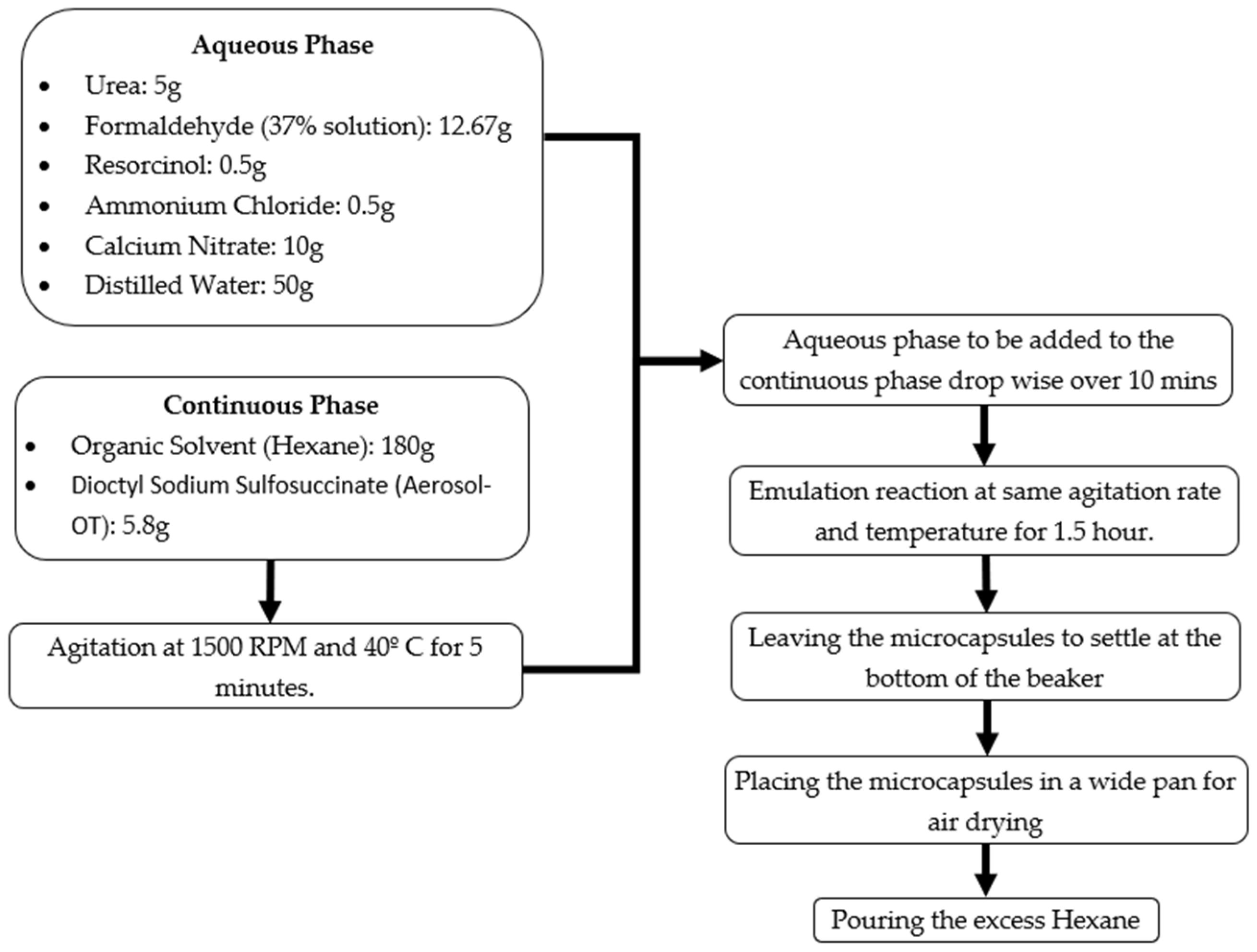
2.1.1. Synthesis

The preparation procedure of self-healing microcapsules developed by Hassan, et al. [4] was used as the basis of this study. A water-in-oil emulsion was used to chemically polymerize the calcium nitrate healing agent in urea-formaldehyde shell. Urea, formaldehyde, resorcinol, ammonium chloride, and calcium nitrate were dissolved in distilled water as core materials to prepare an aqueous phase with 1:1.9 formaldehyde to urea molar ratio. The continuous phase constituents were altered by dissolving AOT in an organic solvent (Hexane) with water to AOT ratio (w) of 10. The water weight is computed by adding the weight of the distilled water to the weight of solution in the formaldehyde (i.e., weight of the 37% solution formaldehyde multiplied by 0.63). The modified composition of both phases is summarized in Table 1.

2.1.2. Emulsification and Polymerization

A high shear rate of 1500 RPM at a high temperature of 40 °C, which was recommended by Hassan, et al. [4] and Milla, et al. [5], was used to agitate the continuous phase components. The aqueous phase was then added dropwise to the AOT-in- hexane continuous phase over 10 min and the emulsion was left to react at the same agitation rate and temperature for 1.5 h. Finally, the emulation was kept for some time to settle, and the excess hexane was decanted. The settled microcapsules were placed in a wide pan for air drying. A schematic illustration of the microcapsules’ preparation method is presented in Figure 1.

2.2. Scanning Electron Microscopy

The microcapsules were characterized using a Nova NanoSEM model scanning electron microscope. The microcapsules were scattered on top of a double-sided tape attached to a pin stub specimen mount. Platinum was then used to coat the samples for four minutes before imaging them under an accelerating voltage of 3 kV secondary electron mode.

2.3. Mortar Testing

Mortar samples were prepared using the original procedure (with sulfonic acid) [6] and with the modified procedure (with AOT) based on 0.75% microcapsule concentration (by weight of cement). Compressive and flexural strength tests were carried out on the prepared samples according to ASTM C109 [22] and ASTM C348 [23], respectively. The mortar mix design is summarized in Table 2.
Two batches were prepared for each mortar mix as the batch volume is approximately 1.5 L. Three 50-mm cubes and three 40 mm × 40 mm × 160 mm prisms were prepared from each batch as per ASTM C109 [22] and ASTM C348 [23], respectively. After 24 h, the specimens were demolded and cured for 7 days in limewater. Table 3 summarizes the testing matrix.
The specimens were tested in compression and three point bending after 7 days of moist curing using a universal testing machine. Scanning electron microscope images of the samples’ fractured surfaces were carried out to investigate microcapsule diameter and shell thickness. Gold was deposited as a conductive material on the samples’ surfaces to increase the image resolution. The image scales were selected to capture the microcapsules and their surrounding regions. The images were obtained under secondary electron mode at an accelerating voltage of 3 kV.

3. Results and Discussion

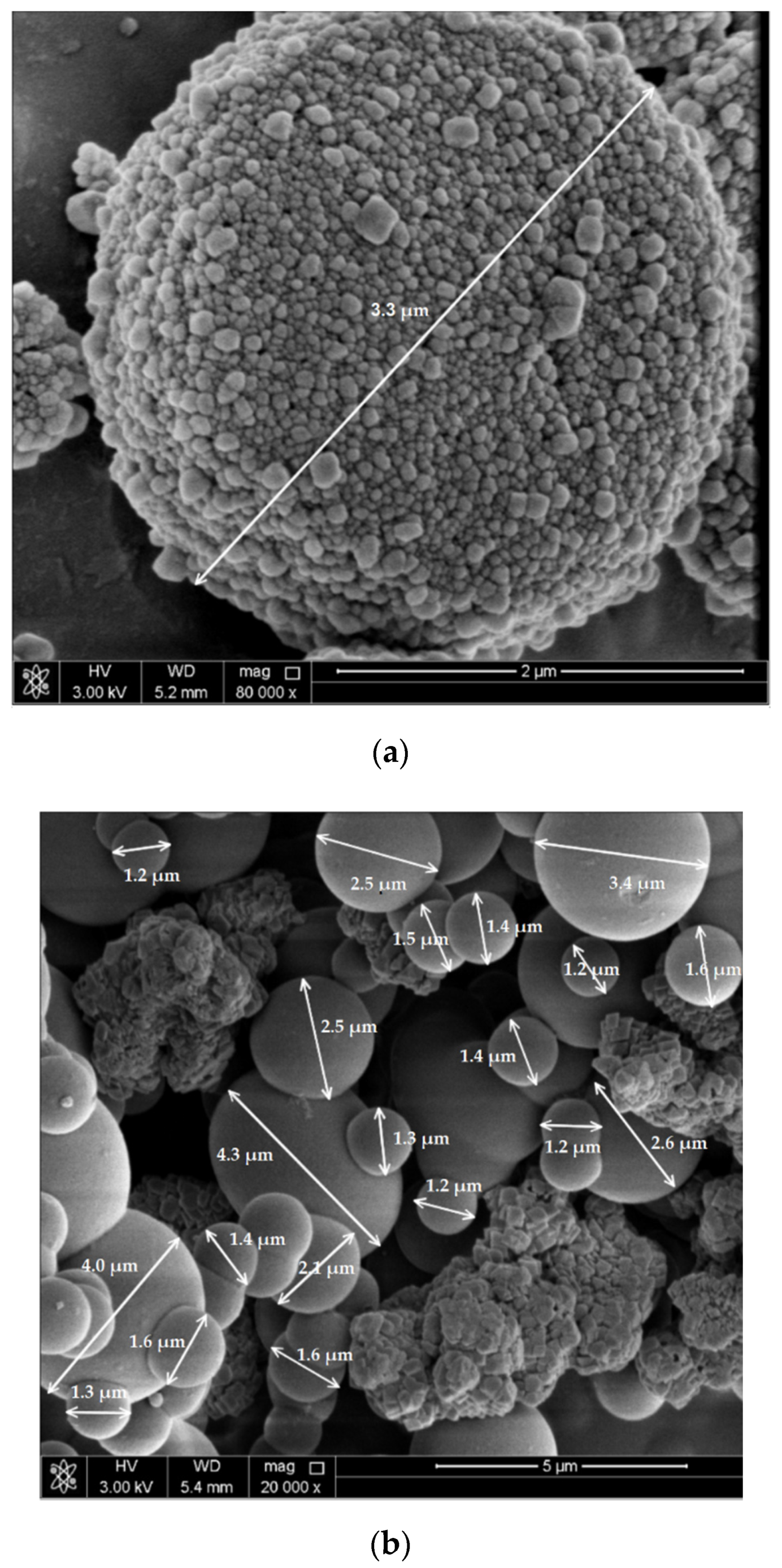
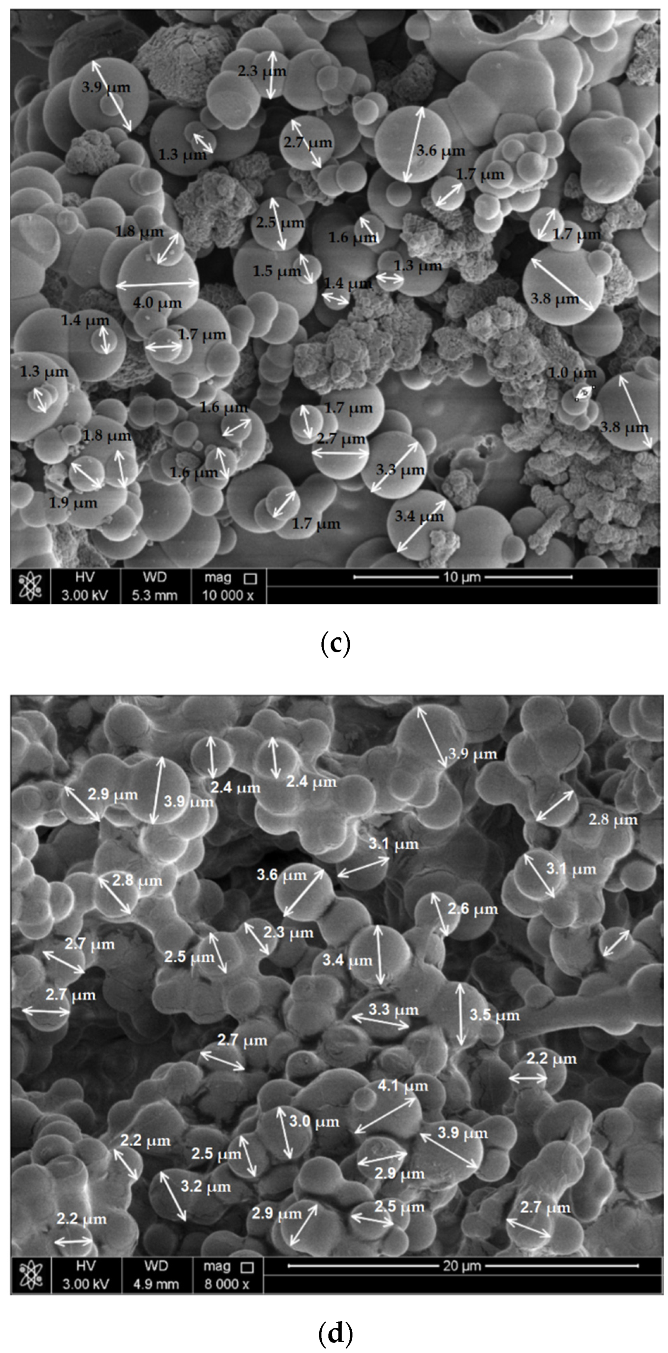
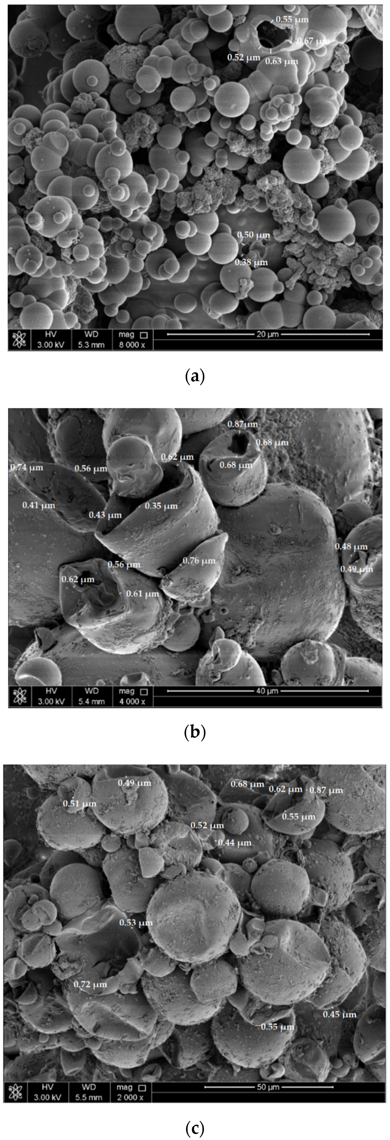
3.1. Microcapsule Scanning Electron Microscopy

The diameter of the microcapsules prepared using Aresol-OT are shown in the scanning electron microscope images presented in Figure 2 at different imaging scales. As the number of the prepared microcapsules shown in the SEM images is large, and considering that the main task herein is to find the average diameter and shell thickness of the microcapsules prepared using AOT-in-hexane solution and to compare them with the same for other previous preparation procedures, statistical analysis was not practical for such huge number of microcapsules. Accordingly, the average values of 150–200 microcapsules randomly selected from the SEM images were considered (as per similar previous works reported in literature [4,5]). It could be noted in Figure 2 that the microcapsules’ diameters fall within the range of 1 µm to almost 5 µm with an approximate average value of 2.5 µm, which is smaller than those prepared using the original procedure developed by Hassan, et al. [4] (51 µm) and Al-Ansari, et al. [6] (70 µm).
It could be also noted that the shape of the produced microcapsules is perfectly spherical and uniform. Figure 2a clearly shows a rough surface of a single microcapsule and has smaller particles attached to it. Such small particles attached to the microcapsule’s shell might be nano sized capsules produced during the polymerization or they could be the remaining of the healing agent that could not be totally polymerized in the continuous phase. Testing the healing efficiency of the microcapsules that were produced using AOT-in-hexane solution should be investigated thoroughly in future work.
Figure 3 presents the scanning electron microscope images showing the produced microcapsule approximate shell thicknesses, which are in the range of 0.35 µm to 0.87 µm with an average value of 0.58 µm. This value range is also smaller than those reported by Hassan, et al. [4] (0.91 µm) and Al-Ansari, et al. [6] (0.81 µm). This may suggest that rupturing the microcapsule’s shell with the crack tip will be easier as the shell thickness is thinner and, hence, the self-healing efficiency will be effectively enhanced.
As previously mentioned, most of the produced microcapsules have regular sizes ranging from 1 to 5 µm in diameter (Figure 2a–c) with an average value of 2.5 µm. However, much bigger microcapsules (with diameters around 40 µm) could be also produced, as shown in Figure 3b,c. It has been observed that such large-sized microcapsules are deformed and broken, which might be attributed to the very thin shell thickness compared to the diameter of such large microcapsules.

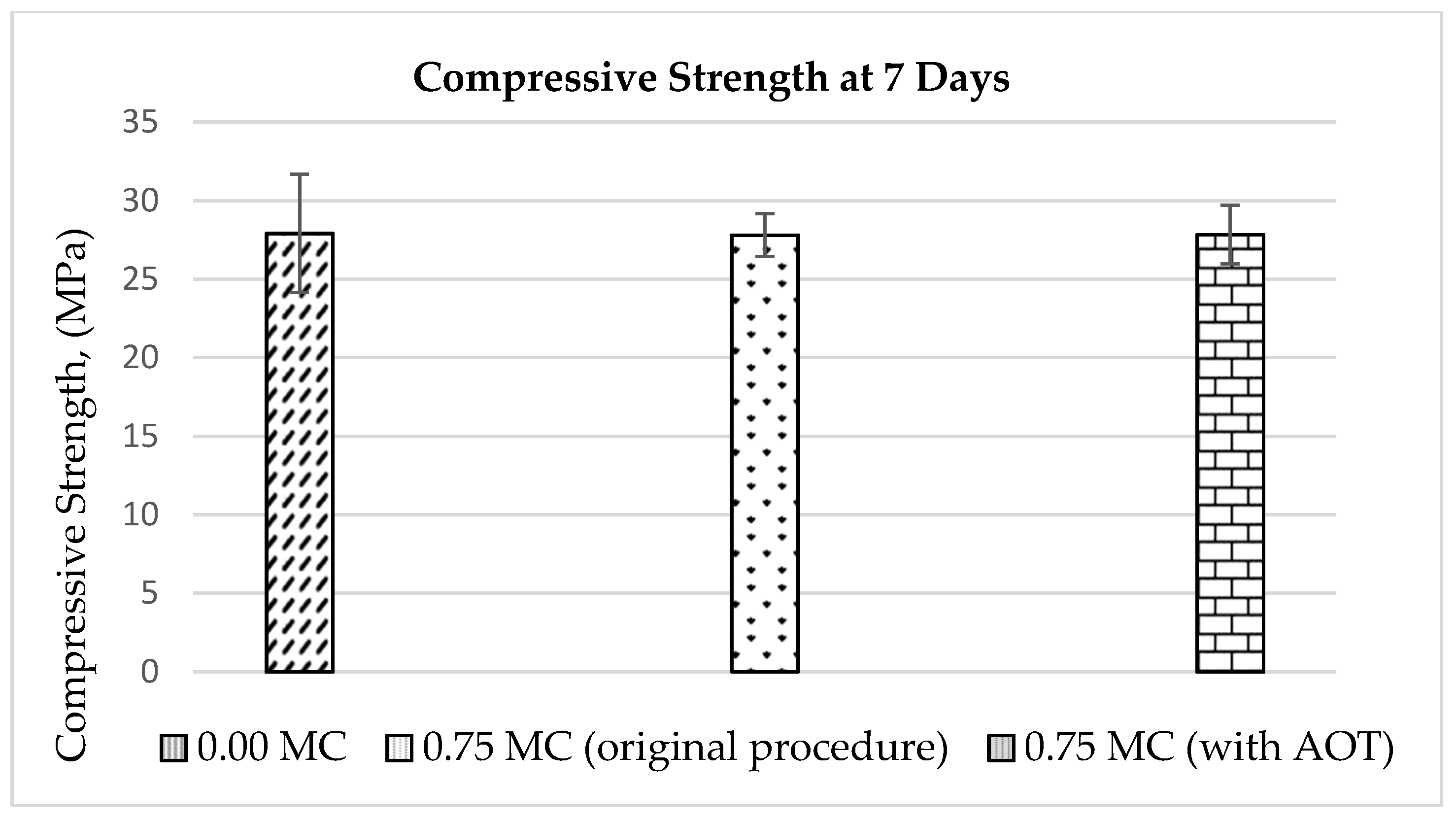
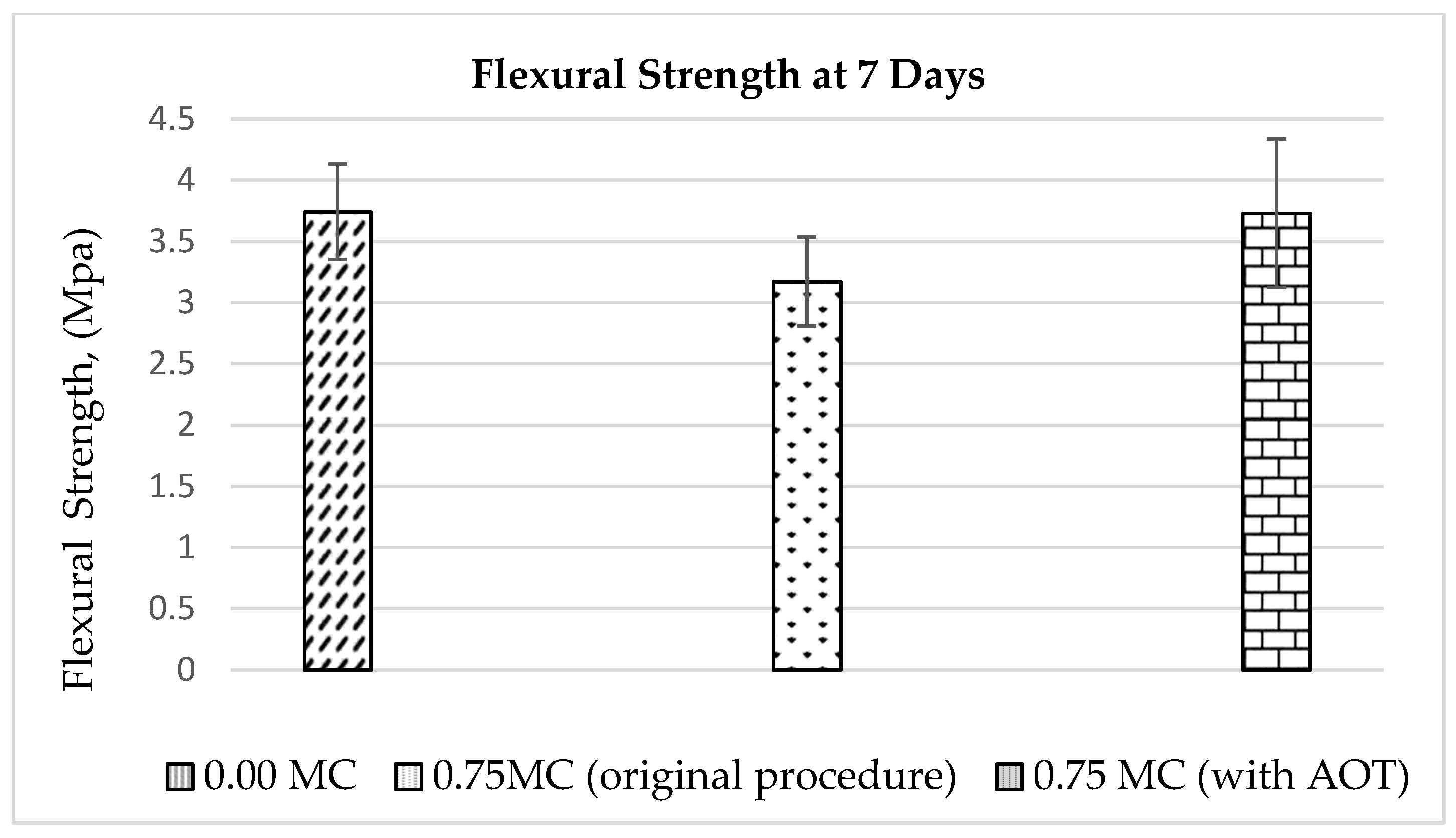
3.2. Compression and Flexural Strengths

Table 4 and Table 5 summarize the compressive and flexural strength results of mortar samples, respectively. The outlier values have been checked according to ASTM E178 [24] before calculating the average and standard deviation values of the results. Figure 4 and Figure 5 show the variation of compressive and flexural strengths of all mortar mixes (without MC, with MC prepared using the original procedure, and with MC prepared using AOT), respectively. The Tables present the results of all tested samples which could not be Figured out from the Figures. The error bars in the Figures indicate the standard deviation.
It could be noted from Figure 4 that the addition of self-healing microcapsules has small effect on the compressive strength as reported in similar previous works [4,5,6]. However, the significant strength reduction is related to the flexural strength, as shown in Figure 5. The results show that the 7th day compressive and flexural strengths were enhanced for the mixes with microcapsules prepared using AOT compared to those prepared using the original preparation procedure. Compared to the control mix, the reductions of the compressive and flexural strengths of mortar mixes containing microcapsules prepared using the modified procedure with AOT were only 0.27% and 0.31%, respectively, which could be considered negligible. However, the reductions of the compressive and flexural strengths were 0.34% and 15.30%, respectively, for the mixes containing microcapsules prepared using the original preparation procedure developed by Hassan, et al. [4]. This indicates that using AOT for the preparation of the self-healing microcapsules can reduce the adverse effects on the mechanical properties, especially the flexural strength, of the cementitious mixes containing such microcapsules.
Figure 6 presents the pictures of the fracture surface of the mortar prisms after carrying out the flexural strength test. From this Figure, one can visually observe the differences in the microcapsule’s distributions within the samples for different preparation procedures. It shows that the distribution of the microcapsules prepared using AOT was more uniform with smaller microcapsule sizes (Figure 6b) compared to the samples with microcapsules prepared using the original preparation procedure (Figure 6a). This visual observation reinforces the aforementioned results presented in Section 3.1, which indicate that using AOT for the preparation of microcapsules refines the mix physical properties and produces much smaller-sized microcapsules (1–5 µm in diameter) compared to the microcapsules prepared using the original preparation procedure (average diameter of 51 µm) [4].

4. Conclusions

Based on experimental study results, the following conclusions can be drawn:
  • The diameters of the microcapsules prepared using the procedure with Aresol-OT were found to be between 1 µm and 5 µm. They were smaller than those of the microcapsules prepared using previous preparation procedures. The microcapsule shape was found to be perfectly spherical and uniform;
  • The scanning electron microscope images have shown that the approximate shell thickness of the microcapsules was in the range of 0.35 µm and 0.87 µm, which was smaller than those reported in the literature. This may suggest that rupturing the microcapsule shell with the crack tip becomes easier when the shell thickness is thinner;
  • The 7th day compressive and flexural strength reductions were alleviated for the mixes with microcapsules prepared using AOT compared to those with microcapsules prepared using the original preparation procedure. The compressive and flexural strengths of the mortar mixes containing microcapsules prepared with AOT were only 0.27% and 0.31% lower than those of the control, which could be considered negligible;
  • However, the compressive and flexural strength reductions were 0.34% and 15.30%, respectively, for the mixes containing microcapsules prepared using the original preparation procedure (i.e., containing sulfonic acid). This indicates that preparing self-healing microcapsules using AOT can reduce the potential adverse effects on the mechanical properties, especially the flexural strength, of the cementitious mixes.
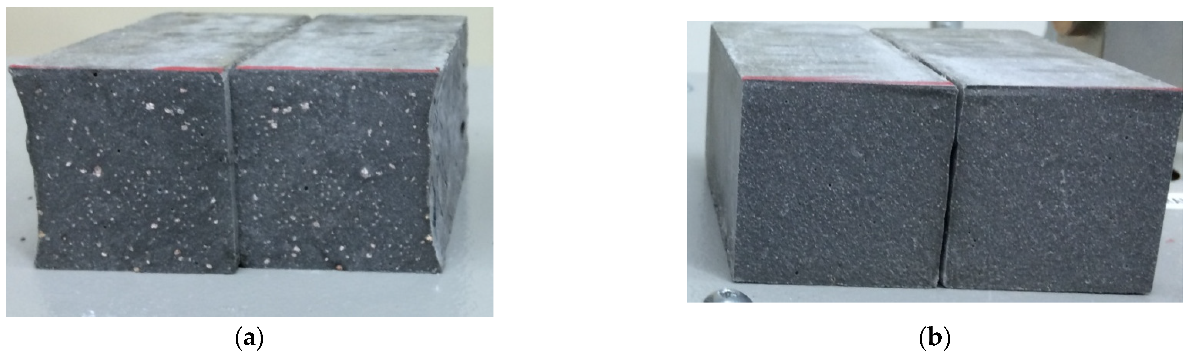
  • The visual observation of the mortar prism fracture surface images has shown that the distribution of the microcapsules prepared using AOT was more uniform than those prepared using the original preparation procedure. This observation suggests that using AOT for the preparation of the microcapsules refines the mix physical properties and may enhance the overall healing efficiency of such microcapsules.

5. Future Work

The effect of incorporating self-healing microcapsule prepared with AOT on the mechanical properties of mortar and large concrete samples at 28 and 56 days with different MC concentrations should be investigated thoroughly in future work. Moreover, the healing efficiency of the microcapsules produced using AOT-in-hexane solution should be also verified.

Author Contributions

The authors contributed to this research article as follows: literature review, A.A.T., G.S. and M.O.M.; experimental work and testing, A.A.T., A.S. and M.O.M.; investigation, A.A.T. and M.O.M.; writing—original draft preparation, A.A.T.; writing—review and editing, G.S., A.S. and M.O.M.; supervision, M.O.M. and A.S.; project administration, G.S., A.S., M.O.M. and A.A.T.; funding acquisition, A.A.T. and G.S. All authors have read and agreed to the published version of the manuscript.

Funding

This research was funded by the deanship of scientific research at Aqaba university of Technology.

Institutional Review Board Statement

Not applicable.

Informed Consent Statement

Not applicable.

Data Availability Statement

The data used to support the findings of this study are available upon request from the corresponding author.

Acknowledgments

This work was made possible by the support and deanship of scientific research at Aqaba university of Technology as well as the collaboration of the university of Houston. The statements made herein are solely the responsibility of the authors.

Conflicts of Interest

The authors declare that they have no known competing financial interest or personal relationships that could have appeared to influence the work reported in this paper.

References

  1. Pelletier, M.; Brown, R.; Shukla, A.; Bose, A. Self-Healing Concrete with a Microencapsulated Healing Agent. Available online: http://energetics.chm.uri.edu/system/files/Self%20healing%20concrete%20-7-11.pdf (accessed on 25 March 2022).
  2. Maes, M.; Van Tittelboom, K.; De Belie, N. The efficiency of self-healing cementitious materials by means of encapsulated polyurethane in chloride containing environments. Constr. Build. Mater. 2014, 71, 528–537. [Google Scholar] [CrossRef]
  3. Van Tittelboom, K.; De Belie, N.; Van Loo, D.; Jacobs, P. Self-healing efficiency of cementitious materials containing tubular capsules filled with healing agent. Cem. Concr. Compos. 2011, 33, 497–505. [Google Scholar] [CrossRef]
  4. Hassan, M.M.; Milla, J.; Rupnow, T.; Al-Ansari, M.; Daly, W.H. Microencapsulation of Calcium Nitrate for Concrete Applications. Transp. Res. Rec. 2016, 2577, 8–16. [Google Scholar] [CrossRef]
  5. Milla, J.; Hassan, M.M.; Rupnow, T.D.; Al-Ansari, M.; Arce, G.A. Effect of Self-Healing Calcium Nitrate Microcapsules on Concrete Properties. Transp. Res. Rec. 2016, 2577, 69–77. [Google Scholar] [CrossRef]
  6. Al-Ansari, M.; Abu-Taqa, A.G.; Hassan, M.M.; Senouci, A.; Milla, J. Performance of modified self-healing concrete with calcium nitrate microencapsulation. Constr. Build. Mater. 2017, 149, 525–534. [Google Scholar] [CrossRef]
  7. Milla, J.; Hassan, M.M.; Rupnow, T.; Daly, W.H. Measuring the crack-repair efficiency of steel fiber reinforced concrete beams with microencapsulated calcium nitrate. Constr. Build. Mater. 2019, 201, 526–538. [Google Scholar] [CrossRef]
  8. He, J.; Shi, X. Laboratory assessment of early-age durability benefits of a self-healing system to cementitious composites. J. Build. Eng. 2021, 44, 102602. [Google Scholar] [CrossRef]
  9. Aïssa, B.; Therriault, D.; Haddad, E.; Jamroz, W. Self-Healing Materials Systems: Overview of Major Approaches and Recent Developed Technologies. Adv. Mater. Sci. Eng. 2012, 2012, 854203. [Google Scholar] [CrossRef] [Green Version]
  10. Blaiszik, B.J.; Sottos, N.R.; White, S.R. Nanocapsules for self-healing materials. Compos. Sci. Technol. 2008, 68, 978–986. [Google Scholar] [CrossRef]
  11. Farshi Azhar, F.; Ahmadinia, A.; Mohammadjafari Sadeghi, A. Modified self-healing cementitious materials based on epoxy and calcium nitrate microencapsulation. J. Microencapsul. 2021, 38, 203–217. [Google Scholar] [CrossRef] [PubMed]
  12. Kalyanram, P.; Valenzuela, P.; Gupta, A.R. Aerosol-Ot Stabilized Micro and Nano-Scale Emulsions for Pharmaceutical Formulations. Org. Med. Chem. Int. J. 2017, 4, 88–90. [Google Scholar] [CrossRef]
  13. Hensel, J.K.; Carpenter, A.P.; Ciszewski, R.K.; Schabes, B.K.; Kittredge, C.T.; Moore, F.G.; Richmond, G.L. Molecular characterization of water and surfactant AOT at nanoemulsion surfaces. Proc. Natl. Acad. Sci. USA 2017, 114, 13351–13356. [Google Scholar] [CrossRef] [Green Version]
  14. La Mesa, C.; Coppola, L.; Ranieri, G.A.; Terenzi, M.; Chidichimo, G. Phase diagram and phase properties of the system water-hexane-Aerosol OT. Langmuir 1992, 8, 2616–2622. [Google Scholar] [CrossRef]
  15. Eastoe, J.; Fragneto, G.; Robinson, B.H.; Towey, T.F.; Heenan, R.K.; Leng, F.J. Variation of surfactant counterion and its effect on the structure and properties of Aerosol-OT-based water-in-oil microemulsions. J. Chem. Soc. Faraday Trans. 1992, 88, 461–471. [Google Scholar] [CrossRef]
  16. Aveyard, R.; Binks, B.P.; Clark, S.; Mead, J. Interfacial tension minima in oil–water–surfactant systems. Behaviour of alkane–aqueous NaCl systems containing aerosol OT. J. Chem. Soc. Faraday Trans. 1 Phys. Chem. Condens. Phases 1986, 82, 125–142. [Google Scholar] [CrossRef]
  17. Kizling, J.; Kronberg, B. On the formation of concentrated stable w/o emulsions. Adv. Colloid Interface Sci. 2001, 89–90, 395–399. [Google Scholar] [CrossRef]
  18. Leong, Y.S.; Candau, F. Inverse microemulsion polymerization. J. Phys. Chem. 1982, 86, 2269–2271. [Google Scholar] [CrossRef]
  19. Srivastava, S.; Sharma, S.K.; Sharma, R.K. Synthesis of gold nanorods using concentrated aerosol OT in hexane and its application as catalyst for the reduction of eosin. Colloids Surf. A Physicochem. Eng. Asp. 2011, 373, 61–65. [Google Scholar] [CrossRef]
  20. Gupta, M.; Gupta, A.K. In vitro cytotoxicity studies of hydrogel pullulan nanoparticles prepared by AOT/N-hexane micellar system. J. Pharm. Pharm. Sci. 2004, 7, 38–46. [Google Scholar]
  21. Eastoe, J.; Robinson, B.H.; Steytler, D.C.; Thorn-Leeson, D. Structural studies of microemulsions stabilised by aerosol-OT. Adv. Colloid Interface Sci. 1991, 36, 1–31. [Google Scholar] [CrossRef]
  22. ASTM C109/C109M–16a; Standard Test Method for Compressive Strength of Hydraulic Cement Mortars (Using 2-in. or [50-mm] Cube Specimens). ASTM International: West Conshohocken, PA, USA, 2016.
  23. ASTM C348–14; Standard Test Method for Flexural Strength of Hydraulic-Cement Mortars. ASTM International: West Conshohocken, PA, USA, 2014.
  24. ASTM E178–08; Standard Practice for Dealing with Outlying Observations. ASTM International: West Conshohocken, PA, USA, 2008.
Figure 1. Microcapsule preparation method schematic illustration.
Buildings 12 00751 g001
Figure 2. Microcapsule scanning electron microscope images at different scales: (a) 2 µm, (b) 5 µm, (c) 10 µm, and (d) 20 µm.
Buildings 12 00751 g002aBuildings 12 00751 g002b
Figure 3. Microcapsule scanning electron microscope images at different scales: (a) 20 µm, (b) 40 µm, and (c) 50 µm.
Buildings 12 00751 g003
Figure 4. Compressive strength versus microcapsule concentration at 7 curing days (Error bars = standard deviation).
Buildings 12 00751 g004
Figure 5. Mortar flexural strength versus microcapsule concentration at 7 curing days (Error bars = standard deviation).
Buildings 12 00751 g005
Figure 6. Samples after flexural breaking (a) microcapsules prepared using the original procedure and (b) microcapsules prepared using AOT.
Buildings 12 00751 g006
Table 1. Modified synthesis of aqueous and continuous phases.
Aqueous PhaseConstituentAmount (g)Continuous PhaseConstituentAmount (g)
Urea5.0Organic Solvent (Hexane)180.0
Formaldehyde (37% solution)12.67
Resorcinol0.5
Ammonium Chloride0.5Dioctyl Sodium Sulfosuccinate (Aerosol-OT)5.8
Calcium Nitrate10.0
Distilled Water50.0
Table 2. Mortar mix design.
ConstituentQuantity
Class 42.5 R Portland cement CEM I, complying with EN 197-1(grams)740
Natural Sand (Conformance to BS EN 12620) (grams)2035
Water (grams)359
Microcapsules% By weight of cement0.00, 0.75
grams0.00, 5.55
Table 3. Testing matrix.
Mix IDBatch No.MC Concentration
(% by Weight of Cement)
No. of Samples (Compression Test)No. of Samples (Flexural Test)
Without MC
(Control)
10.0033
233
With 0.75 MC (original procedure [4])10.7533
233
With 0.75 MC (with AOT)10.7533
233
Table 4. Seven-day mortar compressive strength test results, (MPa).
Sample0.00 MC (Control)0.75 MC (Original Procedure)0.75 MC (with AOT)
127.8228.9726.86
225.6227.8228.73
327.7929.8427.38
430.2327.4129.27
526.9926.3327.77
628.9826.4926.97
Average, (µ)27.9127.8127.83
Stand. Dev.(σ)1.591.380.98
% Reduction-0.340.27
Table 5. Seven-day mortar flexural strength test results, (MPa).
Sample0.00 MC (Control)0.75 MC (Original Procedure)0.75 MC (with AOT)
13.703.213.88
24.283.033.38
33.482.543.78
43.873.113.97
53.622.943.64
63.493.163.83
Average, (µ)3.743.173.73
Stand. Dev.(σ)0.300.200.21
% Reduction - 15.300.31
Publisher’s Note: MDPI stays neutral with regard to jurisdictional claims in published maps and institutional affiliations.

