Article

Improvement of the Oxidation Resistance of FeMnSiCrNi Alloys with a Pre-Oxidation Treatment

by
João Gabriel da Cruz Passos
1,*,
Rodrigo da Silva
2,
Carlos Alberto Della Rovere
2 and
Artur Mariano de Sousa Malafaia
3
1
Engineering School of São Carlos, Materials Science and Engineering Department, University of São Paulo, Av. João Dagnone, 1100 Jd Sta Angelina, São Carlos 13563-120, SP, Brazil
2
Munir Rachid Corrosion Laboratory, Federal University of São Carlos, Rodovia Washington Luis Km 235, São Carlos 13565-905, SP, Brazil
3
Campus Santo Antônio, Mechanical and Production Engineering Department, Federal University of São João del-Rei, Praça Frei Orlando, 170—Centro, São João del Rei 36307-334, MG, Brazil
*
Author to whom correspondence should be addressed.
Metals 2023, 13(12), 1928; https://doi.org/10.3390/met13121928
Submission received: 17 August 2023 / Revised: 12 October 2023 / Accepted: 13 October 2023 / Published: 23 November 2023

Abstract

:
Shape-memory Mn-rich austenitic stainless steels have a low high-temperature oxidation resistance because Mn tends to inhibit the formation of protective oxides. Mn depletion from oxidation also creates a ferritic Mn-depleted layer. A Mn-depleted layer formed via vacuum annealing has been associated with increased oxidation resistance. Thus, in the present study, a Mn-depleted layer was created with a pre-oxidation treatment conducted at 1000 °C for 30 min. Then, pre-treated and untreated samples were oxidized at 800 °C for up to 200 h. The resulting oxide layers were analyzed, as well as the metal/oxide interface roughness and the ferritic layer thickness. After pre-treatment, a 9 μm thick ferritic layer as well as an oxide layer richer in Cr-containing oxides than those usually observed in FeMnSiCrNi alloys oxidized at 800 °C were detected. After 200 h at 800 °C, the metal/oxide interface roughness of pre-treated samples was considerably lower. The oxidation rate of pre-treated samples was one order of magnitude lower for the first 50 h, but the effect significantly decreased afterward. The pre-existing ferritic layer was unable to stop Mn-rich oxides from being incorporated into the oxide layer, making its effect short-lived.

1. Introduction

Due to their unique shape-memory effect, shape-memory alloys have been considered for a large array of potential practical applications [1,2,3,4,5]. They have a high potential for applications in which prestressing of mechanical joining is needed, for example, as the SME effect can be used to facilitate assembly [6]. Although the conventional NiTi SMAs are more popular, Fe-Mn-Si-based SMAs, such as the austenitic FeMnSiCrNi alloys, are of high interest. Their main advantage over the NiTi SMAs is their comparably low content of nickel, which is an expensive element. Furthermore, they present good workability and good weldability [7,8,9,10]. Their development can be associated with a trend of increasing the number of principal elements in metallic alloys, which is also seen through the rising popularity of high-entropy, multi-component, and complex concentrated alloys [11,12]. This trend was expanded from equiatomic to non-equiatomic compositions and was applied recently to steels for the development of high-entropy steels [13,14,15]. A 2020 study associated the FeMnSiCrNi alloys and their promising properties with their multicomponent nature, classifying them as metastable FCC multicomponent alloys [16]. Despite their novelty and promising characteristics, as well as some studies interested in investigating their high-temperature properties [17,18], the usability of FeMnSiCrNi alloys at high temperatures is limited. High-temperature oxidation tests performed on FeMnSiCrNi alloys showed that their innate oxidation resistance is low when compared to conventional stainless steels [19,20,21,22,23]. This is not limited to FeMnSiCrNi alloys, as many of the recently developed multicomponent alloys present poor oxidation resistance due to the formation of complex multi-oxide layers or undesirable oxides that negatively affect protective oxide formation [24]. For FeMnSiCrNi alloys, this discouraging behavior is mainly a consequence of the high manganese (Mn) content, an element that promotes fast-growing and stable oxide formation. Chromium (Cr) and silicon (Si), which are also present in FeMnSiCrNi alloys, are usually associated with an increase in oxidation resistance. In general, Mn has a strong effect on Cr oxide formation, leading to a reduction in chromia formation and the appearance of Mn–Cr–O spinels and pure Mn oxides, which are less protective [25]. Even though Mn, Cr, and Si are the only oxide formers usually observed in these alloys, it is difficult to increase oxidation resistance by increasing or decreasing their contents. This happens because the available composition field for these alloys is limited by the need to retain the attractive shape-memory effect and the austenitic structure (Cr, Mn, and Si affect austenite stability). Also, previous results have shown that changing the amount of these elements beyond a certain range can have unexpected negative effects, with a reduction in the oxidation resistance of an alloy with lower Mn and higher Si content [20].
Among the previously attempted strategies for increasing the oxidation resistance of these alloys, pre-heat-treatment appears to be one of the most successful, as it promotes an appreciable reduction in oxidation rate and does not require compositional changes. A study on the effects of a vacuum pre-heat-treatment on an Fe-17Mn-5Si-10Cr-4Ni-VC alloy showed that pre-treated samples had a significantly lower mass gain rate than untreated ones [21]. This treatment promoted the volatilization of Mn, which is the main austenite-stabilizing element in these alloys. Consequently, a ferritic Mn-depleted zone grew on the surface of the pre-treated samples. The treated samples were then exposed to laboratory air for up to 24 h at 800 °C. During exposure, the ferritic layer reduced Mn activity, promoting the formation of a more protective oxide layer, with a higher Cr-Mn spinel volume compared to pure Mn oxides and even the formation of a protective Cr2O3 oxide. This pre-treatment, however, requires a vacuum atmosphere, which can be hard to create, especially for large-scale applications. Furthermore, the Mn content in the oxide layer increased during the test, showing that the effectiveness of the pre-treatment could be reduced or nullified during long-term exposure. For the first 5 h, only chromia and Mn–Cr–O spinels could be detected via XRD analysis, with the intensity of the spinel peak increasing significantly from 1 to 5 h. After 10 h, pure Mn oxide (Mn2O3) could already be seen, and after 24 h, it heavily predominated over chromia.
Depletion of Mn is also characteristic of oxidation tests due to Mn oxide formation. For this reason, ferritic layers are commonly observed in oxidized FeMnSiCrNi samples [22]. Thus, pre-oxidation could be a viable strategy for creating Mn-depleted layers. The oxide layer formed during pre-oxidation is also potentially beneficial to oxidation resistance. This pre-existing layer can help to protect the material in subsequent exposures to oxidative atmospheres, as seen in other chromia-forming alloys [26,27]. In this study, the effectiveness of a pre-oxidation treatment, in which a Mn-depleted layer was formed by means of a short exposure to air at an elevated temperature, was evaluated in a FeMnSiCrNi alloy.

2. Materials and Methods

The composition of the FeMnSiCrNi alloy investigated in this study is Fe-13.67Mn-8.32Cr-3.46Si-3.04Ni, as determined via inductively coupled plasma optical emission spectroscopy (Vista RL, Varian, Mulgrave, Australia). The alloy studied here has a sufficiently high configurational entropy (1.01 R) to be considered a medium-entropy alloy (between 1 and 1.5 R [11]). This is in line with the earlier observation that these materials can be considered multicomponent alloys. The alloy was cast in a vacuum induction furnace, hot-rolled at 1000 °C, and solution treated at 1050 °C for 1 h, then water quenched. Stainless steel AISI 304L was used as the reference material, to which high-purity elements were added. Samples with dimensions of approximately 2 mm × 4 mm × 11 mm were cut from the ingot and ground with SiC abrasive paper (up to 600 grit). They were then cleaned in an ultrasound bath (Metasom 14, Pantec).
The prepared samples were oxidized at 800 °C, with four different exposure times: 20, 50, 100, and 200 h. One set of samples were oxidized without any prior treatment. A second set were first pre-treated at 1000 °C for 30 min (lab air atmosphere), then exposed at 800 °C. A higher temperature (1000 °C) was chosen for the pre-treatment to accelerate oxide formation and Mn depletion. This allowed the desired pre-oxidation to occur in a shorter timeframe. Also, higher temperatures favor Mn–Cr–O spinel formation in the Mn-Cr-O system [28], which is more protective than pure Mn oxides [21,23]. The samples were weighed before the pre-treatment, after the pre-treatment, and after exposure. The mass variation values presented here concern the difference between the mass of the samples before exposure and after exposure. For the pre-treated samples, the mass variation between the unoxidized and pre-treated states was not considered in this calculation. The samples were weighed before and after oxidation in a 0.1 mg-resolution balance. The final weighing was performed at room temperature after the sample was cooled inside the furnace.
Two samples were exposed to each test condition, and one sample was characterized in the pre-treated state. The oxidized samples were analyzed using X-ray diffraction (XRD) (Shimadzu XRD-6000, Kyoto, Japan) and a scanning electron microscope equipped for energy-dispersive X-ray spectroscopy (SEM/EDS) (SEM—Hitachi TM 3000, Tokyo, Japan and EDS detector from Bruker, Billerica, Massachusetts) analysis. For XRD, the samples were analyzed as oxidized (Cu-Kα radiation, 2°/min step size, 20–90° range in ϴ/2ϴ geometry). For SEM/EDS analysis, samples were cross-sectioned, mounted in Bakelite, and polished (1.0 μm alumina suspension). These procedures were used to characterize the oxide layer and the Mn-depleted layer.
The thickness of the Mn-depleted layer was also measured from images obtained via optical microscopy (Olympus BX51, Tokyo, Japan), as shown in Figure 1. As previously stated, Mn depletion during oxidation destabilizes the austenitic structure, leading to the formation of a ferritic that has a well-defined boundary with the austenitic substrate. The difference in mechanical properties between these two phases causes them to respond differently to the grinding and polishing processes (i.e., reach different degrees of surface finish). Consequently, the ferritic layer can be distinguished from the austenitic layer even in optical microscopy images, as the figure shows. Through digital image analysis with the open-source ImageJ software (first developed by Wayne Rasband at the National Institutes of Health, Bethesda, MD, USA), it was possible to measure the area occupied by ferrite and calculate its average thickness (h). Finally, the same software was also used to measure the metal/oxide interface roughness (Ra and Rz) in these same images. This was achieved via the same technique used previously by our group [20].

3. Results and Discussion

3.1. Pre-Treated Sample Characterization

The pre-treatment formed a Mn-depleted ferritic layer with an average thickness of 9 μm and an average Mn and Cr content of 0.8 and 5.8 wt.%, respectively (measurements done via EDS). As expected, the samples also suffered oxidation, gaining, on average, 0.00263 ± 0.00108 kg/m2 during the pre-treatment. Due to the slow furnace cooling rate at the end of the test and the short exposure time, which formed only a thin oxide layer, it was possible to avoid any observable spallation (no spalled oxide was detected in the crucibles or during the handling of the samples). Furthermore, a similar FeMnSiCrNi alloy was shown to be resistant to spallation under cyclic conditions for almost 300 h of exposure at 1000 °C [29]. Both Mn and Cr contents were lower than in the unoxidized alloy, which is expected, as both Mn and Cr are consumed during oxidation. For this reason, the Cr-to-Mn ratio after pre-treatment is not as favorable for chromia formation as that previously reported and achieved via vacuum annealing [21], in which only Mn was volatilized. The EDS analysis of a pre-treated sample in Figure 2a shows the Mn-depleted region and the Mn-rich oxide layer. An oxide layer rich in Mn and Cr is present below the pure Mn oxide layer, probably consisting of the Mn–Cr–O spinels observed in similar alloys [22,23,29]. No Fe was detected in the oxide layer. The pattern obtained via XRD analysis, shown in Figure 2b, has strong Mn2O3 crystalline peaks and weaker Fe-α (substrate), (Mn, Cr)3O4 and Cr2O3 peaks. Interestingly, the stable Mn oxide species at this temperature is assumed to be Mn3O4, both according to the phase diagram and previous tests at this temperature on a similar alloy [28,29]. This shows that part of the layer seen here was probably formed during heating and could suffer transformation if the treatment ran for longer. Furthermore, the formation of Mn2O3 during a short period (30 to 90 min) of exposure to 1000 °C was previously observed in a similar alloy [19]. In general, it appears that the pre-treatment was successful in generating the (Mn, Cr)3O4 spinels and Cr2O3 oxides (the second being in very small quantities, as indicated by the low diffraction intensity), which have protective capabilities [21,22,23]. However, even with the presence of these oxides, the layer formed during the pre-treatment is not highly protective by itself, as the EDS maps do not show the presence of any continuous pure Cr oxide layer. The Mn–Cr–O spinels are more resistant to further oxidation than pure Mn oxides, but it is still not as protective as chromia [30]. A continuous chromia layer would be hard to create even with different oxidation parameters. As the Mn-Cr-O phase diagram shows, Cr2O3 is only stable at a Cr-to-Mn ratio higher than 3:2, which is not the case for the alloy in its initial state [28].

3.2. Oxidation Kinetics

Mass variation analysis (mass variation data in Figure 3a and square root transform in Figure 3b) shows that the pre-treated samples gained less mass throughout the entire test. However, it is important to highlight that the samples gained more mass during pre-oxidation (0.00263 ± 0.00108 kg/m2) than during the first 20 h of exposure (0.00137 ± 0.00069 kg/m2). This is attenuated throughout the test, as the mass gained through exposure constantly increases, eventually becoming much greater than that gained during pre-treatment. Furthermore, even if the average mass gained during the pre-treatment is added to the mass gained after 20 h of exposure (totaling 0.004 kg/m2), the value would still be smaller than the mass gain of the untreated sample (0.00683 kg/m2).
The square root transform in Figure 3b also shows that both samples followed a parabolic behavior (R2 above 0.99 for all three lines shown in the figure). A single parabolic constant (Kp) was not sufficient to describe the behavior of the pre-treated sample, with an acceleration in the oxidation rate occurring after 50 h of exposure. The calculated Kp was 3.1 × 10⁻¹¹ kg2/m4·s for the first 50 h, increasing to 2.0 × 10⁻¹⁰ kg2/m4·s between 50 and 200 h. The behavior of the untreated samples, on the other hand, can be described by a single parabolic constant (Kp 5.5 × 10⁻¹⁰ kg2/m4·s). This acceleration in the oxidation rate was not seen in the vacuum-annealed Fe-17Mn-5Si-10Cr-4Ni-VC alloy oxidized at 800 °C for 24 h, as the tests were too short to see such changes. However, the authors point out in their conclusions that such an acceleration was a possibility, as Mn was constantly being incorporated into the oxide layer [21]. A similar acceleration in oxidation kinetics was seen in Fe-Cr-Mn alloys with low Mn contents (between 0 and 3 wt.%). It was associated with the presence of Mn in the oxide layer via the formation of Mn–Cr–O spinels or Mn oxides. This only happened when Mn-rich oxides were gradually incorporated into the oxide layer, in the alloys with 0.5 and 1 wt.% Mn. With no Mn (0 wt.) or the highest Mn content (3 wt.%), the alloys followed a single-stage oxidation kinetics, because either only chromia formed or a Mn-rich oxide layer was present from the start. Furthermore, the study on the Fe-Cr-Mn alloys also shows that chromia and the spinels are unable to act as a diffusion barrier for Mn [31].
Although the treated samples had a reduced Kp compared to the untreated ones, the reduction in oxidation rate seen here was much smaller than that achieved through vacuum annealing, from 2.2 × 10⁻⁹ kg2/m4·s to 2.2 × 10⁻¹¹ kg2/m4·s [21]. The reason for this difference is probably the much thicker ferrite layer created by the vacuum pre-treatment (170 μm vs. 9 μm here), which was able to cause a more significant drop in Mn availability. Another probable cause for the worse performance of the pre-oxidation treatment compared to vacuum annealing is Cr consumption during the pre-treatment. Similarly to Mn, Cr diffuses faster in ferrite than in austenite [22,32]. Therefore, having a ferrite layer richer in Cr at the start of the oxidation process probably facilitated the formation of chromia in the vacuum-annealed samples. Both of those possibilities point to significant drawbacks of the pre-oxidation treatment. The thickness of the ferritic layer achieved with this technique is directly associated with Mn and Cr consumption to form oxides. Therefore, a longer test would, in theory, produce a thicker ferrite layer, but it would also eventually consume enough Mn and Cr to induce catastrophic oxidation. In summary, the design of a pre-oxidation treatment that is as efficient in reducing Mn activity (compared to Cr) is probably not feasible.

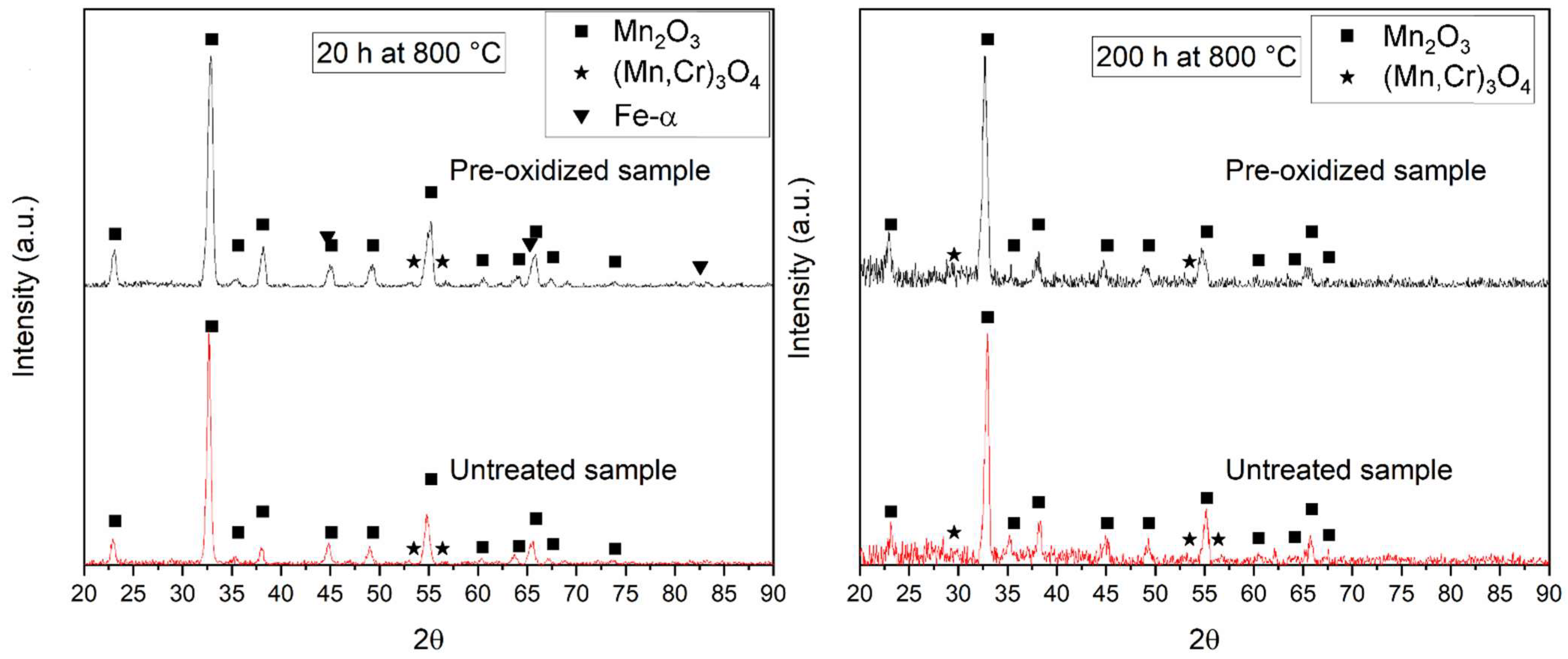
3.3. Oxide Layer Characterization

After oxidation, the same oxides were detected with XRD analysis in treated and untreated samples at all exposure times. The results at the shortest (20 h) and longest (200 h) exposure times are used to illustrate this behavior (Figure 4). The main difference between the results at 20 and 200 h is the easier identification of Fe-α and M3O4 peaks. These structures are located below the external Mn2O3 oxide (as seen in the EDS images) and were observed in previous investigations into this alloy system [20,21,22], which thickens with exposure and thus makes it harder to detect the inner layers. The M2O3 peaks seen in Figure 2 disappeared, which may be a sign that the oxide formed during the pre-treatment is richer in Cr than the oxide formed at the test temperature, but Mn content in the oxide layer quickly began to increase. The formation of Cr2O3 in the oxide layers of FeMnSiCrNi alloys was pointed out as the main factor responsible for the increase in oxidation resistance seen after vacuum annealing [21]. However, the absence of these crystalline peaks in Figure 4 shows that this effect might be specific to the early oxidation stages, after which the oxide composition approaches that of the untreated samples and Cr2O3 disappears or becomes harder and harder to detect. This could explain the first parabolic kinetics stage seen in pre-treated samples (Figure 3b). The main mechanism of oxidation protection here seems to be the same one observed during vacuum annealing: the presence of a Mn-depleted layer facilitates Cr participation in the oxidation process, leading to the formation of a more protective layer and an overall increase in oxidation resistance.
The SEM/EDS analysis confirms that the pre-treatment did not drastically affect the oxide layer configuration, as seen in the Fe, Mn, Cr, Si, and O EDS maps shown in Figure 5. The oxide layer, in all test conditions, was composed of an external Mn-rich oxide, followed by a Mn and Cr-rich oxide layer between the external layer and the substrate. In both cases, a heavy Mn depletion is seen on the substrate (ferritic layer). Cr is also slightly depleted in the ferritic layer. As a result, Fe, Si, and Ni (not shown here) appear enriched.
Therefore, the Cr oxide present in the pre-treated samples was apparently replaced by a Mn-rich oxide layer composed of pure Mn oxides and Mn–Cr–O spinels. This agrees with results from vacuum-annealed FeMnSiCrNi samples, which showed that, after 24 h of exposure at 800 °C, Mn oxides were incorporated into the oxide layer, which also caused the disappearance of pure Cr oxides, as they reacted with Mn to form the less protective Mn–Cr–O spinels [21]. Here, the pre-treatment allowed the formation of some protective oxides and a Mn-depleted layer, which reduced the oxidation rate of the alloy by reducing Mn activity. The element, however, continued to function as an oxide former. This occurs because Mn has a high diffusion rate in ferrite, and the Mn–Cr–O spinels are not a diffusion barrier to Mn. Eventually, Mn-rich oxides become the predominant oxide type, and the oxidation rate accelerates. An indication of this behavior can be seen in the spinel signal in the oxide layers. After the pre-treatment (Figure 2), only the outermost edges of the oxide have no Cr signal. After 20 and 50 h, the spinel layer seems to have a similar thickness in all samples, but it is more continuous in pre-treated ones (this is especially clear when comparing Figure 5a to Figure 5b). After 100 and 200 h, the Mn–Cr–O layer is similar for treated and pre-treated conditions, both in continuity and in thickness. The EDS maps also confirm the XRD results that show no continuous pure Cr oxide layer formation.
Spot EDS measurements performed in several regions of the spinels also show that they are, on average, richer in Cr in the pre-treated samples at 20 and 50 h of exposure. These average values are shown as a function of exposure duration in Figure 6 for treated and untreated samples. The Cr content is not well-distributed in these conditions, which explains the large error bars seen in the figure. At 100 and 200 h, the Cr content seems to be more stable and is only slightly higher than in the untreated samples. For the untreated samples, the Cr content in the spinels remains mostly stable throughout the 200 h of exposure. This result indicates that spinels richer in Cr might be responsible for the increased oxidation resistance during the first 50 h of exposure. This Mn enrichment of the spinel layer during exposure was already reported in the vacuum-treated samples [21]. The reaction of the Cr oxide with Mn to form spinels is also responsible for the changes seen in Figure 6. Even though XRD analysis is no longer able to detect Cr oxide, dispersed particles might still be present for the first 50 h, causing the increased Cr signal in some of the measurements. This would be in agreement with a previous study that has shown that chromia can be present as discontinuous particles inside the spinel layer [33]. Both Cr oxide formation and Cr/Mn ratio alterations in the spinels seem to contribute to the formation of a more protective spinel layer. However, although both pure Cr oxides and Mn–Cr–O spinels are more protective than pure Mn oxides, they are not barriers for Mn diffusion. This is especially true for this case, in which the continuous Cr-rich scale is formed mainly by Mn–Cr–O spinels, which is even less efficient in slowing Mn than pure chromia. The chromia formed after 20 and 50 h of oxidation, although probably efficient in slowing Mn diffusion, is present only as discontinuous particles, severely reducing its protective capabilities. In summary, because the Cr-rich layer in these samples is unable to sufficiently reduce Mn activity, it allows the formation of Mn-rich oxides in the oxide layer. After 100 and 200 h, the only Cr-rich oxide layer left contains only spinels that are richer in Mn and is, therefore, even less capable of reducing Mn diffusion.

3.4. Ferritic Layer Thickness and Composition

Figure 7 shows ferritic layer thickness in pre-treated and untreated samples as a function of mass variation. The values were correlated with mass using a linear model, achieving a good fit in both cases (R2 of 0.963 for pre-treated samples and 0.974 for untreated ones). A previous study has shown that the ferritic layer thickness increase and oxidation rate are related in FeMnSiCrNi alloys, as the layer grows due to Mn consumption through oxidation [20], explaining the good linear fit. The equations for both linear models are included in the figure. The slope (b) of each line shows the tendency for ferrite to grow linearly with mass variation. Therefore, a high slope shows that a small increase in mass variation will cause a high increase in ferrite thickness. Because ferritic thickness is related to Mn depletion and mass variation is related to oxidation, the slope can be said to indirectly represent the relationship between oxidation and Mn depletion. With this interpretation, a high slope would now mean that a small increase in mass variation causes a large Mn depletion. Knowing that the oxide layer is composed mainly of Mn oxide and Mn–Cr–O spinels, a high slope could then indicate that the Mn oxides are predominant in the oxide layer because a high amount of Mn is consumed to form oxides. This requires the assumption that oxidation is the only source of mass variation, which is reasonable for isothermal exposure at a temperature in which none of the formed oxides are volatile [34]. In this study, it is seen that the slope of the line is lower for pre-treated samples. Thus, even though Mn was incorporated into the spinels in favor of Cr, the treatment was sufficient to reduce its general participation in the oxide layer (favoring the Mn–Cr–O spinels over Mn oxides).

3.5. Metal/Oxide Interface Roughness

A major difference between the behaviors of pre-treated and untreated samples is the metal/oxide interface roughness. The Ra and Rz roughness of samples exposed to 100 and 200 h were measured and are shown in Figure 8. The results from the samples exposed to shorter times were omitted, as low interfacial roughness could not be accurately measured via the digital image analysis employed. The results show that the metal/oxide interface roughness is much higher in untreated samples, and the difference increases from 100 to 200 h. A previous study has shown that the roughness increase in these samples occurs due to the inwards growth of Mn oxides and is beneficial for spallation resistance [20], but a higher interface roughness can increase stress in oxide layers [35,36]. This increase should affect the cyclic oxidation behavior of these alloys. In this study, the increased roughness in untreated samples could have promoted the appearance of cracks. These, in turn, would facilitate oxygen diffusion through the oxide layer. The initially more protective layer may have helped to control the inward growth of Mn oxides by acting as a diffusion barrier. Even though this layer is eventually not as protective, its effect at early stages might have been sufficient to delay or even avoid the increase in interface roughness usually seen in these alloys. Furthermore, the amount of Mn consumed in general was lower on the pre-treated alloys, as shown by the lower mass variation. Finally, the result seen here is the opposite of that one obtained with vacuum pre-treatment, in which the pre-treatment caused an increase in surface roughness. Although the authors do not measure roughness evolution along the experiment, some spallation was detected in the treated samples after exposure, indicating that the initial surface roughness was detrimental to oxide adhesion [21].

4. Conclusions

A pre-oxidation treatment (exposure to air for 30 min at 1000 °C) was able to create a ferritic layer due to Mn depletion on samples of an FeMnSiCrNi alloy. The treatment affected its oxidation behavior at 800 °C for up to 200 h compared to untreated samples. After exposure, a significant reduction in the metal/oxide interface roughness of pre-treated samples was observed, which could have prevented crack formation and should influence cyclic oxidation behavior. Finally, the ferritic layer grew at a slower rate in pre-treated samples, indicating a slower consumption of Mn, the main driver for its growth. Mass variation data show that the pre-treatment did indeed increase oxidation resistance, an effect that is clearer between 0 and 50 h of exposure. Between 50 and 200 h, the parabolic constant increases, becoming close (but still lower) to that of untreated samples, apparently an effect of Mn incorporation into the oxide layer. No differences in the oxide phases present could be observed, but the Cr content in ferrite was significantly reduced during the first 50 h of exposure. Thus, even though the pre-treatment influenced the oxidation behavior after 200 h, it lost almost all effectiveness in its main objective of reducing the oxidation rate. Therefore, it appears that pre-existing Mn-depleted layers (formed either via Mn volatilization or via pre-oxidation) are an effective but short-lived solution for increasing the oxidation resistance of FeMnSiCrNi alloys.

Author Contributions

Conceptualization, J.G.d.C.P., R.d.S., C.A.D.R. and A.M.d.S.M.; methodology, J.G.d.C.P. and A.M.d.S.M.; validation, J.G.d.C.P.; formal analysis, J.G.d.C.P. and A.M.d.S.M.; investigation, J.G.d.C.P.; writing—original draft preparation, J.G.d.C.P.; writing—review and editing, J.G.d.C.P., R.d.S., C.A.D.R. and A.M.d.S.M.; supervision, A.M.d.S.M.; project administration, A.M.d.S.M.; resources, R.d.S., C.A.D.R. and A.M.d.S.M. All authors have read and agreed to the published version of the manuscript.

Funding

The authors gratefully acknowledge Fapemig (Research Support Foundation of the State of Minas Gerais—grant no. APQ-01629-21 and APQ-01394-23), CAPES (Coordenação de Aperfeicoamento de Pessoal de Nível Superior, grant no. 88882.427179/2019-01) and CNPq (National Council for Scientific and Technological Development, Brazil—grant no. 312614/2020-9 and 406740/2021-6) for their financial support of this work.

Data Availability Statement

The data presented in this study are available upon request from the corresponding author.

Conflicts of Interest

The authors declare no conflict of interest.

References

  1. Chaudhari, R.; Vora, J.J.; Parikh, D.M. A Review on Applications of Nitinol Shape Memory Alloy. In Recent Advances in Mechanical Infrastructure; Parwani, A.K., Ramkumar, P., Abhishek, K., Yadav, S.K., Eds.; Springer: Singapore, 2021; pp. 123–132. ISBN 978-981-33-4175-3. [Google Scholar]
  2. Costanza, G.; Tata, M.E. Shape Memory Alloys for Aerospace, Recent Developments, and New Applications: A Short Review. Materials 2020, 13, 1856. [Google Scholar] [CrossRef]
  3. Safaei, K.; Abedi, H.; Nematollahi, M.; Kordizadeh, F.; Dabbaghi, H.; Bayati, P.; Javanbakht, R.; Jahadakbar, A.; Elahinia, M.; Poorganji, B. Additive Manufacturing of NiTi Shape Memory Alloy for Biomedical Applications: Review of the LPBF Process Ecosystem. JOM 2021, 73, 3771–3786. [Google Scholar] [CrossRef]
  4. Zareie, S.; Issa, A.S.; Seethaler, R.J.; Zabihollah, A. Recent advances in the applications of shape memory alloys in civil infrastructures: A review. Structures 2020, 27, 1535–1550. [Google Scholar] [CrossRef]
  5. Zhang, Z.-X.; Zhang, J.; Wu, H.; Ji, Y.; Kumar, D.D. Iron-Based Shape Memory Alloys in Construction: Research, Applications and Opportunities. Materials 2022, 15, 1723. [Google Scholar] [CrossRef]
  6. Lee, W.J.; Weber, B.; Leinenbach, C. Recovery stress formation in a restrained Fe–Mn–Si-based shape memory alloy used for prestressing or mechanical joining. Constr. Build. Mater. 2015, 95, 600–610. [Google Scholar] [CrossRef]
  7. Wang, Q.; Luo, Y.; Luo, Q.; Peng, H.; Wen, Y. Aligning Cr23C6 particles to improve shape memory effect in an FeMnSiCrNi alloy by ageing after pre-deformation at different temperatures. Mater. Charact. 2022, 192, 112238. [Google Scholar] [CrossRef]
  8. Pricop, B.; Söyler, A.U.; Özkal, B.; Bujoreanu, L.G. Powder Metallurgy: An Alternative for FeMnSiCrNi Shape Memory Alloys Processing. Front. Mater. 2020, 7, 247. [Google Scholar] [CrossRef]
  9. Alaneme, K.K.; Okotete, E.A. Reconciling viability and cost-effective shape memory alloy options—A review of copper and iron based shape memory metallic systems. Eng. Sci. Technol. Int. J. 2016, 19, 1582–1592. [Google Scholar] [CrossRef]
  10. Otsuka, H.; Yamada, H.; Maruyama, T.; Tanahashi, H.; Matsuda, S.; Murakami, M. Effects of alloying additions on Fe-Mn-Si shape memory alloys. ISIJ Int. 1990, 30, 674–679. [Google Scholar] [CrossRef]
  11. Miracle, D.B.; Senkov, O.N. A critical review of high entropy alloys and related concepts. Acta Mater. 2017, 122, 448–511. [Google Scholar] [CrossRef]
  12. Fu, Y.; Li, J.; Luo, H.; Du, C.; Li, X. Recent advances on environmental corrosion behavior and mechanism of high-entropy alloys. J. Mater. Sci. Technol. 2021, 80, 217–233. [Google Scholar] [CrossRef]
  13. Raabe, D.; Tasan, C.C.; Springer, H.; Bausch, M. From High-Entropy Alloys to High-Entropy Steels. Steel Res. Int. 2015, 86, 1127–1138. [Google Scholar] [CrossRef]
  14. Zhang, H.; Tang, H.; Li, W.H.; Wu, J.L.; Zhong, X.C.; Chen, G.; Guo, S. Novel high-entropy and medium-entropy stainless steels with enhanced mechanical and anti-corrosion properties. Mater. Sci. Technol. 2018, 34, 572–579. [Google Scholar] [CrossRef]
  15. Tsianikas, S.J.; Chen, Y.; Jeong, J.; Zhang, S.; Xie, Z. Forging strength–ductility unity in a high entropy steel. J. Mater. Sci. Technol. 2021, 113, 158–165. [Google Scholar] [CrossRef]
  16. Nie, Y.; Peng, H.; Yong, L.; Wang, D.; Zhang, C.; Wang, S.; Wen, Y. Improvement of shape memory effect via strengthening austenite by virtue of thermally activated process in FCC-type metastable multicomponent alloys. Mater. Sci. Eng. A 2020, 793, 139748. [Google Scholar] [CrossRef]
  17. Jiang, S.; Wang, Y.; Zhang, Y.; Xing, X. Role of stacking faults in martensite transformation of FeMnSiCrNi shape memory alloy subjected to plastic deformation at high temperatures. Intermetallics 2020, 124, 106841. [Google Scholar] [CrossRef]
  18. Jiang, S.; Wang, Y.; Zhang, Y.; Xing, X.; Yan, B. Constitutive behavior and microstructural evolution of FeMnSiCrNi shape memory alloy subjected to compressive deformation at high temperatures. Mater. Des. 2019, 182, 108019. [Google Scholar] [CrossRef]
  19. Guo, Y.; Zhao, J.; Xu, B.; Gu, C.; Feng, K.; Wang, Y. Effect of high-temperature oxidation on the subsurface microstructure and magnetic property of medium manganese austenitic steel. J. Alloys Compd. 2022, 913, 165254. [Google Scholar] [CrossRef]
  20. Da Cruz Passos, J.G.; Rabelo, L.F.P.; de Freitas, B.X.; Da Silva, R.; Della Rovere, C.A.; de Sousa Malafaia, A.M. Effects of silicon and manganese content on the oxidation behavior of FeMnSiCrNi alloys and the correlation between Mn-depleted zone, surface roughness and oxidation resistance. Corros. Sci. 2021, 191, 109724. [Google Scholar] [CrossRef]
  21. de Sousa Malafaia, A.M.; Latu-Romain, L.; Wouters, Y. FeMnSiCrNi Oxidation at 800 °C: Mechanism and Characterization of Improved Oxidation Resistance Generated by Vacuum Annealing Treatment. Oxid. Met. 2021, 96, 17–29. [Google Scholar] [CrossRef]
  22. Jiao, Y.; Zhang, H.; Wen, Y. Influence of Temperature on the Oxidation Behaviour of an Austenitic Stainless FeMnSiCrNi Shape Memory Alloy. Oxid. Met. 2019, 92, 109–121. [Google Scholar] [CrossRef]
  23. Ma, R.; Peng, H.; Wen, Y.; Zhang, L.; Zhao, K. Oxidation behavior of an austenitic stainless FeMnSiCrNi shape memory alloy. Corros. Sci. 2013, 66, 269–277. [Google Scholar] [CrossRef]
  24. Veselkov, S.; Samoilova, O.; Shaburova, N.; Trofimov, E. High-Temperature Oxidation of High-Entropic Alloys: A Review. Materials 2021, 14, 2595. [Google Scholar] [CrossRef] [PubMed]
  25. Wang, Y.; Zhang, M.; Jin, J.; Gong, P.; Wang, X. Oxidation behavior of CoCrFeMnNi high entropy alloy after plastic deformation. Corros. Sci. 2020, 163, 108285. [Google Scholar] [CrossRef]
  26. Li, D.; Chen, J.; Etim, I.P.; Liu, Y.; Wu, C.; Wang, J.; Su, X. High temperature oxidation behavior of Ni-based superalloy Nimonic95 and the effect of pre-oxidation treatment. Vacuum 2021, 194, 110582. [Google Scholar] [CrossRef]
  27. Shen, J.; Liu, S.; Guo, X.H.; Niu, Y. Simultaneous oxidation and carburization of a Fe-9Cr alloy under different oxygen pressures at 800 °C. Corros. Sci. 2017, 129, 1–15. [Google Scholar] [CrossRef]
  28. JUNG, I. Critical evaluation and thermodynamic modeling of the Mn–Cr–O system for the oxidation of SOFC interconnect. Solid State Ion. 2006, 177, 765–777. [Google Scholar] [CrossRef]
  29. deSousa Malafaia, A.M.; Da Silva, R.; Della Rovere, C.A.; Baldan, R.; Suárez-Fernández, L.; Cabrera-Marrero, J.-M.; de Oliveira, M.F. High temperature cyclic oxidation behavior of a low manganese Fe12Mn9Cr5Si4Ni-NbC shape memory stainless steels. J. Alloys Compd. 2021, 857, 158198. [Google Scholar] [CrossRef]
  30. Stott, F.H.; Wei, F.I.; Enahoro, C.A. The influence of manganese on the High-temperature oxidation of iron-chromium alloys. Mater. Corros. Werkst. Korros. 1989, 40, 198–205. [Google Scholar] [CrossRef]
  31. Hua, B.; Kong, Y.; Zhang, W.; Pu, J.; Chi, B.; Jian, L. The effect of Mn on the oxidation behavior and electrical conductivity of Fe–17Cr alloys in solid oxide fuel cell cathode atmosphere. J. Power Sources 2011, 196, 7627–7638. [Google Scholar] [CrossRef]
  32. Smith, A.F.; Hales, R. The diffusion of chromium in a duplex alloy steel. Met. Sci. 1976, 10, 418–423. [Google Scholar] [CrossRef]
  33. de Sousa Malafaia, A.M.; Latu-Romain, L.; Wouters, Y. Initial stages of FeMnSiCrNi shape memory stainless steels oxidation mechanism at 800 °C. Corros. Sci. 2021, 181, 109255. [Google Scholar] [CrossRef]
  34. Stenzel, A.; Fähsing, D.; Schütze, M.; Galetz, M.C. Volatilization kinetics of chromium oxide, manganese oxide, and manganese chromium spinel at high temperatures in environments containing water vapor. Mater. Corros. 2019, 70, 1426–1438. [Google Scholar] [CrossRef]
  35. Noh, W.; Lee, J.; Kim, D.-J.; Song, J.-H.; Lee, M.-G. Effects of the residual stress, interfacial roughness and scale thickness on the spallation of oxide scale grown on hot rolled steel sheet. Mater. Sci. Eng. A 2019, 739, 301–316. [Google Scholar] [CrossRef]
  36. Lee, J.; Noh, W.; Kim, D.-J.; Lee, M.-G. Spallation analysis of oxide scale on low carbon steel. Mater. Sci. Eng. A 2016, 676, 385–394. [Google Scholar] [CrossRef]
Figure 1. Ferrite layer selection and average thickness measurement through digital image analysis of an optical microscope image. In (a), the relevant regions are labeled, in (b), the selected ferritic area is marked in red and in (c) the width (x) and calculated average height (h) are shown in the green rectangle.
Metals 13 01928 g001
Figure 2. XRD analysis (a) and EDS mapping (b) of a pre-treated sample (30 min at 1000 °C).
Metals 13 01928 g002
Figure 3. Average mass variation of pre-treated and untreated FeMnSiCrNi samples oxidized at 800 °C for up to 200 h (a), and square root transform with parabolic constants (b).
Metals 13 01928 g003
Figure 4. XRD of the pre-treated and untreated samples after 20 h at 800 °C and 200 h at 800 °C.
Metals 13 01928 g004
Figure 5. EDS maps of samples after exposure at 800 °C for (a) 20 h, (b) 20 h (pre-treated for 30 min at 1000 °C), (c) 50 h, (d) 50 h (pre-treated), (e) 100 h, (f) 100 h (pre-treated), (g) 200 h, and (h) 200 h (pre-treated).
Metals 13 01928 g005
Figure 6. Average Cr/Mn ratio inside the spinels for treated and untreated samples exposed to a temperature of 800 °C for 200 h.
Metals 13 01928 g006
Figure 7. Average ferritic layer thickness per mass variation for pre-treated and untreated samples. A line fit shows that the ferritic layer thickness increases linearly with the mass variation.
Metals 13 01928 g007
Figure 8. Ra roughness of the metal/oxide interface of pre-treated and untreated samples after 100 and 200 h of oxidation at 800 °C.
Metals 13 01928 g008
Disclaimer/Publisher’s Note: The statements, opinions and data contained in all publications are solely those of the individual author(s) and contributor(s) and not of MDPI and/or the editor(s). MDPI and/or the editor(s) disclaim responsibility for any injury to people or property resulting from any ideas, methods, instructions or products referred to in the content.

