Article

Influence of the Glassy Fraction Surface of a ZrCoAlAg Ribbon Alloy on the Bioactive Response to Simulated Body Fluid and Its Effect on Cell Viability

1
Instituto de Investigación en Metalurgia y Materiales, Universidad Michoacana de San Nicolás de Hidalgo, Ed. U, C. U., Francisco J. Múgica S/N, Col. Felícitas del Río, Morelia C. P. 58030, Mexico
2
Facultad de Medicina Veterinaria y Zootecnia, Universidad Michoacana de San Nicolás de Hidalgo, Carretera Morelia–Zinapécuaro Km. 9.5, Tarímbaro C. P. 58893, Mexico
3
Instituto de Investigaciones en Materiales, Universidad Nacional Autónoma de México, Circuito Exterior S/N, Ciudad Universitaria, Ciudad de México C. P. 04510, Mexico
4
Escuela Nacional de Estudios Superiores, Unidad León, Universidad Nacional Autónoma de México, Boulevard UNAM no. 2011, Col. Predio El Saucillo y El Potrero, Guanajuato C. P. 37689, Mexico
*
Author to whom correspondence should be addressed.
Metals 2023, 13(1), 55; https://doi.org/10.3390/met13010055
Submission received: 17 October 2022 / Revised: 20 December 2022 / Accepted: 20 December 2022 / Published: 25 December 2022

Abstract

:
To purpose of this work is to determine the biocompatibility of the Zr58Co21Al9Ag12 alloy; vitreous ribbons of the Zr58Co21Al9Ag12 alloy were obtained by a chill block melt spinner. They were characterized by X-ray diffraction and transmission electron microscopy. Chemical homogeneity was examined by scanning electron microscopy. Similarly, thermal analysis was performed using differential scanning calorimetry. The nanoindentation test was performed with a Berkovich nanoindenter. Subsequently, the bioactivity was evaluated by chemical immersion in simulated body fluid. After immersion, it was characterized by XRD. A cell adhesion test was performed using mesenchymal stem cells from human dental pulp. Cell viability and proliferation were evaluated with a CCK-8 assay using human lymphocytes. The ribbons have crystalline phases close to 20%. The thermal parameters, Young’s modulus, and hardness were obtained. After the immersion test, Ca and P were identified. Ion release does not exceed critical levels for human cells. The alloy has a higher concentration of adhered cells compared to Ti6Al4V. The alloy remains bio-inert with respect to apatite formation; however, it exhibits excellent cell viability, proliferation, and adhesion behavior.

1. Introduction

Biomaterials currently used for load-bearing implants, hip, and knee prostheses mainly consist of alloys based on titanium, chrome-cobalt, and 316 L stainless steel. They generally perform in hostile environments combined with wear and tear and corrosion within the human body. In order to extend the durability of such implants, new materials have been continuously developed, including as metallic glasses (MG) offering properties such as superior mechanical strength, high elasticity, and excellent resistance to wear and corrosion [1,2,3]. Since Zr is a highly biocompatible element [4,5], Zr-based bulk metallic glasses (BMGs) have been reported with high hardness and elastic limits [6]. Liu et al. [7] investigated the mechanical and biocompatibility properties of Zr60.14Cu22.31Fe4.85Al9.7Ag3 BMG, reporting a Young’s modulus of 82 ± 1.9 GPa and a low release of metal ions. Tan et al. [8] reported that replacing appropriate amounts of Al for Zr on ZrCuNiAlNb bulk amorphous alloy improved the crystallization activation energy, glass forming ability, and mechanical properties. Sun et al. [9] analyzed the Zr62.5Cu22.5Fe5Al10 BMG, and obtained results that showed an elastic modulus of 50 GPa, improved adhesion morphology, and cytoskeletal arrangement of human bone marrow mesenchymal stem cells. Moreover, the most promising cells in the field of tissue engineering are adult mesenchymal stem cells because have the ability to self-renew; they are pluripotent with fibroblast morphology and plasticity to different cell lines. Human dental pulp stem cells (hDPSC) were first isolated by Gronthos et al. [10]. They present characteristic properties of stem cells, rapid proliferation and the ability to form dentin-like mineralized tissue [11]. To date, few reports have demonstrated the interactions of Zr-based BMG with T lymphocytes and hDPSC. Therefore, this research aims to obtain Zr58Co21Al9Ag12 alloy at different amorphous fractions to study its response to cell viability, adhesion behavior, and proliferation using human lymphocytes to generate a precedent for this material and lead to subsequent biocompatibility studies to propose it as a biomaterial. Other important properties of the glassy alloy have also been investigated in this work, including the thermal, hardness, and Young’s modulus.

2. Materials and Methods

2.1. Casting of Ribbons

Alloy ingots were prepared by arc melting of mixtures of the pure metals zirconium (99.5%), silver (99.99%) of high purity, cobalt (99.9%), and aluminum (99.9%) in shot form in an argon (Ar) atmosphere (Edmund Bühler Arc-Melter AM). The ingots were re-melted five times to homogenize the alloy. Three castings were performed by a Chill Block Melt Spinner (CBMS) under different processing conditions, obtaining ribbons with thicknesses of 80–150 µm.

2.2. Structural Characterization

The ribbons were characterized by TEM (Phillips Tecnai F20); the samples were ground by low energy mechanical grinding in a planetary mill and subsequently diluted in alcohol; one drop was poured onto the grid and allowed to dry for 24 h. The structure and phases after the immersion in body fluid immersion were verified by X-ray diffraction (Bruker D8), using Kα radiation of Cu (λ = 0.15406 nm) at 40 kV and scanned within a diffraction angle range of 2θ = 20–120° with a counting time per angular step of 1 s. The corresponding phases detected were matched using the cards of the database installed in the Difrac Suit Eva software installed in the same diffractometer. The chemical homogeneity and phases after the immersion were examined by scanning electron microscopy (JEOL JSM-7600F); the samples were rinsed in an ultrasonic bath using acetone for 10 min and dried in the open air for 24 h [12,13,14].

2.3. Differential Scanning Calorimetry (DSC)

Thermal analysis was performed using differential scanning calorimetry (DSC, TA Instruments SDT Q600, New Castle, DE, USA). The samples smaller than 30 mg were taken, washed in an ultrasonic bath using acetone for 10 min and dried in the open air for 24 h as previously stated. The samples were heated at a rate of 20 K/min from 303 to 1273 K, using an argon flow of 100 mL/min [15,16,17,18].

2.4. Nanoindentation Test

In these tests, it was possible to investigate deformation reversibility by multiple loading and unloading steps [19]. The elastic modulus was measured using a Berkovich nanoindenter (BE0102 Nanovea Indenter, Irvine, CA, USA) using a single loading rate of 100 mN/min; at least 10 indentations were made in each sample, ensuring that there was a separation of 50 µm in order to avoid interferences [20,21,22]. The Young’s Modulus was determined with the Oliver and Pharr method [23] (Equation (1)). The nanoindentation tip penetration was determined from the depth value at the maximum load registered in the load-displacement curve.
1 E r = ( 1 v 2 ) E + ( 1 v i 2 ) E i
where Er is the reduced modulus, E is the Young’s Modulus, and v is the Poisson´s ratio of the specimen. Ei and vi are the same parameters for the indenter.

2.5. Simulated Body Fluid Immersion Test (SBF)

To investigate bioactivity, a simulated body fluid immersion test (SBF) with a pH of 7.4 was performed by ASTM-G31-72 to evaluate apatite’s inductive capacity [24,25]. The immersion was carried out at 309.5 K for 3, 7, 14, 21, and 28 days. The solution was renewed every two days to maintain a relatively stable pH value [25,26,27,28].

2.6. Cell Adherence Assay

A cell adherence test was performed using hDPSC as previously reported [29], in primary culture passage 3, with Dulbeco’s Modified Eagle Medium (DMEM) supplemented with 10% fetal bovine serum (SFB), 1% L-glutamine, and 1% penicillin/streptomycin (10,000 IU/mL and 10,000 μg/mL) as an antibiotic. Samples were placed at the bottom of the plates (1 × 10⁶ cells/well) on a 12-well plate. Plates without material were considered as controls. Nine samples per group were tested. The cell suspension was inoculated (5 × 10⁴ cells/well), then incubated at cell culture standard conditions. In this test, the Zr58Co21Al9Ag12 alloy and Ti6Al4Vgroups were analyzed. Cell adhesion was determined by staining with crystal violet (methyl violet). After 24 h of incubation, the culture medium was removed; the cells were fixed by adding 1 mL of 4% paraformaldehyde and incubated at room temperature for 15 min.
Then, a crystal violet solution (glutaraldehyde 6.0% (vol/vol); crystal violet 0.5% (wt/vol) in H2O) [30,31,32] dye was added during 20 min of incubation to allow cell staining; finally, it was rinsed twice with deionized water. Cells were stained with crystal violet (methyl violet) and viewed under an inverted light microscope Carl Zeiss Axio and photographed. The number of adhered cells was quantified by considering the number of cells adhered to the material per field. At least nine images were analyzed for each experimental group.

2.7. Cell Viability and Proliferation Assay In Vitro

The biocompatibility was evaluated through the viability and cell proliferation behaviors using CCK-8 assay in human T lymphocytes (Jurkat, clone E61 ATCC TIB152). Dulbecco’s Modified Eagle Medium without phenol red medium-high glucose (SIGMA) with 10% fetal bovine serum (SIGMA) was used and the cells were incubated at 37 °C under a 5% CO2 atmosphere and 95% humidity. The cell suspension was inoculated (5 × 10⁵ cells/well) in a 24-well plate. Then, each sample was placed in a well (rectangle form) of 0.5 × 1 × 0.1 cm width, length, and thickness, respectively. Cells without material were considered as control. Nine samples per group were tested. The plate was incubated in a humidified incubator at 310 K and atmosphere of 5% CO2. Then, 10 μL of the CCK-8 solution was added to each well of the plate. Cells were incubated every 24 h, for up to 96 h. The absorbance at 440 nm was measured with a microplate reader.

3. Results

3.1. Structural Characterization

To determine the glass-forming ability of the Zr58Co21Al9Ag12 alloy, three ribbons were obtained by CBMS, and the processing conditions were varied as shown in Table 1. Figure 1 shows the XRD patterns of the ribbons in which some crystalline diffraction peaks can be observed. They are superimposed on a large maximum of amorphous dispersion located at approximately 38°; such a broad peak arises as a result of X-ray scattering in an atomic network, showing no long-range order. This indicates that the ribbons are mainly in the vitreous state. It is known that a glassy/crystalline compound can be produced when the cooling rate is not sufficient for complete amorphization, or the amount of impurity is too high. Increasing the speed of the wheel shows less precipitation of the crystalline phase. A semi quantitative content of the amorphous phase was calculated using Equation (2) [33] by analyzing the X-ray diffraction patterns in the software Diffrac Suite EVA [33,34,35], as shown in Figure 1.
Total   Amorphous   Content   ( % ) = Global   Area   - Reduced   Area Global   Area × 100 %
where the Global area is the total area under the curve and the reduced area is the area total area minus the amorphous area; both areas are determined by the software.

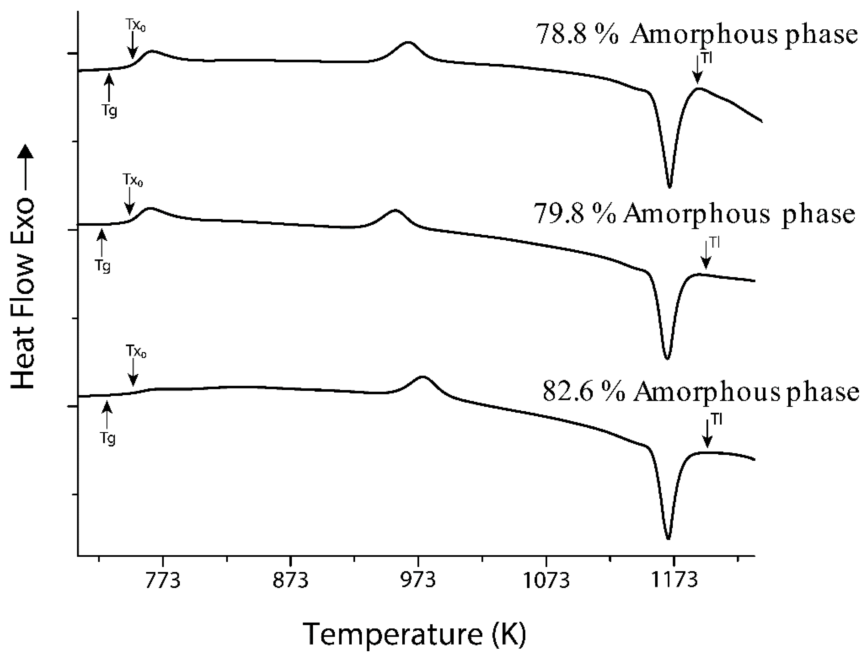
3.2. Differential Scanning Calorimetry (DSC)

Glassy metals cannot be used at high temperatures since the amorphous structure would be lost. Thermal parameters were investigated through differential scanning calorimetry (DSC). Figure 2 shows that the ribbon with 78.8% amorphous phase presents two exothermic peaks at 748 K and at 942 K, which indicates a phase transformation associated with the crystalline fraction. Before the first exothermic peak, the glass transition temperature (Tg) at 733 K and the crystallization temperature (Tx0) at 748 K were identified, followed by a single endothermic peak associated with the melting process (Tm) at 1169 K. The temperature of the liquid (Tl) was 1191 K. When analyzing the ribbon with 79.8% amorphous phase, two exothermic peaks were also observed, the first at 746 K and the second at 933 K, indicating a phase transformation associated with the crystalline fraction; the glass transition temperature (Tg) at 725 K was identified before the first exothermic peak and the crystallization temperature (Tx0) at 746 K followed by a single endothermic peak associated with the melting process (Tm) at 1167 K. The liquid temperature (Tl) was 1198 K. On the other hand, a decrease in heat flow is observed for the ribbon with 82.6% amorphous phase before an exothermic reaction, indicating the onset of crystallization and is defined by the crystallization temperature (T0) at 744 K. This phenomenon is typical of metallic glass and indicates that the glass transition temperature (Tg) in this case at 728 K. This behavior is attributed to the fact that it does not have a purely glassy structure. A single endothermic peak associated with the melting process (Tm) is observed at 1168 K, suggesting that it is a eutectic composition; the liquid temperature (Tl) was 1200 K. Table 2 summarizes the thermal properties, Tg, Tx0, Tm, Tl, the relationship ΔTx = (Tx0 − Tg), and the reduced glass transition temperature Trg = (Tg/Tl). It is important to point out that the liquid temperatures measured in this work corresponds to values reported elsewhere [36].

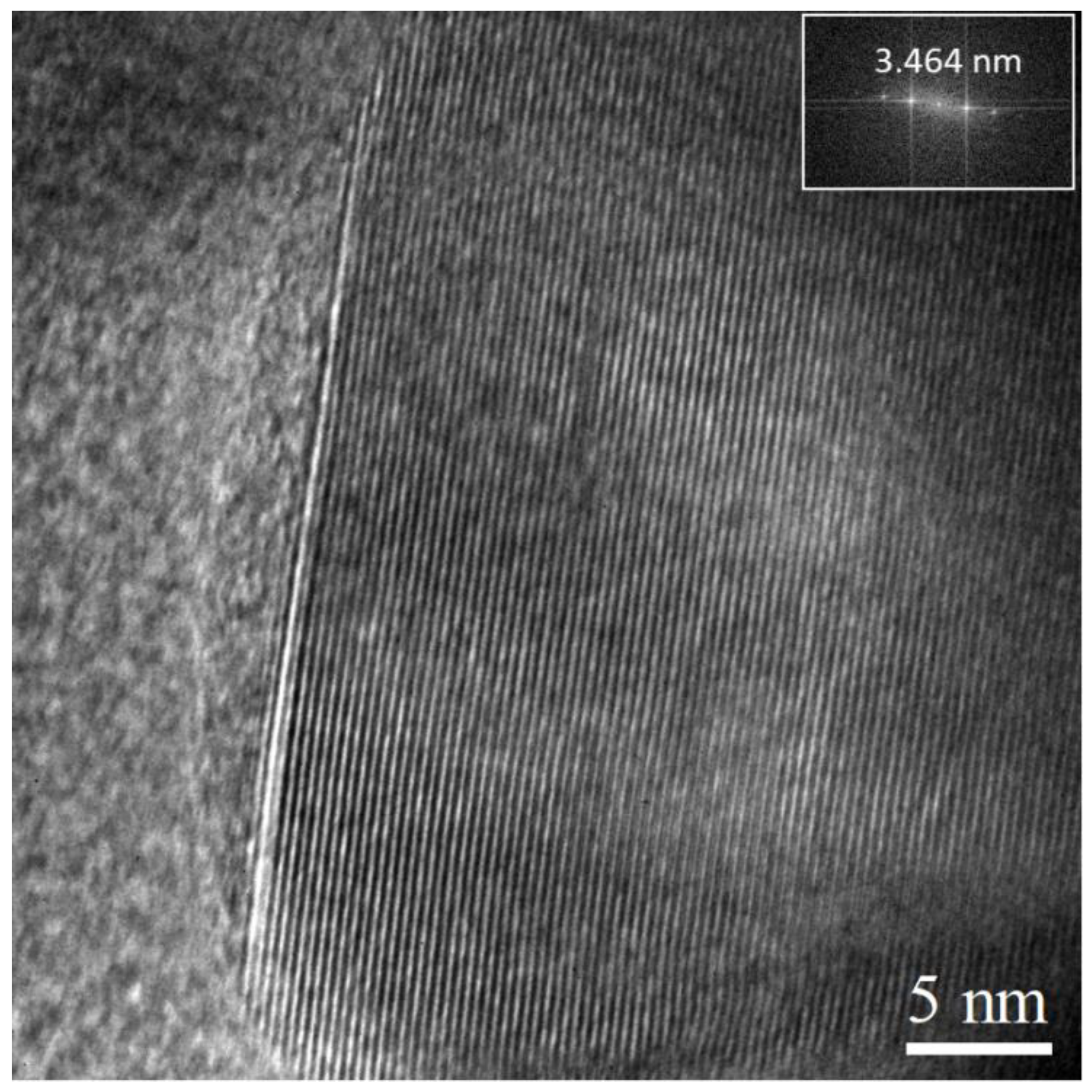
3.3. Transmission Electron Microscopy (TEM)

The ribbons were characterized through TEM. Figure 3 is a high-resolution transmission electron microscopy (HRTEM) image of the ribbon with 78.8% amorphous phase. From this image, it was possible to measure, by means of the fast Fourier transform, the interplanar distances, i.e., it was identified in the crystalline domain the planes (111) of ZrO2. Figure 4 is a HRTEM image of the ribbon with 82.6% amorphous phase; similarly, from the fast Fourier transform, it was possible to measure the interplanar distance, identifying the crystalline plane present—in this case (012) of Al2O3.

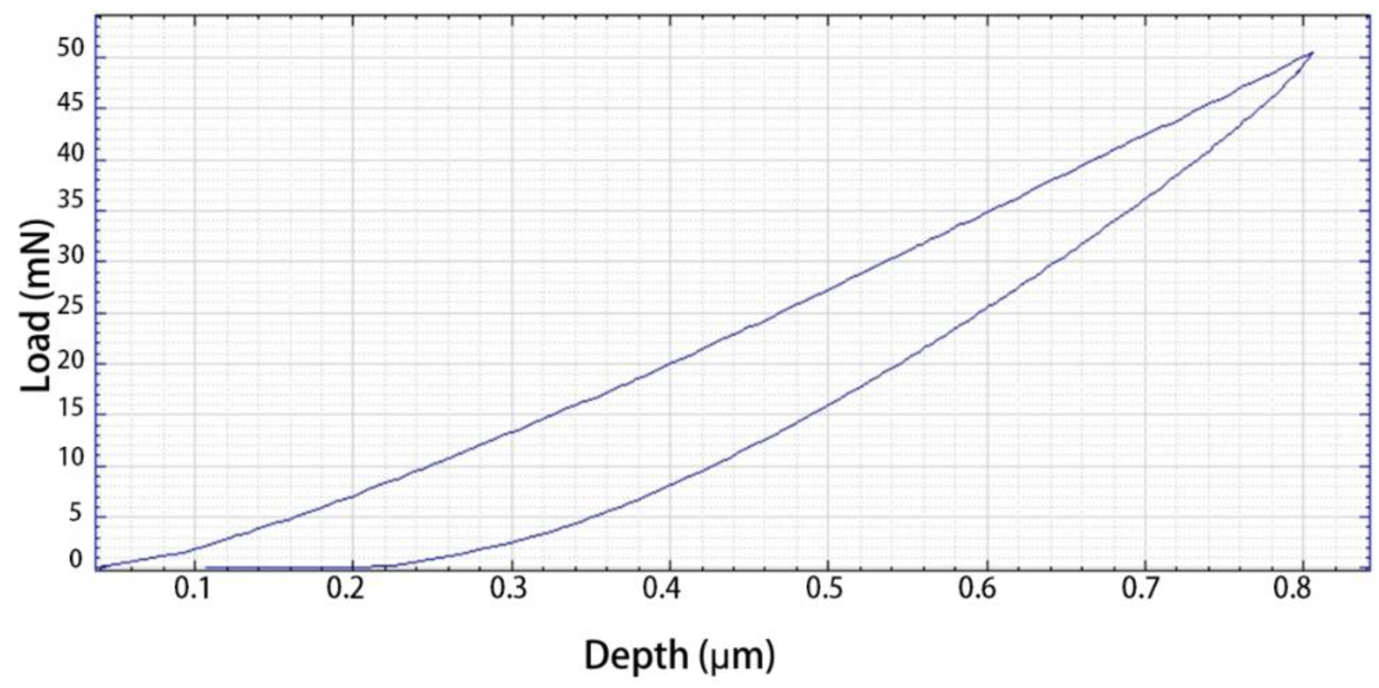
3.4. Nanoindentation

Figure 5 shows an example of the obtained nanoindentation load-displacement curve of a Zr58Co21Al9Ag12 sample subjected to a single load rate of 100 mN/min and reaching a maximum load of 49.84 mN. The Young’s modulus and hardness were 42.43 GPa and 7.72 GPa, respectively. These results showed that the obtained hardness improved significantly from the conventional biomaterials such as 316 L stainless steel (3.34 GPa) and CoCrMo alloy (3.76 GPa). The Young’s modulus was lower than that for commercial biomedical Ti, which is generally higher than 100 GPa [37,38]. Therefore, by having a value closer to that of the average human bone (10–30 GPa) [39,40,41], it can allay the protection stress shielding effect in prosthetic applications.

3.5. Immersion in SBF

SEM examined the surface morphology of the ribbons submerged in SBF. Figure 6 shows the ribbon’s SEM images with 82.6% amorphous phase submerged for 28 days. A smooth and continuous surface can be observed, indicating the absence of attack by corrosion.
Usually, the biological activity of glass is closely related to the growth rate of hydroxyapatite on its surface [42]. It is known that base Zr alloys tend to naturally form a passive layer of ZrO2 oxide [38,43,44,45] and are susceptible to the presence of chlorides, which induce pitting and selective dissolution of the amorphous matrix [46]. It then begins to attract calcium and phosphorus ions, resulting in the formation of apatite on the ZrO2 surface due to a higher specific surface area as mentioned by Liu et al. [27]. It has also been reported that Zr-based metallic glasses are bioinert in nature [47,48] and cannot form a bioactive bond with living bone if implanted into a bony site. This is probably the reason why only small domains are located with no continuous structure along the surface. It is known that the presence of crystalline phases can significantly slow down the rate of formation of the hydroxyapatite layer on the glass surface.
Figure 7a shows the ribbon’s resulting XRD patterns with 82.6% amorphous phase after being immersed in the SBF. The results showed that, at longer immersion times, the presence of crystalline peaks increases. However, this behavior was demonstrated in up to 21 days of immersion. The crystalline peaks subsequently depressed at 28 days. This may be due to the oxide layer not being thick enough, causing it to suffer from pitting corrosion due to the action of the chlorides present in the SBF, finally restarting the formation process. To study this result in detail, an examination of the pattern corresponding to 21 days was carried out (Figure 7b). In this figure, a set of diffraction peaks was detected, matching the following cards: (JCPDS 99-100-7407) AlPO4, (JCPDS 99-101-6720) Ca3(PO4)2, (JCPDS 99-100-0370) ZrC, (JCPDS 99-100-6690) ZrO2, and (JCPDS 99-101-3086) Ag. The presence of Ca and P was identified; these could favor phosphates’ precipitation, accelerating the deposition process of apatite on the surface [49].

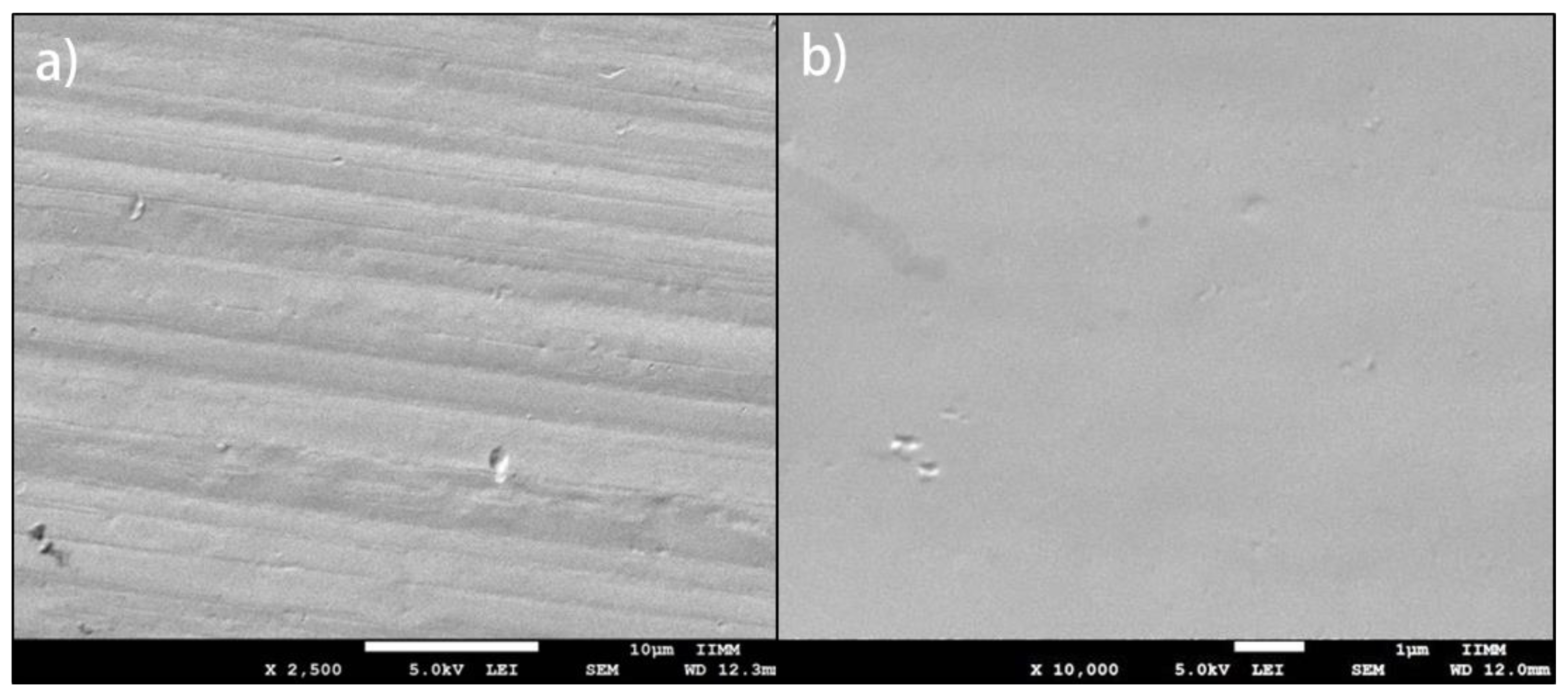
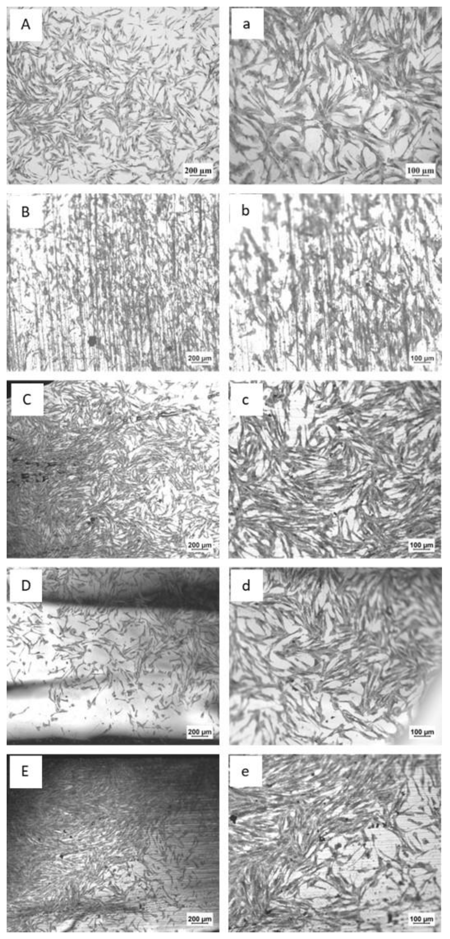
3.6. Cell Adherence Assay

Table 3 summarizes the mean ( X - ) and standard deviation (SD) of the cell adherence assay of three replicates for each sample of the Zr58Co21Al9Ag12 alloy and four replicates of Ti6Al4V alloy. Figure 8A,a shows the micrographs of the control taken at 5 and 10× respectively, where the typical spindle shape of hDPSC cells is observed, and its distribution after an incubation period of 24 h. Figure 8B,b shows the micrographs of the Ti6Al4V alloy. It can be observed there are many areas without adhered cells compared to the ribbon with 78.8% amorphous phase shown in Figure 8C,c, where a fully defined elongated spindle morphology is observed, nearly forming a monolayer, indicating that it is cytocompatible. Similar behavior was observed for the ribbon with the 79.8% amorphous phase, shown in Figure 8D,d where a higher cellular concentration is evident, and almost forming a monolayer on the surface of the sample; the ribbon with 82.6% of amorphous phase shown in E and e, however, shows fewer cells.
Cell adhesion, spread, and proliferation at the implant surface are necessary events for the bone tissue differentiation before bone tissue formation [50]. From an image analysis on the micrographs, a mean quantification of the adherent cell density was obtained, indicated in Table 4. The plotted results are shown in Figure 9.

3.7. Cell Viability and Proliferation Assay In Vitro

The cytotoxicity was evaluated using T lymphocytes through a CCK-8 assay; this type of cell has growth properties in suspension. This type of assay allows sensitive colorimetric assays to be carried out to determine the number of viable cells [51]. CCK-8 detection sensitivity is higher than MTT, XTT, MTS, or WST-1 [52]. The test was started with 40,000 T cells per well using a 24-well plate (COSTAR, flat bottom lid, ultra-low fixation, non-pyrogenic polystyrene surface); T cells were used as a positive control and the culture medium as a negative control. Table 5 summarizes the mean and standard deviation of six experiments for each sample.
Nine replicates per sample were used in the cell proliferation assay. Figure 10 shows a graph of the Y-axis to the optical density (absorbance at 440 nm); this is directly proportional to cell proliferation. It is observed that the behavior of the Zr58Co21Al9Ag12 alloy is similar to the control and shows an increase in proliferation, allowing the cells to proliferate at their regular rate. The samples with 78.8% and 79.8% of the amorphous phase presented a lower optical density at 96 h than that for 82.6%.

4. Discussion

4.1. Structural Characterization

The crystalline phase fraction fluctuated from 21.2% to 17.4%, dependent on the cooling speed. The highest degree of vitreous phase obtained was 82.6% with a tangential speed of the cooling disc of 10 m/s. On the other hand, by using the Ca5P3O13H2.82 apatite card (JCPDS No. 99-100-2294), the formation of apatite was confirmed. It is known that a material capable of forming apatite on its surface in SBF can bind to living bone through the apatite layer formed on its surface in the living body [53].
It is known that the surfaces of Zr-based MGs are commonly bioinert, meaning that direct tissue-implant bonding cannot occur. One approach to solve this problem is to make a bioactive coating on the metal surface [54,55]. X-ray diffraction results show spontaneous formation of bone apatite in SBF, indicating that the ribbons have a bioactive behavior. However, according to ASTM G31-72, in many cases, the corrosion rate is established by gain rather than loss of mass [24]. It has been reported that Zr-based MGs have a passive layer of natural oxide (ZrO2) and thus show good corrosion resistance [38,45,56,57,58]. Liu et al. [7] observed that immersing a Zr60.14Cu22.31Fe4.85Al9.7Ag3 glassy alloy in SBF causes the active metals to dissolve anodically in the solution, causing the presence of metal ions. This generates a layer of ZrO2; this layer in turn promotes the bioactivity of the ribbons.

4.2. Differential Scanning Calorimetry (DSC)

The value of ΔTx is generally an indicator of the thermal stability of the glass produced. In glassy alloys, this temperature range is usually extensive, and values of more than 120 K have been reported. In the case of thin, fast-setting glassy ribbons and marginal glass formers, the value of ΔTx is minimal, if observed at all [59]. According to Figueroa et al. [60], an alloy presents a better GFA due to subcooling, and is as close as possible to the Tg value. Hua and Zhang [61] demonstrated an evident change in the atomic structure of the Zr–Al–Co alloy by adding Ag. This behavior is mainly due to the need for efficient dense packing and the chemical affinity between the component atoms.
In the Zr–Co–Al–Ag alloy, the atomic radii of the component elements are Zr 0.16 nm, Co 0.125 nm, Al 0.143 nm, and Ag, 0.145 nm, respectively. That the atomic size ratios are 1.28 for Zr/Co, 1.12 for Zr/Al, 1.10 for Zr/Ag, 1.16 for Ag/Co, and 1.01 for Ag/Al. Therefore, this combination of atomic sizes can produce an efficiently packed local structure that is associated with low internal energy and high liquid viscosity [62,63,64]. Furthermore, in the present alloy, the heats of mixing for the pairs Zr–Al, Zr–Co, Zr–Ag, Al–Co, and Ag–Al are −44, −41, −20, −19, −4 kJ/mol, respectively. The large negative heat of mixing values improve interactions between components, promote short-range chemical ordering in liquids, improve local random packing density efficiency, and restrict long-term atom diffusion [65,66,67,68].

4.3. Transmission Electron Microscopy (TEM)

The presence of zirconium and aluminum oxides from the crystalline phase was observed. Zr-based MGs have been reported to possess a passive layer of natural oxide (ZrO2), associated with excellent biocompatibility and resistance to corrosion in the physiological environment [12,43,45,56]. Thus, it is expected that this type of oxide will form on the ribbons’ surfaces considering the oxyphilic nature of the Zr and Al elements of the alloy.

4.4. Nanoindentation

Mechanically, the Zr58Co21Al9Ag12 alloy exhibited higher hardness due to its compact atomic configuration and the presence of Zr and Al, which are highly oxyphilic elements that have a much higher hardness than that of the Zr-based alloy, contributing to their excellent wear resistance [44]. On the other hand, the Zr58Co21Al9Ag12 alloy presented a Young’s module of 42 GPa; this result is relatively close to the value reported by Qiu et al. [69] for a Zr60Cu22.5Pd5Al7.5Nb5 alloy, which is approximately 50% less than that of TiAl6V4. These results are promising for the application proposed, since a lower elastic modulus will relieve the stress shielding effect between the implant and human bones that typically have an average Young’s modulus of 10–30 GPa [55].

4.5. Immersion in SBF

The alloy elements and the microstructure mainly influence the corrosion resistance of alloys. Ag is the most effective alloy element to increase corrosion resistance [70,71,72,73]. It has been suggested that the addition of Ag increases the amount of Al2O3 when compared to a Ag-free alloy. Bearing in mind that the main characteristics of Al2O3 are the compact, dense, and stable structure that offer excellent protection than other oxides, Ag favors the formation of a protective surface with better chemical stability [14,71,74].
The presence of some phosphates and aluminum oxides was identified, which may result from the alloy ribbons’ reaction. This was attributed to the fact that the aluminum oxide layer was not thick enough, causing corrosion by pitting in the presence of chlorides [38,54]. This result indicates that the ribbons’ surface is not optimal enough to induce apatite formation, although it is not entirely bioinert. The presence of both aluminum and zirconium oxides does not allow the adhesion of phosphates to induce the formation of a layer of apatite.
It is important to point out that Escamilla et al. [75] found, by means of metal ion release studies, that the concentration of Zr ions during 28 days of immersion was 26,295 ppb. This was mainly attributed to the effect of chloride ions in the solution that is preferably absorbed by chemical and physical defects on the ribbons’ surface. Passive films are very susceptible to attack by chloride ions since they are highly distorted at these interfaces [46,57]. On average, Zr is only present in the human body at 250,000 ppb; daily intake is around 3500 ppb. However, it does not have any natural biological effect. Short-term exposure to zirconium dust has been reported to cause irritation [76]. Zr is a non-toxic metal due to the lack of binding with biomolecules, showing good biocompatibility [47,77]. Al is related to neurotoxicity and senile dementia of the Alzheimer type, but only in very high doses. The daily concentration of Al ions during 28 days of immersion was 733 ppb, which is less than the concentration of the trace element in the human body [76]. Silver is very toxic to some organisms, for instance: bacteria, viruses, algae, and fungi, but does not cause high toxicity for humans.
Silver has no known biological functions. Although the metal itself causes few problems, it has been reported to have low potential for skin irritation, as well as hepatic, renal, neurological, and hematological effects [78]. Several occupational exposure limits and guidelines exist for silver, but the values for each depend on the form of silver as well as the individual agency making the recommendations [79], (p. 575). The concentration of Ag ions during 28 days of immersion was 142 ppb; As this is a high value for human exposure, it would be worthy to conduct more studies on the size and its possible accumulation in the human body from a medical implant.
Cobalt is an important trace metal in the human body. This element promotes the production of red blood cells as a component of vitamin B12 (cyanocobalamin). However, in excess, it can cause intoxication, emesis, diarrhea, paralysis, and hypotension [80]. Co ion release was not detected, which is a positive effect for biomedical uses, since Co is cytotoxic; In vivo studies showed that it could induce damage to the DNA of oral mucosa cells [13]; the median lethal dose value for soluble cobalt salts has been estimated to be between 150 and 500 ppb [39].
The ions released are mainly Zr, and they are within the threshold in the human body. Co release did not occur, indicating that its presence in the composition of the ribbons remains stable due to its double structure face cubic centered-hexagonal close packed (FCC-HCP) that provides resistance. Likewise, Al is kept at a low value that is tolerable for the human body, demonstrating that the ribbons show stable behavior regarding ion release under immersion in SBF.

4.6. Cell Adherence Assay

In Figure 8, it is observed that cells are adhered to the alloy ribbons. Although the 78.8% and 82.6% amorphous phase Zr58Co21Al9Ag12 alloy samples clearly present a slightly higher cellular concentration average compared to the commercial Ti6Al4Valloy [81,82], which were accounted for and represented in Figure 9, there is still a significant standard deviation compared to the ribbon with 78.8% amorphous phase, which has higher adhesion. As can be seen in Figure 10, even though the samples present lower cell proliferation compared to the control, they have a fairly close behavior among them; this might be attributed to the fact that the cells require an adaptation time when they come into contact with the alloy.
This behavior indicates that the Zr58Co21Al9Ag12 alloy has high adherence and allows cell proliferation, and therefore good biocompatibility by itself, without being favored by the presence of superficial defects. This is despite the fact that Zr-based alloys have been reported, due to their bioinert nature, to be unable to form a bioactive bond with the living bone after they are implanted in bone sites [83]. Typically, surface modifications are required to prepare a bioactive coating on the surface of the glassy alloys [54,55,84]. Huang et al. [85] modified the surface of Zr-based alloys, by using the low-energy ion implantation technique of Ar and Ca ions. Their work resulted in simultaneous modifications in atomic structure, nano hardness, surface chemistry, and cell adhesion. Therefore, the result obtained is attributed to the fact that the crystalline phase directly influences the bioactive response without the need of considering a surface modification. However, a less favorable response is observed in the adherent cell density for the ribbon with 82.6% vitreous phase. A similar effect was observed by Baino [42] concerning the presence of crystalline phases that can significantly reduce the formation rate of a hydroxyapatite layer on the glass surface. Considering that the ribbons do not present surface defects, it is possible to deduce that the material’s high adherence is due only to the Zr58Co21Al9Ag12 alloy since the response presented in even the lowest cell adhesion density is comparable with the Ti6Al4V control sample.

4.7. Cell Viability and Proliferation Assay In Vitro

The behavior observed in the results is attributed to the presence of a higher proportion of the amorphous phase which offers a more suitable surface due to the lack of interaction as it is mostly bioinert, allowing cell proliferation. The medium was kept constant with values to a minimum, indicating that no unwanted behavior occurred during the test. These results are similar to those obtained by Li and Ai [77], who evaluated the biocompatibility of Zr61Ti2Cu25Al12 metallic glass. They used human umbilical vein endothelial cells together with the metal’s cellular viability through CCK-8 assays, demonstrating that it has biocompatibility as good as that of commercially pure Ti.
In other research, Li et al. [12] evaluated the biocompatibility of Zr61Ti2Cu25Al12 metallic glass using three cell lines. They obtained a cellular response comparable to Ti and its alloys, suggesting good biocompatibility associated with the formation of a layer of zirconium oxide on the surface and good resistance to corrosion in the physiological environment. On the other hand, Liu et al. [86] studied the biocompatibility of three Ni-free Zr-based alloys, obtaining cytotoxicity and cell viability comparable to the Ti6Al4V alloy. Similarly, Guan et al. [87] reported that for a ZrAlCoNb metallic glass with different Nb contents, cell viability is improved with increasing Nb content.

5. Conclusions

The fraction of the crystalline phase in the Zr-based ribbons seems to exert a favorable behavior regarding the results obtained through the characterization, showing good biocompatibility; from the SBF immersion test, it was observed that the bioinert nature of the surface of the ribbons prevails despite having a fraction of crystalline phases, which allows the attraction of elements that induce the apatite formation. Zr58Co21Al9Ag12 alloy does not present cytotoxicity and allows T lymphocyte cell proliferation, demonstrating good biocompatibility for potential use in biomedical applications since it complies with the fundamental requirement of biocompatibility—not causing any harm to the host. In this sense, the results are consistent in that the Zr58Co21Al9Ag12 alloy has properties of high cell adherence, cytocompatibility, and cell proliferation. The results of the in vitro tests were completely concluded. It remains to carry out in vivo studies in animal models to strengthen our hypothesis of the high adherence, cytocompatibility and proliferation properties exhibited by the new biomaterial in cultured human cells.

Author Contributions

Conceptualization, J.V. and A.E.; methodology, A.E., R.N. and R.G.; validation, J.V., R.N. and I.F.; formal analysis, J.V., R.N. and I.F.; investigation, A.E.; resources, A.E.; data curation, A.E.; writing-original draft preparation, A.E.; writing-review and editing, J.V.; visualization, R.N.; supervision, J.V.; project administration, J.V.; funding acquisition, J.V. All authors have read and agreed to the published version of the manuscript.

Funding

This research was funded by the Universidad Michoacana de San Nicolás de Hidalgo, through the Coordinación de la Investigación Científica, grant number 1.21 and by the Universidad Nacional Autónoma de México through the research project UNAM-DGAPA-PAPIIT-IN102422.

Institutional Review Board Statement

Not applicable.

Informed Consent Statement

Not applicable.

Data Availability Statement

Data available under request.

Acknowledgments

Aish Escamilla acknowledges to CONACYT México, for the economic support grant number 254934; Jorge Verduzco thanks to the Universidad Michoacana de San Nicolás de Hidalgo for the research project grant 1.21. Ignacio Figueroa acknowledges to the Universidad Nacional Autónoma de México for the research project UNAM-DGAPA-PAPIIT-IN102422.

Conflicts of Interest

The authors declare no conflict of interest.

References

  1. Jia, H.; Wang, G.; Chen, S.; Gao, Y.; Li, W.; Liaw, P.K. Fatigue and fracture behavior of bulk metallic glasses and their composites. Prog. Mater. Sci. 2018, 98, 168–248. [Google Scholar] [CrossRef]
  2. Khan, M.M.; Nemati, A.; Rahman, Z.U.; Shah, U.H.; Asgar, H.; Haider, W. Recent Advancements in Bulk Metallic Glasses and Their Applications: A Review. Crit. Rev. Solid State Mater. Sci. 2018, 43, 233–268. [Google Scholar] [CrossRef]
  3. Tang, J.; Yu, L.; Qiao, J.; Wang, Y.; Wang, H.; Duan, M.; Chamas, M. Effect of atomic mobility on the electrochemical properties of a Zr58Nb3Cu16Ni13Al10 bulk metallic glass. Electrochim. Acta 2018, 267, 222–233. [Google Scholar] [CrossRef]
  4. Chu, J.-H.; Lee, J.; Chang, C.-C.; Chan, Y.-C.; Liou, M.-L.; Lee, J.-W.; Jang, J.S.-C.; Duh, J.-G. Antimicrobial characteristics in Cu-containing Zr-based thin film metallic glass. Surf. Coat. Technol. 2014, 259, 87–93. [Google Scholar] [CrossRef]
  5. Hua, N.; Zheng, Z.; Fang, H.; Ye, X.; Lin, C.; Li, G.; Wang, W.; Chen, W.; Zhang, T. Dry and lubricated tribological behavior of a Ni- and Cu-free Zr-based bulk metallic glass. J. Non-Cryst. Solids 2015, 426, 63–71. [Google Scholar] [CrossRef]
  6. Li, H.F.; Zheng, Y.F. Recent advances in bulk metallic glasses for biomedical applications. Acta Biomater. 2016, 36, 1–20. [Google Scholar] [CrossRef]
  7. Liu, Y.; Wang, Y.-M.; Pang, H.-F.; Zhao, Q.; Liu, L. A Ni-free ZrCuFeAlAg bulk metallic glass with potential for biomedical applications. Acta Biomater. 2013, 9, 7043–7053. [Google Scholar] [CrossRef]
  8. Tan, Y.; Wang, Y.W.; Cheng, X.; Fu, Q.; Xin, Z.H.; Xu, Z.Q.; Cheng, H.W. Effects of Al replacement on glass forming ability and mechanical properties of Zr-based bulk metallic glasses. J. Non-Cryst. Solids 2021, 568, 120962. [Google Scholar] [CrossRef]
  9. Sun, Y.-S.; Zhang, W.; Kai, W.; Liaw, P.K.; Huang, H.-H. Evaluation of Ni-free Zr–Cu–Fe–Al bulk metallic glass for biomedical implant applications. J. Alloys Compd. 2014, 586, S539–S543. [Google Scholar] [CrossRef]
  10. Gronthos, S.; Mankani, M.; Brahim, J.; Robey, P.G.; Shi, S. Postnatal human dental pulp stem cells (DPSCs) in vitro and in vivo. Proc. Natl. Acad. Sci. USA 2000, 97, 13625–13630. [Google Scholar] [CrossRef]
  11. Huang, M.; Hill, R.G.; Rawlinson, S.C. Strontium (Sr) elicits odontogenic differentiation of human dental pulp stem cells (hDPSCs): A therapeutic role for Sr in dentine repair? Acta Biomater. 2016, 38, 201–211. [Google Scholar] [CrossRef] [PubMed]
  12. Li, J.; Shi, L.-L.; Zhu, Z.-D.; He, Q.; Ai, H.-J.; Xu, J. Zr61Ti2Cu25Al12 metallic glass for potential use in dental implants: Biocompatibility assessment by in vitro cellular responses. Mater. Sci. Eng. C 2013, 33, 2113–2121. [Google Scholar] [CrossRef] [PubMed]
  13. Li, X.; Xu, H.; Jin, Y.; Zhang, T. Fabrication of highly ordered nanotube layer on Zr-based bulk metallic glass for biomedical uses. Mater. Lett. 2017, 200, 63–66. [Google Scholar] [CrossRef]
  14. Sun, Y.; Huang, Y.; Fan, H.; Wang, Y.; Ning, Z.; Liu, F.; Feng, D.; Jin, X.; Shen, J.; Sun, J.; et al. In vitro and in vivo biocompatibility of an Ag-bearing Zr-based bulk metallic glass for potential medical use. J. Non-Cryst. Solids 2015, 419, 82–91. [Google Scholar] [CrossRef]
  15. Babilas, R.; Bajorek, A.; Radoń, A.; Nowosielski, R. Corrosion study of resorbable Ca 60 Mg 15 Zn 25 bulk metallic glasses in physiological fluids. Prog. Nat. Sci. Mater. Int. 2017, 27, 627–634. [Google Scholar] [CrossRef]
  16. Rakhimova, N.R.; Rakhimov, R.Z.; Morozov, V.P.; Gaifullin, A.R.; Potapova, L.I.; Gubaidullina, A.M.; Osin, Y.N. Marl-based geopolymers incorporated with limestone: A feasibility study. J. Non-Cryst. Solids 2018, 492, 1–10. [Google Scholar] [CrossRef]
  17. Suo, Z.; Qiu, K.; Li, Q.; You, J.; Ren, Y.; Hu, Z. A new parameter to evaluate the glass-forming ability of bulk metallic glasses. Mater. Sci. Eng. A 2010, 528, 429–433. [Google Scholar] [CrossRef]
  18. Wang, W.; Dong, C.; Shek, C. Bulk metallic glasses. Mater. Sci. Eng. R Rep. 2004, 44, 45–89. [Google Scholar] [CrossRef]
  19. Li, X.; Bhushan, B. A review of nanoindentation continuous stiffness measurement technique and its applications. Mater. Charact. 2002, 48, 11–36. [Google Scholar] [CrossRef]
  20. Guo, H.; Jiang, C.; Yang, B.; Wang, J. On the fracture toughness of bulk metallic glasses under Berkovich nanoindentation. J. Non-Cryst. Solids 2018, 481, 321–328. [Google Scholar] [CrossRef]
  21. Jian, S.-R.; Chen, G.-J.; Lin, T.-C. Berkovich nanoindentation on AlN thin films. Nanoscale Res. Lett. 2010, 5, 935–940. [Google Scholar] [CrossRef] [PubMed] [Green Version]
  22. Liu, M.; Hou, D.; Gao, C. Berkovich nanoindentation of Zr55Cu30Al10Ni5 bulk metallic glass at a constant loading rate. J. Non-Cryst. Solids 2021, 561, 120750. [Google Scholar] [CrossRef]
  23. Oliver, W.C.; Pharr, G.M. An improved technique for determining hardness and elastic modulus using load and displacement sensing indentation experiments. J. Mater. Res. 1992, 7, 1564–1583. [Google Scholar] [CrossRef]
  24. ASTM-G31-72; Standard Practice for Laboratory Immersion Corrosion Testing of Metals. ASTM International: West Conshohocken, PA, USA, 2004. [CrossRef]
  25. Zhang, Y.; Li, J.; Li, J. Effects of microstructure transformation on mechanical properties, corrosion behaviors of Mg-Zn-Mn-Ca alloys in simulated body fluid. J. Mech. Behav. Biomed. Mater. 2018, 80, 246–257. [Google Scholar] [CrossRef]
  26. Baino, F.; Yamaguchi, S. The Use of Simulated Body Fluid (SBF) for Assessing Materials Bioactivity in the Context of Tissue Engineering: Review and Challenges. Biomimetics 2020, 5, 57. [Google Scholar] [CrossRef]
  27. Liu, L.; Chan, K.; Yu, Y.; Chen, Q. Bio-activation of Ni-free Zr-based bulk metallic glass by surface modification. Intermetallics 2010, 18, 1978–1982. [Google Scholar] [CrossRef]
  28. Zhang, X.; Yuan, G.; Niu, J.; Fu, P.; Ding, W. Microstructure, mechanical properties, biocorrosion behavior, and cytotoxicity of as-extruded Mg–Nd–Zn–Zr alloy with different extrusion ratios. J. Mech. Behav. Biomed. Mater. 2012, 9, 153–162. [Google Scholar] [CrossRef] [PubMed]
  29. García Contreras Rene, A.T.L.S.; Arenas, A.M.C.; Nuñez, A.R.E. Enseñanza Practica del Aislamiento, Cultivo y Caracterización de Celulas Madre Mesenquimales de la Pulpa Dental Humana; Laboratorio de Investigación Interdisciplinaria (LII), Área de Nanoestructuras y Biomateriales, Escuela Nacional de Estudios Superiores, Unidad León, UNAM: Leon, Gto, Mexico, 2019. [Google Scholar]
  30. Ma, A.; Stratikopoulos, E.; Park, K.-S.; Wei, J.; Martin, T.C.; Yang, X.; Schwarz, M.; Leshchenko, V.; Rialdi, A.; Dale, B. Discovery of a first-in-class EZH2 selective degrader. Nat. Chem. Biol. 2020, 16, 214–222. [Google Scholar] [CrossRef]
  31. Vo, D.T.; Karanam, N.K.; Ding, L.; Saha, D.; Yordy, J.S.; Giri, U.; Heymach, J.V.; Story, M.D. miR-125a-5p Functions as Tumor Suppressor microRNA And Is a Marker of Locoregional Recurrence And Poor prognosis in Head And Neck Cancer. Neoplasia 2019, 21, 849–862. [Google Scholar] [CrossRef]
  32. Yakout, S.M.; Hassan, M.R.; Abdeltawab, A.A.; Aly, M.I. Sono-sorption efficiencies and equilibrium removal of triphenylmethane (crystal violet) dye from aqueous solution by activated charcoal. J. Clean. Prod. 2019, 234, 124–131. [Google Scholar] [CrossRef]
  33. Hui-Teng, N.; Cheng-Yong, H.; Yun-Ming, L.; Abdullah, M.M.A.B.; Hun, K.E.; Razi, H.M.; Yong-Sing, N. Formulation, mechanical properties and phase analysis of fly ash geopolymer with ladle furnace slag replacement. J. Mater. Res. Technol. 2021, 12, 1212–1226. [Google Scholar] [CrossRef]
  34. Daou, I.; Lecomte-Nana, G.; Tessier-Doyen, N.; Peyratout, C.; Gonon, M.; Guinebretiere, R. Probing the Dehydroxylation of Kaolinite and Halloysite by In Situ High Temperature X-ray Diffraction. Minerals 2020, 10, 480. [Google Scholar] [CrossRef]
  35. Tsai, Y.; Huang, G.; Zhao, J.; Shu, C. Dust cloud explosion characteristics and mechanisms in MgH2-based hydrogen storage materials. AIChE J. 2021, 67, e17302. [Google Scholar] [CrossRef]
  36. Soto, C.E.B.; Vargas, I.A.F.; Rodríguez, G.L.; Martínez, J.A.V. Glass Forming Ability and Mechanical Properties of Zr57.52Co21.24Al9.24Ag12 bulk metallic glass. Mater. Res. 2016, 19, 86–91. [Google Scholar] [CrossRef] [Green Version]
  37. Tan, M.H.; Baghi, A.D.; Ghomashchi, R.; Xiao, W.; Oskouei, R.H. Effect of niobium content on the microstructure and Young’s modulus of Ti-xNb-7Zr alloys for medical implants. J. Mech. Behav. Biomed. Mater. 2019, 99, 78–85. [Google Scholar] [CrossRef]
  38. Zhao, G.-H.; Aune, R.E.; Mao, H.; Espallargas, N. Degradation of Zr-based bulk metallic glasses used in load-bearing implants: A tribocorrosion appraisal. J. Mech. Behav. Biomed. Mater. 2016, 60, 56–67. [Google Scholar] [CrossRef]
  39. Chen, Q.; Thouas, G.A. Metallic implant biomaterials. Mater. Sci. Eng. R Rep. 2015, 87, 1–57. [Google Scholar] [CrossRef]
  40. Luo, J.; Huang, Y.; Xu, J.; Sun, J.; Dargusch, M.; Hou, C.; Ren, L.; Wang, R.; Ebel, T.; Yan, M. Additively manufactured biomedical Ti-Nb-Ta-Zr lattices with tunable Young’s modulus: Mechanical property, biocompatibility, and proteomics analysis. Mater. Sci. Eng. C 2020, 114, 110903. [Google Scholar] [CrossRef]
  41. Wu, D.; Isaksson, P.; Ferguson, S.J.; Persson, C. Young’s modulus of trabecular bone at the tissue level: A review. Acta Biomater. 2018, 78, 1–12. [Google Scholar] [CrossRef]
  42. Baino, F. Bioactive glasses—When glass science and technology meet regenerative medicine. Ceram. Int. 2018, 44, 14953–14966. [Google Scholar] [CrossRef]
  43. Akbarpour, A.; Milkova, D.A.; Zanaeva, E.N.; Parkhomenko, M.S.; Cheverikin, V.V.; Lubenchenko, A.; Bazlov, A.I. Influence of Cold Rolling Process and Chemical Composition on the Mechanical Properties and Corrosion Behavior of Zr-Based Metallic Glasses. Metals 2021, 11, 1514. [Google Scholar] [CrossRef]
  44. Hua, N.; Chen, W.; Wang, W.; Lu, H.; Ye, X.; Li, G.; Lin, C.; Huang, X. Tribological behavior of a Ni-free Zr-based bulk metallic glass with potential for biomedical applications. Mater. Sci. Eng. C 2016, 66, 268–277. [Google Scholar] [CrossRef] [PubMed]
  45. Wang, Y.B.; Zheng, Y.F.; Wei, S.C.; Li, M. In vitro study on Zr-based bulk metallic glasses as potential biomaterials. J. Biomed. Mater. Res. B Appl. Biomater. 2011, 96, 34–46. [Google Scholar] [CrossRef] [PubMed]
  46. Tian, H.F.; Qiao, J.W.; Yang, H.J.; Wang, Y.S.; Liaw, P.K.; Lan, A.D. The corrosion behavior of in-situ Zr-based metallic glass matrix composites in different corrosive media. Appl. Surf. Sci. 2015, 363, 37–43. [Google Scholar] [CrossRef]
  47. Aliyu, A.A.; Abdul-Rani, A.M.; Ginta, T.L.; Rao, T.; Axinte, E.; Ali, S.; Ramli, M. Hydroxyapatite Electro Discharge Coating of Zr-Based Bulk Metallic Glass for Potential Orthopedic Application. Key Eng. Mater. 2019, 796, 123–128. [Google Scholar] [CrossRef]
  48. Shi, H.; Zhao, W.; Wei, X.; Ding, Y.; Shen, X.; Liu, W. Effect of Ti addition on mechanical properties and corrosion resistance of Ni-free Zr-based bulk metallic glasses for potential biomedical applications. J. Alloys Compd. 2020, 815, 152636. [Google Scholar] [CrossRef]
  49. Rajan, S.T.; Terada-Nakaishi, M.; Chen, P.; Hanawa, T.; Nandakumar, A.; Subramanian, B. Zirconium-based metallic glass and zirconia coatings to inhibit bone formation on titanium. Biomed. Mater. 2020, 15, 065019. [Google Scholar] [CrossRef]
  50. Koons, G.L.; Diba, M.; Mikos, A.G. Materials design for bone-tissue engineering. Nature Reviews Materials 2020, 5, 584–603. [Google Scholar] [CrossRef]
  51. Shieh, T.-M.; Hsu, S.-M.; Chang, K.-C.; Chen, W.-C.; Lin, D.-J. Calcium Phosphate Cement with Antimicrobial Properties and Radiopacity as an Endodontic Material. Materials 2017, 10, 1256. [Google Scholar] [CrossRef]
  52. Cai, L.; Qin, X.; Xu, Z.; Song, Y.; Jiang, H.; Wu, Y.; Ruan, H.; Chen, J. Comparison of Cytotoxicity Evaluation of Anticancer Drugs between Real-Time Cell Analysis and CCK-8 Method. ACS Omega 2019, 4, 12036–12042. [Google Scholar] [CrossRef] [Green Version]
  53. Nasker, P.; Sinha, A. Titanium based bulk metallic glasses for biomedical applications. In Fundamental Biomaterials: Metals; Woodhead Publishing: Sawston, UK, 2018; pp. 269–283. [Google Scholar] [CrossRef]
  54. Qiu, Z.; Li, Z.; Fu, H.; Zhang, H.; Zhu, Z.; Wang, A.; Li, H.; Zhang, L.; Zhang, H. Corrosion mechanisms of Zr-based bulk metallic glass in NaF and NaCl solutions. J. Mater. Sci. Technol. 2020, 46, 33–43. [Google Scholar] [CrossRef]
  55. Rajan, S.T.; Arockiarajan, A. Thin film metallic glasses for bioimplants and surgical tools: A review. J. Alloys Compd. 2021, 876, 159939. [Google Scholar] [CrossRef]
  56. Jiao, Y. Nanosecond Laser Processing of a Zr-Based Bulk Metallic Glass for Potential Orthopaedic Implant Applications. Ph.D. Thesis, Cardiff University, Cardiff, UK, 2020. [Google Scholar]
  57. Tang, J.; Wang, Y.; Zhu, Q.; Chamas, M.; Wang, H.; Qiao, J.; Zhu, Y.; Normand, B. Passivation Behavior of a Zr60Cu20Ni8Al7Hf3Ti2 Bulk Metallic Glass in Sulfuric Acid Solutions. Int. J. Electrochem. Sci. 2018, 13, 6913–6929. [Google Scholar] [CrossRef]
  58. Wang, W.; Mraied, H.; Diyatmika, W.; Chu, J.P.; Li, L.; Cai, W. Effects of nanoscale chemical heterogeneity on the wear, corrosion, and tribocorrosion resistance of Zr-based thin film metallic glasses. Surf. Coat. Technol. 2020, 402, 126324. [Google Scholar] [CrossRef]
  59. Suryanarayana, C.; Inoue, A. Bulk Metallic Glasses, 2nd ed.; CRC Press: Boca Raton, FL, USA, 2017. [Google Scholar]
  60. Figueroa, I.A.; Carroll, P.A.; Davies, H.A.; Jones, H.; Todd, I. Preparation of Cu-based bulk metallic glasses by suction casting. In Proceedings of the SP07: Proceedings of the 5th Decennial International Conference on Solidification Processing, Sheffield, UK, 23–25 July 2007; pp. 479–482. [Google Scholar]
  61. Hua, N.; Zhang, T. Glass-forming ability, crystallization kinetics, mechanical property, and corrosion behavior of Zr–Al–Ni–Ag glassy alloys. J. Alloys. Compd. 2014, 602, 339–345. [Google Scholar] [CrossRef]
  62. Rashidi, R.; Malekan, M.; Gholamipour, R. Microstructure and mechanical properties of a Cu-Zr based bulk metallic glass containing atomic scale chemical heterogeneities. Mater. Sci. Eng. A 2018, 729, 433–438. [Google Scholar] [CrossRef]
  63. Zhang, W.; Zhang, Q.; Qin, C.; Inoue, A. Synthesis and properties of Cu–Zr–Ag–Al glassy alloys with high glass-forming ability. Mater. Sci. Eng. B 2008, 148, 92–96. [Google Scholar] [CrossRef]
  64. Zhu, J.; Wang, C.; Han, J.; Yang, S.; Xie, G.; Jiang, H.; Chen, Y.; Liu, X. Formation of Zr-based bulk metallic glass with large amount of yttrium addition. Intermetallics 2018, 92, 55–61. [Google Scholar] [CrossRef]
  65. Inoue, A. Stabilization of metallic supercooled liquid and bulk amorphous alloys. Acta Mater. 2000, 48, 279–306. [Google Scholar] [CrossRef]
  66. Rahvard, M.M.; Tamizifar, M.; Boutorabi, S.M.A. Zr-Co(Cu)-Al bulk metallic glasses with optimal glass-forming ability and their compressive properties. Trans. Nonferrous Met. Soc. China 2018, 28, 1543–1552. [Google Scholar] [CrossRef]
  67. Xie, S.; Tu, X.; Kruzic, J. Inducing strain hardening in a Zr-based bulk metallic glass via cobalt mediated phase separations. J. Alloys Compd. 2018, 735, 1576–1581. [Google Scholar] [CrossRef]
  68. Yuan, R.; Yu, Z.; Leng, H.; Chou, K. Thermodynamic evaluation and experimental verification of the glass forming ability of Cu-Zr-based alloys. J. Non-Cryst. Solids 2021, 564, 120835. [Google Scholar] [CrossRef]
  69. Qiu, C.; Chen, Q.; Liu, L.; Chan, K.; Zhou, J.; Chen, P.; Zhang, S. A novel Ni-free Zr-based bulk metallic glass with enhanced plasticity and good biocompatibility. Scr. Mater. 2006, 55, 605–608. [Google Scholar] [CrossRef]
  70. Li, B.; Sun, W.; Qi, H.; Lv, J.; Wang, F.; Ma, M.; Zhang, X. Effects of Ag substitution for Fe on glass-forming ability, crystallization kinetics, and mechanical properties of Ni-free Zr–Cu–Al–Fe bulk metallic glasses. J. Alloys Compd. 2020, 827, 154385. [Google Scholar] [CrossRef]
  71. Rahvard, M.M.; Tamizifar, M.; Boutorabi, S.M.A. The effect of Ag addition on the non-isothermal crystallization kinetics and fragility of Zr56Co28Al16 bulk metallic glass. J. Non-Cryst. Solids 2018, 481, 74–84. [Google Scholar] [CrossRef]
  72. Yang, Y.; Cheng, B.Y.; Lv, J.W.; Li, B.; Ma, M.Z.; Zhang, X.Y.; Li, G.; Liu, R.P. Effect of Ag substitution for Ti on glass-forming ability, thermal stability and mechanical properties of Zr-based bulk metallic glasses. Mater. Sci. Eng. A 2019, 746, 229–238. [Google Scholar] [CrossRef]
  73. Zhang, C.; Li, N.; Pan, J.; Guo, S.; Zhang, M.; Liu, L. Enhancement of glass-forming ability and bio-corrosion resistance of Zr–Co–Al bulk metallic glasses by the addition of Ag. J. Alloys Compd. 2010, 504, S163–S167. [Google Scholar] [CrossRef]
  74. Kim, H.-K.; Ahn, J.-P.; Lee, B.-J.; Park, K.-W.; Lee, J.-C. Role of atomic-scale chemical heterogeneities in improving the plasticity of Cu-Zr-Ag bulk amorphous alloys. Acta Mater. 2018, 157, 209–217. [Google Scholar] [CrossRef]
  75. Escamilla, A.; Verduzco, J.; Figueroa, I. Manufacturing, Mechanical and Electrochemical Characterization of Zr-Based Amorphous Ribbons. Microsc. Microanal. 2020, 26, 2906–2908. [Google Scholar] [CrossRef]
  76. Kumar, R.; Katyal, P. Effects of alloying elements on performance of biodegradable magnesium alloy. Mater. Today Proc. 2022, 56, 2443–2450. [Google Scholar] [CrossRef]
  77. Li, J.; Ai, H.-J. The responses of endothelial cells to Zr61Ti2Cu25Al12 metallic glass in vitro and in vivo. Mater. Sci. Eng. C 2014, 40, 189–196. [Google Scholar] [CrossRef] [PubMed]
  78. Hadrup, N.; Sharma, A.K.; Loeschner, K. Toxicity of silver ions, metallic silver, and silver nanoparticle materials after in vivo dermal and mucosal surface exposure: A review. Regul. Toxicol. Pharmacol. 2018, 98, 257–267. [Google Scholar] [CrossRef] [Green Version]
  79. Drake, P.L.; Hazelwood, K.J. Exposure-Related Health Effects of Silver and Silver Compounds: A Review. Ann. Occup. Hyg. 2005, 49, 575–585. [Google Scholar] [CrossRef] [PubMed] [Green Version]
  80. Hu, G.; Ge, L.; Li, Y.; Mukhtar, M.; Shen, B.; Yang, D.; Li, J. Carbon dots derived from flax straw for highly sensitive and selective detections of cobalt, chromium, and ascorbic acid. J. Colloid Interface Sci. 2020, 579, 96–108. [Google Scholar] [CrossRef] [PubMed]
  81. Asgar, H.; Deen, K.; Rahman, Z.U.; Shah, U.H.; Raza, M.A.; Haider, W. Functionalized graphene oxide coating on Ti6Al4V alloy for improved biocompatibility and corrosion resistance. Mater. Sci. Eng. C 2019, 94, 920–928. [Google Scholar] [CrossRef] [PubMed]
  82. Kazemi, M.; Ahangarani, S.; Esmailian, M.; Shanaghi, A. Investigation on the corrosion behavior and biocompatibility of Ti-6Al-4V implant coated with HA/TiN dual layer for medical applications. Surf. Coat. Technol. 2020, 397, 126044. [Google Scholar] [CrossRef]
  83. Sun, L.; Li, J.; Xu, R.; He, S.; Hua, Z.; Liu, H. One-step rapid synthesis of a 3D porous surface on Zr-based bulk metallic glass. Surf. Coat. Technol. 2021, 418, 127230. [Google Scholar] [CrossRef]
  84. Blanquer, A.; Hynowska, A.; Nogués, C.; Ibáñez, E.; Sort, J.; Baró, M.D.; Özkale, B.; Pané, S.; Pellicer, E.; Barrios, L. Effect of Surface Modifications of Ti40Zr10Cu38Pd12 Bulk Metallic Glass and Ti-6Al-4V Alloy on Human Osteoblasts In Vitro Biocompatibility. PLoS ONE 2016, 11, e0156644. [Google Scholar] [CrossRef]
  85. Huang, L.; Zhu, C.; Muntele, C.I.; Zhang, T.; Liaw, P.K.; He, W. Surface engineering of a Zr-based bulk metallic glass with low energy Ar- or Ca-ion implantation. Mater. Sci. Eng. C 2015, 47, 248–255. [Google Scholar] [CrossRef] [Green Version]
  86. Liu, L.; Qiu, C.L.; Huang, C.Y.; Yu, Y.; Huang, H.; Zhang, S.M. Biocompatibility of Ni-free Zr-based bulk metallic glasses. Intermetallics 2009, 17, 235–240. [Google Scholar] [CrossRef]
  87. Guan, B.; Shi, X.; Dan, Z.; Xie, G.; Niinomi, M.; Qin, F. Corrosion behavior, mechanical properties and cell cytotoxity of Zr-based bulk metallic glasses. Intermetallics 2016, 72, 69–75. [Google Scholar] [CrossRef]
Figure 1. XRD pattern of the three ribbons obtained by CBMS; a higher amorphous phase percentage is observed for the tangential wheel speed of 20 m/s. centered.
Metals 13 00055 g001
Figure 2. DSC curves of the 3 ribbons obtained by CBMS.
Metals 13 00055 g002
Figure 3. High resolution HRTEM image of the ribbon with 78.8% amorphous phase. In the upper right corner, the fast Fourier transform is shown.
Metals 13 00055 g003
Figure 4. High resolution HRTEM image of the ribbon with 82.6% amorphous phase. In the upper right corner, the fast Fourier transform is shown, from which the (012) plane of Al2O3 is identified.
Metals 13 00055 g004
Figure 5. Zr58Co21Al9Ag12 alloy nanoindentation offset curve for a load of 100 mN/min.
Metals 13 00055 g005
Figure 6. SEM image of the ribbon with 82.6% amorphous phase immersed in SBF for (a) 21 days at 2500-times magnification and (b) 28 days at 10,000-times magnification.
Metals 13 00055 g006
Figure 7. XRD pattern of the ribbon with 82.6% amorphous phase, (a) different immersion days; (b) higher magnification for the sample immersed during 21 days in the SBF.
Metals 13 00055 g007
Figure 8. Micrographs of the cell adhesion to the materials. (A,a), spindle shape of hDPSC; (B,b), hDPSC on the Ti6Al4Valloy; (C,c,D,d,E,e), hDPSC on the ribbon with 78.8, 79.8 and 82.6% of amorphous phase, respectively.
Metals 13 00055 g008
Figure 9. Quantification of cell adherence. The data are expressed as mean ± SD (n = 3) by triplicate. * Significantly different from control (Student’s t-test; p < 0.05).
Metals 13 00055 g009
Figure 10. Cell proliferation assay. The data are expressed as mean ± SD (n = 3). * Significantly different from control (Bonferroni pos hoc test; p < 0.05). All experimental groups were statistically significant from the control, except the 79.8% group at 96 h.
Metals 13 00055 g010
Table 1. Conditions of the three Zr based ribbons obtained by CBMS.
RibbonTangential Wheel Speed (m/s)Amorphous Phase (%)
1778.8
21079.8
32082.6
Table 2. Thermal properties of the three ribbons obtained by CBMS.
Zr58Co21Al9Ag12 Alloy RibbonTgTx0TmTlΔTxTrg
78.8%72874411681200160.606
79.8%72574611671198210.6051
82.6%73374811671198150.6118
Table 3. Means and standard deviations of the samples used for the cell adherence test.
Zr58Co21Al9Ag12 Alloy Ribbon X - (g)SD (g)
78.8% amorphous phase0.034230.00598
79.8% amorphous phase0.009880.00022
82.6% amorphous phase0.016740.00088
Ti6Al4V0.268570.07303
Table 4. Average quantification of adherent cell density with hDPSC cells.
ControlTi6Al4VRibbon (78.8% Amorphous)Ribbon (79.8% Amorphous)Ribbon (82.6% Amorphous)
40.84%44.35%51.3%42.38%48.35%
Table 5. Means and standard deviations of the samples used for the cytotoxicity test.
Zr58Co21Al9Ag12 Alloy Ribbon X - (g)SD (g)
78.8% amorphous phase0.033710.01019
79.8% amorphous phase0.008620.00101
82.6% amorphous phase0.017100.00074
Disclaimer/Publisher’s Note: The statements, opinions and data contained in all publications are solely those of the individual author(s) and contributor(s) and not of MDPI and/or the editor(s). MDPI and/or the editor(s) disclaim responsibility for any injury to people or property resulting from any ideas, methods, instructions or products referred to in the content.

