Article

Effects of Pollen Deprivation in Groups of Tellian (Apis mellifera intermissa) and Saharan (Apis mellifera sahariensis) Honey Bees under Controlled Conditions

by
Hassiba Khedidji
1,*,
Khaled Abderrahmani
2,
Hakima Oulebsir-Mohandkaci
1,
Kafia Ladjali-Mohammedi
3 and
Arezki Mohammedi
1
1
Laboratory of Valorization and Conservation of Biological Resources, Faculty of Sciences, University M’hamed Bougara of Boumerdes, Boumerdes 35000, Algeria
2
National Centre for Research and Development of Fisheries and Aquaculture (CNRDPA), Boulevard Colonel Amirouche Bouismail, Tipaza 42415, Algeria
3
Laboratory of Cellular and Molecular Biology, University of Sciences and Technology Houari Boumediene (USTHB), El-Alia, Bab-Ezzouar, Algiers 16110, Algeria
*
Author to whom correspondence should be addressed.
Insects 2022, 13(8), 727; https://doi.org/10.3390/insects13080727
Submission received: 21 June 2022 / Revised: 5 August 2022 / Accepted: 9 August 2022 / Published: 15 August 2022
(This article belongs to the Section Insect Societies and Sociality)

Abstract

:

Simple Summary

Pollen is a key element in bee nutrition, containing proteins, vitamins and lipids that support the vital functions of bees. Nutritional stress affects bees from the larval to the adult stage, both at the individual and colony levels. To face this type of stress, the bees developed behavioral and physiological mechanisms to fight against it. Such type of response can vary depending on the breed and age of the bees. In this study under controlled conditions, we investigated the impact of a protein-free diet on physiological functions, including survival, hypopharyngeal gland development, hemolymph proteins and ovarian development of two local bees, Apis mellifera intermissa and Apis mellifera sahariensis.

Abstract

Worldwide, honey bees are increasingly faced with periods of pollen scarcity, which can lead to nutritional deficiencies, especially of proteins and amino acids. These are essential for the proper functioning of the single organism and the colony. To understand how bees react to protein deficiency, under controlled conditions, we studied the effect of pollen deficiency on the main physiological parameters in two subspecies endemic of Algeria, Apis mellifera intermissa and Apis mellifera sahariensis. Emerging workers of both subspecies were reared with two diets: one was pollen-fed, whereas the other pollen-deprived. Several physiological criteria were measured depending on the type of diet and subspecies: the survival of the bees, the amount of total protein in the hemolymph, hypopharyngeal glands development and the ovary development of workers. These last three parameters were assessed at three different ages (7, 14 and 21 days). At birth, sahariensis workers weighed more than intermissa. With the same protein diet, the average life expectancy of sahariensis was extended by 5.55 days compared to intermissa. Even if deprived of pollen, sahariensis lived longer than intermissa fed with pollen (p < 0.001). In the three age levels, the hypopharyngeal glands were more developed and less affected by pollen deficiency (p < 0.001) in sahariensis than in intermissa (p < 0.001). The total hemolymph protein was higher in intermissa than in sahariensis regardless of the diet, and was also higher in protein-fed than in deprived bees (p < 0.001). The ovaries developed more rapidly with a high proportion in intermissa than in sahariensis (p < 0.05) regardless of the diet, and was also higher in the bees fed with pollen than those deprived (p < 0.05). Pollen deficiency generates physiological alterations and modifications, the amplitude of which varied according to the subspecies of the bee studied.

Graphical Abstract

1. Introduction

The natural habitat of the Apis mellifera bee extends from South Africa through the savannah, rainforest, desert and mild climate of the Mediterranean reaching the limit of its expansion in northern Europe and southern Scandinavia [1]. More than 25 sub-species of bees evolved within such a variety of habitats, climatic conditions and flora, with its own characteristic adaptations to each region [2,3]. Two endemic honey bee subspecies, Apis mellifera intermissa (Tellian bee) and Apis mellifera sahariensis (Saharan bee), are present in Algeria. A. mellifera intermissa inhabits the majority of Algerian apiaries, while A. mellifera sahariensis is confined to the southwestern region of Algeria [4]. A. mellifera sahariensis faces several threats that are of anthropic and natural origin, such as the drought that is increasingly frequent in the steppe and the Sahara. In addition, global warming could aggravate the already precarious situation of A. mellifera sahariensis [5].
The Intergovernmental Panel on Climate Change (IPCC) reports an approximate temperature increase in the range of 1.1–6.4 °C by the end of this century. This climate change can have a negative effect on the bee itself but also on its floral environment [6]. Excessively dry climate reduces pollen. which is the only source of proteins for bees [7]. Actually, the ratio and level of the major nutritional components of pollen, including proteins and their constituent amino acids, lipids (including flavonoids phytosterols), carbohydrates, and vitamins and secondary metabolites such as carotenoids, determine its nutritional value [8]. Pollen with good nutritional value provides high levels of hemolymph proteins [9] and ensures survival and good colony growth [10]. The need for protein is variable; it is linked to age, health and physiological status. The nurses require an adequate protein intake to be able to develop their hypopharyngeal glands (HGPs) and to synthesize royal jelly. Protein-rich pollen permits a good development of HGPs [11], improves the reproductive system, promotes ovarian development and increases drone fertility [12,13], boosts the immune defense [10] and increases the activity of digestive enzymes [14]. Consequently, worker bees that suffer from pollen deprivation have a shorter lifespan [15,16], start foraging earlier [17,18], have less developed hypopharyngeal glands [19], poor ovarian development [12], have lower flight efficiency [20], and have lower levels of vitellogenin and transferrin expression [21].
However, as A. mellifera sahariensis has been evolving for thousands of years in an arid and hostile environment, whose thermal amplitude varies from −4 °C in winter to more than 40 °C in summer, it is probable this honey bee subspecies have developed behavioral and physiological characteristics very different from A. mellifera intermissa, which evolves in a relatively more clement environment [22]. The objective of this work is to test this hypothesis by evaluating the physiological response of the two subspecies of bees when they are completely deprived of a source of pollen. The physiological parameters studied to evaluate this response are the level of protein in the hemolymph, the development of the hypopharyngeal glands and ovaries, and the lifespan of the bees. Apart from a few studies characterizing the A. mellifera sahariensis populations on morphometric and molecular aspects [2,23], very few scientific works have been devoted to the A. mellifera sahariensis. In this work, the physiology of the two main subspecies of A. mellifera endemic to North Africa is studied for the first time under controlled conditions.

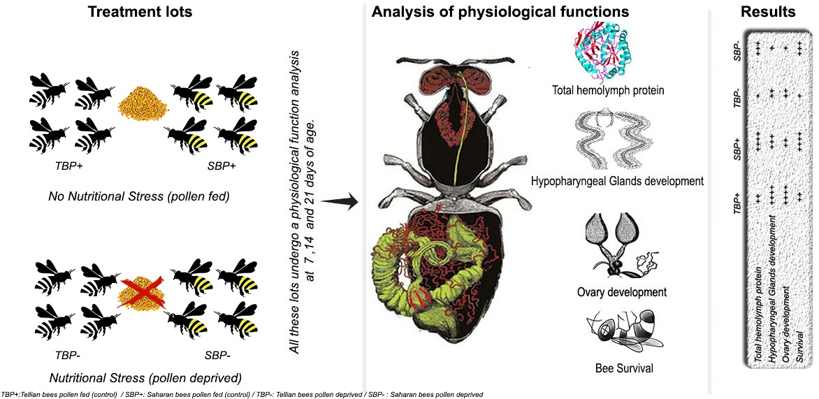
2. Materials and Methods

The experiment was carried out between March and June 2020 in the apiary of the laboratory of The University of Boumerdes (UMBB).

2.1. Source of the Honeybees

The experiment was carried out on the two local subspecies of bees in Algeria. A. mellifera intermissa is typically used in the north of Algeria for beekeeping, and A. mellifera sahariensis by beekeepers in oases in southwest Algeria. The two subspecies can be differentiated by their differences in size and color. A. mellifera sahariensis is slightly smaller than A. mellifera intermissa and is yellowish-reddish, while A. mellifera intermissa is black in color [5]. The bees were taken from two experimental apiaries. The first apiary of A. mellifera intermissa colonies is located on the university campus, and the second apiary of A. mellifera sahariensis colonies is located in an oasis of Ain sefra. This region is a part of the natural habitat of A. mellifera sahariensis. The two apiaries were monitored in the same way and treated against varroa at the appropriate time. For the purposes of the experiment, we moved ten A. mellifera sahariensis colonies and placed them in the experimental apiary on campus.
Three healthy colonies from each subspecies were selected. From each colony, frames containing brood close to emergence were kept in wooden cages (50 × 25 × 14 cm) separately and incubated at 34.5 °C and 75% RH. The bees were collected from the frames during the 24 h following the incubation.

2.2. Average Weight of Emerging Bees

After the emergence of the workers, a batch made up of 100 to 120 individuals randomly taken of each subspecies were weighed. The average weight of each bee (expressed in mg) was calculated based on the total number of individuals weighed.

2.3. Experimental Design

One hundred emerging bees were transferred to the Plexiglas bioassay cages (cage type Pain) [24] (12 × 10 × 4 cm) equipped with transparent and removable sides, ventilation holes and two syringes of 2.5 mL, which were adapted as feeders to provide a water and sucrose solution (50% w/w). A piece of wax was hung in the middle of each cage near the sugar solution and water for the bees to cluster on.
Hoarding bioassay cages were maintained at controlled conditions (34 ± 1 °C, 50–70% relative humidity) and kept in the dark to simulate conditions within the colony. Pollen balls were provided ad libitum for the control bees only; these balls were prepared by mixing pollen (fresh polyfloral harvested the previous year and stored in the freezer) with honey until a soft, cohesive paste was obtained. Water and sucrose were also provided ad libitum for all the cages. A pollen ball of 10 grams was offered to each cage; this quantity allowed the pollen to be constantly available to the bees. The quantity of pollen consumed by caged bees is determined by the difference between the weight of pollen supplied and left after consumption. The diet was provided periodically at 1 or 2 d intervals, and at these time intervals, dead bees were removed and counted. The dead bees and the total number of bees in each cage were considered in the estimation of consumption. The mortality of the bees was registered until the last bee.
Three cages each containing 100 newly emerged A. mellifera intermissa or A. mellifera sahariensis worker bees were registered for each of the following treatments.
Lot 1: A. mellifera sahariensis pollen-deprived (SBP−)
Lot 2: A. mellifera sahariensis pollen-fed (control) (SBP+)
Lot 3: A. mellifera intermissa pollen-deprived (TBP−)
Lot 4: A. mellifera intermissa pollen-fed (control) (TBP+)
For statistical analyses of data, we considered the three repetitions of each treatment as a single lot (one lot consists of three cages that received the same treatment).

2.4. Hemolymph Extraction

After confinement and feeding on the different diets, the hemolymph of 7-, 14- and 21-day-old workers was collected from a small incision at the level of the 3rd dorsal tergite, using microcapillary tubes previously washed in a 0.1% (w:v) phenylthiourea solution in water. The bees were placed in separate 1.5 mL tubes and kept refrigerated until their hemolymph was collected 1–2 h later. The hemolymph of 10 workers fed in each cage was conserved in microcapillary tubes at −20 °C for the later determination of total protein. After the hemolymph was collected, each bee was stored at the same temperature (−20 °C) until the further measurement of the hypopharyngeal glands (HPG) acini and ovary development.

2.5. Total Protein

The quantification of the total protein was carried out using the Bradford method [25]. This is a colorimetric measurement method, based on a reaction between proteins and a dye, Coomassie Brilliant Blue G250 (BBC), which is a red-stranded reagent in the free state that turns blue when bound to proteins, the intensity of the colour being proportional to the concentration of the proteins. The Bradford reagent (BBC) was prepared from 50 mg of Coomassie Brilliant Blue, 25 mL of 95° ethanol, 50 mL of 85% orthophosphoric acid and 500 mL with distilled water. The calibration range was prepared from a stock solution of 10 mg/mL Bovine Serum Albumin (BSA) at 1 mg/mL and a stock solution of 0.1% BSA; then, a series of 6 tubes of increasing concentration were established and incubated for 10 min at room temperature in the dark. The optical density was measured using a spectrophotometer at a wavelength of 595 nm; a calibration curve was then drawn. Samples were thawed in fresh water before a series of tubes were prepared each containing 5 μL of hemolymph made up to 100 μL by adding 95 μL of distilled water. Subsequently, a volume of 4 mL of BBC was added to both sets of tubes, mixed and incubated for 10 min at 4 °C in the dark, before running through the spectrophotometer. The values obtained were plotted on the calibration curve already drawn to determine the protein concentration.

2.6. Extraction and Measurement of Hypopharyngeal Glands (HPG)

HPG were removed with forceps through an incision in the front part of the head and put on a microscope slide in a droplet of sodium chloride solution. The development of the HPG was assessed by calculating the area of the acini according to the protocol established by DeGrandi-Hoffman (2010) [26]. A photo of the glands was taken using a Leica DM 500 microscope with a Leica DC-300 camera and Axio-vision image manager software program. For calibration, a photograph of a 1 mm line was taken at the same magnification used for observing the HPG. The area of each acinus was calculated by multiplying the length by the width and considered as a parameter for development of the acini. Six acini per bee were selected and only acini with clear borders were measured. Acini areas were averaged to obtain an estimated HPG mean size for the cage.

2.7. Ovary Assessments

The ovary development was assessed using a binocular microscope and the classification of the different developmental stages as proposed by Hess (1942) [27] and modified by Velthuis (1970) [28]. Five stages were differentiated: stage 1 is inactive thread-like ovarioles without vitellus; stage 2 is swollen ovarioles without vitellus; stage 3 is swollen ovarioles with visible vitellus but without distinct oocytes; in stage 4, the ovarioles contain distinct, but immature oocytes; and stage 5 is fully activated ovaries with distinct mature oocytes. The two first stages (sizes 1 and 2) correspond to undeveloped ovaries and the last three (sizes 3–5) to developed ovaries.

2.8. Statistical Analysis

Statistical analysis of the data was conducted using the program Statistic, IBM SPSS Statistics 28.0. Results were expressed as means ± standard deviation. The weight of emerging bees was evaluated using a one-way ANOVA. The total protein of the hemolymph were analyzed by using a full-factorial ANOVA. The surface of the hypopharyngeal glands for the three age levels was reanalyzed using ANOVA three ways and Tukey’s HDS test for the post hoc multiple comparisons, to indicate the differences. The level of significance was determined at 5% (normal distribution of the data was verified using the Kolmogrov–Smirnov test before carrying out ANOVA). A chi2 test was performed to study the dependence of ovary development on subspecies and diet.
The survival was analyzed using the Kaplan–Meier survival estimator, and Gehan–Wilcoxon test was conducted to determine the differences. Differences in pollen consumption were investigated using Student’s t-tests.

3. Results

3.1. Average Weight of Emerging Bees

At emergence, sahariensis weighed on average 133 ± 0.06 mg and intermissa 90 ± 0.05 mg (F1,18 = 252.57; p < 0.001).

3.2. Food Consumption

Pollen consumption was statistically the same for bees of both the subspecies (F1,4 = 0.732; p > 0.05): on average, pollen consumption was 2.62 ± 0.33 mg/day for A. mellifera intermissa and 2.13 ± 0.61 mg/day for A. mellifera sahariensis.

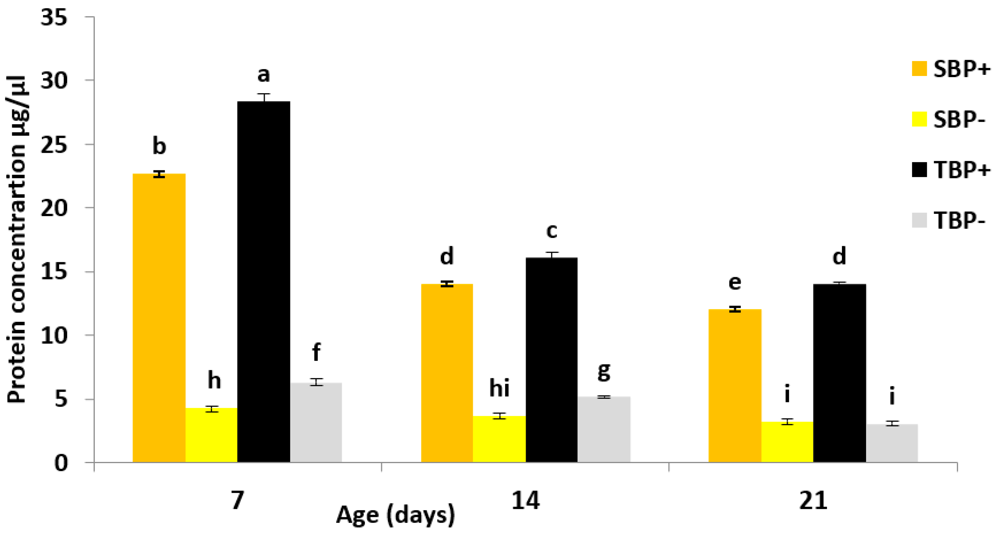
3.3. Total Hemolymph Protein

The amount of protein in the hemolymph (HP) was influenced by the diet (F1,96 = 23,005.41; p < 0.001), the subspecies (F1,96 = 597.62; p < 0.001) and age (F2,96 = 2436.39; p < 0.001).The statistical interactions diet × subspecies (F1,96 = 137.57; p < 0.001), subspecies × age (F2,96 = 97.16; p < 0.001), diet × age (F2,96 = 1378.77; p < 0.001) and subspecies × diet × age (F2,96 = 24.40; p < 0.001) were also significant (Figure 1).
At 7 days, TBP+ had a significantly higher HP content than TBP− (Tukey’s HDS test; p < 0.001) and SBP+ (Tukey’s HDS test; p < 0.001). SBP+ had significantly more HP than SBP− (Tukey’s HDS test; p < 0.001). In the situation of pollen deficiency, the HP content was significantly higher in TBP− than in SBP− (Tukey’s HDS test; p < 0.001).
At 14 days, the amount of HP decreased significantly within all lots and followed the same trend among the different batches. TBP+ had significantly more HP than TBP− (Tukey’s HDS test; p < 0.001) and SBP+ (Tukey’s HDS test; p < 0.001). SBP+ had significantly more HP than SBP− (Tukey’s HDS test; p < 0.001). In a situation of pollen deficiency, the amount of HP was significantly higher in TBP− than in SBP− (Tukey’s HDS test; p < 0.001).
At 21 days, a slight decrease was observed in the amount of HP compared to 14-day-old bees, with the same trend between the four groups. TBP+ had a significantly higher quantity of HP than TBP− (Tukey’s HDS test; p < 0.001) and SBP+ (Tukey’s HDS test; p < 0.001). In the batches of bees deprived of pollen, more HP in SBP− than in TBP− was observed; however, the difference was not significant (Tukey’s HDS test; p > 0.05).

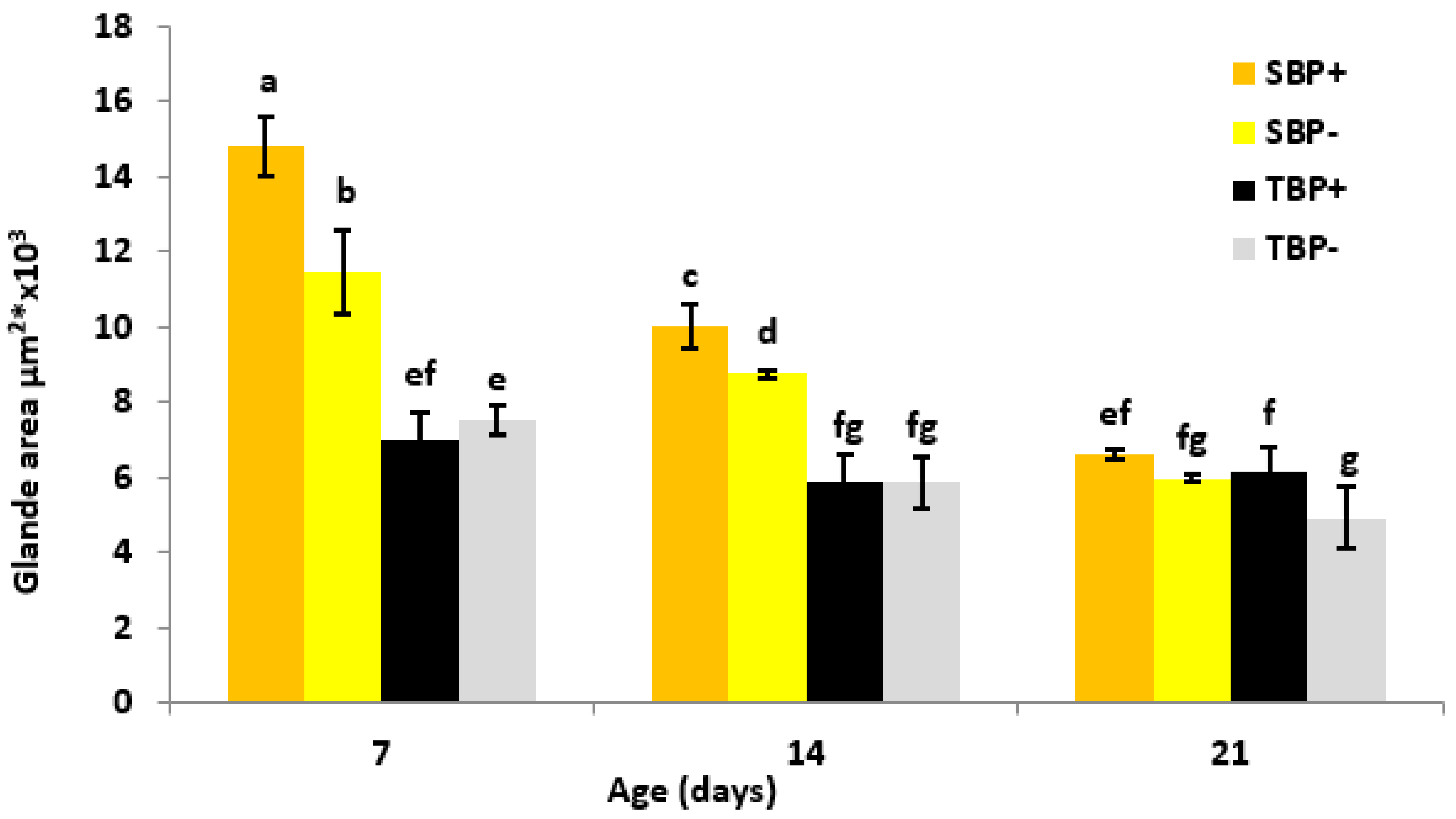
3.4. Hypopharyngeal Glands’ Development

The development of the HPGs was influenced by the diet (F1,348 = 43.69; p < 0.001), the subspecies (F1,348 = 508.45; p < 0.001), the age (F2,348 = 278.43; p < 0.001), the diet × subspecies interactions (F1,348 = 29.99; p < 0.001), the subspecies × age (F2,348 = 97.05; p < 0.001) and the subspecies × diet × age interactions (F2,348 = 18.66; p < 0.001). However, the effect of the diet × age interactions was not significant (F3,348 = 2.03; p < 0.132) (Figure 2).
At 7 days of age, the HPG of SBP+ were significantly more developed than those of SBP− (Tukey’s HDS test; p < 0.001) and TBP+ (Tukey’s HDS test; p < 0.001). The HPGs of SBP− were significantly more developed than those of TBP− (Tukey’s HDS test; p < 0.001). However, in intermissa, there was no difference between TBP+ and TBP− (Tukey’s HDS test; p > 0.05).
At 14 days, the HPG of SBP+ were significantly more developed than those of TBP+ (Tukey’s HDS test; p < 0.001) and SBP− (Tukey’s HDS test; p < 0.033), whereas the HPGs of SBP− were significantly more developed than those of TBP− (Tukey’s HDS test; p < 0.001). In intermissa, there was no significant difference between TBP+ and TBP− (Tukey’s HDS test; p > 0.05).
At 21 days, HPG atrophy was observed in sahariensis (SBP+ and SBP−). At this age, there were no significant differences between the groups except in intermissa where the HPG of TBP+ were significantly more developed than those of TBP− (Tukey’s HDS test; p = 0.045).

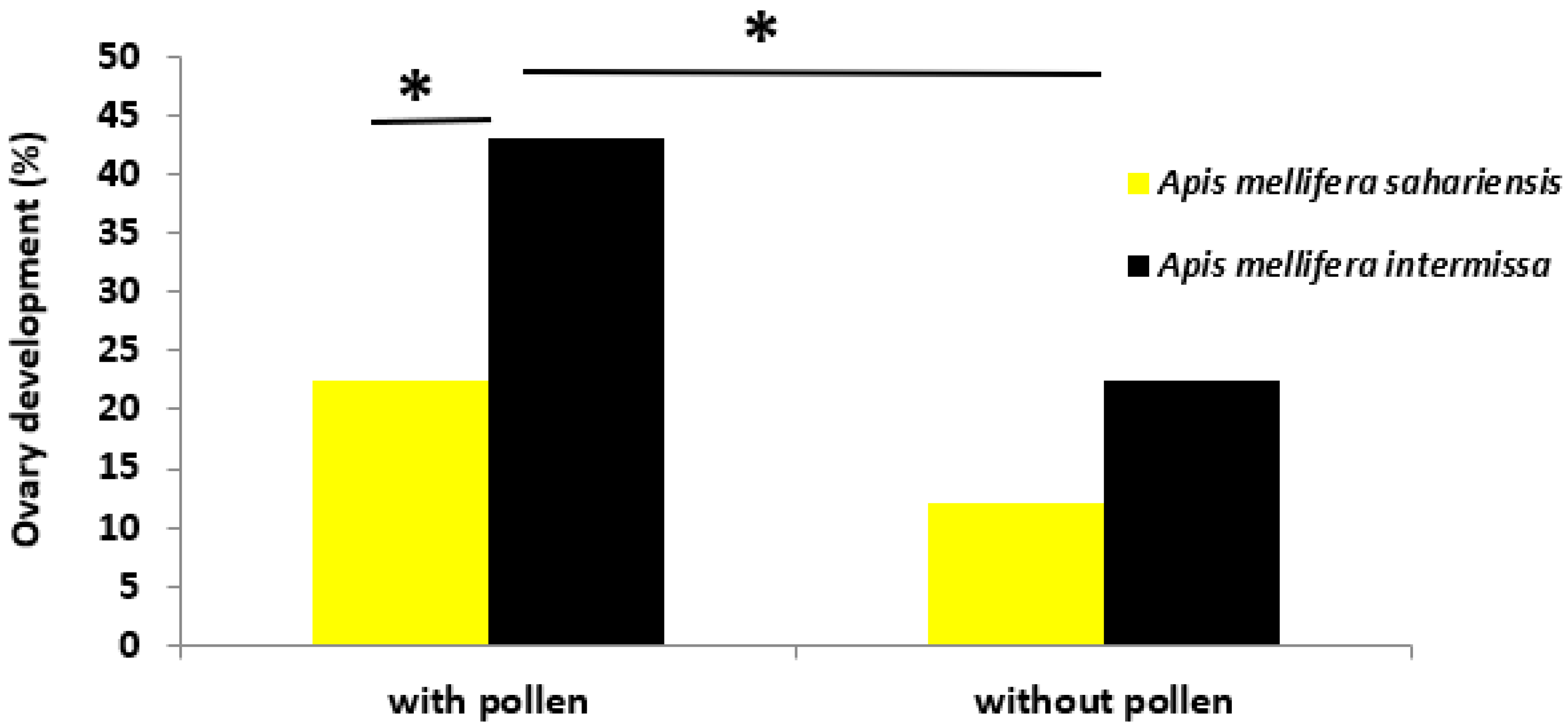
3.5. Ovary Development
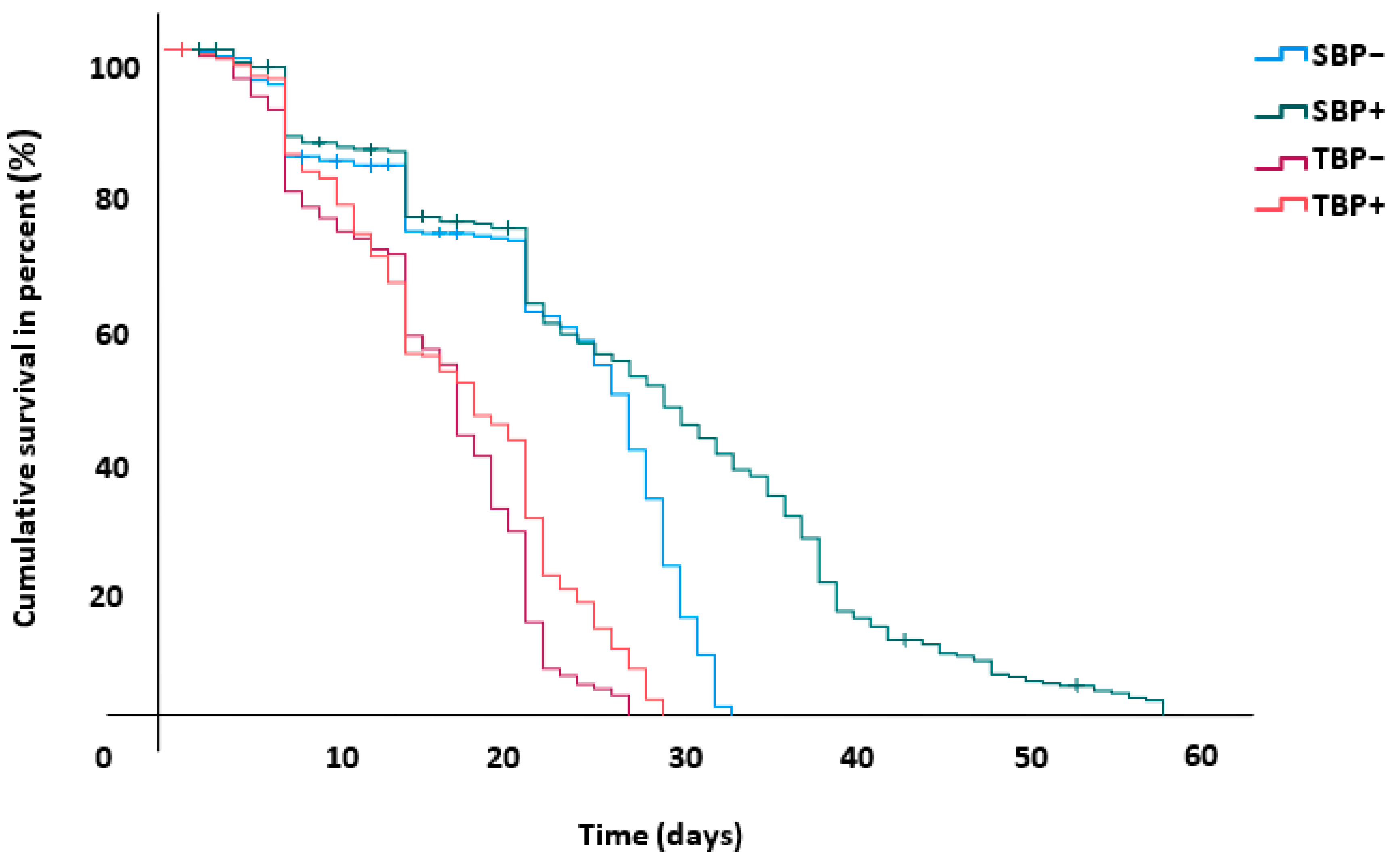
Ovary development was greater in intermissa than in sahariensis. It also depended on the diet of the bees; whether it was rich or deficient in pollen. The result of the chi2 test therefore indicated a dependence of ovary development on diet and subspecies (χ2 (3) = 14.05; p < 0.05) (Figure 3).
A protein diet had a significant effect on the development of the ovaries in intermissa. In TBP+, there were significantly more developed ovaries than in TBP− (χ2 (1) = 4.804; p < 0.05). The comparison between subspecies showed that TBP+ had significantly more developed ovaries than SBP+ (χ2 (1) = 4.804; p < 0.028).
On the other hand, the diet did not seem to have an influence on the ovary development of sahariensis since there was no significant difference in the ovary development of SBP+ and SBP− (χ2 (1) = 2.0235; p > 0.05). When they were both deprived of pollen, there was no significant difference in the ovary development of SBP− and TBP− (χ2 (1) = 2.025; p > 0.05).

3.6. Bee Survival

The survival analysis was conducted over a period of 58 days, the period that corresponded to the date from the start of the trial until the death of the last bee. The Kaplan–Meier estimation results revealed that the survival of sahariensis was superior to intermissa even with a pollen-free diet (Figure 4, Table 1). All pairwise comparisons between groups were significantly different.
SBP+/TBP+ (Wilcoxon test, χ2 (1) = 105.65; p < 0.001)
SBP−/TBP− (Wilcoxon test, χ2 (1) = 119.61; p < 0.001)
SBP+/SBP− (Wilcoxon test, χ2 (1) = 24.286; p < 0.001)
TBP+/TBP− (Wilcoxon test, χ2 (1) = 6.803; p < 0.009)

4. Discussion

This study showed that pollen consumption has an influence on the amount of protein in the hemolymph of caged bees. However, it also depends on the subspecies since, when the same amount of pollen is consumed, intermissa has more hemolymphatic proteins than sahariensis. These results suggest that intermissa optimizes its protein diet much better than sahariensis. A study has shown that, for the same pollen source, the Egyptian subspecies Apis mellifera lamarckii has a higher protein content in its hemolymph than the two subspecies Apis mellifera carnica and Apis mellifera ligustica [29]. In that study, the authors explain this difference by the greater enzymatic secretion in the intestine. In a subsequent study carried out on pre-pupae of Apis mellifera [30], the researchers demonstrated the role of the enzyme acid phosphatase, which would be at the origin of the accumulation of proteins in the hemolymph and in the mobilization of proteins stored in the trophocytes to be used during metamorphosis. Another study [31] has also shown the superiority of Africanized bees in the conversion of pollen proteins into hemolymph proteins compared to European bees. It is therefore possible that intermissa secretes more acid phosphatase enzyme in its intestine than sahariensis. As the two subspecies have evolved under different floral environment, they are maybe more adapted to digest specific pollen diets, and so the diet used in our study may be more adapted to intermissa.
This enzyme could also play a role in the mobilization of body proteins to store them in the hemolymph since even in a situation of pollen deficiency intermissa has more hemolymphatic proteins than sahariensis.
Once stored in the bee’s hemolymph, nutrients (proteins, carbohydrates, lipids and vitamins) as well as hormones are distributed according to various physiological needs [32,33]. These proteins play an essential role in the functioning of the hypopharyngeal glands [12,19,26,34]. This study shows that, even if deprived of pollen, sahariensis has more developed hypopharyngeal glands than intermissa fed with pollen. This trend is observed at 7 and 14 days. This is an age class that corresponds to that of nurses [35]. Field experiments have shown that sahariensis produces more royal jelly than intermissa (unpublished data). These results could support the hypothesis that sahariensis gives priority to the development of the hypoharyngeal glands in order to nourish the brood in an optimal way. This priority would even be to the detriment of the body reserves of the pollen-deficient nurse bee.
According to [13], bees fed a high protein diet have more developed ovaries. This is in agreement with our results, which clearly show that pollen fed bees have the highest proportion of developed ovaries than those fed only with sugar syrup.
However, ovarian development does not depend solely on a diet rich in protein since [36] also demonstrates the role of carbohydrates in ovarian development; the author indicated that a diet with a protein/carbohydrate ratio of (1:3) results in better ovarian development. Even without a protein diet, a greater proportion of developed ovaries is recorded in intermissa. It therefore seems that ovarian development does not only depend on diet but also depends on subspecies. When the queen and brood are experimentally removed, intermissa develops its ovaries more rapidly (5.6 days) than the African subspecies scutellata (9.5 days) or European carnica (30 days) [37]. Likewise, intermissa lays more eggs than scutellata or carnica. The racial aspect is therefore important in the development of the workers ovaries [38].
In our experiment under controlled conditions, the bees of both subspecies consumed the same amount of pollen. The importance of this protein intake in the development of the sahariensis hypopharyngeal glands and the intermissa ovary development is shown. This protein supply is also important in the lifespan of bees [7,39]. sahariensis is much more affected by pollen deficiency than intermissa. Indeed, when the bees are pollen deprived, the life expectancy of intermissa and sahariensis is shortened by just over 8.5% and 18.83%, respectively. On the other hand, when the bees are pollen fed, the average life expectancy of sahariensis is extended by 5.55 days compared to intermissa. It is important to note that even when deprived of pollen, sahariensis lives longer than pollen-fed intermissa. This difference in life expectancy between the two subspecies could have its origin during the larval development of the bee. Indeed, we found that at emergence, sahariensis weighed more than intermissa. This weight difference could be due to a good diet during the larval phase. This hypothesis is confirmed by the propensity of sahariensis to promote the development of its hypopharyngeal glands to the detriment of its ovaries.
Numerous studies have also shown that the phospholipoglycoprotein vitellogenin, known as female-specific egg yolk proteins, accumulates in body reserves and in the hypopharyngeal glands and has a positive effect on the longevity of workers [40,41,42,43,44]. In a recent study carried out on honey bee workers [45], it is demonstrated that telomerase activity and the production of the hormone vitellogenin are positively correlated with body mass and fat cell size. Telomeres, which are involved in the process of survival and aging in eukaryotes [46], bind the ends of chromosomes. They are shortened under the effect of oxidative stress and during cell division [47]. It is possible that, in the case of sahariensis, the oxidative stress is minimal enough not to negatively affect its life expectancy.
These results obtained under controlled conditions made it possible for the first time to highlight certain physiological characteristics of the two subspecies of bees endemic to North Africa.
The two subspecies evolve in two different environments. Intermissa lives in regions characterized by a Mediterranean climate where honey flora is abundant almost all year round. The opulence that benefits intermissa means that this subspecies uses nutrients to promote excessive reproduction [38], while the desert environment of sahariensis is very hostile.
Moreover, it could be said that, in this environment, sahariensis do not have interest to engage in ovary development for workers as it can be a loss of energy for the whole colony.
The sahariensis subspecies seems to be endowed with physiological qualities allowing it to face a hostile environment. Long periods of scarcity are recurrent in the Sahara and sahariensis has learned to live in this environment. Optimal feeding of the larvae as well as longevity of the workers could be a response to this hostility of the nature.

Author Contributions

Conceptualization, H.K., K.L.-M. and A.M.; methodology, H.O.-M., K.L.-M. and A.M.; software, H.K. and K.A.; validation, K.L.-M. and A.M.; formal analysis, H.K. and K.A.; investigation, H.K.; data curation, H.K. and K.A.; writing—original draft preparation, H.K., K.A., H.O.-M., K.L.-M. and A.M.; writing—review and editing, H.K., K.A., H.O.-M., K.L.-M. and A.M.; visualization, H.K. and H.O.-M.; supervision, K.L.-M. and A.M.; project administration, A.M.; funding acquisition, A.M. All authors have read and agreed to the published version of the manuscript.

Funding

Financial support has been provided by Partnership for Research and Innovation in the Mediterranean Area PRIMA (grant number 1812/PLANT-B), a programme supported by the European Union and Ministry of Higher Education and Scientific Research DGRSDT (DO1NO1UN35012018001).

Institutional Review Board Statement

Not applicable.

Informed Consent Statement

Not applicable.

Data Availability Statement

Data are available upon request from the corresponding author.

Acknowledgments

The authors thank Yves Le Conte, Pier Paolo Danieli, Aya Hamiri and the anonymous reviewers for valuable remarks of the manuscript, and many helpful suggestions. The authors also thank PRIMA FOUNDATION and DGRSDT for financing this work.

Conflicts of Interest

The authors declare no conflict of interest.

References

  1. Han, F.; Wallberg, A.; Webster, M.T. From where did the western honeybee (Apis mellifera) originate? Ecol. Evol. 2012, 2, 1949–1957. [Google Scholar] [CrossRef] [PubMed]
  2. Ruttner, F. Biogeography and Taxonomy of Honey Bees; Springer: Heidelberg, Germany, 1988. [Google Scholar]
  3. Sheppard, W.S.; Meixner, M.D. Apis mellifera pomonella, a new honey bee subspecies from Central Asia. Apidologie 2003, 34, 367–375. [Google Scholar] [CrossRef]
  4. Haccour, P. Recherches sur l’abeille saharienne au Maroc. Belg. Apic 1961, 25, 13–18. [Google Scholar]
  5. Mallek, S.; Ladjali, K.; Mohammedi, A. Les Menaces Naturelles et Anthropiques Mettant en Peril l’Existence de l’Abeille Saharienne en Algerie; Bulletin de la Société d‘Histoire Naturelle d‘Afrique du Nord, USTHB: Alger, Algeria, 2018; Volume 74, pp. 165–196. [Google Scholar]
  6. Le Conte, Y.L.; Navajas, M. Climate change: Impact on honey bee populations and diseases. Rev. Sci. Tech. 2008, 27, 499–510. [Google Scholar]
  7. Haydak, M.H. Honey bee nutrition. Annu. Rev. Entomol. 1970, 15, 143–156. [Google Scholar] [CrossRef]
  8. Vaudo, A.D.; Tooker, J.F.; Patch, H.M.; Biddinger, D.J.; Coccia, M.; Crone, M.K.; Fiely, M.; Francis, J.S.; Hines, H.M.; Hodges, M.; et al. Pollen protein: Lipid Macronutrient ratios may guide broad patterns of bee species floral preferences. Insects 2020, 11, 132. [Google Scholar] [CrossRef] [PubMed]
  9. Frias, B.E.; Barbosa, C.D.; Lourenço, A.P. Pollen nutrition in honey bees (Apis mellifera): Impact on adult health. Apidologie 2015, 47, 15–25. [Google Scholar] [CrossRef]
  10. Di Pasquale, G.; Salignon, M.; Le Conte, Y.; Belzunces, L.P.; Decourtye, A.; Kretzschmar, A.; Suchail, S.; Brunet, J.-L.; Alaux, C. Influence of pollen nutrition on honey bee health: Do pollen quality and diversity matter? PLoS ONE 2013, 8, e72016. [Google Scholar] [CrossRef]
  11. DeGrandi-Hoffman, G.; Corby-Harris, V.; Carroll, M.; Toth, A.L.; Gage, S.; Watkins deJong, E.; Graham, H.; Chambers, M.; Meador, C.; Obernesser, B. The importance of time and place: Nutrient composition and utilization of seasonal pollens by european honey bees (Apis mellifera L.). Insects 2021, 12, 235. [Google Scholar] [CrossRef]
  12. Pernal, S.F.; Currie, R.W. Pollen quality of fresh and 1-year-old single pollen diets for worker honey bees (Apis mellifera L.). Apidologie 2000, 31, 387–409. [Google Scholar] [CrossRef]
  13. Hoover, S.E.R.; Higo, H.A.; Winston, M.L. Worker honey bee ovary development: Seasonal variation and the influence of larval and adult nutrition. J. Comp. Physiol. B 2006, 176, 55–63. [Google Scholar] [CrossRef] [PubMed]
  14. Ricigliano, V.A.; Fitz, W.; Copeland, D.C.; Mott, B.M.; Maes, P.; Floyd, A.S.; Dockstader, A.; Anderson, K.E. The impact of pollen consumption on honey bee (Apis mellifera) digestive physiology and carbohydrate metabolism. Arch. Insect Biochem. Physiol. 2017, 96, e21406. [Google Scholar] [CrossRef] [PubMed]
  15. Janmaat, A.F.; Winston, M.L. The Influence of pollen storage area and Varroa jacobsoni Oudemans parasitism on temporal caste structure in honey bees (Apis mellifera L.). Insectes Soc. 2000, 47, 177–182. [Google Scholar] [CrossRef]
  16. Kunert, K.; Crailsheim, K. Seasonal changes in carbohydrate, lipid and protein content in emerging worker honeybees and their mortality. J. Apic. Res. 1988, 27, 13–21. [Google Scholar] [CrossRef]
  17. Mattila, H.R.; Otis, G.W. Effects of pollen availability and Nosema infection during the spring on division of labor and survival of worker honey bees (Hymenoptera: Apidae). J. Econ. Entomol. 2006, 35, 708–717. [Google Scholar] [CrossRef]
  18. Rueppell, O.; Page, R.E.; Fondrk, M.K. Male behavioural maturation rate responds to selection on pollen hoarding in honeybees. Anim. Behav. 2006, 71, 227–234. [Google Scholar] [CrossRef]
  19. Schmickl, T.; Crailsheim, K. Costs of environmental fluctuations and benefits of dynamic decentralized foraging decisions in honey bees. Adapt. Behav. 2004, 12, 263–277. [Google Scholar] [CrossRef]
  20. Crailsheim, K.; Stolberg, E. Influence of diet, age and colony condition upon intestinal proteolytic activity and size of the hypopharyngeal glands in the honeybee (Apis mellifera L.). J. Insect Physiol. 1989, 35, 595–602. [Google Scholar] [CrossRef]
  21. Brodschneider, R.; Crailsheim, K. Nutrition and health in honey bees. Apidologie 2010, 41, 278–294. [Google Scholar] [CrossRef]
  22. Louveaux, J.; Albisetti, M.; Delangue, M.; Theurkauff, M. Les modalités de l’adaptation des abeilles (Apis mellifica) au milieu naturel. Ann. Abeille 1966, 9, 323–350. [Google Scholar] [CrossRef]
  23. Shaibi, T.; Moritz, R.F.A. 10,000 Years in isolation? honeybees (Apis mellifera) in saharan oases. Conserv. Genet. 2010, 11, 2085–2089. [Google Scholar] [CrossRef]
  24. Pain, J. Note technique nouveau modèle de cagtte expérimentales pour le maintien d’abeilles en capativité. Ann. Abeille 1966, 9, 71–76. [Google Scholar] [CrossRef]
  25. Bradford, M.M. A Rapid and sensitive method for the quantitation of microgram quantities of protein utilizing the principle of protein-dye binding. Anal. Biochem. 1976, 72, 248–254. [Google Scholar] [CrossRef]
  26. DeGrandi-Hoffman, G.; Chen, Y.; Huang, E.; Huang, M.H. The effect of diet on protein concentration, hypopharyngeal gland development and virus load in worker Honey Bees (Apis mellifera L.). J. Insect Physiol. 2010, 56, 1184–1191. [Google Scholar] [CrossRef] [PubMed]
  27. Hess, G. Ueber den Einfluss der Weisellosigkeit und des Fruchtbarkeitsvitamins E auf die Ovarien der Bienenarbeiterin: Ein Beitrag zur Frage der Regulationen im Bienenstaat. Ph.D. Thesis, Eidgenössischen Technischen Hochschule Zürich, Zürich, Switzerland, 1942. [Google Scholar]
  28. Velthuis, H.H.W. Ovarian development in Apis mellifera worker bees. Entomol. Exp. Appl. 1970, 13, 377–394. [Google Scholar] [CrossRef]
  29. Zakaria, M.E. Factors affecting on the food metabolism in some honey bee races. J. Appl. Sci. Res. 2007, 3, 311–316. [Google Scholar]
  30. Poiani, S.B.; da Cruz-Landim, C. Storaged products and presence of acid phosphatase in fat body cells at pre-pupal worker stage of Apis mellifera Linnaeus, 1758 (Hymenoptera, Apidae). Micron 2012, 43, 475–478. [Google Scholar] [CrossRef] [PubMed]
  31. Cappelari, F.A.; Turcatto, A.P.; Morais, M.M.; De Jong, D. Africanized honey bees more efficiently convert protein diets into hemolymph protein than do Carniolan bees (Apis mellifera carnica). Genet. Mol. Res. 2009, 8, 1245–1249. [Google Scholar] [CrossRef] [PubMed]
  32. Chan, Q.W.T.; Howes, C.G.; Foster, L.J. Quantitative comparison of caste differences in honeybee hemolymph. Mol. Cell Proteom. 2006, 5, 2252–2262. [Google Scholar] [CrossRef]
  33. Vierstraete, E.; Verleyen, P.; Baggerman, G.; D’Hertog, W.; Van den Bergh, G.; Arckens, L.; De Loof, A.; Schoofs, L. A proteomic approach for the analysis of instantly released wound and immune proteins in Drosophila melanogaster hemolymph. Proc. Natl. Acad. Sci. USA 2004, 101, 470–475. [Google Scholar] [CrossRef]
  34. Free, J.B. Hypopharyngeal gland development and division of labour in hony-bee (Apis mellifera L.) Colonies. Proc. R. Entomol. Soc. Lond. Ser. Gen. Entomol. 1961, 36, 5–8. [Google Scholar] [CrossRef]
  35. Knecht, D.; Kaatz, H.H. Patterns of larval food production by hypopharyngeal glands in adult worker honey bees. Apidologie 1990, 21, 457–468. [Google Scholar] [CrossRef]
  36. Pirk, C.W.W.; Boodhoo, C.; Human, H.; Nicolson, S.W. The importance of protein type and protein to carbohydrate ratio for survival and ovarian activation of caged honeybees (Apis mellifera scutellata). Apidologie 2010, 41, 62–72. [Google Scholar] [CrossRef]
  37. Ruttner, F.; Hesse, B. Rassenspezifische unterschiede in ovarentwicklung und eiablage von weisellosen arbeiterinnen der honibiene Apis mellifera L. Apidologie 1981, 12, 159–183. [Google Scholar] [CrossRef]
  38. Hepburn, H.R.; Radloff, S.E. Honeybees of Africa; Springer: Berlin/Heidelberg, Germany, 1998; ISBN 978-3-642-08389-1. [Google Scholar]
  39. Gregorc, A.; Sampson, B.; Knight, P.R.; Adamczyk, J. Diet quality affects honey bee (Hymenoptera: Apidae) mortality under laboratory conditions. J. Apic. Res. 2019, 58, 492–493. [Google Scholar] [CrossRef]
  40. Fluri, P.; Lüscher, M.; Wille, H.; Gerig, L. Changes in weight of the pharyngeal gland and haemolymph titres of juvenile hormone, protein and vitellogenin in worker honey bees. J. Insect Physiol. 1982, 28, 61–68. [Google Scholar] [CrossRef]
  41. Seehuus, S.-C.; Norberg, K.; Krekling, T.; Fondrk, K.; Amdam, G.V. Immunogold localization of vitellogenin in the ovaries, hypopharyngeal glands and head fat bodies of honeybee workers, Apis mellifera. J. Insect Sci. 2007, 7, 1–14. [Google Scholar] [CrossRef]
  42. Seehuus, S.-C.; Norberg, K.; Gimsa, U.; Krekling, T.; Amdam, G.V. Reproductive protein protects functionally sterile honey Bee workers from oxidative stress. Proc. Natl. Acad. Sci. USA 2006, 103, 962–967. [Google Scholar] [CrossRef]
  43. Amdam, G.V.; Norberg, K.; Omholt, S.W.; Kryger, P.; Lourenço, A.P.; Bitondi, M.M.G.; Simões, Z.L.P. Higher vitellogenin concentrations in honey bee workers may be an adaptation to life in temperate climates. Insect. Soc. 2005, 52, 316–319. [Google Scholar] [CrossRef]
  44. Amdam, G.V.; Simões, Z.L.P.; Arne, H.; Norberg, K.; Schrøder, K.; Mikkelsen, Ø.; Kirkwood, T.B.L.; Omholt, S.W. Hormonal control of the yolk precursor vitellogenin regulates. Exp. Gerontol. 2004, 39, 767–773. [Google Scholar] [CrossRef]
  45. Koubová, J.; Sábová, M.; Brejcha, M.; Kodrík, D.; Čapková Frydrychová, R. Seasonality in telomerase activity in relation to cell size, DNA replication, and nutrients in the fat body of Apis mellifera. Sci. Rep. 2021, 11, 592. [Google Scholar] [CrossRef] [PubMed]
  46. Stindl, R.; Stindl, W. Vanishing honey bees: Is the dying of adult worker bees a consequence of short telomeres and premature aging? Med. Hypotheses 2010, 75, 387–390. [Google Scholar] [CrossRef] [PubMed]
  47. von Zglinicki, T. Oxidative stress shortens telomeres. Trends Biochem. Sci. 2002, 27, 339–344. [Google Scholar] [CrossRef]
Figure 1. Mean ± SE protein concentration in hemolymph of both the subspecies of bees at 7, 14 and 21 days. SBP+: A. mellifera sahariensis with pollen, SBP−: A. mellifera sahariensis pollen-deprived, TBP+: A. mellifera intermissa with pollen, TBP−: A. mellifera intermissa pollen-deprived. The error bars represent the standard deviation from the respective means. Bars with the same letter are not significantly different (p < 0.05).
Insects 13 00727 g001
Figure 2. Mean ± SE acini surface area of the hypopharyngeal glands of both the subspecies of bees, at 7, 14 and 21 days. SBP+: A. mellifera sahariensis pollen-fed, SBP−: A. mellifera sahariensis pollen-deprived, TBP+: A. mellifera intermissa pollen-fed, TBP−: A. mellifera intermissa pollen-deprived. The error bars represent the standard deviation for the mean area of the hypopharyngeal glands. Bars with the same letter are not significantly different (p < 0.05).
Insects 13 00727 g002
Figure 3. Ovary development rate in both subspecies of bee (Apis mellifera intermissa and Apis mellifera sahariensis), pollen-fed (control) and pollen-deprived. The asterisk (*) indicates the significance of the chi-squared (χ2) test at a significance level of p-value < 0.05.
Insects 13 00727 g003
Figure 4. Cumulative survival in percent of worker bees. SBP+: A. mellifera sahariensis pollen-fed, SBP−: A. mellifera sahariensis pollen-deprived, TBP+: A. mellifera intermissa pollen-fed, TBP−: A. mellifera intermissa pollen-deprived.
Insects 13 00727 g004
Table 1. Kaplan–Meier estimate of survival time of Apis mellifera sahariensis and Apis mellifera intermissa according to diet (pollen-fed and pollen-deprived), with confidence interval (95% CI), results expressed as (mean ± standard error), and letters indicate significant difference test (Gehan–Wilcoxon test).
Mean Survival ± Standard Error
DietApis mellifera sahariensisApis mellifera intermissa
With pollen27.50 ± 0.78 a16.76 ± 0.41 c
Without pollen 22.31 ± 0.50 b15.33 ± 0.36 d
Publisher’s Note: MDPI stays neutral with regard to jurisdictional claims in published maps and institutional affiliations.

