Article

Characterization of Chitin Synthase A cDNA from Diaphorina citri (Hemiptera: Liviidae) and Its Response to Diflubenzuron

School of Life Sciences, Nanchang University, Nanchang 330031, China
*
Author to whom correspondence should be addressed.
Insects 2022, 13(8), 728; https://doi.org/10.3390/insects13080728
Submission received: 28 May 2022 / Revised: 9 August 2022 / Accepted: 10 August 2022 / Published: 15 August 2022
(This article belongs to the Section Insect Molecular Biology and Genomics)

Abstract

:

Simple Summary

The transcriptional level of chitin synthase A (DcCHSA) was investigated in Diaphorina citri Kuwayama at various developmental stages and tissues in this work. The survival and molting of D. citri nymphs were negatively impacted by high concentrations of DFB, which could also cause DcCHSA to express itself significantly. We also looked into how CHS knockout affected nymph development and eclosion. The findings revealed that DcCHSA was critical for the eclosion and molting of D. citri nymphs. This study provides a solid theoretical foundation for D. citri management in China.

Abstract

Diaphorina citri Kuwayama is the vector of HLB and one of the most common pests in citrus orchards in southern China. One of the most significant genes in D. citri’s growth and development is the chitin synthase gene. In this study, the CHS gene (DcCHSA) of D. citri was cloned and analyzed by bioinformatics. According to RT-qPCR findings, DcCHSA was expressed at many growth processes of D. citri, with the greatest influence in the fifth-instar nymph. The molting failure rate and mortality of D. citri rose as DFB concentration increased in this research, as did the expression level of DcCHSA. Feeding on DcCHSA caused a large drop in target gene expression, affected nymph molting, caused failure or even death in freshly eclosion adults, increased mortality, and reduced the molting success rate over time. These findings showed that DcCHSA was involved in nymph to adult development and may aid in the identification of molecular targets for D. citri regulation. It provided new ideas for further control of D. citri.

1. Introduction

In China, citrus fruits are mainly Citrus reticulata Blanco, which is the most widely planted citrus fruit in Asia [1]. However, citrus crops are particularly vulnerable to insects, which can cause many diseases [2,3]. Huanglongbing (HLB), sometimes known as “citrus cancer,” is one of the most dangerous illnesses. Diaphorina citri Kuwayama (Hemiptera: Liviidae) is the vector of HLB and one of the most common pests in citrus orchards in southern China. By ingesting the juice from Citrus phloem, they disseminate pathogens (Candidatus Liberibacter asiaticus (CLas)) [4,5]. D. citri are currently found in 10 Chinese provinces, including Jiangxi, Guangdong, Guangxi, Yunnan, Sichuan, and Hainan [6]. HLB has spread more quickly and with a wider range of damage in recent years as a result of the population of D. citri considerably increasing and its geographic distribution moving further north each year due to rising winter temperatures [7]. Wang et al. combined the ecological niche modeling software MaxEnt with ArcGIS to predict the potential geographic distribution of D. citri in China. The results displayed that D. citri had the possibility of further spreading in China in the future [8].
During growth and development, insects, such as D. citri, must molt on a regular basis and replace their stiff exoskeleton [9]. The cuticle of insects’ exoskeleton is vital to their survival because it can provide strength, defend against enemies, and prevent water loss [10]. Most of the cuticles of insects are made of chitin [11]. Insect tissues and organs such as the peritrophic membrane, trachea, and epidermis all contain chitin [12]. Chitin synthase (CHS) and chitin-degrading enzymes are required for chitin synthesis [13]. CHSA and CHSB are the two types of chitin synthase found in most insects (also called CHS1 and CHS2) [14]. Class A enzymes produce chitin in the epidermis, while class B enzymes are responsible for the synthesis of chitin in the intestinal epithelium [15]. Currently, only CHSA has been discovered in Hemiptera insects (Nilaparvata lugens and Aphis glycines) [11,16]. However, whether two chitin synthase genes exist in D. citri is one of the goals of this study. As with other Hemiptera insects, D. citri has only one CHS gene [17].
Chitin has not been found in vertebrates, but only in arthropods, fungi, and nematodes, so it can be used as a target for insecticides [18]. The first commercial insecticide that works by preventing the formation of chitin in insects was called diflubenzuron (DFB) [19]. It is an insecticide made of benzoyl urea (BFU) that interacts with CHS to prevent the production of chitin, preventing insect molting and causing other physiological changes [20,21]. DFB has been widely used in the control of molting and metamorphosis of various pests [22]. The effect of DFB on the growth and development of citrus psyllids has been reported, e.g., according to Tiwar et al. [23], DFB can successfully prevent the emergence of adults. DFB has not, however, been employed to manage D. citri in China. Giving a potential theoretical foundation for DFB application in China is one of the goals of this work.
Both functional genomic research on insects and the management of pest insects can benefit from the use of RNA interference [24,25]. Insecticidal gene silencing by RNA interference (RNAi) is one of the biotechnological advancements expected to revolutionize pest control [24]. The RNA interference pathway begins when Dicer cuts dsRNA into siRNAs and then targets the homologous mRNAs for destruction [26]. RNAi techniques have identified several potential control target genes in D. citri, such as carboxylesterase [27], arginine kinase [28], and muscle protein-20 [29]. However, some potential target genes show varying degrees of sensitivity to RNAi [30]. The findings revealed that genes with greater levels of expression were more susceptible to RNAi [30]. Therefore, we need to examine how much CHS is expressed at different phases of D. citri growth to determine the optimal timing of RNAi. Galdeano et al. [31] studied RNAi technology with the CHS gene of D. citri as the target. Lu et al. [32] knocked-down the CHS gene of D. citri by RNAi technology and only found abnormal wing development in adults. It is not clear what specific effects the reduction of CHS gene transcription will have on the growth and development of nymphs. It is necessary to design more effective dsRNA sequences from the complete sequence of the CHS gene to study the important role of CHS in the growth and development of D. citri.
In this study, we identified a full-length cDNA encoding CHSA and its complete open reading frame (ORF). Based on the amino acid sequence alignment of several insect chitin synthases, a phylogenetic tree was generated. Quantitative real-time PCR (qPCR) was used to explore DcCHSA expression levels at various life stages. The mortality of fifth-instar nymphs significantly increased after being exposed to DFB. We also discovered that dsRNA silencing resulted in a significant reduction in DcCHSA relative expression and nymph molting rate, an increase in mortality, and the failure of adult wing expansion. By studying DcCHSA, we hope to provide a new basis for the field control of D. citri in China.

2. Materials and Methods

2.1. Insect

Insects were collected from Murraya exotica in Quanzhou, Fujian Province, China in 2018. D. citri were reared in mesh cages (60 cm × 60 cm × 90 cm) on Murraya exotica with 27 ± 1 °C, 70 ± 5% relative humidity, and 14:10 h light:dark photoperiods.

2.2. Leaf-Dip Bioassay

DFB was a prepared insecticide used in bioassays (Aladdin®, Aladdin Industrial Corporation, Ontario, CA, USA). We employed a modified leaf-dip bioassay that was based on a prior methodology [33]. As a stock solution, 150 mg of diflubenzuron was weighed, 150 mL of acetone was added, and 1 mg/mL of DFB solution was made. Then, five concentrations (5, 50, 100 mg/L, 200 mg/L, and 500 mg/L) were made with 0.01% surfactant Triton X-100 (Sangon Biotechnology, Shanghai, China). The control was treated with 0.01% surfactant Triton X-100. Isolated fresh leaves (M. exotica) were immersed in DFB solution for 5 min and air-dried for 1 h before 30 5th-instar nymphs were transferred to each leaf. The test insects were maintained at 27 °C with a 14:10 (L:D) photoperiod in a climate-controlled environment. The parameters including the regression equation, median lethal concentration, and correlation coefficient were calculated by SPSS 24.0. According to the virulence equation, three concentrations (5, 100, and 500 mg/L) were selected to evaluate the effect of DFB on the 5th-instar nymphs. At 48 h after the DFB exposure, the number of deaths and molts were counted, and the mortality and abortion molting rates were calculated. The mortality statistics also included the individuals who died due to abortive molting. Dead and surviving individuals were also included in the abortive molting rates. Three replicates were used for each experiment.

2.3. Total RNA Extraction and cDNA Synthesis

Extraction of total RNA from D. citri was conducted with a kit (Eastep® Super Kit, Shanghai Promega Biological Products Co., Ltd., Shanghai, China) to create the DcCHSA cDNA template. A spectrophotometer (NanoDrop2000; Thermo Fisher Scientific, Waltham, MA, USA) was used to measure the concentration of RNA, and the quality of the RNA was determined using 1% agarose gel electrophoresis. The PrimeScriptTMII 1st Strand cDNA Amplification Kit (TaKaRa Bio, Dalian, China) was used to make the first-strand cDNA, and the RACE-PCRs were conducted with a SMARTer™ RACE cDNA Amplification Kit (TaKaRa Bio, Dalian, China). The generated cDNA was maintained at −20 °C after reverse transcription for future use.

2.4. Cloning and Sequencing of DcCHSA

We obtained some CHS gene sequences from the transcriptome data of D. citri, which were highly similar to the CHS Gene previously studied (GenBank: XP_017303059). Degenerate primers and specific primers were designed using Primers 5.0. These primers were used to amplify unidentified sequences and validate obtained sequences (Table 1). Rapid amplification of cDNA ends (RACE) PCR was performed using the SMARTer® RACE 5′/3′ Kit (Takara Biomedical Technology (Beijing) Co., Ltd., Beijing, China ) by the standard protocol. The cDNA end clones for the DcCHSA genes and the 25 µL PCR reaction system included: 12.5 µL of 2 × Tag PCR Master Mix (TaKaRa Bio, Kusatsu, Japan), 9.5 µL of sterile H2O, 1.0 µL each of forward and reverse primers (10 µmol·L−1), and 2 µL of the sample cDNA template (100 ng). The PCR results were detected by electrophoresis (1% agarose gel) and the correct fragment was recovered from the agarose gel using a gel extraction kit (SanPrep Column DNA Gel Extraction Kit, Sangon Biotechnology, Shanghai, China). The recovered products were cloned into the 007VS vector (Tsingke Biotechnology Co., Ltd., Beijing, China) and then transformed into competent Escherichia coli cells (Tsingke Biotechnology Co., Ltd., Beijing, China). The monoclonal colonies were selected for PCR verification, and the positive clones were sequenced (Sangon Biotechnology, Shanghai, China).

2.5. Real-Time Quantitative PCR (RT-qPCR)

To determine the best time of RNAi, D. citri at various times (1st–5th-instar nymphs and adults) were collected for RT-qPCR detection. In addition, to obtain tissue specificity, the heads, wings, integuments, mycetomes, guts (containing foregut, midgut, and hindgut), female ovaries, and male testis of adults were dissected. Under a Leica S9i (Leica Microsystems GmbH, Wetzlar, Germany) stereomicroscope, the pests were dissected in phosphate-buffered saline (PBS) (Sangon Biotech Shanghai Co., Ltd., Shanghai, China). The isolated tissues were put into Trizol (Invitrogen, New York, NY, USA) and stored at −80 °C until RNA was extracted. According to Trizol’s instructions, total extract RNA was from tissues and insects of different ages, and the synthesis method of cDNA was as described above. For nymphs during various stages of growth, each sample was made up of 20–30 pests and was repeated three times. For different tissues, each sample contained 50–60 pests and was repeated 3 times.
RT-PCR primers for DcCHSA are listed in Table 1. The RT-qPCR was run using a StepOnePlusTM Real-Time PCR system (Thermo Fisher Scientific, Singapore). β-Actin (GenBank: DQ675553) and α-tubulin (GenBank: DQ675550) were used as internal reference genes. The reaction system was: 10 µL of TB green Mix, 7.8 µL of ddH2O, 1.0 µL of cDNA, 0.4 µL of Rox dye, and 0.4 µL of each primer. The reaction procedure was: 95 °C for 3 min and 40 cycles at 95 °C for 10 s and 60 °C for 20 s. Three replicates were performed for each reaction. The 2−ΔΔCt was used to determine the relative expression levels of DcCHSA in each sample [34].

2.6. Preparation of dsRNA and Feeding

To investigate the biological roles of DcCHSA in D. citri, RNAi was used. The most distinctive DcCHSA nucleotide regions were chosen for particular dsRNA production. According to the manufacturer’s instructions, DcCHSA was engineered to produce dsRNA using the T7 High Yield Transcription Kit (Vazyme Biotech Co., Ltd., Nanjing, China). Table 1 lists the primers used to manufacture dsRNA. dsGFP was used in the control group. The size of the dsRNA products was confirmed by electrophoresis on a 1% agarose gel and the final concentration of dsRNA was 300 ng/μL. A total of 50 newly emerged 5th-instar nymphs were used in dsRNA treatment. A 20% (w:v) sucrose artificial diet was combined with dsDcCHSA at a final concentration of 500 ng/L. Nymphs were placed on stretchable parafilm with artificial feed (200 μL) between the two membranes. All live insects were collected after 48 h to extract total RNA and synthesize cDNA. RT-qPCR was used to assess the effect of dsDcCHSA on gene expression. Each experiment included three biological replicates.

2.7. Bioinformatic and Phylogenetic Analyses and Statistical Analysis

The sequence similarities were analyzed using blast programs in the NCBI databases (available online: http://www.ncbi.nlm.nih.gov/, accessed on 21 August 2021) and open reading frames were predicted at ORF finder (available online: http://www.ncbi.nlm.nih.gov/gorf/gorf.html, accessed on 14 October 2021). The molecular weight (MW) and isoelectric point (pI) of the deduced protein sequences were obtained using the ExPASy portal (available online: http://web.expasy.org/compute_pi/, accessed on 14 October 2021). The N-glycosylation sites were predicted by the NetNGyc 1.0 Server (http://www.cbs.dtu.dk/services/NetNGyc/), and the transmembrane helices were analyzed using TMHMM V.2.0 (http://www.cbs.dtu.dk/services/TMHMM-2.0/, accessed on 14 October 2021). The NetPhos 3.1 Server (http://www.cbs.dtu.dk/services/NetPhos/, accessed on 14 October 2021) predicted the phosphorylation site of the amino acid sequence and predicted the spatial structure. MEGA 6.0 and the neighbor-joining approach were used to create the phylogenetic tree, with bootstrap values determined on 1000 iterations [35]. Table 2 lists the GenBank IDs of the CHS utilized to create the tree, which came from 21 insects.

2.8. Statistical Analysis

The data were summarized using the mean ± SE for all datasets. A one-way analysis of variance (ANOVA) was performed using SPSS 26.0. The Student–Newman–Keuls (S–N–K) test was used to evaluate mean differences for multiple comparisons. All experiments were performed with three biological replicates.

3. Results

3.1. Cloning and Sequence Analysis of DcCHSA

We identified a full-length 4677 bp long cDNA encoding for a CHS gene (DcCHSA) (Figure 1). The full-length confirmation experiments showed that the complete cDNA sequence of DcCHSA contained an ORF of 4437 bp that encoded a 1478-amino-acid residue with a predicted molecular mass of 169.74 kDa and an isoelectric point of 6.35. This result is different from previous studies [32]. The possible reason is that the previous studies did not obtain a complete gene sequence, or the previous sequences have sequence deletion and duplication regions [17].
Figure 1 shows the nucleotide and projected amino acid sequences of the DcCHSA. Scanning of the deduced amino acid sequence of DcCHSA at TMHMM Server V. 2.0 (predicted 15 hydrophilic, membrane-spanning helices. In addition, the DcCHSA protein was predicted to contain 11 N-glycosylation sites. The putative catalytic domain of DcCHSA was 339 amino-acid-residues long and contained two signature motifs, i.e., EDR and QRRRW. The DcCHSA gene had a total of 20 amino acids, of which Leu was most common (159), and Trp was least common (28). The NetPhos 3.1 Server was used to predict the phosphorylation site of this protein, which had 234 phosphorylation sites.

3.2. Phylogenetic Analysis of DcCHSA

Utilizing MEGA (version 6.0), a phylogenetic tree was created using the entire amino acid sequence of this study and those of other insects (Figure 2). CHS proteins were divided into two branches, A and B. DcCHSA in this study belonged to CHSA. Interestingly, D. citri and the Aphididae family insects gathered into one branch, and their bootstrap value was high, reaching 99. In sum, phylogenetic analysis showed that DcCHSA belonged to orthologs of several hemipteran species including Aphis citricidus and Aphis glycines.

3.3. Gene Expression Profiles of DcCHSA

DcCHSA transcription values were observed using RT-qPCR at different growth stages (Figure 3A). The transcriptional levels of DcCHSA showed differences from the egg to the adult. The expression of DcCHSA in a fifth-instar nymph was highest, 82.3 times that of the egg, 14.7 times that of the first-instar nymph, 18.8 times that of a second-instar nymph, 5.9 times that of the third-instar nymph, 3.6 times that of the fourth-instar nymph, and 1.8 times that of the adult. The adult stage had the second greatest level of expressiveness. The DcCHSA was almost not expressed in the egg stage, but slightly decreased in the second-instar nymph stage, significantly increased in the fourth-instar nymph to the fifth-instar nymph, and significantly decreased in the emergence to the adult. The findings of first- to third-instar nymphs conflict with earlier research (Figure 3A). Examining the transcription level of DcCHSA in the nymph and adult stages, Lu et al. [32] found that the expression of DcCHSA is highest in the first- and second-instar nymphs and lowest in the third-instar nymphs. Tissue-specific results showed that the transcriptional level of DcCHSA was highest in the head and integument (Figure 3B). The expression in the gut, on the other hand, was much lower than in the head and integument. DcCHSA expression was much higher in the ovaries of female adults than in the testis of male adults, second only to the head and integument.

3.4. Effect of DFB on D. citri Survival and DcCHSA Expression Level

The results of the virulence test showed that the median lethal concentration for D. citri fifth-instar nymphs exposed to DFB was 140.9 mg/L (Table 3). With the increase in DFB concentration, the toxic effect of DFB on D. citri became more obvious. When treated with 500 mg/L of DFB, the cumulative mortality of the treatment (83%, including individuals with abortive molting) after 48 h was higher than those of the control and other concentrations (5 mg/L, 8%; 100 mg/L, 46%) (Figure 4A). Among 17% of the surviving individuals, 60% were those who failed to spread their wings. Similarly, at 48 h, the rate of abortive molting in D. citri treated with 500 mg/L of DFB (31%, including dead and living individuals) was greater than that of the control (1%), that treated with 5 mg/L of DFB (3%), and that treated with 100 mg/L of DFB (14%) (Figure 4B). In addition, as the concentration of DFB increased, the proportion of insects that could not completely molt and failed to spread wings increased. Even at 500 mg/L of DFB, blisters appeared on the left front side of the nymphs’ thoraxes (Figure 5B). All of these individuals with blisters could not emerge and died. The relative expression of DcCHSA increased significantly from 48 h after being induced by high DFB concentrations (Figure 4C).

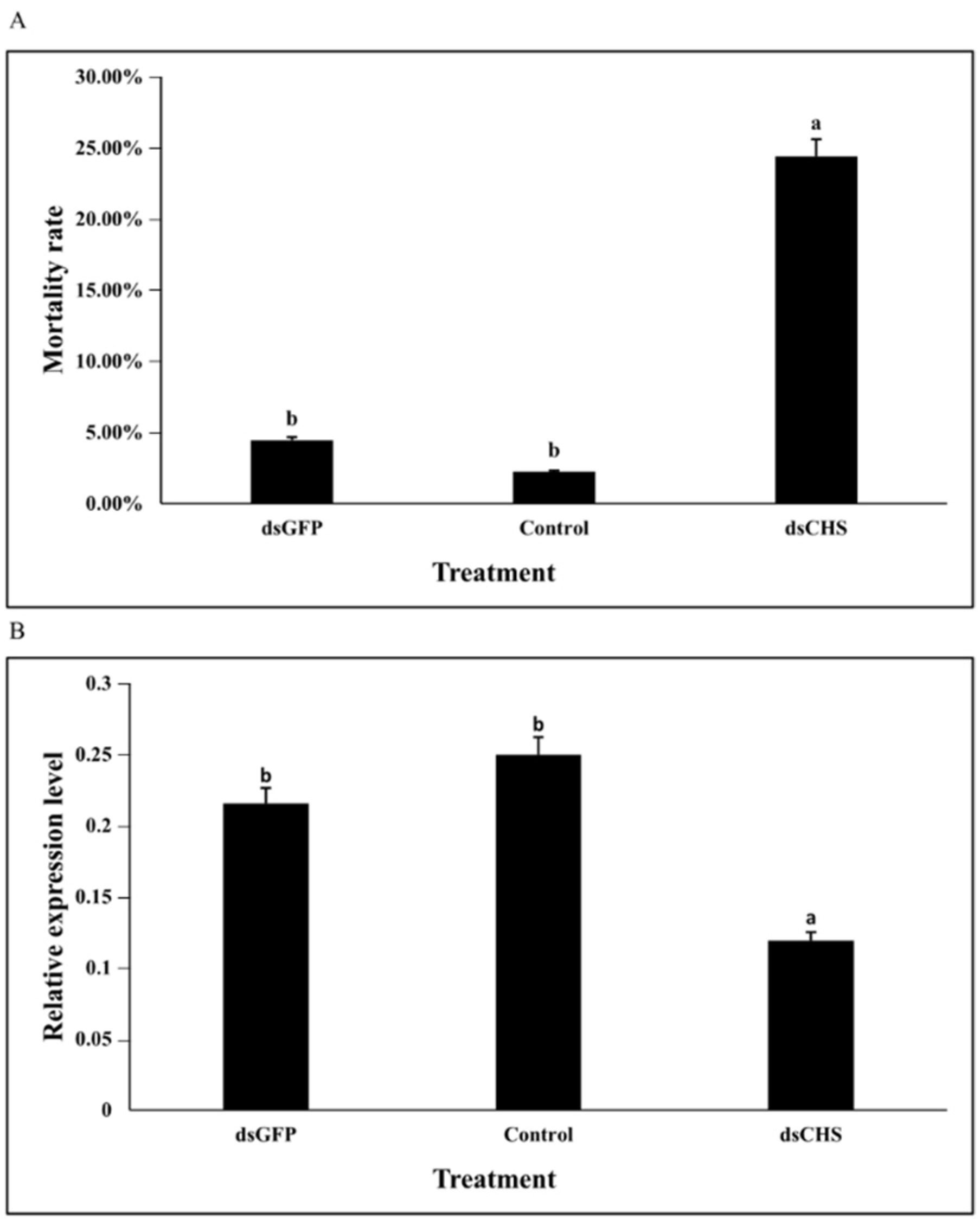
3.5. DcCHSA RNAi Analysis

To explore the DcCHSA effects on the nymph to the adult transition of D. citri, insects were collected for feeding with dsDcCHSA. The fifth-instar nymphs with the greatest gene expression were chosen for RNAi based on the gene expression profile. After 48 h, the cumulative mortality in the dsDcCHSA group was 24%, 4% in the dsGFP group, 2% in the control, and significantly higher in the dsDcCHSA group (Figure 6A). After 48 h of feeding, the expression of the dsDcCHSA decreased by 38.26% compared to the dsGFP and 44.91% compared to the control (Figure 6B). Compared with the control, nymphs had partial molting and even molting failure until death (Figure 7B). The freshly emerged adults failed to spread their wings, and the abdomen was unable to shed the old epidermis (Figure 7C). These findings revealed that inhibiting CHSA expression has a major impact on the nymph-to-adult metamorphosis.

4. Discussion

In this study, the CHS cDNA (DcCHSA) sequence of D. citri was successfully cloned. Sequence alignment analysis revealed that DcCHSA contains two chitin synthase tag sequences, EDR and QRRRW. This indicates that DcCHSA has the structural characteristics of chitin synthase and may have similar physiological functions as CHSA. The CHS protein of insects usually contains 15 transmembrane helices [10]. The CHS protein obtained in this study also has 15 transmembrane helices. This was the same as the annotation results of Miller et al. [17]. Our findings differ from those of Lu et al. [32], in part, because of missing sequences and repetitive areas in the previously predicted D. citri CHS (xp017303059). The phylogenetic analysis also showed that DcCHSA had a higher genetic relationship with the CHS1 gene of brown citrus aphid [36]. These results indicate that the DcCHSA obtained in this study belongs to CHS1. Generally, insects have two CHS genes [13,37]. Interestingly, there was just one CHS gene, according to several studies [11,36], CHSA, in Hemiptera. This study also supports the results that Hemiptera only contains CHSA. In many insects, the midgut epithelium is lined with an acellular, semipermeable structure referred to as the peritrophic matrix (PM) [13,38]. However, the research shows that there is a lack of PM in Hemiptera [11]. This may be the reason that there is only the DcCHSA gene in D. citri. Additionally, this study did not suggest alternative splicing variations in DcCHSA, which is consistent with aphid investigations [11,36]. In Hemiptera insects such as Nilaparvata lugens, Laodelphax striatellus, and Sogatella furcifera, alternate splicing variants do exist [16,39]. In addition, mutations in some loci of CHS genes can enhance insect insecticide resistance [40]. Therefore, it is necessary to further study DcCHSA.
Developmental expression of DcCHSA from eggs to adults was determined by RT-qPCR. Our findings demonstrated that DcCHSA expression was identified in all physiological stages, which is consistent with the findings of Acyrthosiphon pisum [41]. DcCHSA expression, on the other hand, was lowest in eggs and greatest in fifth-instar nymph. The lowest expression level in eggs was opposite to those of the Aphis glycines Ay CHS [11] and PcCHS1 gene of Panonychus citri [33], but it was consistent with that of the Locusta migratoria manilensis LmCHS1 gene [42]. The development and growth of insects are affected by periodic molting, during which the insects expand their exoskeleton to adapt to internal growth. During this process, insects need to significantly increase exoskeletal chitin to form new cuticles [42]. When the D. citri develops to the fifth-instar nymph, its body size increases sharply, and a large amount of chitin needs to be synthesized to meet its own needs. The fifth-instar nymph is also considered the critical period for the D. citri to move from the nymph to the adult [43]. This may be the reason for the highest expression of DcCHSA in the fifth-instar nymph stage. The results of tissue expression were similar to those of other insects. CHSA is exclusively found in epidermal cells originating from ectoderm, such as the epidermis, trachea, and salivary gland, and is responsible for the formation of chitin in these tissues, according to prior research [14,44]. CHSA also exhibits a high level of expression in the ovaries, indicating that it is involved in ovarian development and insect reproduction [45,46]. As a consequence of the findings, thus far, additional research is required to fully comprehend the role of DcCHSA in D. citri.
The incidence of molting failure and mortality increased as the concentration of DFB increased, as did the expression level of DcCHSA, which is consistent with earlier findings [11,36]. The molting failure rate was greatest when the concentration of DFB was 500 mg/L, and a distinct phenotype formed. DFB was the first commercial insecticide that acts by inhibiting chitin synthesis in insects [19]. Insects exposed to this chitin synthesis inhibitor may acquire aberrant cuticle forms, abnormal procuticle depositions, and abortive molting [47]. Blisters developed on the thorax and abdomen of nymphs treated with a high concentration of DFB, which may be because the stratum corneum cannot tolerate the increased turgor pressure, leading to inadequate muscular support during molting [48]. However, DFB did not inhibit the expression of DcCHSA, which may not be the target of DFB. DFB was formerly thought to lower chitin content by decreasing chitin synthase activity [19]. The increased level of CHS expression can also indicate the existence of a feedback regulation mechanism to compensate for low enzyme content [33]. However, upregulation or mutation of CHS expression can improve insect resistance [40,49]. As a result, we should pay more attention to the DcCHSA molecular mechanisms in drug resistance in the future.
RNAi has been widely used in studies to understand how genes operate, and it has shown great promise for the creation of fresh pest control strategies [50]. We performed RNAi tests on the fifth-instar nymphs by feeding them dsDcCHSA. The dsRNA-mediated DcCHSA silencing produced a considerable drop in DcCHSA expression during the nymph to adult metamorphosis, affecting nymph molting and causing death. Toxoptera citricida and Locusta migratoria manilensis had the same outcomes when CHS was silenced [36,42]. Similarly, freshly emerging adults following RNAi were unable to stretch their wings successfully, which was comparable to a study of Glyphodes pyloalis [51]. This suggests that DcCHSA was involved in the synthesis of chitin in the exoskeleton of D. citri, and that it may be a crucial gene for the nymph to adult development, as well as for wing expansion. However, more research is needed to demonstrate how DcCHSA knockdown impacts other stages of D. citri development, including the transition from second- to third-instar nymphs.

5. Conclusions

In summary, we identified a complete sequence of the CHS gene in D. citr. RT-qPCR showed that the relative expression of DcCHSA was highest in the fifth-instar nymph. When exposed to DFB, the molting failure rate and mortality rate of D. citri increased with the increase in DFB concentration, and the expression of DcCHSA also increased. Exposure to dsDcCHSA by ingestion resulted in a significant decline in target gene expression and also affected nymph molting, leading to increased mortality and reducing molting success over time. These results further deepened our understanding of the biological functions of the DcCHSA gene in D. citri and provided new ideas for control.

Author Contributions

C.Z., Z.Z., B.X. and T.X.: Conceptualization, Methodology, Formal Analysis, Investigation, Writing—Original Draft, Methodology, Formal Analysis, Investigation. W.H., Z.Y., X.L. and J.W.: Methodology, Formal Analysis, Investigation. All authors have read and agreed to the published version of the manuscript.

Funding

This work was supported by the National Natural Science Foundation of China (31760621, 31460553, and 31860601); Natural Science Foundation of Jiangxi Province, China (20161ACB20003 and 20181BAB20405); Leadership in Major Subjects Project of Jiangxi Province (20172BCB22004); Jiangxi Key Research & Development Plan, China (20161BBF60117); Jiangxi Provincial Department of Education Technology plan (KJLD14014); Jiangxi Provincial Key R&D Program (20212BBF63042, 20203BBF63041).

Institutional Review Board Statement

Not applicable.

Informed Consent Statement

Not applicable.

Data Availability Statement

The data generated during the study have already been reported in the manuscript.

Conflicts of Interest

The authors declare no conflict of interest.

References

  1. Dong, M.; Wen, G.; Tang, H.; Wang, T.; Zhao, Z.; Song, W.; Wang, W.; Zhao, L. Dissipation and safety evaluation of novaluron, pyriproxyfen, thiacloprid and tolfenpyrad residues in the citrus-field ecosystem. Food Chem. 2018, 269, 136–141. [Google Scholar] [CrossRef]
  2. Ruiz, M.J.; Juárez, M.L.; Alzogaray, R.A.; Arrighi, F.; Arroyo, L.; Gastaminza, G.; Willink, E.; Bardón, A.; Vera, M.T. Oviposition behaviour and larval development of Anastrepha fraterculus from Argentina in citrus. Entomol. Exp. Appl. 2015, 157, 198–213. [Google Scholar] [CrossRef]
  3. Sun, D.; Zhu, Y.; Pang, J.; Zhou, Z.; Jiao, B. Residue level, persistence and safety of spirodiclofen-pyridaben mixture in citrus fruits. Food Chem. 2016, 194, 805–810. [Google Scholar] [CrossRef]
  4. Chu, C.C.; Hoffmann, M.; Braswell, W.E.; Pelz Stelinski, K.S. Genetic variation and potential coinfection of Wolbachia among widespread Asian citrus psyllid (Diaphorina citri Kuwayama) populations. Insect Sci. 2019, 26, 671–682. [Google Scholar] [CrossRef] [PubMed]
  5. Halbert, S.; Manjunath, K. Asian citrus psyllids (Sternorrhyncha: Psyllidae) and greening disease of citrus: A literature review and assessment of risk in Florida. Fla. Entomol. 2004, 3, 330–353. [Google Scholar] [CrossRef]
  6. Yang, Y.; Huang, M.; Beattie, G.A.C.; Xia, Y.; Ouyang, G.; Xiong, J. Distribution, biology, ecology and control of the psyllid Diaphorina citri Kuwayama, a major pest of citrus: A status report for China. Int. J. Pest Manag. 2006, 52, 343–352. [Google Scholar] [CrossRef]
  7. Zhou, C. The status of citrus Huanglongbing in China. Trop. Plant Pathol. 2020, 45, 279–284. [Google Scholar] [CrossRef]
  8. Wang, R.; Yang, H.; Luo, W.; Wang, M.; Lu, X.; Huang, T.; Zhao, J.; Li, Q. Predicting the potential distribution of the Asian citrus psyllid, Diaphorina citri (Kuwayama), in China using the MaxEnt model. PeerJ 2019, 7, e7323. [Google Scholar] [CrossRef]
  9. Merzendorfer, H.; Zimoch, L. Chitin metabolism in insects: Structure, function and regulation of chitin synthases and chitinases. J. Exp. Biol. 2003, 206, 4393–4412. [Google Scholar] [CrossRef]
  10. Merzendorfer, H. Insect chitin synthases: A review. J. Comp. Physiol. B 2006, 176, 1–15. [Google Scholar] [CrossRef] [PubMed]
  11. Bansal, R.; Mian, M.A.R.; Mittapalli, O.; Michel, A.P. Characterization of a Chitin Synthase Encoding Gene and Effect of Diflubenzuron in Soybean Aphid, Aphis glycines. Int. J. Biol. Sci. 2012, 8, 1323–1334. [Google Scholar] [CrossRef]
  12. Zhuo, W.; Fang, Y.; Kong, L.; Li, X.; Sima, Y.; Xu, S. Chitin synthase A: A novel epidermal development regulation gene in the larvae of Bombyx mori. Mol. Biol. Rep. 2014, 41, 4177–4186. [Google Scholar] [CrossRef]
  13. Toprak, U.; Erlandson, M.; Baldwin, D.; Karcz, S.; Wan, L.; Coutu, C.; Gillott, C.; Hegedus, D.D. Identification of the Mamestra configurata (Lepidoptera: Noctuidae) peritrophic matrix proteins and enzymes involved in peritrophic matrix chitin metabolism. Insect Sci. 2016, 23, 656–674. [Google Scholar] [CrossRef] [PubMed]
  14. Arakane, Y.; Muthukrishnan, S.; Kramer, K.J.; Specht, C.A.; Tomoyasu, Y.; Lorenzen, M.D.; Kanost, M.; Beeman, R.W. The Tribolium chitin synthase genes TcCHS1 and TcCHS2 are specialized for synthesis of epidermal cuticle and midgut peritrophic matrix. Insect Mol. Biol. 2005, 14, 453–463. [Google Scholar] [CrossRef]
  15. Moussian, B.; Schwarz, H.; Bartoszewski, S.; Nüsslein-Volhard, C. Involvement of chitin in exoskeleton morphogenesis in Drosophila melanogaster. J. Morphol. 2005, 264, 117–130. [Google Scholar] [CrossRef]
  16. Wang, Y.; Fan, H.; Huang, H.; Xue, J.; Wu, W.; Bao, Y.; Xu, H.; Zhu, Z.; Cheng, J.; Zhang, C. Chitin synthase 1 gene and its two alternative splicing variants from two sap-sucking insects, Nilaparvata lugens and Laodelphax striatellus (Hemiptera: Delphacidae). Insect Biochem. Mol. Biol. 2012, 42, 637–646. [Google Scholar] [CrossRef]
  17. Miller, S.; Shippy, T.D.; Tamayo, B.; Hosmani, P.S.; Flores-gonzalez, M.; Mueller, L.A.; Hunter, W.B.; Brown, S.J.; Elia, T.D.; Surya, S. Annotation of chitin biosynthesis genes in Diaphorina citri, the Asian citrus psyllid. Gigabyte 2021, 2, 1–12. [Google Scholar] [CrossRef]
  18. Grosscurt, A.C.; Jongsma, B. Mode of Action and Insecticidal Properties of Diflubenzuron; Springer: Dordrecht, The Netherlands, 1987; pp. 75–99. [Google Scholar]
  19. Cohen, E. Chitin synthesis and inhibition: A revisit. Pest Manag. Sci. 2001, 57, 946–950. [Google Scholar] [CrossRef]
  20. Merzendorfer, H. Chitin synthesis inhibitors: Old molecules and new developments. Insect Sci. 2013, 20, 121–138. [Google Scholar] [CrossRef]
  21. Douris, V.; Steinbach, D.; Panteleri, R.; Livadaras, I.; Pickett, J.A.; Van Leeuwen, T.; Nauen, R.; Vontas, J. Resistance mutation conserved between insects and mites unravels the benzoylurea insecticide mode of action on chitin biosynthesis. Proc. Natl. Acad. Sci. USA 2016, 113, 14692–14697. [Google Scholar] [CrossRef]
  22. Rösner, J.; Merzendorfer, H. Transcriptional plasticity of different ABC transporter genes from Tribolium castaneum contributes to diflubenzuron resistance. Insect Biochem. Mol. Biol. 2020, 116, 103282. [Google Scholar] [CrossRef]
  23. Tiwari, S.; Clayson, P.J.; Kuhns, E.H.; Stelinski, L.L. Effects of buprofezin and diflubenzuron on various developmental stages of Asian citrus psyllid, Diaphorina citri. Pest Manag. Sci. 2012, 68, 1405–1412. [Google Scholar] [CrossRef]
  24. Huvenne, H.; Smagghe, G. Mechanisms of dsRNA uptake in insects and potential of RNAi for pest control: A review. J. Insect Physiol. 2010, 56, 227–235. [Google Scholar] [CrossRef] [PubMed]
  25. Dias, N.P.; Cagliari, D.; Dos Santos, E.A.; Smagghe, G.; Jurat-Fuentes, J.L.; Mishra, S.; Nava, D.E.; Zotti, M.J. Insecticidal Gene Silencing by RNAi in the Neotropical Region. Neotrop. Entomol. 2020, 49, 1–11. [Google Scholar] [CrossRef]
  26. Tijsterman, M.; Plasterk, R.H.A. Dicers at RISC: The mechanism of RNAi. Cell 2004, 117, 1–3. [Google Scholar] [CrossRef]
  27. Kishk, A.; Anber, H.A.I.; AbdEl Raof, T.K.; El Sherbeni, A.H.D.; Hamed, S.; Gowda, S.; Killiny, N. RNA interference of carboxyesterases causes nymph mortality in the Asian citrus psyllid, Diaphorina citri. Arch. Insect Biochem. 2017, 94, e21377. [Google Scholar] [CrossRef]
  28. Andrade, E.C.; Hunter, W.B. RNAi feeding bioassay: Development of a non-transgenic approach to control Asian citrus psyllid and other hemipterans. Entomol. Exp. Appl. 2017, 162, 389–396. [Google Scholar] [CrossRef]
  29. Yu, X.; Gowda, S.; Killiny, N. Double-stranded RNA delivery through soaking mediates silencing of the muscle protein 20 and increases mortality to the Asian citrus psyllid, Diaphorina citri. Pest Manag. Sci. 2017, 73, 1846–1853. [Google Scholar] [CrossRef] [PubMed]
  30. Chen, J.; Peng, Y.; Zhang, H.; Wang, K.; Tang, Y.; Gao, J.; Zhao, C.; Zhu, G.; Palli, S.R.; Han, Z. Transcript level is a key factor affecting RNAi efficiency. Pestic. Biochem. Phys. 2021, 176, 104872. [Google Scholar] [CrossRef]
  31. Galdeano, D.M.; Breton, M.C.; Lopes, J.R.S.; Falk, B.W.; Machado, M.A. Oral delivery of double-stranded RNAs induces mortality in nymphs and adults of the Asian citrus psyllid, Diaphorina citri. PLoS ONE 2017, 12, e171847. [Google Scholar] [CrossRef] [PubMed]
  32. Lu, Z.; Huang, Y.; Yu, H.; Li, N.; Xie, Y.; Zhang, Q.; Zeng, X.; Hu, H.; Huang, A.; Yi, L.; et al. Silencing of the Chitin Synthase Gene Is Lethal to the Asian Citrus Psyllid, Diaphorina citri. Int. J. Mol. Sci. 2019, 20, 3734. [Google Scholar] [CrossRef] [PubMed]
  33. Xia, W.; Ding, T.; Niu, J.; Liao, C.; Zhong, R.; Yang, W.; Liu, B.; Dou, W.; Wang, J. Exposure to Diflubenzuron Results in an Up-Regulation of a Chitin Synthase 1 Gene in Citrus Red Mite, Panonychus citri (Acari: Tetranychidae). Int. J. Mol. Sci. 2014, 15, 3711–3728. [Google Scholar] [CrossRef]
  34. Livak, K.J.; Schmittgen, T.D. Analysis of Relative Gene Expression Data Using Real-Time Quantitative PCR and the 2−ΔΔCT Method. Methods 2001, 25, 402–408. [Google Scholar] [CrossRef]
  35. Tamura, K.; Stecher, G.; Peterson, D.; Filipski, A.; Kumar, S. MEGA6: Molecular Evolutionary Genetics Analysis Version 6.0. Mol. Biol. Evol. 2013, 30, 2725–2729. [Google Scholar] [CrossRef]
  36. Shang, F.; Xiong, Y.; Xia, W.K.; Wei, D.D.; Wei, D.; Wang, J.J. Identification, characterization and functional analysis of a chitin synthase gene in the brown citrus aphid, Toxoptera citricida (Hemiptera, Aphididae). Insect Mol. Biol. 2016, 25, 422–430. [Google Scholar] [CrossRef]
  37. Merzendorfer, H. The cellular basis of chitin synthesis in fungi and insects: Common principles and differences. Eur. J. Cell Biol. 2011, 90, 759–769. [Google Scholar] [CrossRef] [PubMed]
  38. Hegedus, D.; Erlandson, M.; Gillott, C.; Toprak, U. New insights into peritrophic matrix synthesis, architecture, and function. Annu. Rev. Entomol. 2009, 54, 285–302. [Google Scholar] [CrossRef] [PubMed]
  39. Wang, Z.; Yang, H.; Zhou, C.; Yang, W.; Jin, D.; Long, G. Molecular cloning, expression, and functional analysis of the chitin synthase 1 gene and its two alternative splicing variants in the white-backed planthopper, Sogatella furcifera (Hemiptera: Delphacidae). Sci. Rep. 2019, 9, 1087. [Google Scholar] [CrossRef]
  40. Fotakis, E.A.; Mastrantonio, V.; Grigoraki, L.; Porretta, D.; Puggioli, A.; Chaskopoulou, A.; Osório, H.; Weill, M.; Bellini, R.; Urbanelli, S.; et al. Identification and detection of a novel point mutation in the Chitin Synthase gene of Culex pipiens associated with diflubenzuron resistance. PLoS Negl. Trop. Dis. 2020, 14, e8284. [Google Scholar] [CrossRef] [PubMed]
  41. Ye, C.; Jiang, Y.; An, X.; Yang, L.; Shang, F.; Niu, J.; Wang, J. Effects of RNAi-based silencing of chitin synthase gene on moulting and fecundity in pea aphids (Acyrthosiphon pisum). Sci. Rep. 2019, 9, 3694. [Google Scholar] [CrossRef]
  42. Zhang, J.; Liu, X.; Zhang, J.; Li, D.; Sun, Y.; Guo, Y.; Ma, E.; Zhu, K.Y. Silencing of two alternative splicing-derived mRNA variants of chitin synthase 1 gene by RNAi is lethal to the oriental migratory locust, Locusta migratoria manilensis (Meyen). Insect Biochem. Mol. Biol. 2010, 40, 824–833. [Google Scholar] [CrossRef] [PubMed]
  43. Boina, D.R.; Rogers, M.E.; Wang, N.; Stelinski, L.L. Effect of pyriproxyfen, a juvenile hormone mimic, on egg hatch, nymph development, adult emergence and reproduction of the Asian citrus psyllid, Diaphorina citri Kuwayama. Pest Manag. Sci. 2010, 66, 349–357. [Google Scholar] [CrossRef] [PubMed]
  44. Jiang, L.; Mu, L.; Jin, L.; Anjum, A.A.; Li, G. RNAi for chitin synthase 1 rather than 2 causes growth delay and molting defect in Henosepilachna vigintioctopunctata. Pestic. Biochem. Phys. 2021, 178, 104934. [Google Scholar] [CrossRef] [PubMed]
  45. Guedes, R.N.C.; Beins, K.; Navarro Costa, D.; Coelho, G.E.; Bezerra, H.S.D.S. Patterns of insecticide resistance in Aedes aegypti: Meta-analyses of surveys in Latin America and the Caribbean. Pest Manag. Sci. 2020, 76, 2144–2157. [Google Scholar] [CrossRef] [PubMed]
  46. Li, T.; Chen, J.; Fan, X.; Chen, W.; Zhang, W. MicroRNA and dsRNA targeting chitin synthase A reveal a great potential for pest management of the hemipteran insect Nilaparvata lugens. Pest Manag. Sci. 2017, 73, 1529–1537. [Google Scholar] [CrossRef] [PubMed]
  47. Mulder, R.; Gijswijt, M.J. The laboratory evaluation of two promising new insecticides which interfere with cuticle deposition. Pestic. Sci. 1973, 4, 737–745. [Google Scholar] [CrossRef]
  48. Zhang, J.; Zhu, K.Y. Characterization of a chitin synthase cDNA and its increased mRNA level associated with decreased chitin synthesis in Anopheles quadrimaculatus exposed to diflubenzuron. Insect Biochem. Mol. Biol. 2006, 36, 712–725. [Google Scholar] [CrossRef] [PubMed]
  49. Gong, C.; Wang, X.; Huang, Q.; Zhang, J.; Zhang, Y.; Zhan, X.; Zhang, S.; Hasnain, A.; Ruan, Y.; Shen, L. The fitness advantages of bistrifluron resistance related to chitin synthase A in Spodoptera litura (Fab.) (Noctuidae: Lepidoptera). Pest Manag. Sci. 2021, 77, 3458–3468. [Google Scholar] [CrossRef]
  50. Zhu, K.Y.; Palli, S.R. Mechanisms, Applications, and Challenges of Insect RNA Interference. Annu. Rev. Entomol. 2020, 65, 293–311. [Google Scholar] [CrossRef] [PubMed]
  51. Shao, Z.; Li, Y.; Ding, J.; Liu, Z.; Zhang, X.; Wang, J.; Sheng, S.; Wu, F. Identification, Characterization, and Functional Analysis of Chitin Synthase Genes in Glyphodes pyloalis Walker (Lepidoptera: Pyralidae). Int. J. Mol. Sci. 2020, 21, 4656. [Google Scholar] [CrossRef]
Figure 1. Nucleotide and deduced amino acid sequences of DcCHSA cDNA from D. citri. The start codon (ATG) and the stop codon (TAA) are indicated with black underlines, and the putative polyadenylation signal (AATAAA) in bold with a black ellipse. The 15 hydrophilic, membrane-spanning helices are indicated in gray font, the amino acid sequence of the putative catalytic domains is wavy-lined, and the chitin synthase signature motifs EDR (852–854 bp) and QRRRW (889–894 bp) are indicated in red font. Black boxes are used to indicate the 11 predicted N-glycosylation sites.
Insects 13 00728 g001
Figure 2. Phylogeny of insect chitin synthases. Phylogenetic relationships of DcCHSA in different insect species using the neighbor-joining method with a bootstrap value of 1000. Chitin synthases were from 21 insects.
Insects 13 00728 g002
Figure 3. Relative expression levels of DcCHSA in different developmental stages and tissues of D. citri. (A) The expression of DcCHSA was highest in fifth-instar nymphs, followed by the adult stage. (B) Tissue-specific analysis showed that the transcription level of DcCHSA was highest in the head and integument, and the second highest was in the ovary. Different letters on the bars of the histogram indicate significant difference in gene expression compared to the treatment with lowest expression at p value < 0.05 (ANOVA). Eg, egg; N1, first-instar nymph; N2, second-instar nymph; N3, third-instar nymph; N4, fourth-instar nymph; N5, fifth-instar nymph; Ad, adult; He, Head; Wi, Wing; Gu, guts; My, Mycetome; Te, Testis; Ov, Ovary; In, Integument.
Insects 13 00728 g003
Figure 4. Effect of DFB on D. citri mortality, molting rate, and DcCHSA expression levels. (A) At 48 h, the cumulative mortality of D. citri in 500 mg/L DFB treatment was significantly higher than those in the control, 5 mg/L DFB, and 100 mg/L DFB treatments. (B) The abortive molting rate of D. citri treated with 500 mg/L of DFB at 48 h was significantly higher than that of the control group, that treated with 5 mg/L of DFB, and that treated with 100 mg/L of DFB. (C) The relative expression of DcCHSA in D. citri increased significantly at 48 h after being induced by low and high DFB concentrations. Different letters on the bars of the histogram indicate significant difference at p value < 0.05 (ANOVA).
Insects 13 00728 g004
Figure 5. Exposed to 500 mg/L of DFB, D. citri nymphs showed a special phenotype. (A) The nymphs in the control (0.01% Triton X-100) can normally develop into adults. (B) Blisters appeared on the left front side of the nymph thorax in 500 mg/L of DFB.
Insects 13 00728 g005
Figure 6. Effects on D. citri after RNAi of DcCHSA. (A) Cumulative mortality of D. citri in the treatment and control after RNAi of 48 h. After 48 h, the cumulative mortality of dsDcCHSA group was significantly higher than that of control group. (B) Relative expression levels of DcCHSA when D. citri was treated with dsDcCHSA and dsGFP. After feeding dsDcCHSA for 48 h, the transcript level of DcCHSA was significantly lower than that of the control group. Different letters on the bars of the histogram indicate significant difference at p value < 0.05 (ANOVA).
Insects 13 00728 g006
Figure 7. Fifth-instar nymphs treated with dsRNA. (A) Fifth-instar nymphs fed dsGFP can emerge normally. (B) After feeding dsRNA for 48 h, fifth-instar nymphs could not molt. (C) The freshly emerged adults failed to spread their wings, and the abdomen was unable to shed the old epidermis. A total of 30 fifth-instar nymphs were used for phenotypic observation after RNAi.
Insects 13 00728 g007
Table 1. The DcCHSA expression analysis and cloning primers.
ExperimentsNameSequences
PCRDcCHSA5FTWCTGCAATAAAGAYYTA
DcCHSA5RAKATAWRTGAARTAWCGWGT
DcCHSA3FATGATGGAVATGWTNARSTSKAT
DcCHSA3RCATAAGRAATATHGTKCCHGGDC
DcCHSAZFACTACAACGAGCAAACATTCA
DcCHSAZRGAGAATGGGCGTAATGTTGT
3′-RACEDcCHSAF1GACAAGCAAGCGGTGGACATA
DcCHSAF2AGGTCCGCGTCAACTCTTCTTCCATC
5′-RACEDcCHSAR1AGGCGGGAGGTCTCGGAACA
DcCHSAR2ATGGAGGTGACGAAGATAAA
3′/5′-RACENUPAAGCAGTGGTATCAACGCAGAGT
UPM (mix)TAATACGACTCACTATAGGGCAAGCAGTGGTATCAACGCAGAGT
CTAATACGACTCACTATAGGGC
RT-qPCRDcActinFCCATCTTGGCTTCTCTGTCTAC
DcActinRCATTTGCGGTGAACGATTCC
DcTubulinFTTTCCAACACCACCGCTAT
DcTubulinRAGGTCTTCCCTCGCCTCTG
DcCHSAqFCCACGACTCCTTCTCACAG
DcCHSAqRTCATGGCGAAATTATCCTC
RNAiGFPiFGCCAACACTTGTCACTACTT
GFPiRGGAGTATTTTGTTGATAATGGTCTG
GFPdsFtaatacgactcactatagggGCCAACACTTGTCACTACTT
GFPdsRtaatacgactcactatagggGGAGTATTTTGTTGATAATGGTCTG
DcCHSAiFCGAGTGGTAGACCCGAACAT
DcCHSAiRATGAAAAAGGCGAACACGAC
DcCHSAdsFtaatacgactcactatagggCGAGTGGTAGACCCGAACAT
DcCHSAdsRtaatacgactcactatagggATGAAAAAGGCGAACACGAC
Table 2. Species and Gen Bank accession numbers of chitin synthase genes used in the phylogenetic tree construction.
Gene NameGenBank Accession NumberSpecies
DcCHSAthis studyDiaphorina citri
SeCHSAAAZ03545.1Spodoptera exigua
HaCHSAAKJ54482.1Helicoverpa armigera
EoCHS1ACA50098.1Ectropis obliqua
MsCHSAAAL38051.2Manduca sexta
PxCHS1API61827.1Plutella xylostella
DtCHS1XP_017009970.1Drosophila takahashii
CqCHSAXP_001866798.1Culex quinquefasciatus
AqCHS1ABD74441.1Anopheles quadrimaculatus
NvCHS1XP_001602290Nasonia vitripennis
BtCHSAXP_00339885Bombus terrestris
AmCHS1XP_395677Apis mellifera
AtCHS1AFM38193.1Anasa tristis
LhCHSAJAQ09912.1Lygus hesperus
AcCHSAKR611528.1Aphis citricidus
AgCHSAAFJ00066.1Aphis glycines
NlCHSAJQ040014Nilaparvata lugens
SfCHSAKY987034Sogatella furcifera
LsCHSAJQ040012Laodelphax striatellus
PcCHS1AJQ20794.1Panonychus citri
CmCHS2AJF93428.1Mamestra configurata
MysCHSBASF79498.1Mythimna separate
SeCHSBABI96087.1Spodoptera exigua
HaCHSBAKZ08595.1Helicoverpa armigera
DmCHSBCAC83725.1Drosophila melanogaster
Table 3. Toxicities of DFB to the fifth-instar nymph of D. citri.
Toxic Regression Equation95% Confidence IntervalCorrelation CoefficientChi-Square ValueHalf Lethal Concentration (mg/L)
y = −4.012 + 1.867x110.295–174.7180.9031.861140.992
Publisher’s Note: MDPI stays neutral with regard to jurisdictional claims in published maps and institutional affiliations.

