Article

Novel Technique of Interproximal Enamel Reduction Based on Computer-Aided Navigation Technique—An In Vitro Study

by
María Dolores Cotrina-Peregrín
1,2,
Patricia Arrieta-Blanco
2,
Juan Manuel Aragoneses-Lamas
2,3,
Alberto Albaladejo Martínez
4,
Ana Belén Lobo Galindo
4 and
Álvaro Zubizarreta-Macho
2,4,*
1
Doctoral Student in Cancer Biology and Clinic and Translational Medicine program, Faculty of Medicine, University of Salamanca, 37008 Salamanca, Spain
2
Faculty of Health Sciences, Alfonso X el Sabio University, 28691 Madrid, Spain
3
Department of Dentistry, Universidad Federico Henríquez y Carvajal, Santo Domingo 10106, Dominican Republic
4
Department of Surgery, Faculty of Medicine, University of Salamanca, 37008 Salamanca, Spain
*
Author to whom correspondence should be addressed.
J. Pers. Med. 2024, 14(2), 138; https://doi.org/10.3390/jpm14020138
Submission received: 21 December 2023 / Revised: 17 January 2024 / Accepted: 23 January 2024 / Published: 26 January 2024
(This article belongs to the Special Issue Orthodontics and Oral Surgery in Personalized Medicine)

Abstract

:
The aim of this study was to analyze and compare the accuracy of a novel interproximal enamel reduction (IPR) technique based on a computer-aided static navigation technique with respect to a conventional free-hand-based technique for interproximal enamel reduction. Twenty anatomical-based experimental cast models of polyurethane were randomly distributed into the following IPR techniques: IPR technique based on computer-aided static navigation technique (n = 10) (GI) for Group A and conventional free-hand-based technique for the IPR (n = 10) (FHT) for Group B. The anatomical-based experimental cast models of polyurethane randomly assigned to the GI study group were submitted for a preoperative 3D intraoral surface scan; then, datasets were uploaded into 3D implant-planning software to design virtual templates for the interproximal enamel reduction technique. Afterward, the anatomical-based experimental cast models of polyurethane of both GI and FHT study groups were subjected to a postoperative digital impression by a 3D intraoral surface scan to compare the accuracy of the interproximal enamel reduction techniques at the buccal (mm), lingual/palatal (mm), and angular (◦) levels using the Student t-test. Statistically significant differences between the interproximal enamel reduction technique based on the computer-aided static navigation technique and the conventional free-hand-based technique for the interproximal enamel reduction at the buccal (p = 0.0008) and lingual/palatal (p < 0.0001) levels; however, no statistically significant differences were shown at the angular level (p = 0.1042). The interproximal enamel reduction technique based on computer-aided static navigation technique was more accurate than the conventional free-hand-based technique for interproximal enamel reduction.

1. Introduction

Interproximal enamel reduction (IPR) or “stripping” is an irreversible technique used to reduce controlled amounts of enamel on the proximal surface of the tooth by decreasing its mesiodistal size as an alternative to extraction or expansion in borderline cases [1]. This procedure is recommended to increase the space in the dental arch and align the teeth [2] without an excessive labioversion of incisors or, in some cases, without increasing the intercanine distance, solving many malocclusions caused by dental crowding generated by the difference between the size of the teeth and the length of the dental arch [3]. The IPR is indicated in cases of not very severe crowding, from 4 to 8 mm, to align teeth without retruding the profile and reduce the unsightly interproximal black triangles in gingival improving the periodontal prognosis [3,4,5]. The thickness of the enamel is greater in the posterior than in the anterior region and a little greater in the distal face than in the mesial (0.10 mm (95% CI, 0.09–0.12)), and there is even more in molars than in premolars (0.12 mm (95% CI, 0.07–0.17)). In addition, it is important to highlight that in temporary teeth, the enamel layer is less than in permanent teeth, and they also have a greater tendency to demineralize than permanent teeth due to the low mineral content and high organic content [6].
Stripping procedures can be performed through manual stainless-steel strips, diamond blades, motor-driven abrasive strips (strips connected to a micromotor), or air rotor stripping (milling cutter connected to a turbine). Sheridan and Chudasama created action guides with this last painless, fast, and efficient technique, being able to perform an interproximal reduction of 1 mm at each point of contact in the posterior area and no more than 0.5 mm in the anterior area because the enamel is thinner. After using bur, it is recommended to finish with thin rotating discs Sof-Lex to soften and contour the tooth in the interproximal area. Therefore, these authors also use a thin abrasive strip impregnated with 35% orthophosphoric acid and a water syringe. Finally, they recommend applying fluoride because the reduced enamel surface tends to re-mineralize and when applying fluorinated products, they prevent caries in this area [5]. It is also very important to use cooling water and suction even if visibility decreases to prevent the temperature from rising [7,8,9]. Moreover, Gazzani et al. analyzed the effects on enamel surface after reducing the interproximal enamel surface by the oscillating mechanical system and reported more regular enamel surfaces using a single metallic strip and recommended adequate polishing after IPR procedures to maintain the enamel morphologic integrity [10]. Additionally, Kaauara et al. evaluated the enamel surface after IPR and recommended minimizing the number of abrasions caused by stripping to obtain a surface condition close to an untreated enamel surface using Soft-Lex abrasive discs to obtain a shiny finish and polish of the entire interdental surface [11].
Surgical guides are indispensable biomedical devices for the proper insertion of dental implants. In particular, Sarment et al. highlighted the effectiveness of CAD/CAM stereolithographic rapid prototyping techniques for the manufacturing of surgical guides to increase the accuracy of dental implant placement [10]. In general, diagnostic and surgical guides must have properties of rigidity, stability, and precision to ensure accuracy and safety during implant insertion procedures [11]. Additionally, CAD-CAM procedures require high cost and a detailed and precise planning process, and treatment plan. As a result, it allows the manufacture of personalized guidance templates for minimally invasive treatment and allows accurate rehabilitation. Moreover, Gao JH (2021) assessed the accuracy between different tooth preparation guides for veneer restorations and concluded that personalized guidance templates improved the accuracy of tooth preparation [12].
Furthermore, previous measurement techniques have been used to quantify the amount of enamel reduced such as the optical microscope, the transmission electron microscope (TEM) [13], the scanning electron microscope (SEM) [14], the atomic force microscope (AFM) [15], the standardized intraoral electron photography technique, the electronic photograph measurement technique, and the intraoral scanners [16,17]. Moreover, the roughness of the enamel has been analyzed by prolymphometry [18], although it has been also measured with the scanning electron microscope (SEM) and the atomic force microscope (AFM) [19].
The aim of this study was to analyze and compare the accuracy of a novel interproximal enamel reduction technique based on the computer-aided static navigation technique with respect to a conventional free-hand-based technique for the interproximal enamel reduction, with a null hypothesis (H0) stating that there are no differences between the accuracy of a novel interproximal enamel reduction technique based on computer-aided static navigation technique and the conventional free-hand-based technique for the interproximal enamel reduction.

2. Material and Methods

2.1. Study Design

A randomized controlled experimental trial was conducted in accordance with the principles defined in the International Organization for Standardization (ISO 14801) [20] at the Department of Surgery of the University of Salamanca (Salamanca, Spain), the Stomatology Department of Fundación Jimenez Díaz Hospital (Madrid, Spain) and the Department of Biomedical, Surgical and Dental Sciences of Universitá degli Studi di Milano, (Milan, Italy), between September 2022 and October 2023. In addition, this study was authorized by the Ethical Committee of the Faculty of Health Sciences, University Alfonso X el Sabio (Madrid, Spain) in July 2022 (Process No. 14/2022). Two hundred (200) interproximal enamel reduction procedures were included in this study to ensure a power effect of 80.00% for detecting statistically significant differences. The bilateral Student’s t-test of two independent samples was used to evaluate the null hypothesis H₀: μ₁ = μ₂, with a significance level of 5.00%. The sample size calculation was carried out on the lingual/palatal level variable; to detect differences of 0.2 units with a deviation of 0.1, 10 observations per group were needed.

2.2. Experimental Procedure

Twenty anatomical-based experimental cast models of polyurethane (Ref. 1522-62; Sawbones Europe AB; Malmo, Sweden), with contact points between adjacent teeth, were used in this study. Afterward, the anatomical-based experimental models of polyurethane were randomly distributed (Epidat 4.1, Galicia, Spain) into the following measurement techniques: interproximal enamel reduction technique based on computer-aided static navigation technique (NemoStudio®, Nemotec, Madrid, Spain) (n = 10) (guided IPR (GI)) for Group A and conventional free-hand-based technique for the interproximal enamel reduction (n = 10) (Free-hand IPR (FHT)) for Group B. The use of polyurethane was based on the American Society for Testing and Materials’ (ASTM F-1839-08) approval of the use of polyurethane for testing instruments and dental procedures (“Standard Specification for Rigid Polyurethane Foam for Use as a Standard Material for Test Orthopedic Devices for Instruments”) [21].

2.3. Interproximal Enamel Reduction Procedure

Afterward, the anatomical-based upper and lower experimental cast models of polyurethane were fixed in a phantom imitating the patient’s head and subsequently attached to a dental chair to simulate a real setting. Then, an interproximal enamel reduction of 0.2 mm width was performed using air-rotor burs with 7 mm head length, a total length of 23.5 mm, and a minimum and maximum diameter of 0.2 mm and 0.55 mm, respectively (Code 852-005, E11S Komet Medical, Lemgo, Germany), fixed to the high-speed rotation device (Tornado LK; Bien Air, Le Noirmont, Switzerland) placed on the dental chair at 410,000 rpm with profuse irrigation. In particular, one bur was used on each interproximal enamel reduction technique.
The anatomical-based experimental cast models of polyurethane randomly assigned to the GI study group were submitted for a 3D intraoral surface scan (True Definition, 3M ESPE™, Saint Paul, MN, USA) for a digital impression. Datasets obtained from the digital workflow were uploaded into 3D implant-planning software (NemoStudio®, Nemotec, Madrid, Spain) to design virtual templates for interproximal enamel reduction technique based on computer-aided static navigation technique. Then, two virtual implant drills were designed by crossing the contact point at the interproximal surface of the teeth with a diameter and length of 0.2 and 23.5 mm, respectively, according to the air-rotor bur measurements (Figure 1A) from the recommendations established by Chudasama et Sheridan (2007) [22]. In particular, the bur was placed perpendicular to the axial shaft of the adjacent teeth by buccal to lingual movements up to remove 0.2 mm on mesial and 0.2 mm on distal surface of the adjacent teeth under profuse irrigation. Ten interproximal enamel reduction procedures were performed in each anatomical-based experimental cast model of polyurethane. After designing the virtual templates (Figure 1A,B), they were fabricated using the stereolithography technique (ProJet 6000, 3D Systems, Rock Hill, SC, USA) (Figure 1C). The templates fit the model and did not need further adjustments.
The anatomical-based experimental cast models of polyurethane randomly assigned to the FHT study group were subjected to interproximal enamel reduction procedures following the recommendations established by Chudasama et Sheridan [5] by placing the bur perpendicular to the axial shaft of the adjacent teeth by buccal to lingual movements up to remove 0.2 mm on mesial and 0.2 mm on distal surface of the adjacent teeth under profuse irrigation.
The interproximal enamel reduction procedures were performed by a unique operator with more than 20 years of experience in orthodontics.

2.4. Digital Measurement Procedure

Afterward, the anatomical-based experimental cast models of polyurethane of both GI and FHT study groups were submitted to a postoperative digital impression by an intraoral scan (True Definition, 3M ESPE™, Saint Paul, MN, USA) via a 3D in-motion video imaging technology to generate an STL digital file using a cloud of points that create a tessella network, representing three-dimensional objects as polygons composed of equilateral triangle tessellas [5,23]. The capturing images procedure was performed following manufacturer recommendations by scanning the incisal/occlusal plane and the vestibular and lingual surfaces. Afterward, the preoperative and postoperative “Standard Tesellation Language” (STL) digital files were imported to a 3D implant-planning software (NemoStudio®, Nemotec, Madrid, Spain); a full-arch alignment procedure was conducted. The preoperative STL digital file was considered the reference digital file, and the postoperative STL digital file was superimposed on it using the buccal and palatal/lingual surfaces of the anterior teeth and the occlusal, buccal, and palatal/lingual surfaces of the posterior teeth with the best-fit algorithm (Figure 2A). Afterward, the accuracy of the interproximal enamel reduction was measured at buccal (mm), lingual/palatal (mm), and angular level (◦). This digital measurement procedure was performed in a previous study of Triduo et al. [24]. Additionally, interproximal enamel reduction distance was also measured (Figure 2B).

2.5. Statistical Tests

Statistical analysis of the measurement variables was conducted using SAS 9.4 (SAS Institute Inc., Cary, NC, USA). Descriptive statistics are expressed as mean and SD for the interproximal enamel reduction (mm). Comparative analysis between the interproximal enamel reduction technique based on the computer-aided static navigation technique and the conventional free-hand-based technique for the interproximal enamel reduction at buccal (mm), lingual/palatal (mm), and angular (◦) levels was analyzed by using the Student t-test and the Mann–Whitney non-parametric test. The repeatability and reproducibility of the digital measurement technique were analyzed using Gage R&R statistical analysis.

3. Results

The means and SD values for the interproximal enamel reduction (mm) between the interproximal enamel reduction technique based on the computer-aided static navigation technique and the conventional free-hand-based technique for the interproximal enamel reduction at the buccal level are displayed in Table 1 and Figure 3.
The Student t-test showed statistically significant differences (p = 0.0008) between the interproximal enamel reduction technique based on the computer-aided static navigation technique (0.20 ± 0.09 mm) and the conventional free-hand-based technique (0.39 ± 0.07 mm) for the interproximal enamel reduction at the buccal level (Figure 3).
The Student t-test showed statistically significant differences (p < 0.0001) between the interproximal enamel reduction technique based on the computer-aided static navigation technique (0.24 ± 0.11 mm) and the conventional free-hand-based technique (0.58 ± 0.9 mm) for the interproximal enamel reduction at the lingual/palatal level (Figure 4).
The Student t-test showed statistically significant differences (p = 0.01042) between the interproximal enamel reduction technique based on the computer-aided static navigation technique (3.36 ± 0.58°) and the conventional free-hand-based technique (4.01 ± 0.60°) for the interproximal enamel reduction at the angular level (Figure 5).

4. Discussion

The results presented in this study rejected the null hypothesis (H0) that states there are no differences between the accuracy of a novel interproximal enamel reduction technique based on a computer-aided static navigation technique and a conventional free-hand-based technique for the interproximal enamel reduction.
Sittikornpaiboon et al. assessed the accuracy of computer-assisted implant surgery through surgical templates based on the CBCT scan and STL digital files obtained by digital impressions, concluding that this digital workflow is sensitive to the milling protocol and the design of the device [25]. In addition, it has been reported that fully guided implant surgery is more accurate than partially guided implant surgery and that deviation of the dental implant position may be influenced by the dental implant location; however, it is not affected by implant systems, dental implant software [26], or the manufacturing process of the surgical templates by fused deposition modeling printed in-office or by stereolithography [27]. Moreover, tooth-supported computer-aided static implant surgery by surgical templates has evidenced a predictable treatment outcome for dental implant placement, showing statistically significant differences (p < 0.05) with respect to the number of teeth [28]. Moreover, fixation pins have been also recommended to attach the surgical templates to the maxilla during the drilling procedure; however, Pessoa et al. (2022) reported that the use of surgical templates with or without fixing pins for dental implant placement provided predictable treatment outcomes [29]. In the present study, the surgical template designed for the interproximal enamel reduction procedure did not include fixing pins because it was tooth-supported by fully dentated anatomical-based upper and lower experimental cast models; additionally, the operator checked the surgical template stability before use. In resume, Ngamprasertkit et al. (2022) highlighted that computer-aided implant surgery through fully digital workflow is a practical procedure that provides an accurate dental implant placement [30].
Afterward, the promising results associated with the computer-aided surgical implant technique led to its application to tooth preparation procedures. In particular, Li et al. (2020) reported that 3D-printed surgical templates improved the control and management of the reduction depth of veneer preparations, increasing the accuracy compared to guide milling cutters [31]. Additionally, Jurado et al.’s (2021) custom-fenestrated metal guides have also been used to selectively reduce tooth surface [32]. Moreover, Zong Yi et al. (2020) used a 3D-printed metal alloy guide, which allowed greater accuracy than measuring cutters [33]. Furthermore, Gao JH (2021) analyzed the degree of accuracy between different preparation guides for veneer restorations and highlighted the relevance of using tooth-preparing guides to achieve accurate tooth preparation (Group F was significantly higher than the rest) [34]. In addition, Johner et al. performed an in vitro study to evaluate the predictability of the expected amount of IPR using three common stripping devices on premolar teeth and concluded that for all scenarios, the amount of stripping was less than the predetermined and expected. However, the authors highlighted that traditional hand-held abrasive strips performed an unpredictable IPR in posterior teeth, and motor-driven devices reduce more enamel at the contact point, so this will be flatter and might even show a little edge around the stripped area. In this case, it will be necessary to smooth the edges and reshape the contact point with further devices such as diamond burs [33].
In recent years, numerous studies have been carried out to investigate the amount of enamel tissue removed during stripping procedures, compare the reduction in the distal versus mesial surface, and compare the amount of stripping performed with respect to the planned different digital measurement methods, including the Invisalign Clincheck software [33], the digital set up in the treatment with clear aligner devices [14], and Orthocad digital software [35]. In addition, Kalemaj and Levrini compared the programmed and implemented interproximal enamel reduction in a clinical setting and reported a statistically significant mean difference of 0.15 mm (SD ± 0.14 mm; p = 0.0001). These authors measured the differences in the Orthocad digital software; however, the IPR procedures were performed by free-hand technique without computer-aided static navigation techniques [36]. These authors did not analyze the reliability of the measurement technique and quantified the mesiodistal distances of each tooth, from second premolar to second premolar. However, posterior teeth have been recommended as the enamel thickness increases in these teeth despite presenting worse accessibility [37]
The present study selected the True Definition intraoral since Guth et al. reported that True Definition showed higher trueness (21.8 µm) than Cerec Bluecam (34.2 µm), Cerec Omnicam (43.3 µm), Itero (49.0 µm), Lava C.O.S. (47.7 µm), TRIOS (25.7 µm), and TRIOS color (26.1 µm) digital impression systems for dental nature arch scanning [38]. Furthermore, the present study includes some limitations since Jivanescu et al. reported that the presence of adjacent teeth can decrease the view of interproximal surfaces [36]. Additionally, the full-arch scanning may introduce higher deviations than partial-arch scanning [39], ambient temperature [40], number of teeth and location [41], scanning time [42], lighting conditions [43], and humidity [44]; therefore, further clinical studies are encouraged to increase the results of these procedures.
Meredith et al. analyzed the enamel nanotopography after interproximal enamel reduction using atomic force microscopy (AFM) and reported that the enamel surface becomes progressively smoother from burs to strips and discs to polishers. In addition, surface roughness was higher using a medium roughness strip bur (707 nm) and decreased using medium strip bands (501 nm), fine strip burs (407 nm), fine strip bands (318 nm), mesh strip discs (307 nm), curved strip discs (224 nm), and a Sof-Lex polishing device (37 nm) [39]. However, these measurement procedures did not provide information related to the accuracy of the interproximal enamel reduction procedures or the hard tissue (enamel and/or dentin) affected by tooth preparation. These measurement procedures provide 2D information on a selected area and do not provide 3D information related to the profiles and geometry of the tooth after the interproximal enamel reduction technique.

5. Conclusions

The results show that the interproximal enamel reduction technique based on computer-aided static navigation technique was more accurate than the conventional free-hand-based technique for interproximal enamel reduction.

Author Contributions

Conceptualization, M.D.C.-P., P.A.-B. and Á.Z.-M.; software, A.B.L.G., validation, A.A.M.; data curation, J.M.A.-L.; writing—original draft preparation, J.M.A.-L.; writing—review and editing, J.M.A.-L. All authors have read and agreed to the published version of the manuscript.

Funding

This research received no external funding.

Institutional Review Board Statement

This study was conducted in accordance with the principles defined in the German Ethics Committee’s statement for the use of organic tissues in medical research (Zentrale Ethikkommission, 2003) and was authorized by the Ethical Committee of the Faculty of Health Sciences, University Alfonso X el Sabio (Madrid, Spain), in July 2022 (Process No. 14/2022). All methods were carried out in accordance with the International Organization for Standardization number UNE-EN ISO 14801.

Informed Consent Statement

Not applicable.

Data Availability Statement

Data are available upon request due to restrictions, e.g., privacy or ethical reasons.

Acknowledgments

The authors would like to thank Almudena Sánchez for her invaluable help.

Conflicts of Interest

The authors declare no conflicts of interest.

References

  1. Sheridan, J.J. Air-rotor stripping. J. Clin. Orthod. 1985, 19, 43–59. [Google Scholar] [PubMed]
  2. Peck, H.; Peck, S. An Indexfor assessing tooth shape deviations as applied to the mandibular incisors. Am. J. Orthod. 1972, 61, 384–401. [Google Scholar] [CrossRef] [PubMed]
  3. Zachrison, B.; Nyoygaard, L.; Mobarak, K. Dental health assessed more than 10 years after interproximal enamel reduction of mandibular anterior teeth. Am. J. Orthod. Dentofac. Orthop. 2007, 131, 162–169. [Google Scholar] [CrossRef] [PubMed]
  4. Stroud, J.L.; English, J.; Buschang, P.H. Enamel thickness of the posterior dentition: Its implications for nonextraction treatment. Angle Orthod. 1998, 68, 141–146. [Google Scholar]
  5. Chudasama, D.; Sheridan, J.J. Guidelines for contemporary air-rotor stripping. J. Clin. Orthod. 2007, 41, 315–320. [Google Scholar]
  6. Kailasam, V.; Rangarajan, H.; Easwaran, H.N.; Muthu, M.S. Proximal enamel thickness of the permanent teeth: A systematic review and meta-analysis. Am. J. Orthod. Dentofac. Orthop. 2021, 160, 793–804.e3. [Google Scholar] [CrossRef]
  7. Baysal, A.; Uysal, T.; Usumez, S. Temperature rise in the pulp chamber during different stripping procedures. Angle Orthod. 2007, 77, 478–482. [Google Scholar] [CrossRef]
  8. d’Ornellas Pereira, J.C., Jr.; Weissheimer, A.; de Menezes, L.M.; de Lima, E.M.; Mezomo, M. Change in the pulp chamber temperature with different stripping techniques. Prog. Orthod. 2014, 15, 55. [Google Scholar] [CrossRef]
  9. Sehgal, M.; Sharma, P.; Juneja, A.; Kumar, P.; Verma, A.; Chauhan, V. Effect of different stripping techniques on pulpal temperature: In vitro study. Dent. Press J. Orthod. 2019, 24, 39–43. [Google Scholar] [CrossRef]
  10. Gazzani, F.; Bellisario, D.; Fazi, L.; Balboni, A.; Licoccia, S.; Pavoni, C.; Cozza, P.; Lione, R. Effects of IPR by mechanical oscillating strips system on biological structures: A quantitative and qualitative evaluation. Prog. Orthod. 2023, 24, 9. [Google Scholar] [CrossRef] [PubMed]
  11. Kaaouara, Y.; Mohind, H.B.; Azaroual, M.F.; Zaoui, F.; Bahije, L.; Benyahia, H. In vivo enamel stripping: A macroscopic and microscopic analytical study. Int. Orthod. 2019, 17, 235–242. [Google Scholar] [CrossRef]
  12. Sarment, D.P.; Sukovic, P.; Clinthorne, N. Accuracy of implant placement with a stereolithographic surgical guide. Int. J. Oral Maxillofac. Implant. 2003, 18, 571–577. [Google Scholar]
  13. De Kok, I.J.; Thalji, G.; Bryington, M.; Cooper, L.F. Radiographic stents: Integrating treatment planning and implant placement. Dent. Clin. N. Am. 2014, 58, 181–192. [Google Scholar] [CrossRef]
  14. Gao, J.; He, J.; Fan, L.; Lu, J.; Xie, C.; Yu, H. Accuracy of reduction Depths of Tooth Preparation for Porcelain Laminate Veneers Assisted by Different Tooth Preparation Guides: An in vitro study. J. Prosthodont. 2022, 31, 593–600. [Google Scholar] [CrossRef]
  15. Arroyave Franco, M. Revista Universidad; EAFIT: Medellín, Colombia, 2011; Volume 44, pp. 68–83. [Google Scholar]
  16. Suga, M.; Asahina, S.; Sakuda, Y.; Kazumori, H.; Nishiyama, H.; Nokuo, T.; Alfredsson, V.; Kjellman, T.; Stevens, S.M.; Cho, H.S.; et al. Recent progress in scanning electron microscopy for the characterization of fine structural details of nano materials. Prog. Solid State Chem. 2014, 42, 1–21. [Google Scholar] [CrossRef]
  17. Eghiaian, F.; Rico, F.; Colom, A.; Casuso, I.; Scheuring, S. High-speed atomic force microscopy: Imaging and force spectroscopy. FEBS Lett. 2014, 588, 3631–3638. [Google Scholar] [CrossRef]
  18. Arango, C.; Paz Quintero, C. Technique for computerized measurement of occlusal areas and perimeters on standardized photographic images. Measurement technique and design of the photostandardizer. CES Dent. 1993, 6, 117–119. [Google Scholar]
  19. Richert, R.; Goujat, A.; Venet, L.; Viguie, G.; Viennot, S.; Robinson, P.; Farges, J.C.; Fages, M.; Ducret, M. Intraoral Scanner Technologies: A Review to Make a Successful Impression. J. Healthc. Eng. 2017, 2017, 8427595. [Google Scholar] [CrossRef] [PubMed]
  20. Conry, J.P.; Beyer, J.P.; Pintado, M.R. Technical note: An initial comparison of odontometric methods: Caliper versus computer guided profilometry. Am. J. Phys. Anthropol. 1992, 87, 231–233. [Google Scholar] [CrossRef] [PubMed]
  21. Sugsompian, K.; Tansalarak, R.; Piyapattamin, T. Comparison of the Enamel Surface Roughness from Different Polishing Methods: Scanning Electron Microscopy and Atomic Force Microscopy Investigation. Eur. J. Dent. 2020, 14, 299–305. [Google Scholar] [CrossRef] [PubMed]
  22. ISO 14801:2016; Dentistry. Implants. Dynamic Loading Test for Endosseous Dental Implants. ISO: Geneva, Switzerland, 2016.
  23. Comuzzi, L.; Tumedei, M.; Pontes, A.E.; Piattelli, A.; Iezzi, G. Primary Stability of Dental Implants in Low-Density (10 and 20 pcf) Polyurethane Foam Blocks: Conical vs. Cylindrical Implants. Int. J. Environ. Res. Public Health 2020, 17, 2617. [Google Scholar] [CrossRef]
  24. Renne, W.; Ludlow, M.; Fryml, J.; Schurch, Z.; Mennito, A.; Kessler, R.; Lauer, A. Evaluation of the accuracy of 7 digital scanners: An in vitro analysis based on 3-dimensional comparisons. J. Prosthet. Dent. 2017, 118, 36–42. [Google Scholar] [CrossRef]
  25. Medina-Sotomayor, P.; Pascual-Moscardo, A.; Camps, A.I. Accuracy of 4 digital scanning systems on prepared teeth digitally isolated from a complete dental arch. J. Prosthet. Dent. 2019, 121, 811–820. [Google Scholar] [CrossRef]
  26. Triduo, M.; Zubizarreta-Macho, Á.; Pérez-Barquero, J.A.; Guinot Barona, C.; Alvarado Lorenzo, A.; Vicente-Galindo, P.; Albaladejo Martínez, A. A Novel Digital Technique to Quantify the Area and Volume of Enamel Removal after Interproximal Enamel Reduction. Appl. Sci. 2020, 9, 1098. [Google Scholar] [CrossRef]
  27. Sittikornpaiboon, P.; Arunjaroensuk, S.; Kaboosaya, B.; Subbalekha, K.; Mattheos, N.; Pimkhaokham, A. Comparison of the accuracy of implant placement using different drilling systems for static computer-assisted implant surgery: A simulation-based experimental study. Clin. Implant. Dent. Relat. Res. 2021, 23, 635–643. [Google Scholar] [CrossRef] [PubMed]
  28. Bencharit, S.; Staffen, A.; Yeung, M.; Whitley, D., 3rd; Laskin, D.M.; Deeb, G.R. In Vivo Tooth-Supported Implant Surgical Guides Fabricated with Desktop Stereolithographic Printers: Fully Guided Surgery Is More Accurate Than Partially Guided Surgery. J. Oral Maxillofac. Surg. 2018, 76, 1431–1439. [Google Scholar] [CrossRef] [PubMed]
  29. Sun, Y.; Ding, Q.; Yuan, F.; Zhang, L.; Sun, Y.; Xie, Q. Accuracy of a chairside, fused deposition modeling three-dimensional-printed, single tooth surgical guide for implant placement: A randomized controlled clinical trial. Clin. Oral Implants Res. 2022, 33, 1000–1009. [Google Scholar] [CrossRef]
  30. Derksen, W.; Wismeijer, D.; Flügge, T.; Hassan, B.; Tahmaseb, A. The accuracy of computer-guided implant surgery with tooth-supported, digitally designed drill guides based on CBCT and intraoral scanning. A prospective cohort study. Clin. Oral Implants Res. 2019, 30, 1005–1015. [Google Scholar] [CrossRef] [PubMed]
  31. Pessoa, R.; Siqueira, R.; Li, J.; Saleh, I.; Meneghetti, P.; Bezerra, F.; Wang, H.L.; Mendonça, G. The Impact of Surgical Guide Fixation and Implant Location on Accuracy of Static Computer-Assisted Implant Surgery. J. Prosthodont. 2022, 31, 155–164. [Google Scholar] [CrossRef] [PubMed]
  32. Ngamprasertkit, C.; Aunmeungthong, W.; Khongkhunthian, P. The implant position accuracy between using only surgical drill guide and surgical drill guide with implant guide in fully digital workflow: A randomized clinical trial. Oral Maxillofac. Surg. 2022, 26, 229–237. [Google Scholar] [CrossRef]
  33. Li, Z.Y.; Bai, H.F.; Zhao, Y.J.; Wang, Y.; Ye, H.Q.; Sun, Y.C. 3D Evaluation of Accuracy of Tooth Preparation for Laminate Veneers Assisted by Rigid Constraint Guides Printed by Selective Laser Melting. Chin. J. Dent. Res. 2020, 23, 183–189. [Google Scholar] [CrossRef]
  34. Jurado, C.A.; AlResayes, S.; Sayed, M.E.; Villalobos-Tinoco, J.; Llanes-Urias, N.; Tsujimoto, A. A customized metal guide for controllable modification of anterior teeth contour prior to minimally invasive preparation. Saudi Dent. J. 2021, 33, 518–523. [Google Scholar] [CrossRef]
  35. Johner, A.M.; Pandis, N.; Dudic, A.; Kiliaridis, S. Quantitative comparison of 3 enamel-stripping devices in vitro: How precisely can we strip teeth? Am. J. Orthod. Dentofac. Orthop. 2013, 143 (Suppl. S4), S168–S172. [Google Scholar] [CrossRef]
  36. Kalemaj, Z.; Levrini, L. Quantitative evaluation of implemented interproximal enamel reduction during aligner therapy. Angle Orthod. 2021, 91, 61–66. [Google Scholar] [CrossRef] [PubMed]
  37. De Felice, M.E.; Nucci, L.; Fiori, A.; Flores-Mir, C.; Perillo, L.; Grassia, V. Accuracy of interproximal enamel reduction during clear aligner treatment. Prog. Orthod. 2020, 21, 28. [Google Scholar] [CrossRef] [PubMed]
  38. Laganà, G.; Malara, A.; Lione, R.; Danesi, C.; Meuli, S.; Cozza, P. Enamel interproximal reduction during treatment with clear aligners: Digital planning versus OrthoCAD analysis. BMC Oral Health 2021, 21, 199. [Google Scholar] [CrossRef] [PubMed]
  39. Sarig, R.; Vardimon, A.D.; Sussan, C.; Benny, L.; Sarne, O.; Hershkovitz, I.; Shpack, N. Pattern of maxillary and mandibular proximal enamel thickness at the contact area of the permanent dentition from first molar to first molar. Am. J. Orthod. Dentofac. Orthop. 2015, 147, 435–444. [Google Scholar] [CrossRef]
  40. Güth, J.F.; Runkel, C.; Beuer, F.; Stimmelmayr, M.; Edelhoff, D.; Keul, C. Accuracy of five intraoral scanners compared to indirect digitalization. Clin. Oral Investig. 2017, 21, 1445–1455. [Google Scholar] [CrossRef]
  41. Jivanescu, A.; Rotar, P.; Hategan, S.; Pricop, C.; Rus, R.; Goguta, L. Clinical Factors Influence the Trueness of Intra-oral Scanning. Eur. J. Prosthodont. Restor. Dent. 2019, 27, 51–55. [Google Scholar] [CrossRef]
  42. Abduo, J.; Elseyoufi, M. Accuracy of Intraoral Scanners: A Systematic Review of Influencing Factors. Eur. J. Prosthodont. Restor. Dent. 2018, 26, 101–121. [Google Scholar] [CrossRef] [PubMed]
  43. Revilla-León, M.; Gohil, A.; Barmak, A.B.; Gómez-Polo, M.; Pérez-Barquero, J.A.; Att, W.; Kois, J.C. Influence of ambient temperature changes on intraoral scanning accuracy. J. Prosthet. Dent. 2023, 130, 755–760. [Google Scholar] [CrossRef] [PubMed]
  44. Revilla-León, M.; Alonso Pérez-Barquero, J.; Zubizarreta-Macho, Á.; Barmak, A.B.; Att, W.; Kois, J.C. Influence of the Number of Teeth and Location of the Virtual Occlusal Record on the Accuracy of the Maxillo-Mandibular Relationship Obtained by Using An Intraoral Scanner. J. Prosthodont. 2023, 32, 253–258. [Google Scholar] [CrossRef] [PubMed]
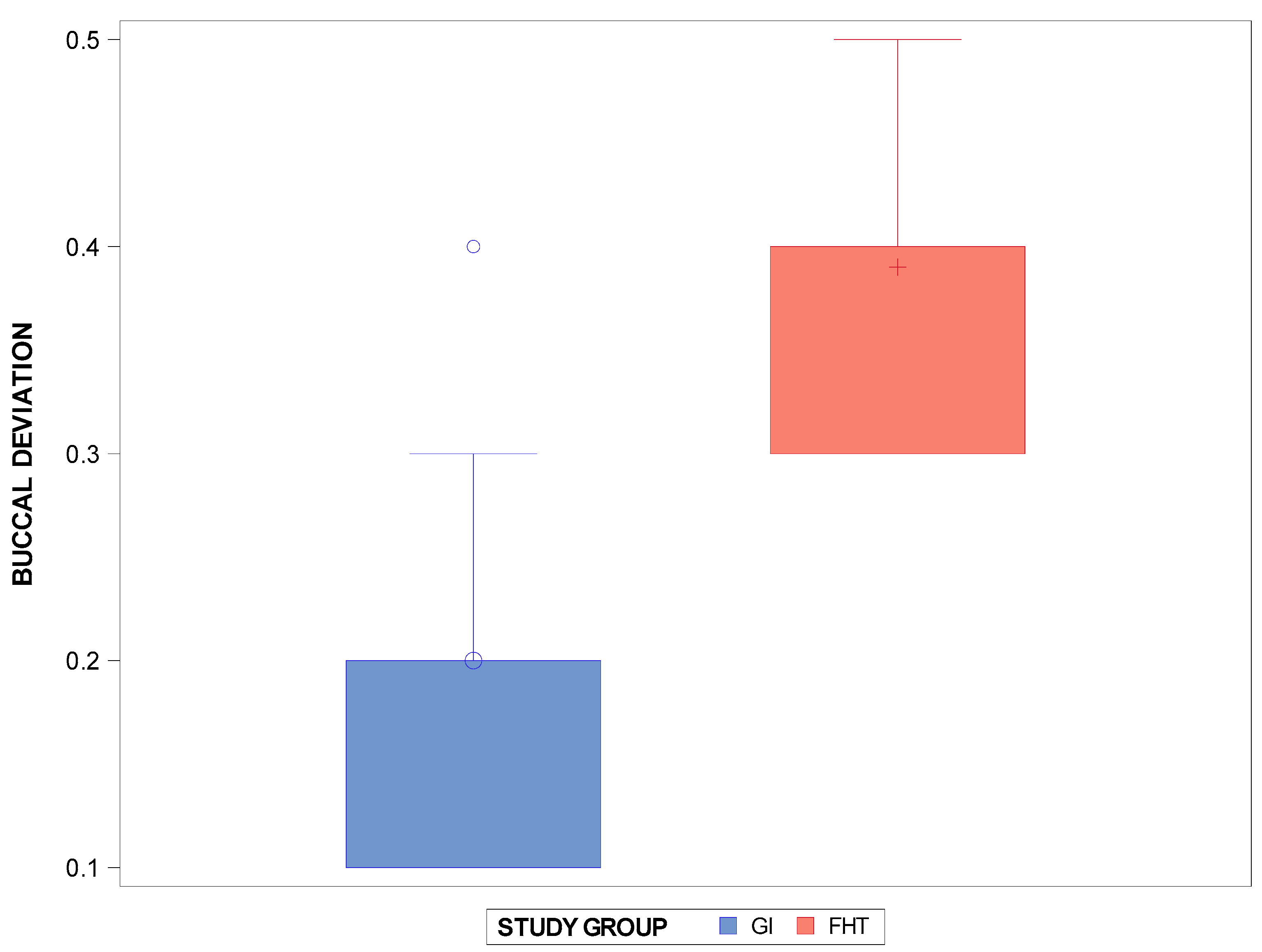
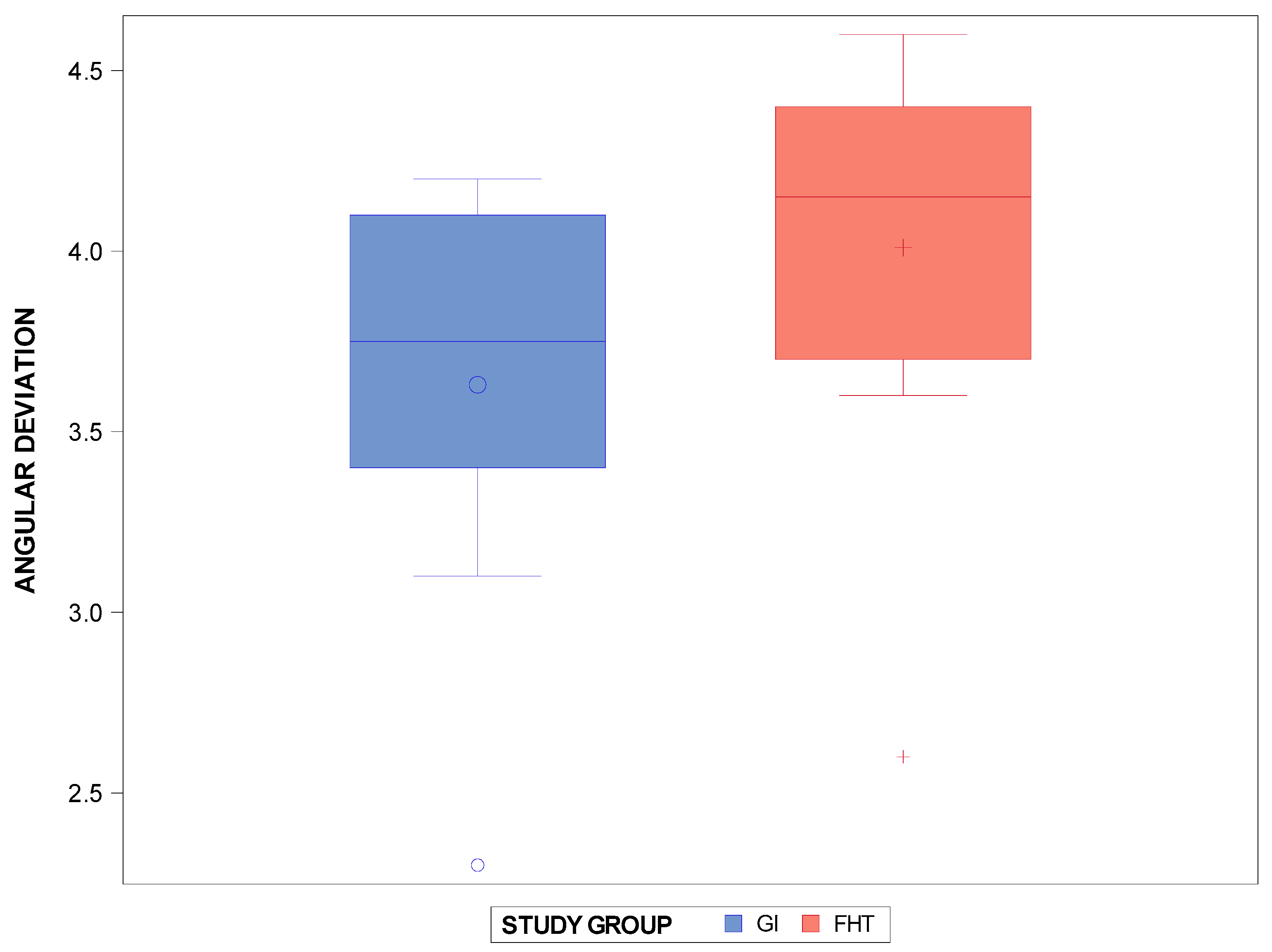
Figure 1. (A) Virtual implant drills designing crossing the contact point at the interproximal surface of the teeth, (B) virtual template designing, and (C) stereolithographic template attached on the GI experimental cast models of polyurethane.
Jpm 14 00138 g001
Figure 2. (A) Alignment procedure between preoperative planning (green cylinders) and postoperative STL digital files. (B) Lineal measurements of the interproximal enamel reduction procedures.
Jpm 14 00138 g002
Figure 3. Box plots of the buccal deviation of the interproximal enamel reduction (mm) between the computer-aided static navigation technique and the conventional free-hand-based technique. The horizontal line in each box represents the median value. +,◦: represent the mean value.
Jpm 14 00138 g003
Figure 4. Box plots of the lingual/palatal deviation in the interproximal enamel reduction (mm) between the computer-aided static navigation technique and the conventional free-hand-based technique. The horizontal line in each box represents median value. +,◦: represent the mean value.
Jpm 14 00138 g004
Figure 5. Box plots of the angular deviation of the interproximal enamel reduction (°) between the computer-aided static navigation technique and the conventional free-hand-based technique. The horizontal line in each box represents median value. +,◦: represent the mean value.
Jpm 14 00138 g005
Table 1. Descriptive statistics of the interproximal enamel reduction (mm) between the interproximal enamel reduction technique based on computer-aided static navigation technique and the conventional free-hand-based technique for the interproximal enamel reduction at buccal level.
MeasureTechniquenMean (mm)SD (mm)Minimum (mm)Maximum (mm)
BuccalGI1000.200.090.100.40
FHT1000.390.070.300.50
Lingual/PalatalGI1000.240.110.100.40
FHT1000.580.90.400.70
AngularGI1003.630.582.304.20
FHT1004.010.602.604.60
SD: standard deviation.
Disclaimer/Publisher’s Note: The statements, opinions and data contained in all publications are solely those of the individual author(s) and contributor(s) and not of MDPI and/or the editor(s). MDPI and/or the editor(s) disclaim responsibility for any injury to people or property resulting from any ideas, methods, instructions or products referred to in the content.

