Article

Establishment of Adenomyosis Organoids as a Preclinical Model to Study Infertility

1
Fundación IVI, Instituto de Investigación Sanitaria La Fe, 46026 Valencia, Spain
2
Departamento de Pediatría, Obstetricia y Ginecología, Universidad de Valencia, 46010 Valencia, Spain
3
Departament of Gynecology, IVIRMA-Valencia, 46015 Valencia, Spain
4
Departament of Gynecology, IVIRMA-Rome, 00197 Rome, Italy
*
Author to whom correspondence should be addressed.
These authors contributed equally to this work.
J. Pers. Med. 2022, 12(2), 219; https://doi.org/10.3390/jpm12020219
Submission received: 23 December 2021 / Revised: 1 February 2022 / Accepted: 2 February 2022 / Published: 4 February 2022
(This article belongs to the Section Clinical Medicine, Cell, and Organism Physiology)

Abstract

:
Adenomyosis is related to infertility and miscarriages, but so far there are no robust in vitro models that reproduce its pathological features to study the molecular mechanisms involved in this disease. Endometrial organoids are in vitro 3D models that recapitulate the native microenvironment and reproduce tissue characteristics that would allow the study of adenomyosis pathogenesis and related infertility disorders. In our study, human endometrial biopsies from adenomyosis (n = 6) and healthy women (n = 6) were recruited. Organoids were established and hormonally differentiated to recapitulate midsecretory and gestational endometrial phases. Physiological and pathological characteristics were evaluated by immunohistochemistry, immunofluorescence, qRT-PCR, and ELISA. Secretory and gestational organoids recapitulated in vivo glandular epithelial phenotype (pan-cytokeratin, Muc-1, PAS, Laminin, and Ki67) and secretory and gestational features (α-tubulin, SOX9, SPP1, PAEP, LIF, and 17βHSD2 expression and SPP1 secretion). Adenomyosis organoids showed higher expression of TGF-β2 and SMAD3 and increased gene expression of SPP1, PAEP, LIF, and 17βHSD2 compared with control organoids. Our results demonstrate that organoids derived from endometria of adenomyosis patients and differentiated to secretory and gestational phases recapitulate native endometrial-tissue-specific features and disease-specific traits. Adenomyosis-derived organoids are a promising in vitro preclinical model to study impaired implantation and pregnancy disorders in adenomyosis and enable personalized drug screening.

1. Introduction

Adenomyosis is a benign uterine disease affecting 35% of women of reproductive age [1], characterized by invagination of endometrial glands and stroma into the myometrium [2]. Many mechanisms have been postulated to be involved in adenomyosis’ development and its associated symptoms, such as altered sex steroid signaling, excessive proliferation and invasiveness of the endometrium, and an abnormal immune response [3]. To date, the exact trigger of the disease is not known but there are two main hypotheses that attempt to explain adenomyosis origin [4,5]. The first and most commonly accepted theory lies in the invasiveness of the basal endometrium into the inner myometrium [6]. This is due to a combination of two events. On the one hand, endometrial epithelial cells undergo a process called epithelial–mesenchymal transition (EMT), in which they lose their cell polarity and intercellular junctions are disrupted, facilitating the transition to a mesenchymal state and increasing the invasive capacity [7,8]. On the other hand, there is a partial loss of continuity of the junctional zone (JZ) that facilitates the invasion and establishment of adenomyotic lesions [6]. The second theory is that adenomyotic lesions are generated de novo rather than originating from the eutopic endometrium [4,5]. This would be due to a differentiation of the misplaced Müllerian remnants into tissue that resembles endometrial tissue and would therefore grow in ectopic sites [9,10]. However, other authors support that ectopic lesions could be due to endometrial stem cells (ESCs) that are transported in retrograde menstruation [11,12], which have the ability to adhere, implant, differentiate, and propagate in ectopic locations [13]. The incidence and prevalence of adenomyosis is unknown due to the lack of an adequate and standardized diagnostic criteria [14,15]. The main diagnosis of adenomyosis is made either by imaging or by histological examination after hysterectomy, which implies the need to establish a diagnosis compatible with the woman’s reproductive intentions [16,17,18]. Moreover, no clear pattern of coexistence of adenomyosis with uterine diseases such as endometriosis, leiomyomas, and other uterine conditions has been established [18]. Although one third of women affected by adenomyosis is asymptomatic, most of them may present abnormal uterine bleeding, chronic pelvic pain, dysmenorrhea, or dyspareunia [19]. This condition can also result in infertility and miscarriages [19], leading most affected women to undergo assisted reproductive techniques [20]. Meta-analyses found that implantation, clinical and ongoing pregnancy, and live birth rates are significantly lower in patients with adenomyosis compared with healthy women, while miscarriage rates are higher [21,22,23].
Local inflammation [24], oxidative stress [25], impaired vascularization [26], hyperestrogenism [27], and epithelial–mesenchymal transition (EMT) [28] may be mechanisms involved in adenomyosis pathogenesis. Steroid hormones disruption, particularly hyperestrogenism, which ultimately results in progesterone resistance, are known to play a key role in its pathogenesis [3,27]. Since many of the mechanisms involved in the pathogenesis of adenomyosis are driven by estrogen upregulation, adenomyosis is widely known as an estrogen-dependent disease [27]. Estrogen induces proliferation of endometrial cells, which, in the case of adenomyotic endometrium, results in an overproliferation [29]. In this regard, it has been hypothesized that estrogen is responsible for triggering EMT in adenomyosis [30]. Transforming growth factor (TGF)-β1 and TGF-β2 are growth and differentiation factors involved in EMT induction and regulation [31], which are upregulated in secretory endometrium from patients with adenomyosis [32,33,34], suggesting dysfunctionality during the secretory phase. Further, TGF-β/SMAD (SMAD Family Member 3) signaling is implicated in adenomyosis’ pathology [35]. SMAD2/3 are downstream proteins of TGF-β pathway involved in endometrial function maintenance, including early glandular formation, appropriate endometrial hormonal response, and tumor suppression [36]. Specifically, it has been reported that SMAD3 is overexpressed in epithelial cells from eutopic adenomyosis endometrium in secretory phase [37]. Since SMAD3 is involved in endometrial receptivity and embryo implantation [38], these findings suggest SMAD3 as a key protein in adenomyosis-related infertility [37].
SPP1 (Secreted Phosphoprotein 1) is an adhesion protein secreted by ECM (extracellular matrix) involved in endometrial-embryo signaling and embryo attachment [39], which is upregulated in the receptive phase human uterus [40]. PAEP (Progestagen Associated Endometrial Protein), also called Glycodelin-A (GdA), is a marker of morphological differentiation and an immunosuppressive molecule [41] secreted from luminal epithelial cells and localized in endometrial glands during pinopodes formation [42] in the secretory phase [43]. PAEP has been found to be elevated in the decidua throughout early pregnancy [44], being essential in the first processes of placentation [45]. LIF (Leukemia Inhibitory Factor) is a glycoprotein cytokine considered as an endometrial receptivity biomarker [46] that mediates implantation and immune response in several species [47], being crucial in decidualization regulation. 17βHSD2 (Hydroxysteroid 17-Beta Dehydrogenase) is an enzyme that metabolizes estradiol [48] and is overexpressed in both midgestation and placenta [49]. Furthermore, its activity is elevated during the secretory phase in diseased endometrium and estrogen metabolism is altered in the endometria of patients with estrogen-dependent benign diseases [50].
Patients with adenomyosis experience defective decidualization [51], altered uterine peristaltic activity [52] and endometrial receptivity [53], impaired embryo-maternal communication [54], and delayed pinopode formation, resulting in unsuccessful embryo implantation [55]. However, molecular mechanisms underlying these embryo implantation and pregnancy defects remain largely unknown, mainly due to the difficulty in obtaining secretory and gestational endometrium samples and the lack of reliable preclinical study models. Overcoming these challenges is crucial to significantly improve adenomyosis-related infertility therapies.
Organoids are self-organized in vitro in 3D structures that are genetically stable during long-term culture and viable after cryopreservation [56,57,58,59]. Organoids overcome the disadvantages of 2D culture by recapitulating the native microenvironment and reproducing native tissue characteristics [60]. Endometrial organoids may be differentiated, mimicking the midsecretory phase, in response to ovarian hormones estradiol (E2), progesterone (P4), and 8-Bromoadenosine3′,5′-cyclicmonophosphate (cAMP) [61,62] and acquire an early gestation phenotype with pregnancy hormones prolactin (PRL) and human placental lactogen (hPL) [61]. There are several patient-derived organoid models of endometrial diseases, such as endometriosis and endometrial cancers [63], which can be cryopreserved and used for preclinical studies [59].
Human organoids derived from adenomyosis eutopic endometrium and their differentiation to midsecretory and early pregnancy phases represent a powerful platform to study the dysregulated molecular mechanisms involved in implantation and pregnancy disorders. Generation of an organoid biobank representing healthy and pathological conditions would provide innovative and powerful preclinical study models for drug screening and personalized medicine.

2. Materials and Methods

2.1. Patient Samples

Endometrial biopsies were obtained from adenomyosis patients and healthy women (n = 6/group) at IVI Valencia. The study population underwent pelvic ultrasound examination as the routine workout in infertile women. All patients, cases, and controls were carefully scanned by transvaginal ultrasound. A heterogeneous myometrium with blurring of the endometrial border is the key for diagnosis of adenomyosis. In case of suspected adenomyosis, MRI (Magnetic Resonance Imaging) or hysteroscopic evaluation of the endometrial cavity was performed. Hysteroscopic findings were superficial openings on the endometrial cavity, endometrial hypervascularization, and cystic hemorrhagic lesions. Control group is based on young healthy women included in an egg donation (ED) program with a standard uterine volume, with no evidence of adenomyotic lesions by ultrasound, who were free from other gynecologic (endometrial or myometrial or ovarian) pathologies and without medication during the previous 3 months, as condition to be included in the ED program. Human tissue use was approved by the Clinical Ethics Committee at Hospital La Fe (2004-FIVI-039-HF; Valencia, Spain). Informed consent was provided.

2.2. Derivation and Culture of Organoids from Human Endometrial Tissue Samples

Endometrial biopsies from adenomyosis patients (n = 6) and healthy (n = 6) women were processed to isolate the epithelial glandular fractions [61]. Biopsies were mechanically and enzymatically digested with 50 U/mL Dispase II (Sigma-Aldrich, St. Louis, MO, USA, D4693) and 4 mg/mL Collagenase-V (Sigma-Aldrich, St. Louis, MO, USA, C9263). Supernatant was passed through 100 µm cell sieves (Corning, Tewksbury, MA, USA, 431752) to retain glandular elements; the pellet was resuspended in 15% DMEM/F12 (Invitrogen, Invitrogen, Paisley, UK, 12634010) and 85% Matrigel (Corning, Bedford, MA, USA, 354234) and 20 μL droplets were seeded. A total 250 μL of organoid Expansion Medium (ExM) (Table 1) were added and changed every 2 days. Organoid passage was made every 7 days, with pipetting up and down forcefully and centrifuging to disaggregate organoids and remove Matrigel. Pellets containing organoid fragments were resuspended in 15% DMEM/F12–85% Matrigel.

2.3. Differentiation of Endometrial Organoids to Secretory and Gestational Phases

Organoids in passage 3 (p3) were cultured with ExM for 7 days to maintain undifferentiated status (derived-organoids). For differentiation experiments [61], after 4 days in ExM, culture media was enriched with 10 nM E2 (Sigma-Aldrich, St. Louis, MO, USA, Sigma E4389) for 48 h; and with 10 nM E2, 1 µM P4 (Sigma-Aldrich, St. Louis, MO, USA, Sigma P7556), and 1 µM cAMP (Sigma-Aldrich, St. Louis, MO, USA, Sigma B7880) for 4 days to promote differentiation to the secretory phase (sec-organoids). To induce gestational phase (gest-organoids) differentiation, 20 ng/mL PRL (Peprotech, Cranbury, NJ, USA, 100-07) and 20 ng/mL hPL (R&D, Minneapolis, MN, USA, 5757-PL) were added to previously supplemented ExM for 8 days.

2.4. Immunohistochemical (IHC) and Immunofluorescence (IF) Characterization

To verify that Control- and adenomyosis (Adeno) derived-organoids reproduced biological and pathological features of the native tissue, organoids were fixed, paraffin embedded [64], and cut into 4-µm sections. Periodic acid-Schiff (PAS) (glycogen secretion) (Sigma-Aldrich, St. Louis, MO, USA, 395B) staining was performed according to manufacturer’s protocol. For assessment of MUC-1 (mucin-1) (glandular secretion), Ki67 (cell proliferation), SOX9 (SRY-Box Transcription Factor 9) (progenitor cells markers), TGF-β2, and SMAD3 (adenomyosis development) protein expression, samples were incubated with primary antibodies (Table 2) overnight at 4 °C. After endogenous peroxidase activity blockage, slides were incubated with labeled-polymer HRP (horseradish peroxidase), substrate-chromogen and counterstained with hematoxylin. To evaluate pan-cytokeratin (epithelial marker), vimentin (stromal component), laminin (basoapical polarity), and acetylated α-tubulin (cilia presence) expression, samples were incubated with primary antibodies overnight at 4 °C and their correspondent secondary antibody 45 min at RT (room temperature) (Table 2). Samples were visualized using a Nikon Eclipse 80i microscope. To measure SOX9, α-tubulin, TGF-β2, and SMAD3 protein expression levels, four images per sample were quantitatively assessed with Image ProPlus (Media Cybernetics, Rockville, MD, USA). Quantification was made by calculating the ratio between the area stained by the signal and the total area of the histological section.

2.5. Gene Expression Analysis

To confirm organoid differentiation into secretory and gestational phases, gene expression of implantation and placentation biomarkers SPP1, PAEP, LIF, and 17βHSD2 was evaluated by quantitative real time PCR (qRT-PCR) using a StepOnePlus system (Applied Biosystems, Waltham, MA, USA, 4376600). To evaluate the possible role of these biomarkers in implantation and pregnancy in adenomyosis, their expression was assessed in Control and Adeno differentiated organoids. Total RNA was extracted from organoids (n = 6/group) using Trizol reagent (Qiagen, Gilde, Germany, 79306). Gene expression levels were normalized with housekeeping gene GAPDH (Glyceraldehyde-3-Phosphate Dehydrogenase), quantified by the ΔΔCt method, and represented as fold-change in each group. Primers were designed using Primer Quest Tool (DNA Integrated Technologies) (Table 3).

2.6. ELISA

To assess proper sec-organoid differentiation, we evaluated SPP1 protein secretion. Culture media from Adeno and Control derived- and sec-organoids were collected and supernatants were concentrated with Vivaspin2-concentrators (Generon, Slough, UK, VS0291). Osteopontin (SPP1) Human ELISA (Enzyme-Linked ImmunoSorbent Assay) (Invitrogen, Eugene, OR, USA, BMS2066) was performed in duplicate according to the manufacturer’s instructions.

2.7. Chromosomal Stability

Chromosomal stability of Control and Adeno derived-, sec-, and gest-organoids (n = 3/group) was evaluated using a Genome-Wide high-resolution Affymetrix-Cytoscan 750K-array (Affymetrix Inc, Santa Clara, CA, USA). DNA was extracted using the Cells and Tissue DNA-Isolation Micro-Kit (Norgen, Thorold, ON, Canada, 57300). Data were analyzed using Affymetrix Chromosome-Analysis Suite (ChAS4.2).

2.8. Statistical Analysis

Graphpad Prism 6.0 was used for statistical analyses. Two-tailed Student’s t-test and one-way ANOVA were used for comparisons between two and three groups, and p < 0.05 was considered statistically significant.

3. Results

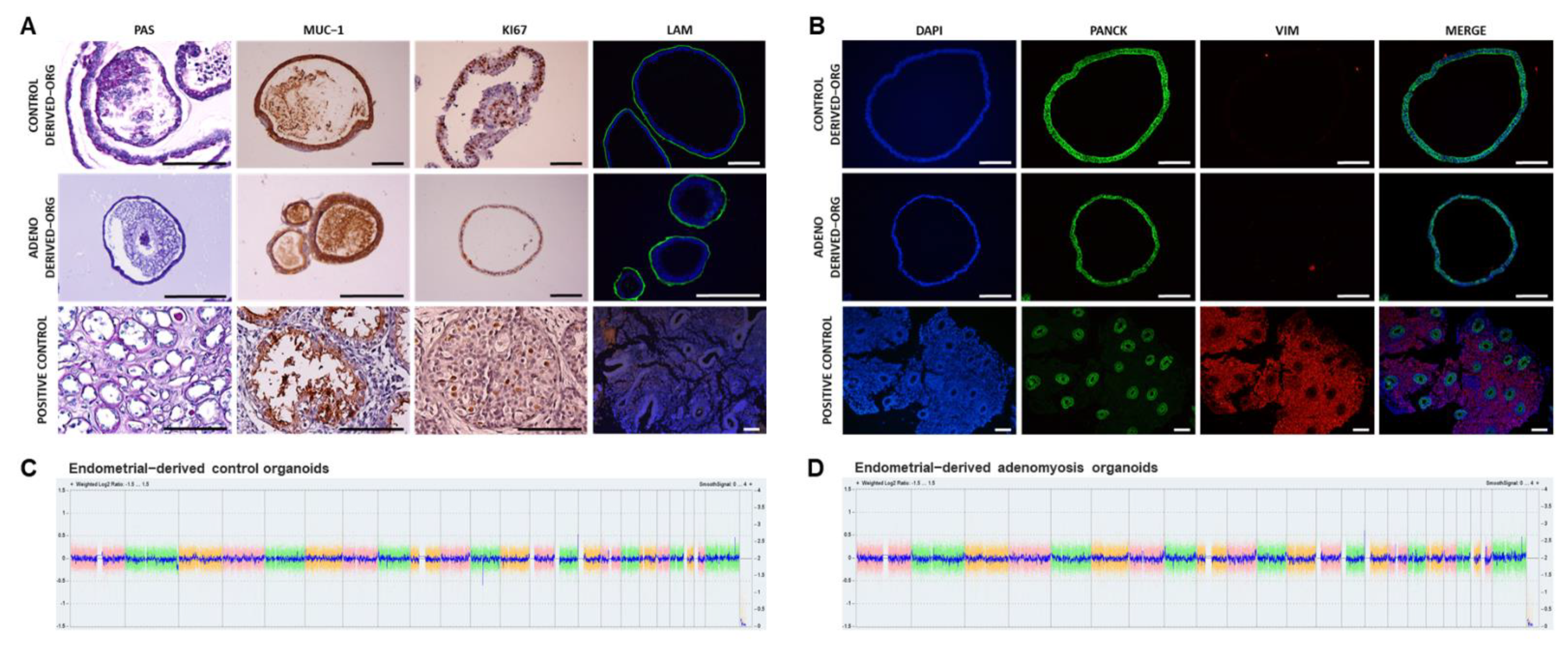
3.1. Human Endometrial Organoids Can Be Derived from Adenomyosis Patients and Recapitulate Endometrial Gland Biology In Vivo

To evaluate if Adeno derived-organoids recapitulate the biological characteristics of the native endometrium, we determined their glandular epithelial origin, organoid structure, secretions, proliferation capacity, and apicobasal polarity maintenance (Figure 1A). Endometrial organoids were derived from healthy women for the control group. PAS staining confirmed organoid production of epithelial glycogen, a main component of endometrial glandular secretions [61]. MUC-1 was secreted by organoids into the luminal compartment, as observed in human endometrial gland lumen. Ki67 expression in the organoids demonstrated maintenance of proliferative capacity. Laminin presence along the basolateral membrane confirmed that organoid epithelial cells maintain apicobasal polarity.
Pan-cytokeratin (glands) and vimentin (stroma) expression confirmed correct isolation of the epithelial glands (Figure 1B). Pan-cytokeratin was expressed in organoid cell cytoplasmic compartment while vimentin was not expressed, corroborating the epithelial origin of the organoids.

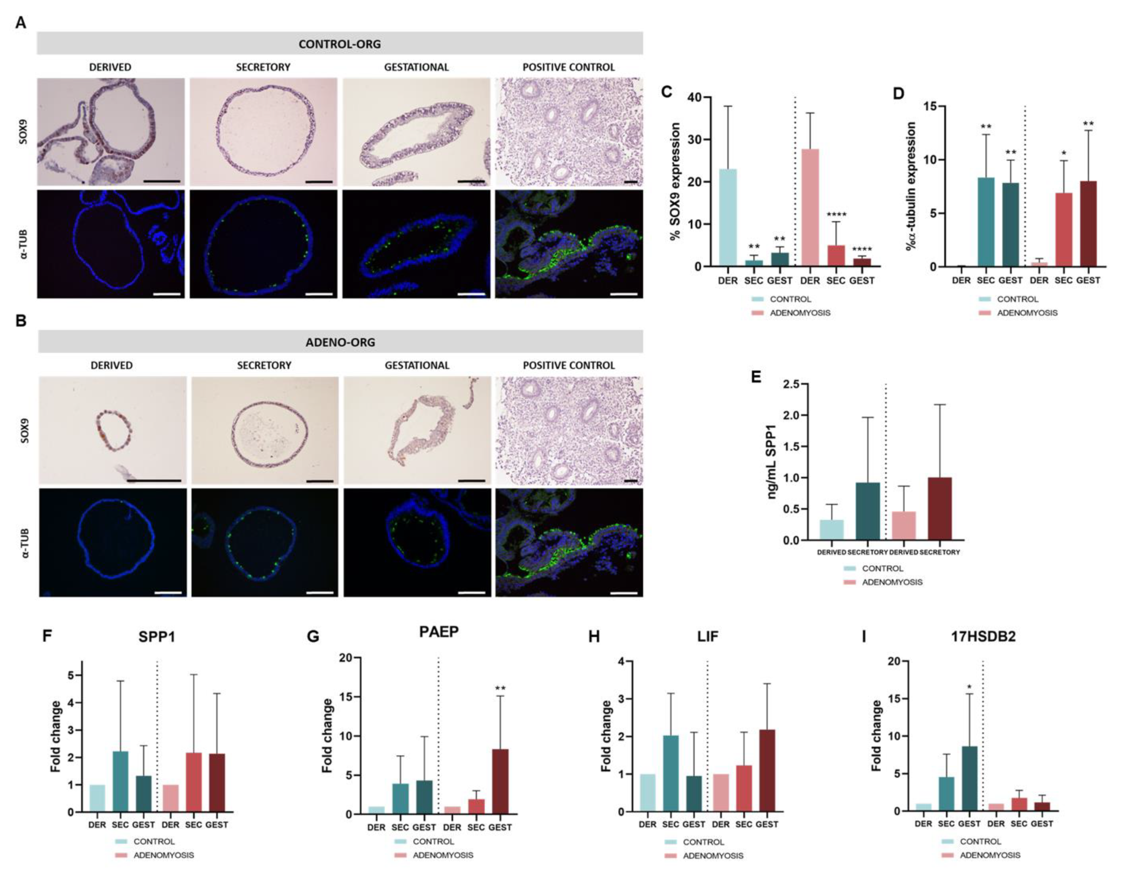
3.2. Differentiation to Secretory and Gestational Phases of Human Adenomyosis-Derived Organoids in Response to Hormonal Treatments

To reproduce secretory (sec) and gestational (gest) in vivo conditions, human-derived organoids were exposed to E2, P4, and cAMP to promote transition to receptive state, and to PRL and hPL to mimic the early gestational phase. We confirmed that glandular epithelial origin was preserved after differentiation in Control and Adeno sec- and gest-organoids (Supplementary Figure S1A,B).
SOX9 and α-tubulin expression was evaluated by IHC and IF to confirm differentiation of Control (Figure 2A) and Adeno (Figure 2B) sec- and gest-organoids. SOX9, a progenitor cell marker, was increased in derived organoids and, after differentiation, its expression was significantly reduced in Control (p = 0.0012 and 0.0025) and Adeno (p < 0.0001 for both) sec- and gest-organoids (Figure 2C), as occurs in decidual glands in vivo. Secretory- and gestational-phase hormonal treatment significantly promoted formation of ciliated cells, indicated by expression of acetylated α-tubulin, in Control (p = 0.0031 and 0.0048) and Adeno (p = 0.0146 and 0.0056) sec- and gest-organoids (Figure 2D), as occurs in vivo. Differentiation was evaluated at the protein level by ELISA of implantation marker SPP1 secretion into the culture media (Figure 2E). Secreted SPP1 levels were higher in Control and Adeno sec-organoids compared with derived organoids, confirming differentiation.
Higher expression of the secretory and gestational markers SPP1, PAEP, LIF, and 17βHSD2 in sec- and gest-organoids compared with derived-organoids from Control and Adeno corroborated the differentiation to secretory and gestational phase in both conditions (Figure 2F–I). Hormonal treatment to induce secretory and gestational phases in Control organoids increased expression of SPP1 (Fold Change (FC) = 2.215 ± 2.577; FC = 1.330 ± 1.102), PAEP (FC = 3.926 ± 3.535; FC = 4.331 ± 5.599), LIF (FC = 2.031 ± 1.116; FC = 0.9502 ± 1.161), and 17βHSD2 (FC = 4.520 ± 3.080; FC = 8.641 ± 7.006, p = 0.0168) compared with Control untreated, derived organoids. In sec- and gest-phase Adeno organoids, expression of SPP1 (FC = 2.172 ± 2.853; FC = 2.137 ± 2.195), PAEP (FC = 1.948 ± 1.0.72; FC = 8.330 ± 6.775, p = 0.0145), LIF (FC = 1.234 ± 0.8801; FC = 2.187 ± 1.218), and 17βHSD2 (FC = 1.760 ± 1.021; FC = 1.163 ± 0.9616) were also increased compared with Adeno untreated, derived organoids.

3.3. Human Adenomyosis-Derived Organoids Maintain Chromosomal Stability after Differentiation

Chromosomal stability of Control (n = 3) and Adeno (n = 3) derived-organoids (Figure 1C,D), sec-organoids, and gest-organoids (Supplementary Figure S1C,D) were assessed using a cytogenetics microarray and compared against a reference genome. No DNA copy number alterations were observed after derived-organoids culture and passage until p3. Exposure to secretory and gestational phase hormonal treatment had no effect on chromosomal stability. All established organoid lines from women with adenomyosis and controls showed a normal 46, XX karyotype.

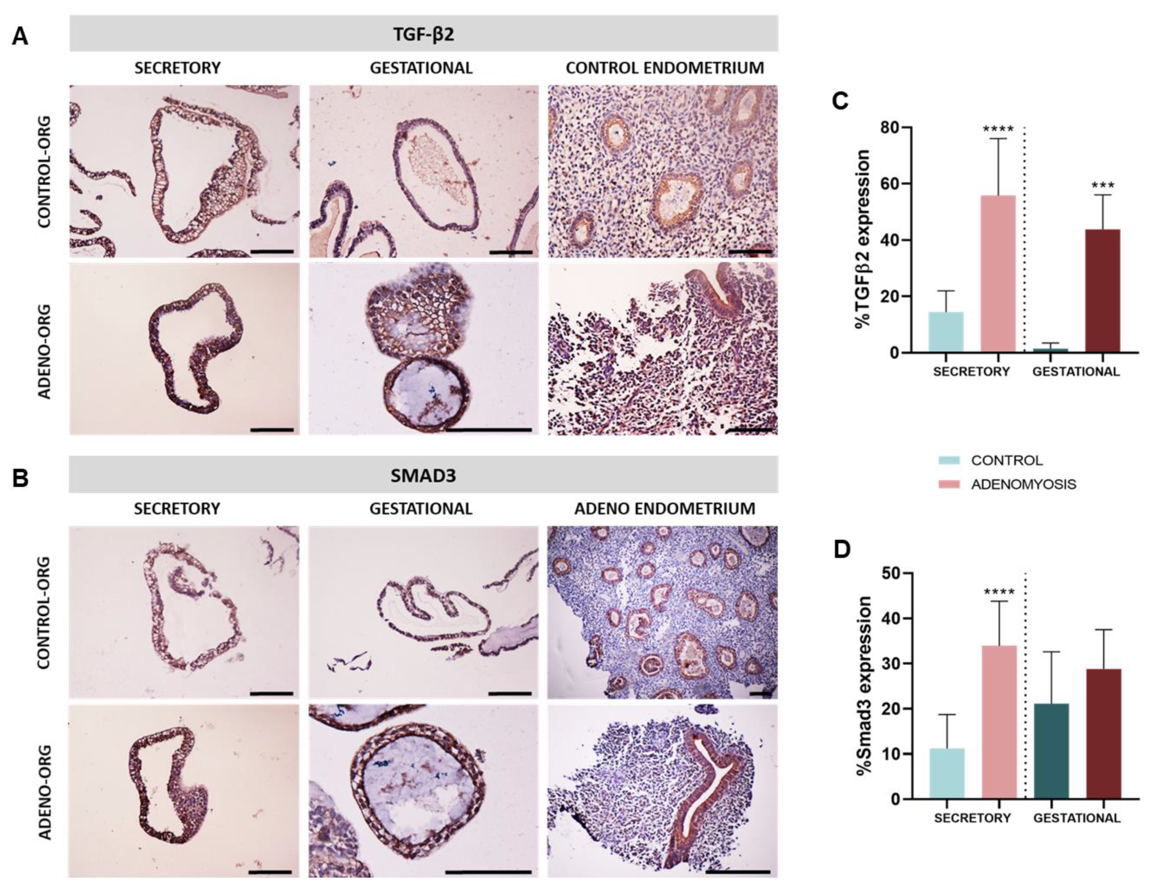
3.4. Human Adenomyosis Secretory and Gestational Organoids Recapitulate Disease-Specific Traits

To determine whether organoids are a suitable in vitro model of the in vivo pathological features of adenomyosis, TGF-β2 and SMAD3 expression were evaluated by IHC in Control and Adeno (n = 6/group) sec- and gest-organoids. TGF-β2 and SMAD3 (Figure 3A,B) expression were upregulated in Adeno compared with Control sec-organoids (TGF-β2: 55.78 ± 20.26% vs. 14.45 ± 7.51%, p < 0.0001 and SMAD3: 33.95 ± 9.88% vs. 11.22 ± 7.51%, p < 0.0001), and gest-organoids (TGF-β2: 43.81 ± 12.22% vs. 1.41 ± 2.00%, p = 0.0003 and SMAD3: 28.81 ± 87.69% vs. 21.16 ± 11.44%, p = 0.3282) (Figure 3C,D), as observed in adenomyosis [32,37].

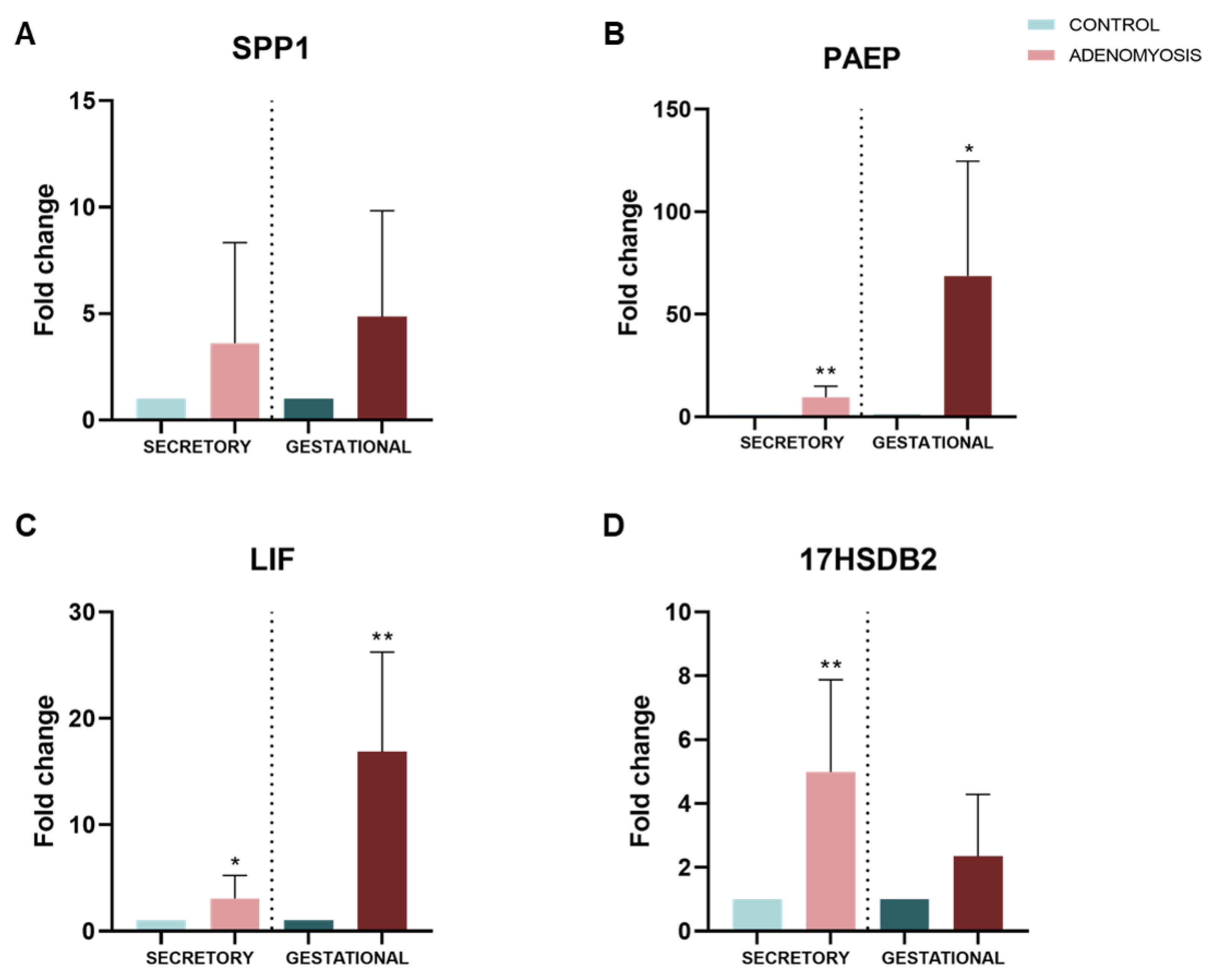
3.5. Dysregulation of Secretory and Gestational Biomarkers in Human Adenomyosis Organoids

As a first approach in understanding impaired implantation and pregnancy disorders characteristic of women with adenomyosis, expressions of secretory and gestational endometrial biomarkers SPP1, PAEP, LIF, and 17βHSD2 were evaluated in adenomyosis sec- and gest-organoids by qRT-PCR and compared with Control sec- and gest-organoids (Figure 4A–D). These biomarkers are involved in regulation of implantation (midsecretory phase) and placentation (early pregnancy); their expression was increased in Adeno sec- and gest-organoids compared with Control organoids in the same phases ((SPP1 FC = 3.603 ± 4.733, p = 0.3636; FC = 4.850 ± 4.983, p = 0.0879), (PAEP FC = 9.610 ± 5.292, p = 0.0030; FC = 68.70 ± 55.88, p = 0.0141); (LIF FC = 3.054 ± 2.179, p = 0.0436; FC = 16.85 ± 9.388, p = 0.0020); 17βHSD2 FC = 4.984 ± 2.892, p = 0.0071; FC = 2.345 ± 1.939, p = 0.1201)).

4. Discussion

Adenomyosis is one of the most widespread uterine conditions among women of reproductive age, but so far, there have been no robust in vitro models that reproduce its pathological features to study the molecular mechanisms involved in its pathogenesis and infertility disorders. We have been able to develop a human organoid model of the adenomyosis secretory and gestational endometrium, recapitulating specific native tissue features and disease traits. These organoids will provide powerful preclinical research models to study adenomyosis-impaired implantation and increased miscarriages as well as to enable personalized medicine.
Previous studies in the field of reproductive medicine have relied on organoids such as 3D in vitro models to study endometrial physiology and disease [60,63,65,66,67]. In this regard, organoids have been exploited in the study of defective endometrial proliferation, such as endometriosis or endometrial cancer [63,68], disorders affecting decidualization [67], endocrine disruptors [67], or gynecological infections [69,70]. In addition, its potential in personalized medicine or as a source of biological material in regenerative therapy is becoming increasingly evident [71,72,73].
We derived endometrial organoids from healthy women, as previously described [61,64], and for the first time, have reproducibly established organoids from endometria of adenomyosis patients. Organoids recapitulated the molecular signatures of in vivo endometrial glands. Histology confirmed expression of several cytokeratins in Control and Adeno organoids, which exert structural function in epithelial cells and have an important role in differentiation and tissue function [74]. Likewise, glycogen (glandular secretions) and MUC-1 (mucus release) presence in the luminal compartments of Adeno organoids suggests that they mimic glandular tissue functioning in the same way as Control organoids. Lastly, both Control and Adeno organoids maintained proliferative capacity (Ki67), cell apicobasal polarity (laminin), and chromosomal stability after successive passages.
Hormone responsiveness of healthy endometrial organoids has been reported [75,76,77]; so, we wanted to evaluate this response ability in Adeno organoids towards the differentiation into secretory and gestational endometrium. Similar to our Control organoids, Adeno organoids showed E2 and P4 treatment sensitivity, acquiring a secretory phenotype, and when further stimulated with pregnancy (hPL) and stroma (PRL) signals, adopted gestational endometrium characteristics. This was substantiated by decreased expression of progenitor cell marker SOX9, indicating differentiation processes, appearance of ciliated cells (α-tubulin), and increased synthesis of SPP1. Acquisition of differentiated phenotypes was further verified by upregulation of SPP1, PAEP, LIF, and 17βHSD2, which are expressed by secretory endometrium and decidua. SPP1 levels are high in the human uterus in the receptive phase [40] and luminal epithelium in early pregnancy in pigs [75], suggesting that SPP1 is essential for endometrial receptivity and implantation [78]. PAEP secretion is increased by P4 midpregnancy [79,80], relating it to endometrial receptivity and early pregnancy. Reported increased LIF expression in mouse endometrium during late diestrus phase, and in the human endometrium during the secretory phase and mid- to late-pregnancy [47,81,82], suggest that LIF is an endometrial receptivity biomarker. High 17βHSD2 expression and activity is found in secretory phase, midgestation, and term human placentas [49].
Adenomyosis is thought to be promoted by EMT, which is induced and regulated by factors including TGF-β1 and TGF-β2 [31,34]. The TGF-β/SMAD3 pathway participates in embryo implantation, as TGF-βs and SMADs largely expressed in human endometrium during implantation window [38]. TGF-β/SMAD3 signaling is a major mechanism involved in endometrial fibrosis [37] and plays a key role in adenomyosis development [35]. Further, patients with adenomyosis present increased TGF-β2 and SMAD3 levels in their eutopic endometrium during the secretory phase compared with disease-free women [32,33,37]] SMAD3. Remarkably, our secretory adenomyosis organoids showed significant TGF-β2 and SMAD3 upregulation compared with controls, confirming successful secretory-phase differentiation and accurate reproduction of specific disease traits. Thus, this adenomyosis in vitro model appears suitable for studying patients with impaired implantation.
Adenomyosis causes defective placentation [83], which is significantly associated with increased risk of preeclampsia [84,85]. TGF-βs—through activation of downstream signaling mediators—and SMAD2/3 are triggering factors for preeclampsia, resulting in abnormal placental development [86,87]. SMAD3 is further involved in key gestational processes, immune regulation, and inflammation and its altered expression may be associated with recurrent pregnancy loss [88] and preterm birth [89]. Accordingly, our gestational adenomyosis organoids (imitating early pregnancy) showed increased levels of TGF-β2 and SMAD3, recapitulating adenomyosis tissue characteristics. This implies that gestational differentiated organoids represent a potent preclinical platform and research approach for studying placentation and early-pregnancy disorders in women with adenomyosis.
SPP1, PAEP, LIF, and 17βHSD2 expression were upregulated in adenomyosis sec- and gest-organoids compared with control organoids, indicating possible molecular mechanisms involved in adenomyosis-impaired implantation and pregnancy disorders. SPP1, which is involved in endometrial-embryo signaling and embryo attachment [39], was upregulated in adenomyosis secretory organoids compared with healthy organoids, as it was described in adenomyosis women ectopic endometrium [90]. Increased SPP1 in our gestational adenomyosis organoids compared with control suggests abnormal endometrial SPP1 expression during implantation window [90] and placentation [91] and could contribute to adenomyosis-related infertility. PAEP, a morphological differentiation marker and immunosuppressive molecule [41] secreted from luminal epithelial cells and localized in endometrial glands during pinopode formation [42], was significantly upregulated in our Adeno secretory organoids. Abnormal PAEP expression during the secretory phase in endometriosis eutopic endometrium [43] suggests that dysregulated PAEP expression could be related to impaired endometrial receptivity. PAEP was also increased in Adeno gestational organoids compared with healthy organoids. This marker is abundant in the decidua during early pregnancy and is crucial in placentation events and fetomaternal defense, regulating trophoblast and immune cell functions during early pregnancy [45]. Thus, its upregulation in adenomyosis sec- and gest-organoids suggests that abnormal endometrial PAEP levels could be involved in early pregnancy loss, preeclampsia, and recurrent miscarriage in women with adenomyosis, as previously suggested for endometriosis disease [43].
Our data showed significant upregulation of LIF—a glycoprotein cytokine involved in decidualization and immune response [47]—in secretory and gestational adenomyosis organoids compared with control. These results are not in line with previous works reporting lower LIF levels in adenomyosis patients’ endometrium, but the critical point is that the control group in these studies included women with other gynecological disorders [92,93]. As LIF regulates the Wnt/β-catenin pathway, which is involved in uterine preparation for implantation and EMT regulation [94], altered expression of this marker may be related to impaired implantation and altered EMT, possibly driving endometrial gland and stroma invagination into the myometrium, characteristic of adenomyosis [28].
Finally, 17βHSD2 is altered in the eutopic endometrium of adenomyosis, endometriosis, and leiomyoma patients [95,96], leading to estrogen metabolism alteration in estrogen-dependent benign disease patient endometria [50]. The observed 17βHSD2 upregulation in secretory adenomyosis organoids aligns with previous studies demonstrating that 17βHSD2 activity is increased in the endometrial secretory phase in diseased but not in disease-free endometrium [50]. In mice, 17βHSD2 disruption results in placentation defects and embryonic lethality [97]. Thus, the observed increase in 17βHSD2 expression in Adeno gestational organoids in the present study suggests a relationship between 17βHSD2 dysregulation in the endometrial gestational phase and associated early-pregnancy alterations in women with adenomyosis. However, further studies are needed in order to determine more accurately the involvement of these genes, as well as to describe other potential genes implicated in implantation and early pregnancy disorders in patients with adenomyosis.
The endometrial organoid model does not include stromal cells, which are involved in decidualization and other important processes, implying a lack of epithelial–stromal paracrine and autocrine crosstalk [67,98]. Regarding microenvironment communication, Matrigel does not allow us to reliably simulate tissue-specific cell–ECM interactions, with its replacement by decellularized endometrial hydrogels being a potential alternative [99]. In addition, adenomyosis organoid model only reproduces the endometrial component, leaving unstudied all the mechanisms involved in the damage that occurs in the JZ and myometrium [6]. However, in recent years, these limitations are being addressed; for example, organoid implantation models have been implemented in such a way that would allow accessibility to the luminal compartment [98,100]. Beyond that, our adenomyosis organoid model opens insights to the development of microfluidic devices and sensor systems that would help to optimize and standardize organoid cultures [101], solving then some of the mentioned limitations and improving the study of this condition.
Many authors have discussed the possibility that adenomyosis and endometriosis have a common origin and are therefore different manifestations of the same disease [102,103,104]. Since endometriosis organoids models have been already established [63] but to date there is, to our knowledge, no model of adenomyosis disease, this study could provide a new insight into the possible common mechanisms involved in the development of both diseases, as well as the associated infertility.
In conclusion, our adenomyosis organoid model maintains biological and pathological characteristics observed in secretory and gestational adenomyosis patients’ eutopic endometria. This model provides new knowledge about the possible role of implantation and early gestational biomarkers in adenomyosis-related infertility, opening avenues for further studies of these biomarkers and for development of therapeutic options for personalized treatments.

5. Conclusions

Here, we have successfully derived organoids from adenomyosis patients for the first time. Patient-derived adenomyosis organoids can be established and cryopreserved, allowing generation of a patient-specific biobank that would permit their use as a preclinical model for drug screening and promoting development of personalized medicine to improve implantation and avoid pregnancy disorders in adenomyosis patients. This is the first model demonstrating recapitulation of adenomyosis tissue origin characteristics at molecular and histological levels, which entails a step forward in generation of robust preclinical models that faithfully mimic this human endometrial pathology.

Supplementary Materials

The following supporting information can be downloaded at: https://www.mdpi.com/article/10.3390/jpm12020219/s1, Figure S1: Characterization of glandular origin, proliferation, and epithelial polarity in human secretory and gestational organoids; Table S1: Expansion medium (ExM) composition; Table S2: Primary and secondary antibodies; Table S3: Primers sequences.

Author Contributions

Conceptualization, E.J.-B., E.F.-H., A.C., I.C. and H.F.; methodology, E.J.-B., E.F.-H., A.C., C.V., J.G., P.A. and A.F.; validation E.J.-B., E.F.-H., A.C. and A.F.; formal analysis, E.J.-B., E.F.-H. and A.C.; investigation, E.J.-B., E.F.-H, A.C., A.F., I.C. and H.F.; resources, C.V., J.G., P.A., A.F., I.C. and H.F.; data curation, E.J.-B., E.F.-H. and A.C.; writing—original draft preparation, E.J.-B., E.F.-H., A.C. and H.F.; writing—review and editing, A.C., A.P., I.C. and H.F.; visualization, E.J.-B., E.F.-H., and A.C.; supervision, A.C., A.P., I.C. and H.F.; project administration, I.C. and H.F.; funding acquisition, E.J.-B., E.F.-H., A.C., I.C. and H.F. All authors have read and agreed to the published version of the manuscript.

Funding

This research was funded by Health Institute Carlos III (FI19/00110 awarded to E.J.-B.), Miguel Servet Program (CP20/00120 awarded to H.F.; CP19/00149 awarded to I.C.), Generalitat Valenciana (APOSTD/2020/123 awarded to A.C.) and by the Spanish Ministry of Education (FPU18/06327 to E.F.-H.)

Institutional Review Board Statement

The study was conducted in accordance with the Declaration of Helsinki and approved by the Clinical Ethics Committee at Hospital La Fe (2004-FIVI-039-HF; on 18 June 2020, Valencia, Spain).

Informed Consent Statement

Informed consent was obtained from all subjects involved in the study.

Data Availability Statement

The data presented in this study are available on request from the corresponding author.

Acknowledgments

The authors would like to thank the participants, clinic, and laboratory staff of IIS La Fe and IVI Valencia.

Conflicts of Interest

The authors declare no conflict of interest.

References

  1. Munro, M.G. Uterine polyps, adenomyosis, leiomyomas, and endometrial receptivity. Fertil. Steril. 2019, 111, 629–640. [Google Scholar] [CrossRef] [PubMed]
  2. Vercellini, P.; Bonfanti, I.; Berlanda, N. Adenomyosis and infertility: Is there a causal link? Expert Rev. Endocrinol. Metab. 2019, 14, 365–367. [Google Scholar] [CrossRef] [PubMed]
  3. Donnez, J.; Stratopoulou, C.A.; Dolmans, M.-M. Uterine Adenomyosis: From Disease Pathogenesis to a New Medical Approach Using GnRH Antagonists. Int. J. Environ. Res. Public Health 2021, 18, 9941. [Google Scholar] [CrossRef]
  4. Stratopoulou, C.A.; Donnez, J.; Dolmans, M.-M. Origin and Pathogenic Mechanisms of Uterine Adenomyosis: What Is Known So Far. Reprod. Sci. 2021, 28, 2087–2097. [Google Scholar] [CrossRef]
  5. García-Solares, J.; Donnez, J.; Donnez, O.; Dolmans, M.-M. Pathogenesis of uterine adenomyosis: Invagination or metaplasia? Fertil. Steril. 2018, 109, 371–379. [Google Scholar] [CrossRef] [PubMed]
  6. Leyendecker, G.; Wildt, L. A new concept of endometriosis and adenomyosis: Tissue injury and repair (TIAR). Horm. Mol. Biol. Clin. Investig. 2011, 5, 125–142. [Google Scholar] [CrossRef] [Green Version]
  7. Dongre, A.; Weinberg, R.A. New insights into the mechanisms of epithelial–mesenchymal transition and implications for cancer. Nat. Rev. Mol. Cell Biol. 2019, 20, 69–84. [Google Scholar] [CrossRef]
  8. Chen, Y.-J.; Li, H.-Y.; Huang, C.-H.; Twu, N.-F.; Yen, M.-S.; Wang, P.-H.; Chou, T.-Y.; Liu, Y.-N.; Chao, K.-C.; Yang, M.-H. Oestrogen-induced epithelial-mesenchymal transition of endometrial epithelial cells contributes to the development of adenomyosis. J. Pathol. 2010, 222, 261–270. [Google Scholar] [CrossRef]
  9. Batt, R.E.; Yeh, J. Müllerianosis. Reprod. Sci. 2013, 20, 1030–1037. [Google Scholar] [CrossRef]
  10. Signorile, P.G.; Baldi, F.; Bussani, R.; D’Armiento, M.; De Falco, M.; Baldi, A. Ectopic endometrium in human foetuses is a common event and sustains the theory of müllerianosis in the pathogenesis of endometriosis, a disease that predisposes to cancer. J. Exp. Clin. Cancer Res. 2009, 28, 49. [Google Scholar] [CrossRef] [Green Version]
  11. Gargett, C.E.; Schwab, K.E.; Brosens, J.J.; Puttemans, P.; Benagiano, G.; Brosens, I. Potential role of endometrial stem/progenitor cells in the pathogenesis of early-onset endometriosis. Mol. Hum. Reprod. 2014, 20, 591–598. [Google Scholar] [CrossRef] [PubMed] [Green Version]
  12. Gargett, C.E.; Schwab, K.E.; Deane, J.A. Endometrial stem/progenitor cells: The first 10 years. Hum. Reprod. Update 2015, 22, 137–163. [Google Scholar] [CrossRef] [PubMed] [Green Version]
  13. Sampson, J.A. Metastatic or Embolic Endometriosis, due to the Menstrual Dissemination of Endometrial Tissue into the Venous Circulation. Am. J. Pathol. 1927, 3, 93–110.43. [Google Scholar]
  14. Morassutto, C.; Monasta, L.; Ricci, G.; Barbone, F.; Ronfani, L. Incidence and Estimated Prevalence of Endometriosis and Adenomyosis in Northeast Italy: A Data Linkage Study. PLoS ONE 2016, 11, e0154227. [Google Scholar] [CrossRef] [Green Version]
  15. Yu, O.; Schulze-Rath, R.; Grafton, J.; Hansen, K.; Scholes, D.; Reed, S.D. Adenomyosis incidence, prevalence and treatment: United States population-based study 2006–2015. Am. J. Obstet. Gynecol. 2020, 223, 94.e1–94.e10. [Google Scholar] [CrossRef]
  16. Chapron, C.; Vannuccini, S.; Santulli, P.; Abrão, M.S.; Carmona, F.; Fraser, I.S.; Gordts, S.; Guo, S.-W.; Just, P.-A.; Noël, J.-C.; et al. Diagnosing adenomyosis: An integrated clinical and imaging approach. Hum. Reprod. Update 2020, 26, 392–411. [Google Scholar] [CrossRef]
  17. Tellum, T.; Nygaard, S.; Lieng, M. Noninvasive Diagnosis of Adenomyosis: A Structured Review and Meta-analysis of Diagnostic Accuracy in Imaging. J. Minim. Invasive Gynecol. 2020, 27, 408–418.e3. [Google Scholar] [CrossRef]
  18. Upson, K.; Missmer, S.A. Epidemiology of Adenomyosis. Semin. Reprod. Med. 2020, 38, 089–107. [Google Scholar] [CrossRef]
  19. Peric, H.; Fraser, I.S. The symptomatology of adenomyosis. Best Pract. Res. Clin. Obstet. Gynaecol. 2006, 20, 547–555. [Google Scholar] [CrossRef]
  20. Puente, J.M.; Fabris, A.; Patel, J.; Patel, A.; Cerrillo, M.; Requena, A.; Garcia-Velasco, J.A. Adenomyosis in infertile women: Prevalence and the role of 3D ultrasound as a marker of severity of the disease. Reprod. Biol. Endocrinol. 2016, 14, 60. [Google Scholar] [CrossRef] [Green Version]
  21. Vercellini, P.; Consonni, D.; Dridi, D.; Bracco, B.; Frattaruolo, M.P.; Somigliana, E. Uterine adenomyosis and in vitro fertilization outcome: A systematic review and meta-analysis. Hum. Reprod. 2014, 29, 964–977. [Google Scholar] [CrossRef] [PubMed] [Green Version]
  22. Tamura, H.; Kishi, H.; Kitade, M.; Asai-Sato, M.; Tanaka, A.; Murakami, T.; Minegishi, T.; Sugino, N. Clinical outcomes of infertility treatment for women with adenomyosis in Japan. Reprod. Med. Biol. 2017, 16, 276–282. [Google Scholar] [CrossRef] [PubMed] [Green Version]
  23. Younes, G.; Tulandi, T. Effects of adenomyosis on in vitro fertilization treatment outcomes: A meta-analysis. Fertil. Steril. 2017, 108, 483–490.e3. [Google Scholar] [CrossRef] [Green Version]
  24. Carrarelli, P.; Yen, C.-F.; Funghi, L.; Arcuri, F.; Tosti, C.; Bifulco, G.; Luddi, A.; Lee, C.-L.; Petraglia, F. Expression of Inflammatory and Neurogenic Mediators in Adenomyosis. Reprod. Sci. 2017, 24, 369–375. [Google Scholar] [CrossRef]
  25. Benagiano, G.; Brosens, I.; Habiba, M. Structural and molecular features of the endomyometrium in endometriosis and adenomyosis. Hum. Reprod. Update 2014, 20, 386–402. [Google Scholar] [CrossRef]
  26. Ota, H.; Tanaka, T. Stromal vascularization in the endometrium during adenomyosis. Microsc. Res. Tech. 2003, 60, 445–449. [Google Scholar] [CrossRef] [PubMed]
  27. Zhai, J.; Vannuccini, S.; Petraglia, F.; Giudice, L.C. Adenomyosis: Mechanisms and Pathogenesis. Semin. Reprod. Med. 2020, 38, 129–143. [Google Scholar] [CrossRef] [PubMed]
  28. Bourdon, M.; Santulli, P.; Jeljeli, M.; Vannuccini, S.; Marcellin, L.; Doridot, L.; Petraglia, F.; Batteux, F.; Chapron, C. Immunological changes associated with adenomyosis: A systematic review. Hum. Reprod. Update 2021, 27, 108–129. [Google Scholar] [CrossRef] [PubMed]
  29. Yang, J.H.; Wu, M.Y.; Chen, C.D.; Chen, M.J.; Yang, Y.S.; Ho, H.N. Altered apoptosis and proliferation in endometrial stromal cells of women with adenomyosis. Hum. Reprod. 2007, 22, 945–952. [Google Scholar] [CrossRef] [Green Version]
  30. Hu, R.; Peng, G.Q.; Ban, D.Y.; Zhang, C.; Zhang, X.Q.; Li, Y.P. High-Expression of Neuropilin 1 Correlates to Estrogen-Induced Epithelial-Mesenchymal Transition of Endometrial Cells in Adenomyosis. Reprod. Sci. 2020, 27, 395–403. [Google Scholar] [CrossRef]
  31. Moustakas, A.; Heldin, C.-H. Signaling networks guiding epithelial?mesenchymal transitions during embryogenesis and cancer progression. Cancer Sci. 2007, 98, 1512–1520. [Google Scholar] [CrossRef] [PubMed]
  32. Yoo, J.-Y.; Ku, B.J.; Kim, T.H.; Il Ahn, J.; Ahn, J.Y.; Yang, W.S.; Lim, J.M.; Taketo, M.M.; Shin, J.-H.; Jeong, J.-W. β-catenin activates TGF-β-induced epithelial–mesenchymal transition in adenomyosis. Exp. Mol. Med. 2020, 52, 1754–1765. [Google Scholar] [CrossRef]
  33. Liu, X.; Shen, M.; Qi, Q.; Zhang, H.; Guo, S.-W. Corroborating evidence for platelet-induced epithelial-mesenchymal transition and fibroblast-to-myofibroblast transdifferentiation in the development of adenomyosis. Hum. Reprod. 2016, 31, 734–749. [Google Scholar] [CrossRef] [PubMed]
  34. An, M.; Li, D.; Yuan, M.; Li, Q.; Zhang, L.; Wang, G. Different macrophages equally induce EMT in endometria of adenomyosis and normal. Reproduction 2017, 154, 79–92. [Google Scholar] [CrossRef] [PubMed]
  35. Letamendia, A.; Labbé, E.; Attisano, L. Transcriptional regulation by Smads: Crosstalk between the TGF-beta and Wnt pathways. J. Bone Joint Surg. Am. 2001, 83 (Suppl. 1), S31–S39. [Google Scholar] [CrossRef] [PubMed]
  36. Kriseman, M.; Monsivais, D.; Agno, J.; Masand, R.P.; Creighton, C.J.; Matzuk, M.M. Uterine double-conditional inactivation of Smad2 and Smad3 in mice causes endometrial dysregulation, infertility, and uterine cancer. Proc. Natl. Acad. Sci. 2019, 116, 3873–3882. [Google Scholar] [CrossRef] [PubMed] [Green Version]
  37. Zhang, Z.; Li, S.; Deng, J.; Yang, S.; Xiang, Z.; Guo, H.; Xi, H.; Sang, M.; Zhang, W. Aspirin inhibits endometrial fibrosis by suppressing the TGF-β1-Smad2/Smad3 pathway in intrauterine adhesions. Int. J. Mol. Med. 2020, 45, 1351–1360. [Google Scholar] [CrossRef]
  38. Li, J.; Dong, X.; Yang, P.; Yang, S.; Hu, D.; Zhang, H.; Sui, C. Activation of Uterine Smad3 Pathway Is Crucial for Embryo Implantation. Curr. Med. Sci. 2019, 39, 997–1002. [Google Scholar] [CrossRef]
  39. Lessey, B.A. Adhesion molecules and implantation. J. Reprod. Immunol. 2002, 55, 101–112. [Google Scholar] [CrossRef]
  40. White, F.J.; Burghardt, R.C.; Hu, J.; Joyce, M.M.; Spencer, T.E.; Johnson, G.A. Secreted phosphoprotein 1 (osteopontin) is expressed by stromal macrophages in cyclic and pregnant endometrium of mice, but is induced by estrogen in luminal epithelium during conceptus attachment for implantation. Reproduction 2006, 132, 919–929. [Google Scholar] [CrossRef] [Green Version]
  41. Jeschke, U.; Kuhn, C.; Mylonas, I.; Schulze, S.; Friese, K.; Mayr, D.; Speer, R.; Briese, V.; Richter, D.-U.; Haase, M.; et al. Development and characterization of monoclonal antibodies for the immunohistochemical detection of glycodelin A in decidual, endometrial and gynaecological tumour tissues. Histopathology 2006, 48, 394–406. [Google Scholar] [CrossRef] [PubMed]
  42. Stavreus-Evers, A.; Mandelin, E.; Koistinen, R.; Aghajnova, L.; Hovatta, O.; Seppälä, M. Glycodelin is present in pinopodes of receptive-phase human endometrium and is associated with down-regulation of progesterone receptor B. Fertil. Steril. 2006, 85, 1803–1811. [Google Scholar] [CrossRef] [PubMed]
  43. Focarelli, R.; Luddi, A.; De Leo, V.; Capaldo, A.; Stendardi, A.; Pavone, V.; Benincasa, L.; Belmonte, G.; Petraglia, F.; Piomboni, P. Dysregulation of GdA Expression in Endometrium of Women With Endometriosis: Implication for Endometrial Receptivity. Reprod. Sci. 2018, 25, 579–586. [Google Scholar] [CrossRef] [PubMed]
  44. Tee, M.K.; Vigne, J.-L.; Yu, J.; Taylor, R.N. Natural and recombinant human glycodelin activate a proapoptotic gene cascade in monocyte cells. J. Leukoc. Biol. 2008, 83, 843–852. [Google Scholar] [CrossRef] [Green Version]
  45. Lee, C.-L.; Lam, K.K.W.; Vijayan, M.; Koistinen, H.; Seppala, M.; Ng, E.H.Y.; Yeung, W.S.B.; Chiu, P.C.N. The Pleiotropic Effect of Glycodelin-A in Early Pregnancy. Am. J. Reprod. Immunol. 2016, 75, 290–297. [Google Scholar] [CrossRef] [Green Version]
  46. Prašnikar, E.; Kunej, T.; Knez, J.; Repnik, K.; Potočnik, U.; Kovačič, B. Determining the molecular background of endometrial receptivity in adenomyosis. Biomolecules 2020, 10, 1311. [Google Scholar] [CrossRef]
  47. Yoo, I.; Chae, S.; Han, J.; Lee, S.; Kim, H.J.; Ka, H. Leukemia inhibitory factor and its receptor: Expression and regulation in the porcine endometrium throughout the estrous cycle and pregnancy. Asian-Australasian J. Anim. Sci. 2019, 32, 192. [Google Scholar] [CrossRef]
  48. Bulun, S.E.; Cheng, Y.H.; Yin, P.; Imir, G.; Utsunomiya, H.; Attar, E.; Innes, J.; Julie Kim, J. Progesterone resistance in endometriosis: Link to failure to metabolize estradiol. Mol. Cell. Endocrinol. 2006, 248, 94–103. [Google Scholar] [CrossRef]
  49. Drolet, R.; Simard, M.; Plante, J.; Laberge, P.; Tremblay, Y. Human type 2 17beta-hydroxysteroid dehydrogenase mRNA and protein distribution in placental villi at mid and term pregnancy. Reprod. Biol. Endocrinol. 2007, 5, 30. [Google Scholar] [CrossRef] [Green Version]
  50. Kitawaki, J.; Koshiba, H.; Ishihara, H.; Kusuki, I.; Tsukamoto, K.; Honjo, H. Progesterone induction of 17β-hydroxysteroid dehydrogenase type 2 during the secretory phase occurs in the endometrium of estrogen-dependent benign diseases but not in normal endometrium. J. Clin. Endocrinol. Metab. 2000, 85, 3292–3296. [Google Scholar] [CrossRef] [Green Version]
  51. Jiang, Y.; Jiang, R.; Cheng, X.; Zhang, Q.; Hu, Y.; Zhang, H.; Cao, Y.; Zhang, M.; Wang, J.; Ding, L.; et al. Decreased expression of NR4A nuclear receptors in adenomyosis impairs endometrial decidualization. Mol. Hum. Reprod. 2016, 22, 655–668. [Google Scholar] [CrossRef] [Green Version]
  52. Kissler, S.; Hamscho, N.; Zangos, S.; Wiegratz, I.; Schlichter, S.; Menzel, C.; Doebert, N.; Gruenwald, F.; Vogl, T.; Gaetje, R.; et al. Uterotubal transport disorder in adenomyosis and endometriosis—a cause for infertility. BJOG An Int. J. Obstet. Gynaecol. 2006, 113, 902–908. [Google Scholar] [CrossRef] [PubMed]
  53. Fischer, C.P.; Kayisili, U.; Taylor, H.S. HOXA10 expression is decreased in endometrium of women with adenomyosis. Fertil. Steril. 2011, 95, 1133–1136. [Google Scholar] [CrossRef]
  54. Campo, S.; Campo, V.; Benagiano, G. Infertility and Adenomyosis. Obstet. Gynecol. Int. 2012, 2012, 786132. [Google Scholar] [CrossRef] [PubMed] [Green Version]
  55. Sudoma, I. The evaluation of pinopode formation in patients with adenomyosis. Fertil. Steril. 2002, 77, S27. [Google Scholar] [CrossRef]
  56. Lancaster, M.A.; Knoblich, J.A. Generation of cerebral organoids from human pluripotent stem cells. Nat. Protoc. 2014, 9, 2329–2340. [Google Scholar] [CrossRef] [PubMed] [Green Version]
  57. Clevers, H. Modeling Development and Disease with Organoids. Cell 2016, 165, 1586–1597. [Google Scholar] [CrossRef] [Green Version]
  58. Maru, Y.; Tanaka, N.; Itami, M.; Hippo, Y. Efficient use of patient-derived organoids as a preclinical model for gynecologic tumors. Gynecol. Oncol. 2019, 154, 189–198. [Google Scholar] [CrossRef] [PubMed]
  59. Deane, J.A.; Cousins, F.L.; Gargett, C.E. Endometrial organoids: In vitro models for endometrial research and personalized medicine. Biol. Reprod. 2017, 97, 781–783. [Google Scholar] [CrossRef]
  60. Heidari-Khoei, H.; Esfandiari, F.; Hajari, M.A.; Ghorbaninejad, Z.; Piryaei, A.; Baharvand, H. Organoid technology in female reproductive biomedicine. Reprod. Biol. Endocrinol. 2020, 18, 64. [Google Scholar] [CrossRef]
  61. Turco, M.Y.; Gardner, L.; Hughes, J.; Cindrova-Davies, T.; Gomez, M.J.; Farrell, L.; Hollinshead, M.; Marsh, S.G.E.; Brosens, J.J.; Critchley, H.O.; et al. Long-term, hormone-responsive organoid cultures of human endometrium in a chemically defined medium. Nat. Cell Biol. 2017, 19, 568–577. [Google Scholar] [CrossRef] [PubMed]
  62. Boretto, M.; Cox, B.; Noben, M.; Hendriks, N.; Fassbender, A.; Roose, H.; Amant, F.; Timmerman, D.; Tomassetti, C.; Vanhie, A.; et al. Development of organoids from mouse and human endometrium showing endometrial epithelium physiology and long-term expandability. Development 2017, 144, 1775–1786. [Google Scholar] [CrossRef] [PubMed] [Green Version]
  63. Boretto, M.; Maenhoudt, N.; Luo, X.; Hennes, A.; Boeckx, B.; Bui, B.; Heremans, R.; Perneel, L.; Kobayashi, H.; Van Zundert, I.; et al. Patient-derived organoids from endometrial disease capture clinical heterogeneity and are amenable to drug screening. Nat. Cell Biol. 2019, 21, 1041–1051. [Google Scholar] [CrossRef] [PubMed]
  64. Francés-Herrero, E.; Juárez-Barber, E.; Campo, H.; López-Martínez, S.; de Miguel-Gómez, L.; Faus, A.; Pellicer, A.; Ferrero, H.; Cervelló, I. Improved models of human endometrial organoids based on hydrogels from decellularized endometrium. J. Pers. Med. 2021, 11, 504. [Google Scholar] [CrossRef]
  65. Alzamil, L.; Nikolakopoulou, K.; Turco, M.Y. Organoid systems to study the human female reproductive tract and pregnancy. Cell Death Differ. 2021, 28, 35–51. [Google Scholar] [CrossRef]
  66. Ojosnegros, S.; Seriola, A.; Godeau, A.L.; Veiga, A. Embryo implantation in the laboratory: An update on current techniques. Hum. Reprod. Update 2021, 27, 501–530. [Google Scholar] [CrossRef]
  67. Nikolakopoulou, K.; Turco, M.Y. Investigation of infertility using endometrial organoids. Reproduction 2021, 161, R113–R127. [Google Scholar] [CrossRef]
  68. Hapangama, D.K.; Drury, J.; Da Silva, L.; Al-Lamee, H.; Earp, A.; Valentijn, A.J.; Edirisinghe, D.P.; Murray, P.A.; Fazleabas, A.T.; Gargett, C.E. Abnormally located SSEA1+/SOX9+ endometrial epithelial cells with a basalis-like phenotype in the eutopic functionalis layer may play a role in the pathogenesis of endometriosis. Hum. Reprod. 2019, 34, 56–68. [Google Scholar] [CrossRef]
  69. Bishop, R.C.; Boretto, M.; Rutkowski, M.R.; Vankelecom, H.; Derré, I. Murine Endometrial Organoids to Model Chlamydia Infection. Front. Cell. Infect. Microbiol. 2020, 10, 416. [Google Scholar] [CrossRef]
  70. Dolat, L.; Valdivia, R.H. An endometrial organoid model of interactions between Chlamydia and epithelial and immune cells. J. Cell Sci. 2021, 134, jcs252403. [Google Scholar] [CrossRef]
  71. Yui, S.; Nakamura, T.; Sato, T.; Nemoto, Y.; Mizutani, T.; Zheng, X.; Ichinose, S.; Nagaishi, T.; Okamoto, R.; Tsuchiya, K.; et al. Functional engraftment of colon epithelium expanded in vitro from a single adult Lgr5+ stem cell. Nat. Med. 2012, 18, 618–623. [Google Scholar] [CrossRef] [PubMed]
  72. Huch, M.; Gehart, H.; van Boxtel, R.; Hamer, K.; Blokzijl, F.; Verstegen, M.M.A.; Ellis, E.; van Wenum, M.; Fuchs, S.A.; de Ligt, J.; et al. Long-Term Culture of Genome-Stable Bipotent Stem Cells from Adult Human Liver. Cell 2015, 160, 299–312. [Google Scholar] [CrossRef] [Green Version]
  73. Dossena, M.; Piras, R.; Cherubini, A.; Barilani, M.; Dugnani, E.; Salanitro, F.; Moreth, T.; Pampaloni, F.; Piemonti, L.; Lazzari, L. Standardized GMP-compliant scalable production of human pancreas organoids. Stem Cell Res. Ther. 2020, 11, 94. [Google Scholar] [CrossRef] [PubMed] [Green Version]
  74. Kumar, A.; Jagannathan, N. Cytokeratin: A review on current concepts. Int. J. Orofac. Biol. 2018, 2, 6. [Google Scholar] [CrossRef]
  75. White, F.J.; Ross, J.W.; Joyce, M.M.; Geisert, R.D.; Burghardt, R.C.; Johnson, G.A. Steroid Regulation of Cell Specific Secreted Phosphoprotein 1 (Osteopontin) Expression in the Pregnant Porcine Uterus1. Biol. Reprod. 2005, 73, 1294–1301. [Google Scholar] [CrossRef] [PubMed] [Green Version]
  76. Robb, L.; Li, R.; Hartley, L.; Nandurkar, M.H.; Koentgen, F.; Glenn Begley, C. Infertility in female mice lacking the receptor for interleukin 11 is due to a defective uterine response to implantation. Nat. Med. 1998, 4, 303–308. [Google Scholar] [CrossRef] [PubMed]
  77. Haider, S.; Gamperl, M.; Burkard, T.R.; Kunihs, V.; Kaindl, U.; Junttila, S.; Fiala, C.; Schmidt, K.; Mendjan, S.; Knöfler, M.; et al. Estrogen Signaling Drives Ciliogenesis in Human Endometrial Organoids. Endocrinology 2019, 160, 2282–2297. [Google Scholar] [CrossRef]
  78. Liu, N.; Zhou, C.; Chen, Y.; Zhao, J. The involvement of osteopontin and β3 integrin in implantation and endometrial receptivity in an early mouse pregnancy model. Eur. J. Obstet. Gynecol. Reprod. Biol. 2013, 170, 171–176. [Google Scholar] [CrossRef]
  79. Bell, S.C.; Drife, J.O. 4 Secretory proteins of the endometrium—potential markers for endometrial dysfunction. Baillieres. Clin. Obstet. Gynaecol. 1989, 3, 271–291. [Google Scholar] [CrossRef]
  80. Soni, C.; Karande, A.A. Glycodelin A suppresses the cytolytic activity of CD8+ T lymphocytes. Mol. Immunol. 2010, 47, 2458–2466. [Google Scholar] [CrossRef]
  81. Serafini, P.C.; Silva, I.D.C.G.; Smith, G.D.; Motta, E.L.A.; Rocha, A.M.; Baracat, E.C. Endometrial claudin-4 and leukemia inhibitory factor are associated with assisted reproduction outcome. Reprod. Biol. Endocrinol. 2009, 7, 30. [Google Scholar] [CrossRef] [PubMed] [Green Version]
  82. De Ruijter-Villani, M.; Deelen, C.; Stout, T.A.E. Expression of leukaemia inhibitory factor at the conceptus–maternal interface during preimplantation development and in the endometrium during the oestrous cycle in the mare. Reprod. Fertil. Dev. 2016, 28, 1642. [Google Scholar] [CrossRef] [PubMed]
  83. Hasdemir, P.; Farasat, M.; Aydin, C.; Ozyurt, B.; Guvenal, T.; Pekindil, G. The Role of Adenomyosis in the Pathogenesis of Preeclampsia. Geburtshilfe Frauenheilkd. 2016, 76, 882–887. [Google Scholar] [CrossRef] [PubMed] [Green Version]
  84. Nirgianakis, K.; Kalaitzopoulos, D.R.; Schwartz, A.S.K.; Spaanderman, M.; Kramer, B.W.; Mueller, M.D.; Mueller, M. Fertility, pregnancy and neonatal outcomes of patients with adenomyosis: A systematic review and meta-analysis. Reprod. Biomed. Online 2021, 42, 185–206. [Google Scholar] [CrossRef]
  85. Hashimoto, A.; Iriyama, T.; Sayama, S.; Nakayama, T.; Komatsu, A.; Miyauchi, A.; Nishii, O.; Nagamatsu, T.; Osuga, Y.; Fujii, T. Adenomyosis and adverse perinatal outcomes: Increased risk of second trimester miscarriage, preeclampsia, and placental malposition. J. Matern. Neonatal Med. 2018, 31, 364–369. [Google Scholar] [CrossRef]
  86. Xu, X.-H.; Jia, Y.; Zhou, X.; Xie, D.; Huang, X.; Jia, L.; Zhou, Q.; Zheng, Q.; Zhou, X.; Wang, K.; et al. Downregulation of lysyoxidase and lysyl oxidase-like protein 2 suppressed the migration and invasion of trophoblasts by activating the TGF-β/collagen pathway in preeclampsia. Exp. Mol. Med. 2019, 51, 1–12. [Google Scholar] [CrossRef]
  87. Brkić, J.; Dunk, C.; Shan, Y.; O’Brien, J.A.; Lye, P.; Qayyum, S.; Yang, P.; Matthews, S.G.; Lye, S.J.; Peng, C. Differential Role of Smad2 and Smad3 in the Acquisition of an Endovascular Trophoblast-Like Phenotype and Preeclampsia. Front. Endocrinol. 2020, 11, 1–15. [Google Scholar] [CrossRef]
  88. Matheus Bremm, J.; Michels, M.; Duarte Rengel, B.; Gomes, F.G.; Fraga, L.R.; Sanseverino, M.T.V. Genetic and in silico analysis show a role of SMAD3 on recurrent pregnancy loss. Hum. Fertil. 2021. [Google Scholar] [CrossRef]
  89. Lappas, M. Identification of SMAD3 as a Novel Mediator of Inflammation in Human Myometrium In Vitro. Mediators Inflamm. 2018, 2018, 3140420. [Google Scholar] [CrossRef] [Green Version]
  90. Xiao, Y.; Yang, X.; Sun, X.; Peng, C.; Li, X.; Wang, M.; Zhou, Y. Expression of integrin beta3 and osteopontin in endometrium of patients with adenomyosis. Zhonghua Fu Chan Ke Za Zhi 2009, 44, 354–358. [Google Scholar]
  91. Batorfi, J.; Fulop, V.; Kim, J.H.; Genest, D.R.; Doszpod, J.; Mok, S.C.; Berkowitz, R.S. Osteopontin is down-regulated in hydatidiform mole. Gynecol. Oncol. 2003, 89, 134–139. [Google Scholar] [CrossRef]
  92. Xiao, Y.; Sun, X.; Yang, X.; Zhang, J.; Xue, Q.; Cai, B.; Zhou, Y. Leukemia inhibitory factor is dysregulated in the endometrium and uterine flushing fluid of patients with adenomyosis during implantation window. Fertil. Steril. 2010, 94, 85–89. [Google Scholar] [CrossRef]
  93. Yen, C.F.; Liao, S.K.; Huang, S.J.; Tabak, S.; Arcuri, F.; Lee, C.L.; Arici, A.; Petraglia, F.; Wang, H.S.; Kayisli, U.A. Decreased Endometrial Expression of Leukemia Inhibitory Factor Receptor Disrupts the STAT3 Signaling in Adenomyosis during the Implantation Window. Reprod. Sci. 2017, 24, 1176–1186. [Google Scholar] [CrossRef] [PubMed]
  94. Rosario, G.X.; Stewart, C.L. The Multifaceted Actions of Leukaemia Inhibitory Factor in Mediating Uterine Receptivity and Embryo Implantation. Am. J. Reprod. Immunol. 2016, 75, 246–255. [Google Scholar] [CrossRef] [PubMed] [Green Version]
  95. Kitawaki, J.; Kado, N.; Ishihara, H.; Koshiba, H.; Kitaoka, Y.; Honjo, H. Endometriosis: The pathophysiology as an estrogen-dependent disease. J. Steroid Biochem. Mol. Biol. 2002, 83, 149–155. [Google Scholar] [CrossRef]
  96. Kitawaki, J. Adenomyosis: The pathophysiology of an oestrogen-dependent disease. Best Pract. Res. Clin. Obstet. Gynaecol. 2006, 20, 493–502. [Google Scholar] [CrossRef]
  97. Rantakari, P.; Strauss, L.; Kiviranta, R.; Lagerbohm, H.; Paviala, J.; Holopainen, I.; Vainio, S.; Pakarinen, P.; Poutanen, M. Placenta defects and embryonic lethality resulting from disruption of mouse hydroxysteroid (17-β) dehydrogenase 2 gene. Mol. Endocrinol. 2008, 22, 665–675. [Google Scholar] [CrossRef] [Green Version]
  98. Abbas, Y.; Brunel, L.G.; Hollinshead, M.S.; Fernando, R.C.; Gardner, L.; Duncan, I.; Moffett, A.; Best, S.; Turco, M.Y.; Burton, G.J.; et al. Generation of a three-dimensional collagen scaffold-based model of the human endometrium. Interface Focus 2020, 10, 20190079. [Google Scholar] [CrossRef] [Green Version]
  99. Hernandez-Gordillo, V.; Kassis, T.; Lampejo, A.; Choi, G.; Gamboa, M.E.; Gnecco, J.S.; Brown, A.; Breault, D.T.; Carrier, R.; Griffith, L.G. Fully synthetic matrices for in vitro culture of primary human intestinal enteroids and endometrial organoids. Biomaterials 2020, 254, 120125. [Google Scholar] [CrossRef] [PubMed]
  100. Rawlings, T.M.; Makwana, K.; Taylor, D.M.; Molè, M.A.; Fishwick, K.J.; Tryfonos, M.; Odendaal, J.; Hawkes, A.; Zernicka-Goetz, M.; Hartshorne, G.M.; et al. Modelling the impact of decidual senescence on embryo implantation in human endometrial assembloids. Elife 2021, 10, e69603. [Google Scholar] [CrossRef]
  101. Campo, H.; Murphy, A.; Yildiz, S.; Woodruff, T.; Cervelló, I.; Kim, J.J. Microphysiological Modeling of the Human Endometrium. Tissue Eng. Part A 2020, 26, 759–768. [Google Scholar] [CrossRef] [PubMed]
  102. Donnez, J.; Dolmans, M.-M.; Fellah, L. What if deep endometriotic nodules and uterine adenomyosis were actually two forms of the same disease? Fertil. Steril. 2019, 111, 454–456. [Google Scholar] [CrossRef] [PubMed] [Green Version]
  103. Leyendecker, G.; Bilgicyildirim, A.; Inacker, M.; Stalf, T.; Huppert, P.; Mall, G.; Böttcher, B.; Wildt, L. Adenomyosis and endometriosis. Re-visiting their association and further insights into the mechanisms of auto-traumatisation. An MRI study. Arch. Gynecol. Obstet. 2015, 291, 917–932. [Google Scholar] [CrossRef] [PubMed] [Green Version]
  104. Chapron, C.; Tosti, C.; Marcellin, L.; Bourdon, M.; Lafay-Pillet, M.-C.; Millischer, A.-E.; Streuli, I.; Borghese, B.; Petraglia, F.; Santulli, P. Relationship between the magnetic resonance imaging appearance of adenomyosis and endometriosis phenotypes. Hum. Reprod. 2017, 32, 1393–1401. [Google Scholar] [CrossRef] [Green Version]
Figure 1. Characterization of glandular origin, proliferation, and epithelial polarity in human endometrial-derived organoids. Representative images of (A) PAS staining as well as MUC-1, Ki67, and laminin expression by IHC; (B) pan-cytokeratin and vimentin staining by IF; and chromosomal stability in Control (C) and Adeno (D) derived-organoids. Scale bars are 100 µm. Kidney, endometrium, and breast cancer samples were used as positive controls.
Jpm 12 00219 g001
Figure 2. Characterization of sec-organoids and gest-organoids. Representative images of SOX9 and α-tubulin expression in sec-organoids and gest-organoids derived from (A) Control and (B) Adenomyosis patient samples by IHC and IF. Quantification of (C) SOX9 and (D) α-tubulin protein expression in derived, secretory, and gestational Control and Adeno organoids. (E) SPP1 protein secretion levels in derived, secretory, and gestational Control and Adeno organoids by ELISA. (F) SPP1, (G) PAEP, (H) LIF, and (I) 17βHSD2 gene expression in derived, secretory, and gestational Control and Adeno organoids by qRT-PCR. Scale bars are 100 µm. Endometrium was used as a positive control. * p < 0.05, ** p < 0.01, **** p < 0.0001.
Jpm 12 00219 g002
Figure 3. Pathological characterization of adenomyosis secretory and gestational organoids. Representative images of (A) TGF-β2 and (B) SMAD3 in Adeno and Control sec-organoids and gest-organoids by IHC. Quantification of (C) TGF-β2 and (D) SMAD3 protein expression in Adeno and Control sec-organoids and gest-organoids. *** p < 0.001, **** p < 0.0001.
Jpm 12 00219 g003
Figure 4. Gene expression of secretory and gestational endometrial biomarkers involved in regulation of implantation and placentation. (A) SPP1, (B) PAEP, (C) LIF, and (D) 17βHSD2 gene expression in Adeno and Control secretory and gestational organoids by qRT-PCR. Scale bars are 100 µm. Endometrium was used as a positive control. * p < 0.05, ** p < 0.01.
Jpm 12 00219 g004
Table 1. Expansion medium (ExM) composition.
ProductCompanyProduct NumberConcentration in ExM
Advance DMEM/F12Life Technologies126340101X
N2 supplementLife Technologies175020481X
B27 supplement minus vitamin ALife Technologies125870101X
PrimocinInvivogenant-pm-1100 μg/ml
N-Acetyl-L-cysteineSigmaA91651.25 mM
L-glutamineSigmaG75132 mM
Recombinant human EGFPeprotechAF-100-1550 ng/ml
Recombinant human NogginPeprotech120-10c100 ng/ml
Recombinant human Rspondin-1Peprotech120-38500 ng/ml
Recombinant human FGF-10Peprotech100-26100 ng/ml
Recombinant human HGFPeprotech100-3950 ng/ml
ALK-4, -5, -7 inhibitor, A83-01Peprotech9094360500 nM
NicotinamideSigmaN063610 nM
Abbreviations: DMEM/F12 (Dulbecco’s Modified Eagle Medium: Nutrient Mixture F-12), EGF (Epidermal Growth Factor), FGF-10 (Fibroblast Growth Factor 10), HGF (Hepatocyte Growth Factor), ALK (Activin Receptor-like Kinase).
Table 2. Primary and secondary antibodies.
AntibodyCompanyProduct NumberConcentration
Anti-MUC-1Abcamab1091851:250
Anti-SOX9Abcamab1859661:100
Anti-Ki67DakoM72401:100
Anti-TGF-β2Abcamab364951:1000
Anti-Smad3Abcamab408541:500
Anti-PanCKAbcamab867341:100
Anti-VimentinAbcamab925471:250
Anti-LamininAbcamab115751:200
Anti-acetylated α-tubulinSanta Cruz Bt611B11:500
AlexaFluor 488 goat antimouse IgG1InvitrogenA211211:500
AlexaFluor 555 goat antirabbit IgGInvitrogenA214291:500
AlexaFluor 488 goat antirabbit IgGInvitrogenA110341:500
AlexaFluor 488 goat antimouse IgGInvitrogenA110291:500
Abbreviations: MUC-1 (mucin-1), SOX9 (SRY-Box Transcription Factor 9), TGF Transforming growth factor), Smad3 (SMAD Family member 3), PanCK (Pancytokeratin), IgG (Immunoglobulin G).
Table 3. Primers sequences.
GENEForward SequenceReverse Sequence
SPP1CGAGGTGATAGTGTGGTTTATGGTCTGTAGCATCAGGGTACT
PAEPATGGCGACCAACAACATCCTCTCCAAGGACCTTCTTCT
LIFAACTGGCACAGCTCAATGATAGCTTGTCCAGGTTGTTG
17HSDß2TGAATGTCAGCAGCATGGGGAAAGCTCCAGTCTCATAAC
GAPDHAACGTGTCAGTGGTGGACCTGAACCACCCTGTTGCTGTAGCCAA
Abbreviations: SPP1 (Secreted Phosphoprotein 1), PAEP (Progestagen Associated Endometrial Protein), LIF (Leukemia Inhibitory Factor), 17HSDβ2 (Hydroxysteroid 17-Beta Dehydrogenase), GAPDH (Glyceraldehyde-3-Phosphate Dehydrogenase).
Publisher’s Note: MDPI stays neutral with regard to jurisdictional claims in published maps and institutional affiliations.

