
Early-Stage Oral Tongue Squamous Cell Carcinoma and a Positive Sentinel Lymph Node Biopsy: Description of a Prognostic Correlation between Pre-Treatment Inflammatory Biomarkers, the Depth of Invasion and the Worst Pattern of Invasion
 , , , , , , ,
  ,
,  ,
,
Abstract
:1. Introduction
2. Materials and Methods
- a histologically-proven case of early-stage OTSCC (T1-T2);
- OTSCC with no regional or distant metastasis detectable at presentation, clinically and radiographically (neck ultrasound, CT, and/or MRI) (cN0-cM0);
- treatment with excision of the primary tongue tumor and SLNB;
- primary OTSCC that had not been previously treated;
- no clinical history of radiotherapy or chemotherapy treatment;
- no previous cancers at any other sites;
- a WPOI evaluation on a post-operative specimen using previously published guidelines: WPOI 1: pushing border, WPOI 2: finger-like growth, WPOI 3: large islands, >15 cells each; WPOI 4: small islands, ≤15 cells each; WPOI 5: satellites ≥1 mm away from the main mass or other satellites;
- no clinical conditions that might affect the neutrophil-to-lymphocyte ratio (NLR), platelet-to-lymphocyte ratio (PLR), and systemic immune-inflammatory index (SII), (infection, autoimmune hematological disease, history of corticosteroid therapy or chronic renal insufficiency).
Statistical Analysis
3. Results
4. Discussion
5. Conclusions
Supplementary Materials
Author Contributions
Funding
Institutional Review Board Statement
Informed Consent Statement
Data Availability Statement
Conflicts of Interest
References
- Head and Neck Cancers Incidence Statistics|Cancer Research UK. Available online: https://www.cancerresearchuk.org/health-professional/cancer-statistics/statistics-by-cancer-type/head-and-neck-cancers/incidence#heading-Four (accessed on 19 July 2022).
- Abu-Ghanem, S.; Yehuda, M.; Carmel, N.N.; Leshno, M.; Abergel, A.; Gutfeld, O.; Fliss, D.M. Elective neck dissection vs observation in early-stage squamous cell carcinoma of the oral tongue with no clinically apparent lymph node metastasis in the neck: A systematic review and meta-analysis. JAMA Otolaryngol. Head Neck Surg. 2016, 142, 857–865. [Google Scholar] [CrossRef] [PubMed]
- Yii, N.W.; Patel, S.G.; Rhys-Evans, P.H.; Breach, N.M. Management of the N0 neck in early cancer of the oral tongue. Clin. Otolaryngol. 1999, 24, 75–79. [Google Scholar] [CrossRef] [PubMed]
- Weiss, M.H.; Harrison, L.B.; Isaacs, R.S. Use of decision analysis in planning a management strategy for the stage N0 neck. Arch. Otolaryngol. Head Neck Surg. 1994, 120, 699–702. [Google Scholar] [CrossRef]
- Schilling, C.; Stoeckli, S.J.; Vigili, M.G.; De Bree, R.; Lai, S.Y.; Alvarez, J.; Christensen, A.; Cognetti, D.M.; D’Cruz, A.K.; Frerich, B.; et al. Surgical consensus guidelines on sentinel node biopsy (SNB) in patients with oral cancer. Head Neck 2019, 41, 2655–2664. [Google Scholar] [CrossRef]
- Tsushima, N.; Sakashita, T.; Homma, A.; Hatakeyama, H.; Kano, S.; Mizumachi, T.; Kakizaki, T.; Suzuki, T.; Fukuda, S. The role of prophylactic neck dissection and tumor thickness evaluation for patients with cN0 tongue squamous cell carcinoma. Eur. Arch. Oto-Rhino-Laryngol. 2016, 273, 3987–3992. [Google Scholar] [CrossRef] [Green Version]
- Amin, M.B.; Greene, F.L.; Edge, S.B.; Compton, C.C.; Gershenwald, J.E.; Brookland, R.K.; Meyer, L.; Gress, D.M.; Byrd, D.R.; Winchester, D.P. The eighth edition AJCC cancer staging manual: Continuing to build a bridge from a population-based to a more “personalized” approach to cancer staging. CA Cancer J. Clin. 2017, 67, 93–99. [Google Scholar] [CrossRef] [PubMed]
- Caldeira, P.C.; Soto, A.L.; De Aguiar, M.C.F.; Martins, C.C. Tumor depth of invasion and prognosis of early-stage oral squamous cell carcinoma: A meta-analysis. Oral Dis. 2019, 26, 1357–1365. [Google Scholar] [CrossRef]
- Tam, S.; Amit, M.; Zafereo, M.; Bell, D.; Weber, R.S. Depth of invasion as a predictor of nodal disease and survival in patients with oral tongue squamous cell carcinoma. Head Neck 2018, 41, 177–184. [Google Scholar] [CrossRef] [Green Version]
- van Lanschot, C.G.; Klazen, Y.P.; de Ridder, M.A.; Mast, H.; Hove, I.T.; Hardillo, J.A.; Monserez, D.A.; Sewnaik, A.; Meeuwis, C.A.; Keereweer, S.; et al. Depth of invasion in early stage oral cavity squamous cell carcinoma: The optimal cut-off value for elective neck dissection. Oral Oncol. 2020, 111, 104940. [Google Scholar] [CrossRef]
- Warburton, G.; Nikitakis, N.G.; Roberson, P.; Marinos, N.J.; Wu, T.; Sauk, J.J.; Ord, R.A.; Wahl, S.M. Histopathological and Lymphangiogenic Parameters in Relation to Lymph Node Metastasis in Early Stage Oral Squamous Cell Carcinoma. J. Oral Maxillofac. Surg. 2007, 65, 475–484. [Google Scholar] [CrossRef]
- Hori, Y.; Kubota, A.; Yokose, T.; Furukawa, M.; Matsushita, T.; Oridate, N. Association between pathological invasion patterns and late lymph node metastases in patients with surgically treated clinical N0 early oral tongue carcinoma. Head Neck 2020, 42, 238–243. [Google Scholar] [CrossRef] [PubMed]
- Turri-Zanoni, M.; Salzano, G.; Lambertoni, A.; Giovannardi, M.; Karligkiotis, A.; Battaglia, P.; Castelnuovo, P. Prognostic value of pretreatment peripheral blood markers in paranasal sinus cancer: Neutrophil-to-lymphocyte and platelet-to-lymphocyte ratio. Head Neck 2016, 39, 730–736. [Google Scholar] [CrossRef] [PubMed]
- Salzano, G.; Orabona, G.D.; Abbate, V.; Vaira, L.A.; Committeri, U.; Bonavolontà, P.; Piombino, P.; Maglitto, F.; Russo, C.; Russo, D.; et al. The prognostic role of the pre-treatment neutrophil to lymphocyte ratio (NLR) and tumor depth of invasion (DOI) in early-stage squamous cell carcinomas of the oral tongue. Oral Maxillofac. Surg. 2021, 26, 21–32. [Google Scholar] [CrossRef]
- Layland, M.K.; Sessions, D.G.; Lenox, J. The Influence of Lymph Node Metastasis in the Treatment of Squamous Cell Carcinoma of the Oral Cavity, Oropharynx, Larynx, and Hypopharynx: N0 Versus N+. Laryngoscope 2005, 115, 629–639. [Google Scholar] [CrossRef]
- Ahmed, F.; Islam, K.M. Site predilection of oral cancer and its correlation with chewing and smoking habit--a study of 103 cases. Bangladesh Med. Res. Counc. Bull. 1990, 16, 17–25. [Google Scholar] [PubMed]
- Amit, M.; Yen, T.C.; Liao, C.T.; Binenbaum, Y.; Chaturvedi, P.; Agarwal, J.P.; Kowalski, L.P.; Ebrahimi, A.; Clark, J.R.; Gil, Z.; et al. Clinical Nodal Stage is a Significant Predictor of Outcome in Patients with Oral Cavity Squamous Cell Carcinoma and Pathologically Negative Neck Metastases: Results of the International Consortium for Outcome Research. Ann. Surg. Oncol. 2013, 20, 3575–3581. [Google Scholar] [CrossRef]
- Zanoni, D.K.; Patel, S.G.; Shah, J.P. Changes in the 8th Edition of the American Joint Committee on Cancer (AJCC) Staging of Head and Neck Cancer: Rationale and Implications. Curr. Oncol. Rep. 2019, 21, 1–7. [Google Scholar] [CrossRef]
- Pu, Y.; Ding, L.; Wang, Y.; Wang, Y.; Chen, S.; Huang, X.; He, Z.; Ni, Y.; Hu, Q. Biopsy pattern of invasion type to determine the surgical approach in early-stage oral squamous cell carcinoma. Virchows Arch. 2021, 479, 109–119. [Google Scholar] [CrossRef]
- Huang, S.H.; Hwang, D.; Lockwood, G.; Goldstein, D.P.; O’Sullivan, B. Predictive value of tumor thickness for cervical lymph-node involvement in squamous cell carcinoma of the oral cavity: A meta-analysis of reported studies. Cancer 2009, 115, 1489–1497. [Google Scholar] [CrossRef]
- Iwai, H.; Kyomoto, R.; Kil Ha-Kawa, S.; Lee, S.; Yamashita, T. Magnetic Resonance Determination of Tumor Thickness as Predictive Factor of Cervical Metastasis in Oral Tongue Carcinoma. Laryngoscope 2002, 112, 457–461. [Google Scholar] [CrossRef]
- Rocchetti, F.; Tenore, G.; Montori, A.; Cassoni, A.; Cantisani, V.; Di Segni, M.; Di Gioia, C.R.T.; Carletti, R.; Valentini, V.; Polimeni, A.; et al. Preoperative evaluation of tumor depth of invasion in oral squamous cell carcinoma with intraoral ultrasonography: A retrospective study. Oral Surgery Oral Med. Oral Pathol. Oral Radiol. 2020, 131, 130–138. [Google Scholar] [CrossRef] [PubMed]
- Bundgaard, T.; Rossen, K.; Henriksen, S.D.; Charabi, S.; Søgaard, H.; Grau, C. Histopathologic parameters in the evaluation of T1 squamous cell carcinomas of the oral cavity. Head Neck 2002, 24, 656–660. [Google Scholar] [CrossRef] [PubMed]
- Bryne, M.; Jenssen, N.; Boysen, M. Histological grading in the deep invasive front of T1 and T2 glottic squamous cell carcinomas has high prognostic value. Virchows Arch. 1995, 427, 277–281. [Google Scholar] [CrossRef] [PubMed]
- Li, Y.; Bai, S.; Carroll, W.; Dayan, D.; Dort, J.C.; Heller, K.; Jour, G.; Lau, H.; Penner, C.; Prystowsky, M.; et al. Validation of the Risk Model: High-Risk Classification and Tumor Pattern of Invasion Predict Outcome for Patients with Low-Stage Oral Cavity Squamous Cell Carcinoma. Head Neck Pathol. 2012, 7, 211–223. [Google Scholar] [CrossRef] [Green Version]
- Brandwein-Gensler, M.; Teixeira, M.S.; Lewis, C.M.; Lee, B.; Rolnitzky, L.; Hille, J.J.; Genden, E.; Urken, M.L.; Wang, B.Y. Oral squamous cell carcinoma: Histologic risk assessment, but not margin status, is strongly predictive of local disease-free and overall survival. Am. J. Surg. Pathol. 2005, 29, 167–178. [Google Scholar] [CrossRef]
- Almangush, A.; Bello, I.O.; Coletta, R.D.; Mäkitie, A.A.; Mäkinen, L.K.; Kauppila, J.H.; Pukkila, M.; Hagström, J.; Laranne, J.; Soini, Y.; et al. For early-stage oral tongue cancer, depth of invasion and worst pattern of invasion are the strongest pathological predictors for locoregional recurrence and mortality. Virchows Arch. 2015, 467, 39–46. [Google Scholar] [CrossRef]
- Marinelli, L.M.; Chatzopoulos, K.; Marinelli, J.P.; Chen, T.; Collins, A.R.; Sotiriou, S.; Raslan, S.W.; Vencio, E.; Price, D.L.; Garcia, J.J.; et al. Clinicopathologic predictors of survival in buccal squamous cell carcinoma. J. Oral Pathol. Med. 2020, 49, 857–864. [Google Scholar] [CrossRef]
- Seki, M.; Sano, T.; Yokoo, S.; Oyama, T. Tumour budding evaluated in biopsy specimens is a useful predictor of prognosis in patients with cN0 early stage oral squamous cell carcinoma. Histopathology 2017, 70, 869–879. [Google Scholar] [CrossRef]
- D’Cruz, A.K.; Vaish, R.; Kapre, N.; Dandekar, M.; Gupta, S.; Hawaldar, R.; Agarwal, J.P.; Pantvaidya, G.; Chaukar, D.; Deshmukh, A.; et al. Elective versus Therapeutic Neck Dissection in Node-Negative Oral Cancer. N. Engl. J. Med. 2015, 373, 521–529. [Google Scholar] [CrossRef]
- Fukano, H.; Matsuura, H.; Hasegawa, Y.; Nakamura, S. Depth of invasion as a predictive factor for cervical lymph node metastasis in tongue carcinoma. Head Neck 1997, 19, 205–210. [Google Scholar] [CrossRef]
- Hernando, J.; Villarreal, P.; Álvarez-Marcos, F.; Gallego, L.; García-Consuegra, L.; Junquera, L. Comparison of related complications: Sentinel node biopsy versus elective neck dissection. Int. J. Oral Maxillofac. Surg. 2014, 43, 1307–1312. [Google Scholar] [CrossRef] [PubMed]
- Krenn-Pilko, S.; Langsenlehner, U.; Stojakovic, T.; Pichler, M.; Gerger, A.; Kapp, K.S.; Langsenlehner, T. The elevated preoperative derived neutrophil-to-lymphocyte ratio predicts poor clinical outcome in breast cancer patients. Tumor Biol. 2015, 37, 361–368. [Google Scholar] [CrossRef] [PubMed]
- Kågedal, Å.; Margolin, G.; Held, C.; Da Silva, P.F.N.; Piersiala, K.; Munck-Wikland, E.; Jacobsson, H.; Häyry, V.; Cardell, L.O. A Novel Sentinel Lymph Node Approach in Oral Squamous Cell Carcinoma. Curr. Pharm. Des. 2020, 26, 3834–3839. [Google Scholar] [CrossRef] [PubMed]
- Sun, W.; Zhang, L.; Luo, M.; Hu, G.; Mei, Q.; Liu, D.; Long, G.; Hu, G. Pretreatment hematologic markers as prognostic factors in patients with nasopharyngeal carcinoma: Neutrophil-lymphocyte ratio and platelet-lymphocyte ratio. Head Neck 2015, 38, E1332–E1340. [Google Scholar] [CrossRef]
- He, J.; Shen, G.-P.; Ren, Z.-F.; Qin, H.; Cui, C.; Zhang, Y.; Zeng, Y.-X.; Jia, W.-H. Pretreatment levels of peripheral neutrophils and lymphocytes as independent prognostic factors in patients with nasopharyngeal carcinoma. Head Neck 2012, 34, 1769–1776. [Google Scholar] [CrossRef]
- Abbate, V.; Orabona, G.D.; Salzano, G.; Bonavolontà, P.; Maglitto, F.; Romano, A.; Tarabbia, F.; Turri-Zanoni, M.; Attanasi, F.; Di Lauro, A.E.; et al. Pre-Treatment Neutrophil-to-Lymphocyte Ratio as a predictor for occult cervical metastasis in early stage (T1-T2 cN0) squamous cell carcinoma of the oral tongue. Surg. Oncol. 2018, 27, 503–507. [Google Scholar] [CrossRef]


| Gender | Age | Smoking | Drinking | Level | Type and Grade | Vascular Invasion | Neural Invasion | DOI | WPOI | N | L | P | NLR | PLR | SII |
|---|---|---|---|---|---|---|---|---|---|---|---|---|---|---|---|
| M | 45 | − | − | III | SCC, G2 | − | − | 5.2 mm | II | 2.9 | 2.2 | 271 | 1.31 | 123.18 | 355.01 |
| M | 42 | − | − | II | SCC, G2 | − | − | 6 mm | I | 2.3 | 1.7 | 222 | 1.35 | 130.5 | 299.7 |
| M | 46 | − | − | III | SCC, G3 | − | − | 3.2 mm | III | 2.6 | 1.7 | 194 | 1.52 | 114.11 | 196.7 |
| F | 79 | − | − | II | SCC, G2 | − | − | 4 mm | III | 3.1 | 1.7 | 225 | 1.82 | 132.35 | 409.5 |
| M | 56 | − | − | II | SCC, G3 | + | − | 5 mm | II | 3.4 | 1.8 | 251 | 1.88 | 139.44 | 474.1 |
| F | 48 | + | − | II | SCC, G3 | + | − | 4 mm | II | 4.2 | 2.2 | 272 | 1.9 | 123.6 | 516.8 |
| F | 81 | − | − | II | SCC, G2 | − | − | 3.8 mm | I | 1.8 | 0.9 | 143 | 2 | 158.8 | 286 |
| F | 86 | − | − | III | SCC, G3 | + | − | 9 mm | III | 3.3 | 1.5 | 148 | 2.2 | 98.66 | 325.6 |
| M | 84 | + | − | III | SCC; G2–G3 | − | − | 0.9 mm | II | 4.2 | 1.9 | 200 | 2.21 | 105.26 | 442 |
| F | 71 | + | − | II | SCC, G3 | − | + | 8 mm | III | 3 | 1.3 | 233 | 2.3 | 179.2 | 542.9 |
| F | 53 | + | − | II | SCC, G2 | − | − | 7 mm | IV | 3.5 | 1.5 | 192 | 2.33 | 128 | 448 |
| M | 80 | + | − | II | SCC, G2–G3 | + | − | 3.5 mm | IV | 5.3 | 2.2 | 194 | 2.4 | 88.18 | 467.36 |
| M | 70 | − | − | III | SCC, G3 | + | + | 8.5 mm | V | 5.4 | 2.2 | 199 | 2.45 | 90.45 | 487.55 |
| F | 65 | − | − | I | SCC, G3 | − | − | 1.6 mm | III | 4.8 | 1.9 | 253 | 2.52 | 133.15 | 637.56 |
| F | 70 | − | − | V | SCC, G3 | − | − | 3.6 mm | IV | 7.3 | 2.8 | 262 | 2.6 | 93.57 | 681.2 |
| M | 54 | + | − | III | SCC, G2 | − | − | 4 mm | II | 7.5 | 2.8 | 306 | 2.67 | 109.28 | 819.6 |
| F | 42 | − | − | II | SCC, G3 | + | + | 6 mm | III | 3.3 | 1.2 | 381 | 2.75 | 317.5 | 1047.75 |
| F | 65 | − | + | III | SCC, G3 | − | − | 2 mm | V | 2.5 | 0.9 | 137 | 2.77 | 152.22 | 379.49 |
| M | 74 | + | − | III | SCC, G2 | − | − | 2.9 mm | IV | 3.8 | 1.3 | 208 | 2.92 | 160 | 607.36 |
| F | 81 | − | − | II | SCC, G3 | − | − | 6 mm | IV | 4.8 | 1.6 | 338 | 3 | 211.25 | 1014 |
| F | 68 | + | − | II | SCC, G3 | − | − | 8 mm | IV | 5.7 | 1.9 | 275 | 3 | 144.73 | 825 |
| M | 55 | + | − | II | SCC, G3 | − | + | 8 mm | V | 4.5 | 1.4 | 233 | 3.21 | 166.42 | 747.93 |
| M | 70 | + | − | II | SCC, G3 | − | + | 8.5 mm | IV | 5.4 | 1.6 | 171 | 3.37 | 106.87 | 577.12 |
| F | 72 | + | − | II | SCC, G3 | − | + | 8 mm | V | 4.4 | 1.3 | 212 | 3.38 | 163.07 | 717.58 |
| F | 79 | − | − | II | SCC, G2 | − | + | 8.4 mm | IV | 5.2 | 1.5 | 326 | 3.46 | 217.3 | 1127.96 |
| F | 62 | + | − | II | SCC, G3 | − | − | 9 mm | V | 6 | 1.7 | 956 | 3.52 | 562.3 | 3365.12 |
| F | 71 | + | − | II | SCC, G3 | + | − | 4 mm | IV | 5.8 | 1.5 | 251 | 3.86 | 167.33 | 970.53 |
| M | 91 | + | + | III | SCC, G3 | + | − | 7 mm | III | 4.3 | 1.1 | 322 | 3.9 | 292.72 | 1258.7 |
| F | 42 | + | − | II | SCC, G3 | − | − | 5 mm | IV | 3.6 | 0.9 | 255 | 4 | 283.33 | 1020 |
| M | 77 | + | − | IV | SCC, G2 | + | − | 1.7 mm | IV | 8.2 | 2 | 278 | 4.1 | 139 | 1139.80 |
| F | 53 | + | − | II | SCC, G3 | + | − | 8 mm | IV | 4.1 | 0.9 | 228 | 4.55 | 253.33 | 1037.40 |
| F | 74 | − | + | II | SCC, G2 | − | − | 5 mm | V | 3.6 | 0.7 | 179 | 5.14 | 255.71 | 920.06 |
| M | 61 | − | − | II | SCC, G3 | − | − | 8 mm | V | 8.7 | 1.3 | 198 | 6.69 | 123.75 | 1324.60 |
| M | 33 | + | + | II | SCC, G2 | − | + | 10 mm | IV | 12.6 | 1 | 259 | 12.6 | 259 | 3263.40 |
| F | 52 | − | − | II | SCC, G3 | − | − | 5 mm | V | 9.4 | 0.7 | 170 | 13.42 | 242.85 | 2281.40 |
| Linear Regression Model | Coefficients | p-Value |
|---|---|---|
| Intercept | 1.786626656 | 4.29488 × 10⁻⁶ |
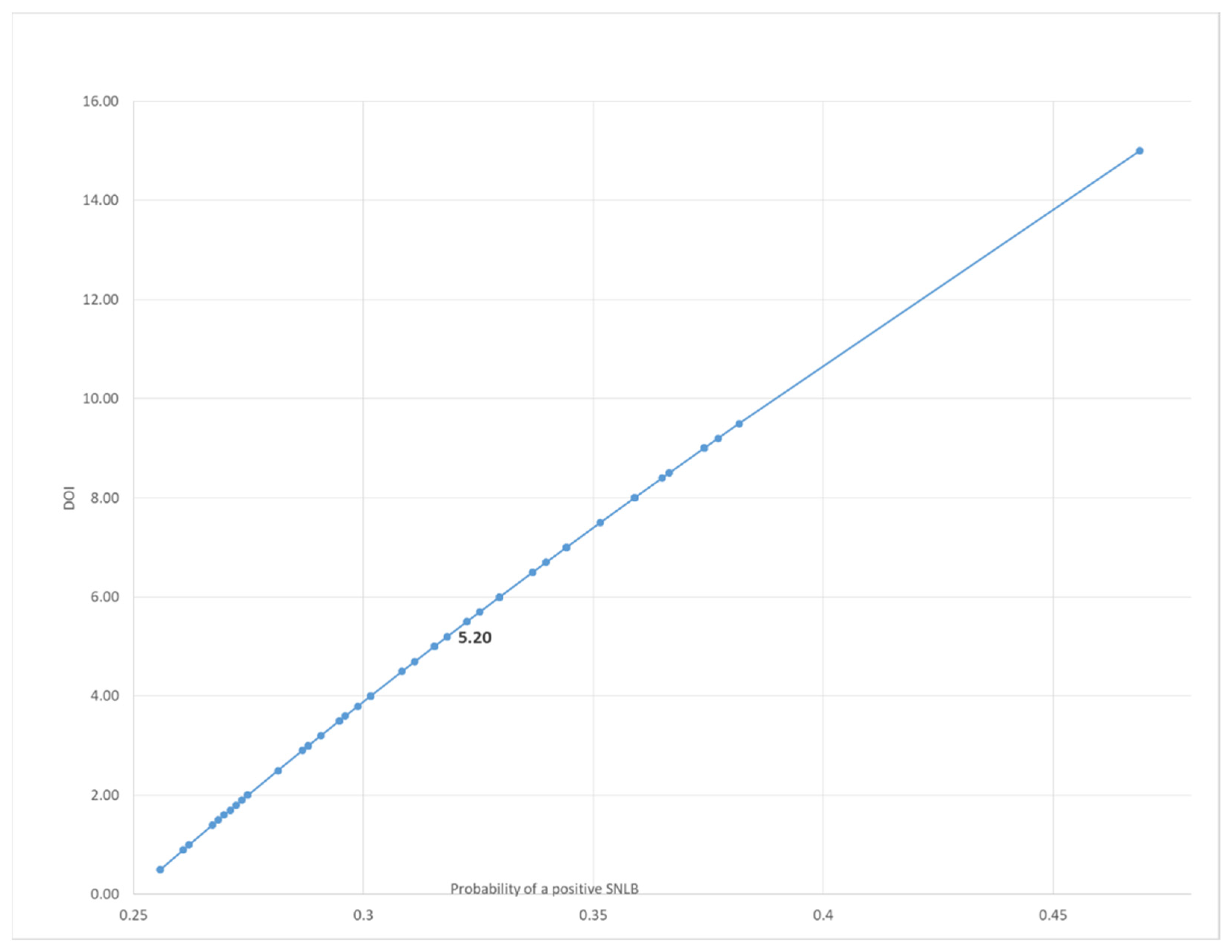
| DOI | 0.173377001 | 4.78043 × 10⁻⁵ |
| PLR | −0.001428808 | 0.50874393 |
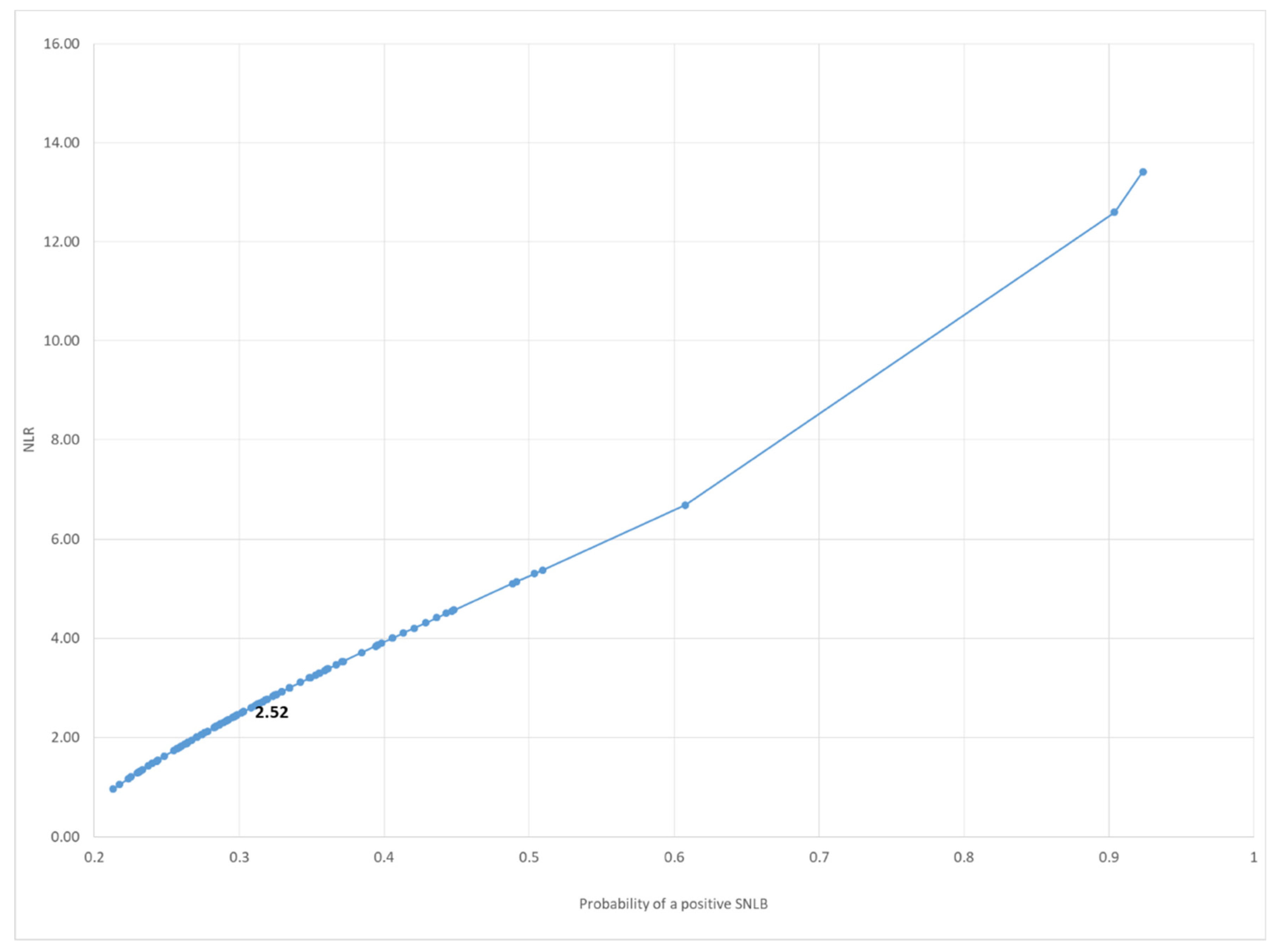
| NLR | 0.130388283 | 0.018422181 |
| SII | 0.000190121 | 0.690418221 |
| Linear Regression Model | Coefficients | p-Value |
|---|---|---|
| Intercept | 2.074314876 | 0.002333873 |
| DOI | 0.115348849 | 0.149478113 |
| PLR | 0.001657338 | 0.652856978 |
| NLR | 0.179003139 | 0.016464027 |
| SII | −0.00010145 | 0.879207812 |
| WPOI | Positive SLNB | Negative SLNB | Probability | 95% CI | p-Value at Chi Square Test |
|---|---|---|---|---|---|
| 1 | 2 | 7 | 10.2% | 3.9–23.8 | 0.003 |
| 2 | 5 | 24 | 18.3% | 10.3–30.5 | |
| 3 | 7 | 25 | 30.7% | 22.1–40.9 | |
| 4 | 13 | 10 | 46.6% | 34.2–59.4 | |
| 5 | 8 | 5 | 63.2% | 43.2–79.5 |
Publisher’s Note: MDPI stays neutral with regard to jurisdictional claims in published maps and institutional affiliations. |
© 2022 by the authors. Licensee MDPI, Basel, Switzerland. This article is an open access article distributed under the terms and conditions of the Creative Commons Attribution (CC BY) license (https://creativecommons.org/licenses/by/4.0/).
Share and Cite
Salzano, G.; Togo, G.; Maffia, F.; Vaira, L.A.; Maglitto, F.; Committeri, U.; Fusco, R.; Maglione, M.G.; Nocini, R.; De Luca, P.; et al. Early-Stage Oral Tongue Squamous Cell Carcinoma and a Positive Sentinel Lymph Node Biopsy: Description of a Prognostic Correlation between Pre-Treatment Inflammatory Biomarkers, the Depth of Invasion and the Worst Pattern of Invasion. J. Pers. Med. 2022, 12, 1931. https://doi.org/10.3390/jpm12111931
Salzano G, Togo G, Maffia F, Vaira LA, Maglitto F, Committeri U, Fusco R, Maglione MG, Nocini R, De Luca P, et al. Early-Stage Oral Tongue Squamous Cell Carcinoma and a Positive Sentinel Lymph Node Biopsy: Description of a Prognostic Correlation between Pre-Treatment Inflammatory Biomarkers, the Depth of Invasion and the Worst Pattern of Invasion. Journal of Personalized Medicine. 2022; 12(11):1931. https://doi.org/10.3390/jpm12111931
Chicago/Turabian StyleSalzano, Giovanni, Giulia Togo, Francesco Maffia, Luigi Angelo Vaira, Fabio Maglitto, Umberto Committeri, Roberta Fusco, Maria Grazia Maglione, Riccardo Nocini, Pietro De Luca, and et al. 2022. "Early-Stage Oral Tongue Squamous Cell Carcinoma and a Positive Sentinel Lymph Node Biopsy: Description of a Prognostic Correlation between Pre-Treatment Inflammatory Biomarkers, the Depth of Invasion and the Worst Pattern of Invasion" Journal of Personalized Medicine 12, no. 11: 1931. https://doi.org/10.3390/jpm12111931