Review

Clinical Improvement in Depression and Cognitive Deficit Following Electroconvulsive Therapy

by
Ahmad Mus’ab Ahmad Hariza
1,
Mohd Heikal Mohd Yunus
1,*,
Jaya Kumar Murthy
1 and
Suzaily Wahab
2
1
Department of Physiology, Faculty of Medicine, UKM Medical Centre, Jalan Yaacob Latiff, Bandar Tun Razak, Kuala Lumpur 56000, Malaysia
2
Department of Psychiatry, Faculty of Medicine, UKM Medical Centre, Jalan Yaacob Latiff, Bandar Tun Razak, Kuala Lumpur 56000, Malaysia
*
Author to whom correspondence should be addressed.
Diagnostics 2023, 13(9), 1585; https://doi.org/10.3390/diagnostics13091585
Submission received: 20 March 2023 / Revised: 17 April 2023 / Accepted: 24 April 2023 / Published: 28 April 2023
(This article belongs to the Section Clinical Laboratory Medicine)

Abstract

:
Electroconvulsive therapy (ECT) is a long-standing treatment choice for disorders such as depression when pharmacological treatments have failed. However, a major drawback of ECT is its cognitive side effects. While numerous studies have investigated the therapeutic effects of ECT and its mechanism, much less research has been conducted regarding the mechanism behind the cognitive side effects of ECT. As both clinical remission and cognitive deficits occur after ECT, it is possible that both may share a common mechanism. This review highlights studies related to ECT as well as those investigating the mechanism of its outcomes. The process underlying these effects may lie within BDNF and NMDA signaling. Edema in the astrocytes may also be responsible for the adverse cognitive effects and is mediated by metabotropic glutamate receptor 5 and the protein Homer1a.

1. Introduction

Major depressive disorder (MDD) is a debilitating illness that affects one out of five people at some point in their lives [1]. MDD is associated with higher treatment costs across all age groups, especially when presented as a comorbidity [2]. While the DSM-IV defines MDD as the presence of a defined set of symptoms that persists for nearly 2 weeks [3], it also encompasses a number of subtypes based on severity, onset, and clinical features [1].
Electroconvulsive therapy (ECT) is a procedure that uses an electrical current to induce seizures in patients. The seizures are then followed by a clinical remission of the psychiatric symptoms. The seizures induced by ECT are typically generalized tonic–clonic (grand mal) seizures [4]. The tonic phase occurs first; clinically, this is presented as a stiffening of the body that lasts for a brief few seconds. It is subsequently followed by the clonic phase, which involves shaking or jerking of the body and lasts for tens of seconds [5]. Although ECT is used to induce seizures, it does not typically cause epilepsy in patients [6].
ECT has long been regarded as a safe and effective treatment option for clinical depression, especially in patients who are resistant to antidepressant medications [7]. When used in conjunction with antidepressants, it produces better results compared to pharmacotherapy management alone [8,9,10]. Other types of depressive disorders, such as suicidal depression, bipolar depression, psychotic depression, depression in the elderly, and postpartum depression, have benefited from ECT as well [11,12]. ECT is also indicated for bipolar disorders, treatment-resistant schizophrenia, catatonia, and neuroleptic malignant syndrome [13]. The administration of ECT alone or in combination with antidepressant medications is more effective in reducing depressive symptoms than antidepressant medications alone [14]. Continuing ECT with adjunct pharmacotherapy after an acute ECT course reduced the likelihood of relapse and recurrence of depression for up to a year [8]. According to a meta-analysis study, it is a more effective treatment for depression compared to repetitive transcranial magnetic stimulation, another non-pharmacological treatment for major depression [15].
The clinical benefits of ECT also come with some adverse effects, such as cognitive deficit [16]. Although the current trend in ECT practice is to reduce the cognitive side effects and maximize the therapeutic effects [11], the mechanism of ECT remains poorly understood. The interplay between neurogenesis and the inflammatory process is implied in the therapeutic mechanism [17,18,19], but little is known about their relation to the cognitive outcome.
The hippocampus is involved in memory and emotion regulation [20]. It can be anatomically divided into the hippocampus proper, which includes the cornu ammonis subfields (CA1 to CA3), and the dentate gyrus (DG), which includes a molecular layer, granule cell layer, and hilus (also known as CA4). Functionally, the hippocampus can be divided along the longitudinal axis into the anterior, middle, and posterior segments. The posterior hippocampus provides spatial learning, memory, and perceptual functions, while the middle segment is involved in cognitive functions [21,22]. The anterior hippocampus regulates the emotional and affective processes [23]. The hippocampal role in emotion regulation is due to its extensive connectivity with the amygdala and the prefrontal cortex (PFC). The PFC increases hippocampal activity during memory elaboration, with memories eliciting stronger effects carrying greater activity [24].
The hippocampus is central to the pathophysiology of depression. For example, chronic stress, which is the often-cited cause of depression, causes glucocorticoid dysregulation, resulting in hippocampal dysfunction [25]. Diminished monoamine neurotransmitters such as norepinephrine, serotonin, and dopamine in regions such as the hippocampus, cerebral cortex, and midbrain have also been implicated in the mechanism of depression [26]. The hippocampus is also a component of the brain’s default mode network and salience network. Depression symptoms are associated with hyperactivity in the default mode network and hypoactivity in the salience network [27,28,29,30]. While hippocampal changes are known to occur following ECT, little is known about their relationship to clinical and cognitive outcomes.
Numerous works in the literature have discussed the effects of ECT and the neurogenesis mechanism that underpins these effects, but very little has been written about the cognitive side effects and their mechanisms, especially given the current state of knowledge. In a recent commentary by Cavaleri and Bartoli, despite the various biomolecular mechanisms implied in the therapeutic and cognitive outcomes of ECT, much of what is known is still very little, due to the fact that most related studies are limited by their small sample size and methodological heterogeneity [31].
This review attempts to highlight the various changes caused by ECT on various brain structures, with an emphasis on the hippocampus, and to form a narrative as to how the changes are related to the therapeutic and cognitive effects of ECT and the potential mechanism underlying these effects. Literature searches were conducted by inputting relevant keywords into several different databases. Studies are limited to within 5 years, but older studies were included to support the narrative.

2. ECT Alters the Brain Structures

2.1. Structural Changes in the Hippocampus in Depression and Following ECT

The hippocampus is susceptible to dystrophic changes caused by depression, which is mediated by chronic stress. When measuring morning serum cortisol levels in MDD patients, it was discovered that higher levels of cortisol were associated with lower volumes of the hippocampal subfields, especially on the right side [32]. Children and adolescents also suffer from the adverse effects of early life stress, which may lead to depression. In a study measuring hair cortisol levels in children, higher hair cortisol levels were significantly associated with reduced volumes in the CA3 and DG, but not in the CA1 or subiculum [33]. Higher perceived stress in adolescents was associated with reduced total hippocampal volume and the left hippocampal volume [34]. Early life stress and greater depression severity in children were also linked to smaller hippocampal volumes [35,36,37]. Other inflammatory markers, such as IL-6 and CRP, were also elevated in MDD patients, but they had no association with changes in the hippocampal subfields [38]. In a preclinical study, rats subjected to chronic unpredictable restraint stress for 4 weeks had a smaller total hippocampal volume. This reflects volume reductions in all subfields in both the dorsal and ventral hippocampus [39].
ENIGMA meta-analyses of magnetic resonance imaging (MRI) studies on subcortical structures in MDD patients revealed significantly reduced hippocampal volumes in MDD patients, of which the effect was driven by recurrent MDD [40]. Other studies found MDD to be related to either left, right, or both hippocampal volume reductions [41,42], with a more substantial loss in the left hippocampus of MDD and bipolar disorder (BD) patients [43]. Variations in the findings may be attributed to factors such as the patients’ age or sex, the different spectra of depressive illnesses, or the duration of the illness. Within the hippocampal subfields, volume effects were confined to the principal subfields such as CA1-CA4, DG, and the subiculum, which were more pronounced on the left side and in recurrent depression [44]. While recurrent depression had a more marked reduction in the CA1 area, only the CA2-CA4 areas were reduced in the first episode of MDD [44], which may be due to a reduction in the bilateral parahippocampal gyrus [45]. A meta-analysis of voxel-based morphometry (VBM) studies revealed smaller gray matter volume in the left hippocampus in MDD and BD patients relative to the control group [43]. Adolescent BD patients with a history of suicide attempts had more GMV reduction in the bilateral hippocampus compared to adolescent MDD patients with a history of suicide attempts [46]. Despite being generally associated with volume reductions, the volume of the hippocampal tail was found to be larger in MDD patients compared to healthy controls. This correlated positively with clinical remission, implying that it could be used as a biomarker for antidepressant treatment [47].
ECT induces structural changes in the hippocampus. Longitudinal imaging studies in depressed patients receiving ECT treatment revealed a significant volumetric increase in the medial temporal lobe regions [48,49,50,51], where the hippocampus resides. ECT has been reported to significantly increase hippocampal volume [49,52]. T1 images obtained from the 3T MRI scanner revealed a significant volume increase in most of the hippocampal subfields, including both left and right DG [53], the right GCL and ML [54,55], the left GCL, the right CA3 and CA4, and the right subiculum [54], as well as the right hippocampal–amygdala transition area [55]. The volume increase in the right CA4 and DG distinguishes remitters from non-remitters [56], as do neuroplastic changes in the neural fiber connections between the hippocampus and other brain regions [57]. While ECT resulted in a significant volume increase in all subfields except the right fimbria [53], another study employing a more powerful MRI at 7T determined that significant volume increases occurred exclusively within the DG, while the other subfields were unaffected [58]. Although this study reaffirms that neuroplastic changes, specifically neurogenesis, occur solely in the DG as a result of ECT, other processes may be attributed to other subfields or regions, namely synaptogenesis or edema.
An interesting disparity was seen between the CA1 subfield, which is affected most by depression, and the DG, which receives a robust volume increase after ECT. As the CA1 is involved in the hippocampal consolidation and retrieval of episodic and autobiographical memory [59], and the DG is the site of adult hippocampal neurogenesis, the relationship between these sites in the context of depression and the ECT mechanism should be further explored.

2.2. Structural Changes in the Amygdala in Depression and Following ECT

Similar to the hippocampus, the amygdala is associated with emotional dysregulation, and has structural changes in depression [60]. According to a meta-analysis, patients with their first episode of MDD who presented with a higher Hamilton Depression Rating Scale (HDRS) score were more likely to have reduced GMV in the right amygdala [45]. An earlier meta-analysis revealed that an earlier age of onset of MDD was linked to a smaller amygdala volume [40]. However, there is evidence stating otherwise. Treatment-resistant depression (TRD) patients had higher GMV in both the right and left amygdala than non-TRD patients [61]. As this difference was not related to medication or bipolar or unipolar features, it may reflect a vulnerability to chronicity [61]. Increased right amygdala volume may occur first in the early stages of the disease, which then progresses to an increase in both as the illness’s chronicity increases. Another meta-analysis determined that depressed patients with comorbid anxiety showed significantly larger amygdala volumes [62]. This may be attributed to the amygdala being the site for the emotion processing of fear and anxiety [63]. Mice subjected to chronic restraint stress (CRS) experienced the persistent activation of projecting neurons from the basolateral amygdala to the ventral hippocampus, which is correlated with stress-induced anxiety-like behavior [64]. From this, it can be deduced that amygdala involvement in MDD is correlated with anxiety symptoms. The involvement of both the right and left amygdala may also differ with the illness’s chronicity, with only the right amygdala experiencing a volume increase in the early stages of the illness and then both sides increasing as the illness progresses.
As ECT influences morphometric changes in the medial temporal regions, other structures within these regions besides the hippocampus are also affected. The parahippocampus, insula, fusiform gyrus, and the amygdala are among these structures [48,49]. The amygdala, in particular, received similar volume increments together with the hippocampus [48,51,65], as both sites are associated with emotional processing and memory [51]. Within the amygdala, these changes were localized in the nuclei within the basolateral and centromedial amygdala and the corticoamygdaloid transition area [55,65]. In addition, VBM and resting state functional connectivity analysis revealed increased GMV in the left superficial amygdala, as well as enhanced connectivity between the left amygdala and the left fusiform area [66]. Similarly, a high-density EEG study revealed significantly increased outgoing functional connectivity in the right amygdala in patients with moderate to severe depression compared to healthy controls [67].

2.3. Structural Changes in Other Brain Regions in Depression and Following ECT

Multiple systematic reviews and meta-analyses have determined cortical thinning in MDD patients, including the bilateral orbitofrontal gyrus [68,69,70], the left calcarine fissure and lingual gyrus [70], the middle temporal gyrus [68,69], the gyrus rectus extending into the right striatum [45,68], the dorsolateral aspect of the right superior frontal gyrus, the medial aspect of the left superior frontal gyrus, and the left superior parietal gyrus [45]. Increased cortical thickness was also found in the anterior and posterior cingulate cortex, as well as the ventromedial PFC [68,69]. Another meta-analysis found that while both MDD and BD patients had lower GMV relative to healthy controls in areas such as the dorsomedial and ventromedial PFC, the anterior cingulate cortex (ACC), and bilateral insula, MDD patients had a more robust GMV reduction in the right dorsomedial PFC compared to the healthy controls. This is in addition to a reduction in the cerebellar, temporal, and parietal regions [43]. Both MDD and BD patients were discovered to have lower GMV in the left ACC when compared to healthy controls, and MDD patients had smaller GMV in the left superior frontal gyrus and the left ACC when compared to BD patients [71]. Besides the cortical changes, volume reductions were also found in the putamen, pallidum, and thalamus of MDD patients without comorbid anxiety [62].
Changes in the brain after ECT were also found in other brain regions besides the medial temporal region. One meta-analysis noted that volumetric increases after ECT occurred in the cortical and subcortical areas [72]. An imaging study showed a similar volumetric gray matter area increase in both general areas [73], implying a widespread effect on the brain. The specific areas that are noted to have a significant increase include the bilateral inferior temporal cortices, the supramarginal gyrus extending to the postcentral gyrus, the middle part of the cingulate gyrus, the right anterior cingulate gyrus and the right subgenual anterior cingulate gyrus, the left insula, and the left fusiform gyrus [48,49,74]. Other notable changes include increased volume in the left superior and inferior temporal gyrus, as well as hypergyrification in the left middle temporal gyrus [74,75]. There are also regions that are spared from any changes post-ECT, namely the brain stem and the bilateral cerebellar cortex [76].

2.4. Influence of Electrode Placement and Number of ECT Sessions on Brain Structural Changes

The magnitude and laterality of the structural changes in the brain are mediated by electrode placement. While bilateral electrode placement has been shown to increase the volume of certain hippocampal subfields on both sides [53,54], it was also reported by other studies to produce a larger volume increase in the right hippocampus and its subfields [48,56,77]. Bitemporal electrode placement was noted to differ from bifrontal placement in that it produces greater direct electrical stimulation of deep midline structures, the left and inferior temporal regions, the central and lateral sulci, and the ventral parts of the brain. It also affected a larger area of the left hemisphere, extending to the inferior frontal cortex, the entire left temporal lobe, and a large part of the left parietal lobe [74]. Studies employing RUL ECT produced a more consistent outcome in which the right hippocampus developed a more significant volumetric increase [49,50,55,75]. Structures deep within the brain or that are further away from the electrodes on the scalp may show a proportional decrease in ECT influence on brain matter [76]. As electric signals from the electrode travel across different tissues (skin, cerebrospinal fluid (CSF), and brain matter) with varying conductive properties, any structures further away from the electrode should receive fewer electrical signals compared to those nearer the electrodes, resulting in regional differences in volume changes [78]. Even though this may in part explain the greater volume changes with bilateral electrode placement as it covers more brain regions [79], another factor to consider is the relationship between individual brain structures and their own respective electric fields. A strong relationship was found within the left hippocampus and left amygdala [78], explaining why these structures changed the most with bilateral ECT. The influence of the electric field was supported by a recent study that determined that there is a stark difference in the electric fields between electrode placements. The highest electric fields for bilateral ECT were found in the white matter of both temporal lobes and the frontal lobes, whereas RUL ECT displayed the highest electric fields in the right hemisphere and the corpus callosum [80].
The number of ECT sessions was found to influence hippocampal volume change in a dose-dependent manner. This means that there is an increasing hippocampal volume that correlates with the number of ECTs administered over the course of an ECT treatment series [75]. This relationship holds true regardless of ECT parameters such as electrode placement and number of sessions, as well as the current, frequency, and duration of the electrical stimulation [75,81]. Another study, which only included subjects who received RUL ECT, found a dose-dependent effect between the strength of the electric field and hippocampal volume change [78]. The strength of the electric field is related to the number of ECT sessions and is also influenced by electrode placement, since RUL ECT produces a more robust volume increase that is lateralized to the right side [78]. It is worth noting that the dose-dependent relationship between the electric field and hippocampal neuroplasticity may be confined to a specific range between 30–40 V/m and 100 V/m [78]. This phenomenon, termed “neuroplasticity threshold,” means that a minimum electric field of 30–40 V/m is required for induction of neurogenesis, while any value above 100 V/m represents a less robust dose-response relationship and may be unrelated to further volumetric increase and antidepressant response [78]. Thus, there is a dose-response relationship between the number of ECT administrations and the increase in hippocampal volume, which is influenced by the electric field and the electrode placement modality.

2.5. The Relationship between Post-ECT Volume Changes and Clinical Outcome

Several studies have pointed out a significant association between post-ECT volumetric changes and improved clinical outcomes. Increased volumes of both the hippocampus and amygdala with ECT are related to improved depression symptoms [65,72,73]. Among the hippocampal subfields, a positive correlation with improved clinical outcome was found to be localized within the DG [58], although other subfields were also mentioned to have similar correlations, such as the CA3, CA4, and subiculum [54,56]. A positive correlation with improved symptoms was found to occur within almost all of the hippocampal subfields, except the right subiculum and fimbria, and the left presubiculum, parasubiculum, fimbria, and the hippocampal tail [53]. These findings supported the idea of hippocampal neuroplasticity as the mechanism necessary for improved clinical outcomes.
In contrast with the previous statement, there is also evidence that suggests that post-ECT volumetric changes may not be correlated with improved clinical outcome. A few meta-analyses determined that volume changes in the hippocampus after ECT had no correlation with reduced depression symptoms [52,82]. Similarly, one study found no link between an increase in left amygdala volume after ECT and an antidepressant response [78]. It seems that the volumetric increase of gray matter in the cortical and subcortical areas [79], especially in the temporal regions [50], had no association with any clinical improvement. Patients with greater treatment-related volume increases had poorer outcomes, though these effects were not significant after controlling the number of ECTs given to these patients [75]. These findings challenge the notion of hippocampal and amygdala volume increase following ECT as the underlying mechanism of clinical improvement. Additionally, when taking the influence of electrode placement and the electric field into account, stronger electric fields in the temporal lobes were associated with poorer clinical responses in bilateral ECT, while no differences in electric field distributions were found between RUL ECT responders and non-responders [80]. It could be inferred here that the greater electrical stimulation of bilateral ECT on the temporal lobe structures, particularly the left hippocampus and amygdala, may reach a certain threshold that impedes clinical remission. It could also explain why a greater increase in hippocampal volume post-ECT was associated with a poorer outcome [75].

2.6. The Relationship between Post-ECT Volume Changes and Cognitive Outcome

A growing body of evidence demonstrates that impaired cognitive performance occurs alongside a volume increase in the brain following ECT (Table 1). Enhanced cortical neuroplasticity after ECT has been linked to both clinical remission and impaired delayed memory, more specifically in the left inferior parietal gyrus and the right inferior temporal gyrus [83]. Increased total hippocampal volume and its subfields in depressed patients who underwent bitemporal ECT were shown to significantly correlate with a reduction in cognitive performance and worsening verbal memory immediately after completing their treatment [17,84]. However, after a 6-month follow-up, there was a reduction in the hippocampal subfields and improved cognitive performance. Improved delayed verbal memory was significantly associated with volumetric reduction in the right hippocampus and its subfields, namely the right DG, the right molecular layer, the right CA3, and the presubiculum, as well as the left subiculum [17]. Another study on RUL ECT noted that a larger absolute right hippocampal volume change is associated with less improvement in visual memory when assessed 6 months after completing the ECT course [85]. While this may indicate that absolute left hippocampal volume change was not significantly correlated with cognitive change [85], an earlier study explained that the greater clinical remission and cognitive deficit that occurred in bilateral ECT were due to greater direct electrical stimulation of the left hippocampus and inferior frontal cortex [74]. In a recent cohort study by Argyelan et al., it was reported that hippocampal enlargement following ECT is associated with poorer cognitive outcomes, regardless of diagnoses, electrode placements, and number of ECT sessions [86].
Another aspect worth mentioning is that hippocampal changes following ECT are transient. After the initial volume increase following ECT, the hippocampal volume gradually shrinks within 10–36 months after ECT has ended [77], though it may also occur as early as within the first 3 months [56]. As cognitive deficits after ECT are also transient, it is possible that changes in hippocampal volumes after ECT and cognitive impairment have some degree of relationship. In addition, impaired cognitive performance after ECT was also correlated with reduced resting-state functional connectivity (RSFC) of the hippocampal middle subregion with the bilateral angular gyrus [21]. Because the angular gyrus is involved in memory functions [87], RSFC reduction implies impaired connectivity between the hippocampus and the angular gyrus, which contributes to impaired cognition after ECT [21]. Taken together, these results showed a possible dual outcome of ECT-induced hippocampal volume increase, in which the mechanism necessary for the volume increase is also responsible for the cognitive deficit [84]. A possible explanation is that ECT is a potent stimulator of neurogenesis, which may increase the likelihood of causing aberrant or excessive neurogenesis that could disrupt hippocampal function [17]. The fact that both volume change and cognitive deficit are influenced by electrode placement and are temporary strengthens this argument.

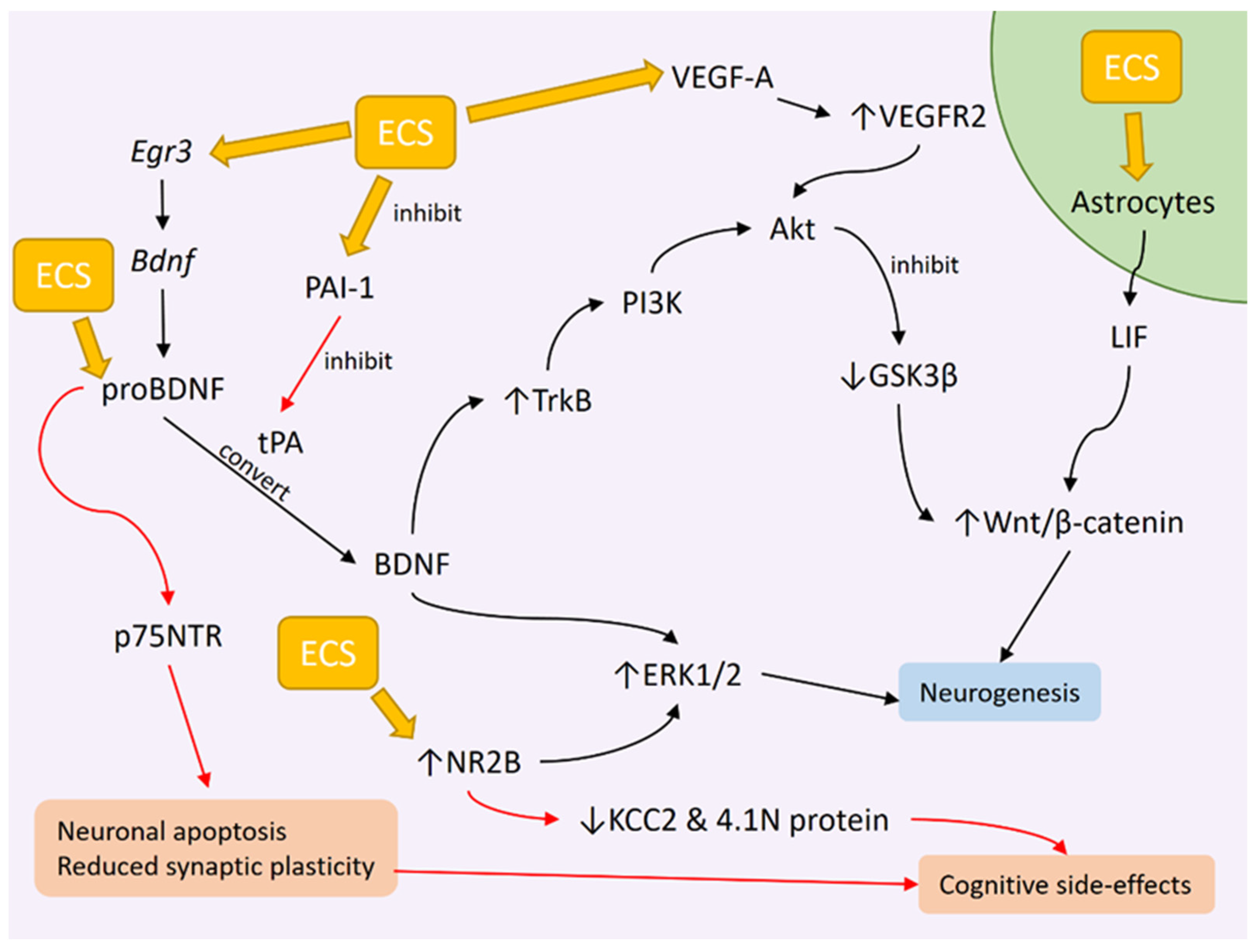
3. ECT Induces Neurogenesis

3.1. ECS Promotes Hippocampal Neurogenesis in Animals

Structural neuroplasticity, especially pertaining to the hippocampus, has been explored in preclinical studies. Rats subjected to CRS displayed depressive behavior, but this was not reflected by any significant change in the rats’ hippocampal volume or in the total number of neurons [88]. In a later study, only the hilus showed a significant volume reduction [89]. The intervention of electroconvulsive stimulation (ECS), the animal equivalent of ECT, alleviated the depressive behavior. While ECS does increase neurogenesis in the hippocampal subgranular zone, it does not alter the total number of neurons or the volume of the hippocampus and its subdivisions [88,89], which may indicate damaging effects from ECS administration. On the other hand, ECS does stimulate neurogenesis, as indicated by the formation of new neuronal cells within the hippocampus [88,90].
The implied relationship between an increased number of ECT administrations and the magnitude of volumetric change in the hippocampus has been proven to occur on a cellular level. An increased number of ECSs given to rats produced more newborn neurons within the hippocampus [91]. However, ECS-induced neurogenesis was not fully efficient since there was a decline of around 40% of the newborn cells stained with the BrdU marker from day 1 post-ECS until 3 months, after which no further decline in newborn neuronal cells was observed for up to 12 months [90]. This early period of the first 3 months after ECS may be crucial for the survival of the newborn neuronal cells. Another study found that the number of cells stained with Ki-67 and NeuroD increased 1 day after ECS administration. However, after 8 days, the number of Ki-67-positive cells reduced while the number of NeuroD-positive cells increased [92]. Ki-67 is an intrinsic marker for recent cell proliferation which is expressed in all proliferating cells [93] and is used to identify the early stages of adult neurogenesis [94]. NeuroD is a transient transcription factor restricted to developing neurons that is essential for the survival and maturation of adult-born neurons [95].
ECS also has varying effects on the cell cycle. During the differentiation phase, ECS was found to increase cell survival, promote the differentiation of newborn cells, and significantly increase the expression of the immature neuronal marker calretinin. Conversely, ECS significantly suppressed the expression of the mature neuronal marker calbindin in newborn neurons during the late maturation phase [96]. It is apparent that ECS has a prominent impact on hippocampal neurogenesis. However, not all newborn neurons survive and mature before being incorporated into the functional hippocampal network. Hence, in order to improve neurogenesis potential, the effects and relationships between the various aspects of neuronal cell synthesis may be further explored.
Regarding the connectivity of new neurons formed after ECS, studies are rather sparse, even in animals. However, similar information could be extrapolated from status epilepticus (SE) studies. New neurons formed 2 weeks after SE was induced in rats were able to survive for up to 6 months and constitute a significant portion of the population of mature granule cells in the DG [97]. Although these neurons are integrated into the hippocampal neurocircuitry, they replace the pre-existing neurons rather than add to the population [97]. Neuronal degeneration was noted to occur in the DG, CA1, and CA3, alongside neurogenesis, after SE was electrically induced in rats in another study [98]. The electrophysiology of the new neurons formed after SE showed reduced excitatory and increased inhibitory synaptic input [98]. Additionally, neurons born after partial SE tend to have altered synaptic scaffolding proteins and adhesion molecules expression that favor the reduced excitability of these new neurons [99]. These changes could either reflect mechanisms to attenuate the epileptic dysfunction [98,99] or indicate immature inhibitory transmission due to the transient nature of elevated inhibitory molecules [99]. These findings could imply similar mechanisms occurring post-ECS and may also explain the cognitive outcome.

3.2. ECT Effects on BDNF Levels

The neurotrophic hypothesis of depression describes dysfunctional neuronal plasticity as the main mechanism underlying depression development. BDNF is a key component of this mechanism [100]. Rats subjected to unpredictable chronic mild stress (UCMS) display behavioral defects akin to those of depression, and this is accompanied by reduced BDNF concentrations in the CA1 and CA3 regions of the hippocampus [101] and also the PFC [102]. In a clinical setting, patients suffering from TRD displayed low serum BDNF levels [103,104]. Elderly patients with late-life depression displayed a similar reduction in serum BDNF that was significantly correlated with reduced hippocampal volume [105]. Cavaleri et al. noted that MDD patients have lower peripheral and central BDNF levels than non-depressed individuals, which have negative correlations to symptom severity but are not linked with suicidality [106].
Studies exploring serum BDNF changes after ECT produced varying outcomes, which may be partly due to the different methods of measuring peripheral BDNF levels. Serum and plasma BDNF have been explored as potential biomarkers in ECT studies. It was found that depressed patients who had undergone ECT displayed increased BDNF concentration in both their serum [107,108] and plasma [107]. However, serum BDNF is preferred over plasma BDNF. A study revealed no difference in baseline plasma BDNF levels between depressed patients and healthy controls [109]. The post-ECT readings either show no changes in concentrations [109,110] or display variations throughout an ECT course [111]. The tendency of plasma BDNF to display greater standard deviations in measurements [112], in addition to being influenced by the pre-analytical processing of blood samples [113], may lie in the manner in which BDNF is stored in serum and plasma [107]. Human BDNF is primarily stored in the platelets and is released mainly via platelet activation and degranulation during the clotting process. This explains why serum BDNF levels are higher than plasma levels [111]. Therefore, serum BDNF is the preferred method for peripheral BDNF measurement. Another recommendation is to employ serum-to-whole blood concentration of BDNF as a more accurate measurement of peripheral BDNF [114].
Even though increased serum BDNF after acute ECT [110] may indicate that ECT corrected the BDNF changes in depression, several studies have reported a lack of any significant changes in serum BDNF concentrations after ECT [104,105,111]. One study reported an insignificant trend of increasing BDNF [115], while another found that BDNF increased up to a certain value only in ECT non-remitters, with the end value resembling that of baseline remitters and no significant change in ECT remitters [116]. A recent overview of meta-analyses indicated that increases in blood BDNF levels can be seen after pharmacological treatment but are absent in non-pharmacological interventions, including ECT [106].
One possibility for the lack of change is that serum BDNF could have a delayed effect, requiring some time before any significant changes could be detected. According to a meta-analysis study by Luan et al., a significant increase in BDNF occurred one month after the completion of an ECT course [107]. Continuation ECT (c-ECT) is a course of less frequent ECT given after an index ECT course that can last up to 6 months [117]. The addition of c-ECT robustly increased serum BDNF concentrations [110,118], which may also support the idea of a serum BDNF increment time delay. However, the delayed increase in peripheral BDNF may not be attributed to the immediate clinical improvement after ECT, as the peripheral BDNF increase should have preceded the clinical response [104].
An alternative to both serum and plasma sampling is studying the cerebrospinal fluid (CSF). One study measured the BDNF concentration in CSF of MDD patients before and within a week after a course of ECT. The results indicate an increase in BDNF concentration in the CSF, which was absent in the corresponding serum samples [119]. However, the results are preliminary since the study is limited by its small sample size due to the exceptional difficulty in recruiting patients, especially for multiple CSF sampling [119]. Post-mortem sampling of brain tissue, CSF, and plasma from individuals with mood disorders and substance or alcohol abuse disorders indicated a significant positive correlation between BDNF in the brain and CSF, and between BDNF in the brain and plasma [120]. Considering BDNF changes in the serum [107], an increase in BDNF in the CSF may either occur concurrently with an increase in serum BDNF or it may slightly precede the changes in the serum. Nonetheless, future studies may explore the BDNF changes in CSF, serum, or plasma across different time points to validate the correlation.
The preclinical setting allowed for a better look at hippocampal BDNF levels. A study employed Flinders sensitive and resistant line rats, the genetic phenotypes that model depression. ECS significantly increased BDNF protein in both lines, and the increase was associated with a volumetric increase in the stratum radiatum in the CA1 regions in the sensitive line [121]. An earlier study found that ECS mediated the upregulation of BDNF protein in Wistar rats previously subjected to UCMS without altering the number of neurons in the CA1 and DG [122]. This effect was notably absent in the genetic variant [122]. While animal studies may provide a more accurate measurement of central BDNF, more studies are required in order to establish a direct relationship between hippocampal BDNF and volume increase.

3.3. Genetic Changes Influencing BDNF Expression as a Result of ECS

Although clinical studies are lacking, looking into animal models may shed some light on the ECS-mediated genetic changes influencing BDNF expression. Previous studies have established that ECS upregulated several immediate early genes (IEGs) such as Bdnf, Egr1, c-Fos, and Nrn1 [123]. IEGs are genes that are rapidly activated in the brain in response to external stimuli and influence many neurobiological processes. One IEG, the early growth response 3 (Egr3) gene, was discovered as crucial for the expression of BDNF in the DG of the hippocampus in rats. The presence of several Egr3 binding sites in the Bdnf promoter implies that Egr3 directly mediates ECS-induced Bdnf gene expression, specifically exons IV and VI of the Bdnf gene [124]. Another IEG, Narp, was found to be an essential regulator for the ECS’s antidepressant effect. A study using Narp knockout mice discovered that Narp deletion inhibits the antidepressant effect of ECS in mice without blocking seizure activity. While the Narp protein was previously known to be secreted from pre-synaptic terminals to regulate AMPA receptor clustering and mediate BDNF-dependent synaptic modulation [125], Narp deletion neither suppressed hippocampal BDNF expression nor suppressed neurogenesis in the DG. However, it does block the dendritic outgrowth of the immature granule cell neurons. Therefore, although Narp protein does not influence ECS-induced hippocampal neurogenesis via BDNF synthesis, it may regulate the BDNF downstream effects on neuronal survival and maturation [126]. These discoveries add to the growing evidence that BDNF synthesis and the downstream pathway activities are regulated by a complex interplay of various genetic factors.

3.4. ECT Effects on BDNF Downstream Signaling

BDNF performs neurotrophic functions by binding to and phosphorylating its high-affinity receptor, tropomyosin receptor kinase B (TrkB) [127]. TrkB regulated hippocampal neural stem cell proliferation and neurogenesis in mice [128]. Multiple ECS performed in rats enhanced BDNF/TrkB signaling, evident by the increased expression of mature BDNF proteins as well as phosphorylated TrkB receptors in the dorsal and ventral regions of the hippocampus despite the reduced full-length TrkB receptor expression [129]. Increased TrkB signaling seems to occur during the resting period after intense epileptic activity has subsided [130]. In another study on mice, the period of slow EEG upon the discontinuation of N2O was discovered to have antidepressant effects with a concurrent increase in TrkB phosphorylation. This effect was also seen with the discontinuation of ketamine and flurothyl-induced convulsions, which mimic the effects of ECS [131].
Upon TrkB activation, BDNF causes the phosphorylation of protein kinase B (Akt) via the phosphatidylinositol 3-kinase (PI3K) pathway [127]. Both in vivo and in vitro studies have determined that PI3K-Akt signaling is involved in neuronal growth and survival since the upregulation of BDNF, TrkB, PI3K, and Akt proteins promotes axonal regeneration [132] and inhibits neuronal apoptosis [133,134]. While evidence connecting ECT and PI3K is currently lacking, it could be stipulated that ECT may promote PI3K upregulation as shown by its upstream and downstream factors, TrkB and Akt, respectively. In patients with TRD, there is reduced baseline expression of the microRNAs (miRNAs) let-7b and let-7c. Both miRNAs mediate the expression of genes in PI3K-Akt signaling, such as the gene for insulin growth factor 1 receptor (IGF-1R) [135]. IGF-1 is another neurotrophin besides BNDF, and it has been hypothesized that hippocampal BNDF upregulation is dependent on IGF-1 [136]. Although both miRNAs were unaffected by ECT [135], the evidence presented here suggests that depression may be influenced by PI3K-Akt activity.
In a manner similar to TrkB, there is a gradual upregulation of glycogen synthase kinase 3 beta (GSK3β) protein phosphorylation during the rebound of slow brain EEG oscillations upon the discontinuation of the antidepressants ketamine and N2O to produce their antidepressant effects [131]. This phenomenon implies a similar outcome in ECS and possibly ECT upon subsidence of their electroconvulsive activity. GSK3β activity is inhibited upon its phosphorylation by Akt and protein kinase C (PKC). GSK3β itself inhibits Wnt/β-catenin signaling by promoting β-catenin degradation via phosphorylation [137]. Wnt/β-catenin signaling is crucial for hippocampal neurogenesis [138] and protection from neuronal apoptosis [139]. The microRNA mIR-155 induced depression-like behaviors in mice, possibly via the inhibition of Wnt/β-catenin signaling [139]. ECS in mice is proven to affect multiple components of a complex pathway, as ECS promotes Akt activation and PKC and Wnt/β-catenin signaling, besides reducing GSK3β activity by increasing its phosphorylated form [140].
Besides Akt signaling, BDNF is also the immediate upstream regulator of extracellular-signal-regulated kinase 1/2 (ERK1/2) signaling. Reduced ERK1/2 signaling was found in the PFC and hippocampus of both human and animal depression models [141]. ERK1/2 is involved in several neurobiological processes, such as promoting neurogenesis and synaptogenesis in the early stages of life and long-term potentiation in adulthood [142]. Elevated levels of phosphorylated ERK1/2 (p-ERK1/2) and ERK1/2 have been associated with the amelioration of depressive symptoms and improved cognitive performance in rats previously subjected to UCMS [143]. Repeated ECS administration in rats causes an ECS-induced cognitive deficit, which is associated with reduced p-ERK1/2 levels and excessive activation of the N-methyl D-aspartate (NMDA) receptor subunit NR2B [144]. Both ERK1/2 and NR2B seem to be influenced by the number of ECS given. A single, but not repeated, ECS was able to induce a transient increase in p-ERK1/2, and this increase occurred without involving the activation of its upstream factor TrkB [145]. The possible reason given is that a single ECS induces an acute membrane depolarization in the PFC and this eventually leads to the activation of ERK1/2 signaling via increased NMDA activity [145]. In contrast, repeated ECS has been shown to reduce NMDA receptor 1 and increase NR2, with the former being associated with cognitive deficit while the latter is associated with improved long-term memory by neurogenesis [140].
The role of NR2B is somewhat muddled by the fact that increased NR2B activity has been linked to both cognitive dysfunction [146] and improvement [147,148]. For cognitive improvement, the administration of piperlongumine, a compound derived from Piper sp., to aged female C57BL/6J mice improved their cognitive performance. The improvement was attributed to the increased phosphorylation of NR2B, ERK1/2, and CamKIIα in the mouse hippocampus, as well as increased neurogenesis in the DG [147]. Another study used a rat model of hepatic ischemia/reperfusion that displayed cognitive dysfunction features and reduced mRNA and protein expression of NR2B in the hippocampus. Treatment with hydrogen sulfide improved their performance on the Morris water maze test while also increasing the NR2B protein levels [148]. On the other hand, one study exposed rats to the anesthetic isoflurane, which increased calpain-2 expression in the hippocampus secondary to increased NR2B expression [146]. Calpain is a Ca2+-activated neural cellular cysteine protease, and it has the isoforms calpain-1 and calpain-2. While calpain-1 is neuroprotective, calpain-2 has neurodegenerative effects [149]. The increased expression of calpain-2 reduced the potassium-chloride cotransporter (KCC2) and total 4.1N protein expression [146]. Both KCC2 and 4.1N proteins, the latter being a cytoskeletal-associated protein, are significant in the formation of dendritic spines [150], and their suppression results in spatial memory deficits in rats [146]. In turn, NR2B inhibition reduced calpain-2 expression and increased KCC2 and 4.1N protein in the rats, alleviating the spatial memory deficit [146]. NR2B may have multiple pathways leading to both outcomes, which may also be affected by other neurogenic pathways. No study thus far has linked ECT and calpain-2, so it is interesting to see whether calpain-2 has a role in the cognitive side effects post-ECT. A diagram of the effects of ECC on BDNF signaling, based on the results of preclinical studies, is shown in Figure 1.

3.5. Potential Role of proBDNF in Disrupting Cognitive Function Following ECT

Mature BDNF arises from the proteolytic cleavage of its isoform precursor, proBDNF [151], a process regulated by tissue plasminogen activator (tPA) [152]. While mature BDNF promotes neurotrophic effects by binding to the TrkB receptor, proBDNF depresses synaptic plasticity and induces neuronal apoptosis by binding to the neutrophin receptor p75NTR [153]. The protein p75 induces the internalization of the intracellular domain (ICD) into the nucleus, resulting in cell apoptosis [154]. Clinical studies have shown that serum BDNF and tPA levels were lower in depressed patients, while serum proBDNF and p75NTR levels were higher [155,156]. Another study reported that plasma BDNF levels were significantly reduced in patients with BD compared to MDD patients, and that MDD patients had a higher ratio of mature BDNF to proBDNF in the plasma than BD patients or healthy controls. This indicates the mature BDNF to proBDNF ratio may be a differentiating factor between MDD and BD [157].
In preclinical studies, mice exposed to UCMS displayed increased protein expression of proBDNF and p75NTR in the neocortex and hippocampus [158,159]. The chronic administration of corticosterone in mice induced depressive-like behaviors, which are accompanied by an elevated proBDNF level in the ventral DG and a reduced BDNF level in the dorsal and ventral DG [160]. Exogenous proBDNF administration was also found to induce depression-like symptoms. Lin et al. employed an adeno-associated virus (AAV) vector to deliver proBDNF via injection into the bilateral gluteus maximus in mice. The AAV was able to induce depression-like behaviors in mice after about 4 weeks, and the administration of sheep anti-proBDNF antibody was able to alleviate these symptoms [161]. The direct injection of exogenous proBDNF into the hippocampal CA1 in rats via an implanted steel cannula produced depressive-like symptoms, similar to the effect of UCMS [162]. On a cellular level, the exogenous delivery of proBDNF reduced the density of dendritic spines in the hippocampal CA1 region [162], the DG, and also the amygdala [161]. Finally, as proBDNF and its receptor are expressed in neural stem cells, exogenous proBDNF treatment reduced the neural stem cells’ proliferation, migration, and overall viability [163]. Thus, it can be deduced that proBDNF may be a potential mediator in the development of depression symptoms.
ECS in rats was shown to upregulate the expression of mature BDNF and TrkB receptors without altering the overall proBDNF expression in both the dorsal and ventral hippocampus [129]. However, an earlier study demonstrated an upregulation of proBDNF in the CA1 regions following the ECS of depressed rats, alongside increased mature BDNF and tPA levels, as well as an increased proBDNF/mature BDNF ratio. The levels of proBDNF and mature BDNF were previously elevated and reduced, respectively, in these rats after being subjected to UCMS, which may be due to tPA inhibition secondary to increased expression of plasminogen activator inhibitor-1 (PAI-1), as there are increased PAI-1 levels after UCMS [152]. It has been suggested that elevated proBDNF in the CA1 regions causes impaired long-term potentiation and may be responsible for the cognitive deficit following ECT [152]. Increased proBDNF levels after UCMS were associated with impaired cognitive ability, whereas ECS was associated with a further increase in proBDNF levels and a worsening cognitive deficit [152]. Elevated proBDNF and reduced mature BDNF have been associated with altered cognitive functioning in mice following radiation therapy, in which irradiated mice engaged in a cognitive task at a slower pace [164]. However, the irradiated mice did not show any reduction in terms of their engagement with the cognitive task, implying that fatigue after irradiation could also influence the cognitive behaviors [164]. Diminishing cognitive ability, as seen in Alzheimer’s disease (AD), has been associated with elevated proBDNF and p75 activity [154]. CSF from AD patients was proven to induce apoptosis in cultured hippocampal neurons, and this effect was mediated by proBDNF found in the CSF [154]. Taken together, the role of proBDNF can be investigated further. While it is clear that proBDNF plays a role in depression, both proBDNF and mature BDNF were also increased after ECS in rats. The exact mechanism by which both proBDNF and mature BDNF increase concurrently after ECS is unknown, but other mechanisms besides the tPA system might be involved in temporarily elevating proBDNF after ECT. It could also explain why proBDNF is elevated in both depression and after ECT. Further studies are warranted to solidify the role of proBDNF-p75 signaling in cognitive deficit, as well as to determine whether there is a temporal change in this signaling that coincides with the temporal changes in cognitive ability following ECT.

4. VEGF Changes after ECT

Another growth factor of interest besides BDNF is the vascular endothelial growth factor (VEGF). Originally known as an angiogenic factor, VEGF also exhibited neurotrophic properties. It has been linked to depression since patients given anti-VEGF medications exhibited depression and anxiety behaviors [165,166]. In an animal study, VEGF treatment was able to ameliorate depression-like behavior in mice [167]. According to a review by Deyama and Duman, ketamine, an NMDA receptor antagonist, produced robust antidepressant effects that were associated with increased BDNF and VEGF expression in the medial PFC and hippocampus [168]. However, clinical studies have produced inconsistent results. Some studies reported no difference in blood VEGF levels before ECT between depressed patients and healthy controls [169,170], while another found lower peripheral VEGF in TRD patients compared to healthy controls [103]. A similar finding was also found in the CSF of TRD patients [171]. On the other hand, several meta-analysis studies pointed out that blood VEGF levels in MDD patients were significantly higher compared to healthy controls [172,173,174]. Higher baseline VEGF levels were also found in treatment-resistant bipolar depression patients [175]. A plausible explanation for the difference in VEGF levels between TRD and non-TRD is that higher VEGF levels in non-TRD are reflective of the brain’s neuroprotective mechanism in response to stress. This mechanism is absent in TRD patients, preventing antidepressant medication response [103]. This is evident from lower VEGF levels that were also seen in treatment-resistant schizophrenia patients; they were increased following ECT [170,176]. Plasma VEGF samples taken 1–3 days after ECT treatment were shown to be unaltered [169]. However, a later study showed that there was an increment in plasma VEGF level when taken 2 h after ECT, and the level decreased when taken 4 h post-ECT [177]. Hence, it could be deduced from here that increased VEGF levels after ECT might be temporary, but VEGF has some degree of involvement in the mechanism of ECT.
Among the VEGF mammalian subtypes, VEGF-A is of particular interest. VEGF-A induces vascular changes such as promoting angiogenesis, vasodilation, and increased vascular permeability. It also possesses neurotrophic actions in that it induces neurogenesis via its receptor VEGFR2 and also promotes neuronal survival via the PI3K-Akt pathway and the mitogen-activated protein kinase (MAPK) pathway [178]. While VEGF is expressed in different cell populations in the hippocampus, VEGFR2 is specifically expressed in the pyramidal neurons of the CA3 subfield, as revealed in an animal study [179]. VEGF-A/VEGFR2 signaling also promoted axonal branching in the rats’ hippocampal neurons [179]. Another study revealed that VEGF-A/VEGFR2 signaling mediates neural stem cell proliferation and migration in immature rats after SE [180]. The increased expression of the VEGF-A downstream proteins VEGFR2 and phosphorylated Akt in the hippocampus following SE supports this [180]. Thus far, there have been no studies investigating the effects of ECS on VEGF-A. The aforementioned findings in animal studies contradict a recent clinical study. Peripheral mRNA expression of VEGF-A was found to be higher in patients with psychotic and non-psychotic depression compared to healthy controls, which was then reduced following ECT [181]. However, the small sample size, as well as ongoing medications that were taken by patients during the study, may confound this outcome [181]. Regardless, VEGF-A is a promising target for future ECT-related studies. Figure 1 depicts the role of VEGF-A in neurogenesis following ECT.
Additionally, it is worth mentioning the study by Van Den Bossche et al. regarding genetic polymorphisms influencing changes in hippocampal volume after ECT. They noted that the expression of VEGF can be regulated by certain single-nucleotide polymorphisms (SNPs), particularly rs699947 in the promoter region of VEGF. It is possible that more C alleles at rs699947 lead to higher VEGF expression following ECT, resulting in a higher increase in hippocampal volume. The association between SNP and hippocampal volume change is not found in rs6265 for BDNF [182]. Genetic studies may provide a further understanding of the role of VEGF in the ECT response.

5. ECT and Cerebral Edema

5.1. Compromised Neuronal Integrity Post-ECT

According to diffusion tensor imaging (DTI) studies, white matter structure can be affected by ECT. A reduction in hippocampal fractional anisotropy (FA) occurred a week after ECT [115], and is associated with seizure duration [183]. As FA is an index measurement of white matter integrity [184], a reduction in FA may indicate compromised white matter integrity as a result of ECT. A lower FA was also found in depressed patients with childhood trauma [185], as well as reduced FA in the peripheral neurons of patients with spinal cord injury [186]. Both indicate neuronal dysfunction with reduced FA, and a reduction in FA following ECT could indicate potential cognitive dysfunction due to compromised neuronal integrity. In contrast, other studies showed no significant changes in FA following ECT [57,187]. However, FA is not always a reliable marker of white matter integrity [184], as it is a non-specific measure of diffusion. Other diffusion metrics, such as mean diffusivity (MD), axial diffusivity (AD), and radial diffusivity (RD), can change without significantly affecting FA [57].
Another marker of neuronal integrity is N-acetyl aspartate (NAA), obtained from proton magnetic resonance spectroscopy (H-MRS) imaging. Reduced NAA levels were found in the left hippocampus of depressed patients [188] and in youths with TRD [189], as well as in the posterior cingulate cortex in elderly patients with late-life depression [190]. Post-ECT measurements indicate that these changes were not corrected with ECT, as evidenced by reduced NAA concentrations in the left medial temporal lobe [76], the dorsal anterior cingulate cortex [188], and the PFC and orbitofrontal cortex [191] after ECT treatment. Interestingly, NAA was also decreased in the right hippocampus following RUL ECT, which occurs in depressed patients with an already-reduced NAA in the left hippocampus [188]. In a recent review, it was revealed that NAA reduction provided the strongest evidence of neurochemical alterations with ECT [192]. This evidence further supports the notion of disturbed neuronal integrity following ECT. It is possible that NAA reduction is a predominant factor in the stimulation of subsequent neurotrophic processes [188] or that it reflects a transient neuronal loss that leads to cognitive side effects due to PFC dysfunction [191]. In patients with bipolar type II depression, reduced NAA relative to creatinine ratios in the bilateral prefrontal white matter were associated with impaired cognitive performance [193].

5.2. Could ECT Result in Cerebral Edema?

Measurements of other DTI indices post-ECT point to a predisposition for increased neuronal diffusivity in the right hemisphere [57,183,187], which could be due to the utilization of RUL ECT [187]. RUL ECT increased MD, AD, and RD in the right hippocampus, and this effect was specific to treatment responders only [57]. ECT-induced reduced white matter integrity may reflect increased permeability of the blood–brain barrier (BBB) [183], suggesting a potential glial or inflammatory response mechanism affecting neuronal integrity [57], which drives fluid shift into the cerebral interstitium. However, the notion of post-ECT cerebral edema is unsupported by other studies. Imaging studies with the fluid attenuated inversion recovery (FLAIR) sequence indicated no edematous changes in the hippocampus post-ECT [194,195]. Another study found a significant decrease in MD in the bilateral hippocampus after ECT, which was attributed to the extracellular space in the hippocampus becoming more occupied due to neurogenesis or synaptogenesis, leaving less space for free water diffusion [196]. It was also determined that gray matter volume changes mainly drove the effects of ECT on the medial temporal lobe, while myelin changes and tissue free water drove the effects in the ventromedial PFC and the anterior cingulate cortex [197]. These findings suggest that neither vasogenic edema nor angiogenesis is responsible for the ECT-induced volume increase in the hippocampus [196,197].

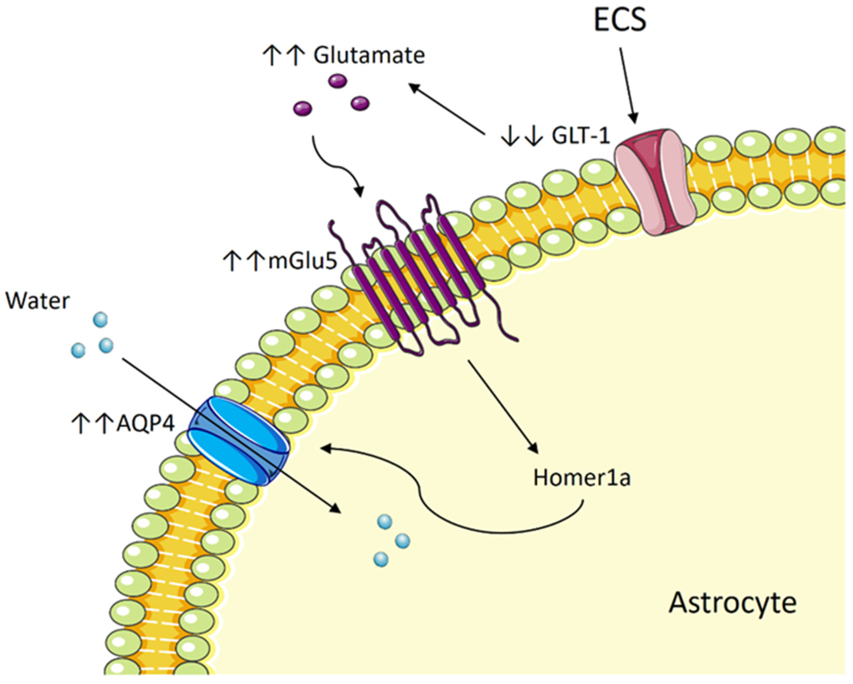
5.3. ECT Induces Cytotoxic Edema in Astrocytes by Way of Aquaporin and Glutamate

Astrocytes are a group of glial cells that support the neuronal cells in the central nervous system (CNS). Astrocytes are involved in homeostatic functions, neuroprotective mechanisms, and synaptic modulation [198]. They are linked to neurotrophic processes by increasing leukemia inhibitory factor (LIF) expression after receiving electrical stimulation, in which LIF stimulates Wnt signaling [199]. Other astrocyte-derived factors such as VEGF and glutamate enhance the permeability of the BBB [200]. Astrocytes possess endfeet processes that ensheath blood vessels in the brain to form gliovascular units that integrate the neural circuitry with local blood flow [201]. Following an acute insult, cytotoxic edema occurs in all CNS cell types but is especially prevalent in astrocytes [202].
ECS in rats transiently enhanced the permeability of the BBB together with swelling in the astrocyte endfeet [201]. A study with Gunn rats, a phenotypic model of affective disorders, showed that ECS improved the depressive-like behavior in the rats. This is accompanied by increased astrocytic blood vessel coverage, which was previously reduced in the model, and also elevated aquaporin-4 (AQP4) expression [203]. These post-ECS changes are not accompanied by any disruption in the tight junctions [203]. Gunn rats also exhibited elevated astrocyte activation in the hippocampus, as evidenced by increased immunoreactivity for glial fibrillary acidic protein (GFAP), which was attenuated with ECS [204]. However, a later review ascertained that chronic ECS induced the transient activation of inflammation and a longer-lasting activation of neurogenesis and astrocytes, as indicated by a prolonged elevation of GFAP for astrocyte activation [205]. Glial activation occurs alongside neurogenesis and temporary inflammation, all of which may partake in the therapeutic effect of ECT.
AQP4 is the main water channel protein in the CNS and is densely expressed in astrocyte endfeet [206]. Increased AQP4 activity, which causes neuronal swelling, may be dependent on the glutamate level in the brain. Glutamate acts on the metabotropic glutamate receptor 5 (mGlu5), whose expression was highest among the metabotropic glutamate receptors in cultured astrocytes and was found to be co-expressed with AQP4 [207]. Glutamate increased AQP4 expression and caused neuronal swelling by binding to mGLu5 [207]. Reduced FA, indicative of reduced neuronal integrity, is also associated with an increased number of swollen astrocytes, immunophenotyped by AQP4, GFAP, and the glucose transporter GLT-1 [208]. GLT-1 is responsible for transporting glutamate into astrocytes before it is converted into glutamine [209]. GLT-1 deficiency has been linked to temporal lobe epilepsy, which promotes an increase in glutamate [209]. Given that AQP4 is expressed within the DG and CA1 of the hippocampus [210], it could be implied that a similar mechanism exists in ECT. The epileptic state induced by ECT may lead to an enhanced activity of glutamate on AQP4, increasing BBB permeability and resulting in edema in the neuronal cells, which may account for the transient cognitive loss post-ECT.
After H-MRS analysis of depressed patients, researchers discovered a reduction in glutamate and glutamine (Glx) concentrations in the subgenual anterior cingulate cortex [188] and the medial frontal cortex [211], as well as an increase in the left hippocampus [188]. Elevated Glx levels in the right hippocampus were also found in adolescents and young adults diagnosed with TRD [189]. Both the subgenual anterior cingulate cortex and the medial PFC have been implicated in the pathophysiology of depression [212,213]. Following ECT, the outcomes varied. Some studies reported increased Glx concentrations after ECT [76,192], while others found no difference between pre- and post-ECT concentrations in the PFC, OFC [191], and the anterior cingulate cortex [214]. The effectiveness of ketamine also contradicts glutamate’s role in mediating cognitive decline. Ketamine, an NMDA receptor antagonist, opposes the action of glutamate and is thought to provide some alleviation of cognitive impairment. When used alone or combined with haloperidol, however, ketamine administration was unable to prevent cognitive decline either in patients undergoing an ECT course or in patients undergoing surgery [215,216]. However, it is possible that the role of glutamate in mediating cognitive decline is due to its action on glial cells rather than synaptic processes. Nevertheless, in a scenario without a reduced baseline Glx in depressed patients, an elevated Glx post-ECT may reflect the inflammatory process leading to neuronal swelling [76,217].

5.4. The Potential Role of mGlu5 and Homer1a

Homer1 is a scaffolding protein that mediates metabotropic glutamate receptors [218,219], including mGlu1 and mGlu5, signaling in neurons. Both stress and ECS administration in mice were noted to induce upregulation of Homer1 expression, which was attributed to the prevention of stress-induced dendritic changes in the hippocampal CA3c region [220]. Homer1a has been implicated in antidepressant mechanisms. In another preclinical study, Homer1a was fused to the protein transduction domain (TAT) to allow cell permeability and delivery of the protein to the brain through the BBB. The peripheral administration of TAT-Homer1a in mice to resemble Homer1a upregulation produced antidepressant effects via enhanced mGlu5/mTOR signaling, resulting in enhanced AMPA receptor expression [221]. Increased Homer1a expression by reactive astrocytes reduced the intensity and duration of glutamate release by astrocytes secondary to a reduction in Ca2+ signaling [222]. Given that mGlu5 expression in astrocytes was accompanied by AQP4 expression [207], both the mGlu5 receptor and the Homer1a protein may be potentially involved in the mechanism of cognitive decline post-ECT. One study reported that mGLu5 receptor antagonists improved cognitive performance. Mice with a genetic model similar to human phenylketonuria (PKU) had higher mGLu5 receptor protein levels in the hippocampus, and they also displayed behavioral impairments in object exploration and spatial memory. The administration of 2-methyl-6-phenylethynyl-pyridine hydrochloride (MPEP), a mGlu5 receptor antagonist [223], significantly ameliorated the behavioral impairments and improved the mice’s cognitive performance [224]. Hence, further investigations are warranted to delve into the potential correlation between mGLu5 receptor and Homer1a expression and clinical and cognitive outcomes after ECT.

6. Conclusions

While it is clear that ECT does indeed cause volume changes in the hippocampus, the exact mechanism that underpins these changes still needs further elucidation. It is most likely that the mechanism for volume changes is also responsible for causing the cognitive side effects, as both the volume increase and the cognitive deficit are temporary and are influenced by electrode placement. ECS in animal studies has been shown to promote both neurogenesis and neuronal cell death (Figure 1). One possibility is that BDNF and VEGF mediate ECT-induced hippocampal neurogenesis, which helps to allay depressive symptoms while also causing excessive neurogenesis in other areas such as the PFC, disturbing the hippocampal cognitive function. Besides BDNF, ECT may also stimulate proBDNF and NR2B signaling that leads to neuronal cell death and degeneration, disturbing the cognitive network. Besides neurogenesis, we also evaluate the potential role of edema in mediating cognitive deficits post-ECT. While FLAIR imaging may have ruled out vasogenic or ionic edema, we cannot ignore evidence showing increased diffusivity in the white matter. Therefore, we theorize that ECT may cause astrocyte swelling that is transient enough not to cause ionic edema but widespread and prevalent enough to impede signal transmission and disturb neural function. As shown in Figure 2, this effect could be mediated by AQP4 and mGLu5, as well as the protein Homer1a. Although this theory is based on current results from animal studies, it may help direct future studies with ECT, especially in regard to improving the cognitive outcome.

Author Contributions

Conceptualization, A.M.A.H., J.K.M., S.W. and M.H.M.Y.; methodology, A.M.A.H. and J.K.M.; software, A.M.A.H.; validation, A.M.A.H., J.K.M., S.W. and M.H.M.Y.; formal analysis, A.M.A.H., J.K.M. and M.H.M.Y.; investigation, A.M.A.H. and J.K.M.; resources, A.M.A.H. and M.H.M.Y.; data curation, A.M.A.H.; writing—original draft preparation, A.M.A.H.; writing—review and editing, A.M.A.H., J.K.M., S.W. and M.H.M.Y.; visualization, A.M.A.H.; supervision, J.K.M. and M.H.M.Y.; project administration, J.K.M. and M.H.M.Y.; funding acquisition, M.H.M.Y. All authors have read and agreed to the published version of the manuscript.

Funding

This research received no external funding.

Institutional Review Board Statement

Not applicable.

Informed Consent Statement

Not applicable.

Data Availability Statement

Not applicable.

Acknowledgments

The authors would like to thank UKM for access to the electronic databases.

Conflicts of Interest

The authors declare no conflict of interest.

References

  1. Malhi, G.S.; Mann, J.J. Depression. Lancet 2018, 392, 2299–2312. [Google Scholar] [CrossRef]
  2. König, H.; König, H.H.; Konnopka, A. The excess costs of depression: A systematic review and meta-analysis. Epidemiol. Psychiatr. Sci. 2019, 29, e30. [Google Scholar] [CrossRef]
  3. Diagnostic and Statistical Manual of Mental Disorders DSM-IV, 4th ed.; American Psychiatric Association: Washington, DC, USA, 1994.
  4. Enev, M.; McNally, K.A.; Varghese, G.; Zubal, I.G.; Ostroff, R.B.; Blumenfeld, H. Imaging Onset and Propagation of ECT-induced Seizures. Epilepsia 2007, 48, 238–244. [Google Scholar] [CrossRef] [PubMed]
  5. Wang, C.C.; Lin, C.H.; Chiu, Y.C.; Tseng, C.C. The Clonic Phase of Seizures in Patients Treated with Electroconvulsive Therapy is Related to Age and Stimulus Intensity. Front. Psychiatry 2013, 4, 166. [Google Scholar] [CrossRef]
  6. Ray, A.K. Does electroconvulsive therapy cause epilepsy? J. ECT 2013, 29, 201–205. [Google Scholar] [CrossRef] [PubMed]
  7. Ross, E.L.; Zivin, K.; Maixner, D.F. Cost-effectiveness of Electroconvulsive Therapy vs Pharmacotherapy/Psychotherapy for Treatment-Resistant Depression in the United States. JAMA Psychiatry 2018, 75, 713–722. [Google Scholar] [CrossRef]
  8. Elias, A.; Phutane, V.H.; Clarke, S.; Prudic, J. Electroconvulsive therapy in the continuation and maintenance treatment of depression: Systematic review and meta-analyses. Aust. N. Z. J. Psychiatry 2018, 52, 415–424. [Google Scholar] [CrossRef] [PubMed]
  9. Hermida, A.P.; Glass, O.M.; Shafi, H.; McDonald, W.M. Electroconvulsive Therapy in Depression: Current Practice and Future Direction. Psychiatr. Clin. N. Am. 2018, 41, 341–353. [Google Scholar] [CrossRef] [PubMed]
  10. Venkatasalam, J.; Suzaily, W. Agitated Depression: The Challenges in Management and the Role of Maintenance Electroconvulsive Therapy. IIUM Med. J. Malays. 2019, 18, 166–168. [Google Scholar] [CrossRef]
  11. Park, M.J.; Kim, H.; Kim, E.J.; Yook, V.; Chung, I.W.; Lee, S.M.; Jeon, H.J. Recent Updates on Electro-Convulsive Therapy in Patients with Depression. Psychiatry Investig. 2021, 18, 1–10. [Google Scholar] [CrossRef]
  12. van Diermen, L.; van den Ameele, S.; Kamperman, A.M.; Sabbe, B.C.G.; Vermeulen, T.; Schrijvers, D.; Birkenhäger, T.K. Prediction of electroconvulsive therapy response and remission in major depression: Meta-analysis. Br. J. Psychiatry J. Ment. Sci. 2018, 212, 71–80. [Google Scholar] [CrossRef]
  13. Weiss, A.; Hussain, S.; Ng, B.; Sarma, S.; Tiller, J.; Waite, S.; Loo, C. Royal Australian and New Zealand College of Psychiatrists professional practice guidelines for the administration of electroconvulsive therapy. Aust. N. Z. J. Psychiatry 2019, 53, 609–623. [Google Scholar] [CrossRef]
  14. Dong, M.; Zhu, X.M.; Zheng, W.; Li, X.H.; Ng, C.H.; Ungvari, G.S.; Xiang, Y.T. Electroconvulsive therapy for older adult patients with major depressive disorder: A systematic review of randomized controlled trials. Psychogeriatrics 2018, 18, 468–475. [Google Scholar] [CrossRef] [PubMed]
  15. Chen, J.J.; Zhao, L.B.; Liu, Y.Y.; Fan, S.H.; Xie, P. Comparative efficacy and acceptability of electroconvulsive therapy versus repetitive transcranial magnetic stimulation for major depression: A systematic review and multiple-treatments meta-analysis. Behav. Brain Res. 2017, 320, 30–36. [Google Scholar] [CrossRef] [PubMed]
  16. Andrade, C.; Arumugham, S.S.; Thirthalli, J. Adverse Effects of Electroconvulsive Therapy. Psychiatr. Clin. N. Am. 2016, 39, 513–530. [Google Scholar] [CrossRef]
  17. Gbyl, K.; Stottrup, M.M.; Raghava, J.M.; Jie, S.X.; Videbech, P. Hippocampal volume and memory impairment after electroconvulsive therapy in patients with depression. Acta Psychiatr. Scand. 2021, 143, 238–252. [Google Scholar] [CrossRef]
  18. Ramlan, H.; Shafri, N.I.; Wahab, S.; Kamarudin, M.A.; Rajikan, R.; Abdul Wahab, N.A.; Damanhuri, H.A. Depression, Anxiety and Stress in Medical Students: An Early Observation Analysis. Mediterr. J. Clin. Psychol. 2020, 8, 1–16. [Google Scholar] [CrossRef]
  19. van Buel, E.M.; Patas, K.; Peters, M.; Bosker, F.J.; Eisel, U.L.M.; Klein, H.C. Immune and neurotrophin stimulation by electroconvulsive therapy: Is some inflammation needed after all? Transl. Psychiatry 2015, 5, e609. [Google Scholar] [CrossRef] [PubMed]
  20. Phillips, M.L.; Swartz, H.A. A Critical Appraisal of Neuroimaging Studies of Bipolar Disorder: Toward a New Conceptualization of Underlying Neural Circuitry and a Road Map for Future Research. Am. J. Psychiatry 2014, 171, 829–843. [Google Scholar] [CrossRef]
  21. Bai, T.; Wei, Q.; Xie, W.; Wang, A.; Wang, J.; Ji, G.J.; Wang, K.; Tian, Y. Hippocampal-subregion functional alterations associated with antidepressant effects and cognitive impairments of electroconvulsive therapy. Psychol. Med. 2019, 49, 1357–1364. [Google Scholar] [CrossRef] [PubMed]
  22. Bannerman, D.M.; Sprengel, R.; Sanderson, D.J.; McHugh, S.B.; Rawlins, J.N.P.; Monyer, H.; Seeburg, P.H. Hippocampal synaptic plasticity, spatial memory and anxiety. Nat. Rev. Neurosci. 2014, 15, 181–192. [Google Scholar] [CrossRef]
  23. Barkus, C.; McHugh, S.B.; Sprengel, R.; Seeburg, P.H.; Rawlins, J.N.P.; Bannerman, D.M. Hippocampal NMDA receptors and anxiety: At the interface between cognition and emotion. Eur. J. Pharmacol. 2010, 626, 49–56. [Google Scholar] [CrossRef] [PubMed]
  24. Nawa, N.E.; Ando, H. Effective connectivity within the ventromedial prefrontal cortex-hippocampus-amygdala network during the elaboration of emotional autobiographical memories. NeuroImage 2019, 189, 316–328. [Google Scholar] [CrossRef] [PubMed]
  25. McEwen, B.S.; Nasca, C.; Gray, J.D. Stress Effects on Neuronal Structure: Hippocampus, Amygdala, and Prefrontal Cortex. Neuropsychopharmacology 2016, 41, 3–23. [Google Scholar] [CrossRef]
  26. Hei, M.; Chen, P.; Wang, S.; Li, X.; Xu, M.; Zhu, X.; Wang, Y.; Duan, J.; Huang, Y.; Zhao, S. Effects of chronic mild stress induced depression on synaptic plasticity in mouse hippocampus. Behav. Brain Res. 2019, 365, 26–35. [Google Scholar] [CrossRef]
  27. Kandilarova, S.; Stoyanov, D.; Kostianev, S.; Specht, K. Altered Resting State Effective Connectivity of Anterior Insula in Depression. Front. Psychiatry 2018, 9, 83. [Google Scholar] [CrossRef]
  28. Picó-Pérez, M.; Radua, J.; Steward, T.; Menchón, J.M.; Soriano-Mas, C. Emotion regulation in mood and anxiety disorders: A meta-analysis of fMRI cognitive reappraisal studies. Prog. Neuro-Psychopharmacol. Biol. Psychiatry 2017, 79, 96–104. [Google Scholar] [CrossRef] [PubMed]
  29. Sambataro, F.; Wolf, N.D.; Pennuto, M.; Vasic, N.; Wolf, R.C. Revisiting default mode network function in major depression: Evidence for disrupted subsystem connectivity. Psychol. Med. 2014, 44, 2041–2051. [Google Scholar] [CrossRef]
  30. Whitfield-Gabrieli, S.; Ford, J.M. Default Mode Network Activity and Connectivity in Psychopathology. Annu. Rev. Clin. Psychol. 2012, 8, 49–76. [Google Scholar] [CrossRef]
  31. Cavaleri, D.; Bartoli, F. Biomolecular Research on Electroconvulsive Therapy for Mental Disorders: State of the Art and Future Directions. Alpha Psychiatry 2022, 23, 57–58. [Google Scholar] [CrossRef]
  32. Moica, T.; Gligor, A.; Moica, S. The Relationship between Cortisol and the Hippocampal Volume in Depressed Patients—A MRI Pilot Study. Procedia Technol. 2016, 22, 1106–1112. [Google Scholar] [CrossRef]
  33. Merz, E.C.; Desai, P.M.; Maskus, E.A.; Melvin, S.A.; Rehman, R.; Torres, S.D.; Meyer, J.; He, X.; Noble, K.G. Socioeconomic Disparities in Chronic Physiologic Stress Are Associated With Brain Structure in Children. Biol. Psychiatry 2019, 86, 921–929. [Google Scholar] [CrossRef] [PubMed]
  34. Piccolo, L.R.; Noble, K.G. Perceived stress is associated with smaller hippocampal volume in adolescence. Psychophysiology 2018, 55, e13025. [Google Scholar] [CrossRef] [PubMed]
  35. Barch, D.M.; Harms, M.P.; Tillman, R.; Hawkey, E.; Luby, J.L. Early childhood depression, emotion regulation, episodic memory, and hippocampal development. J. Abnorm. Psychol. 2019, 128, 81–95. [Google Scholar] [CrossRef]
  36. Barch, D.M.; Tillman, R.; Kelly, D.; Whalen, D.; Gilbert, K.; Luby, J.L. Hippocampal volume and depression among young children. Psychiatry Res. Neuroimaging 2019, 288, 21–28. [Google Scholar] [CrossRef] [PubMed]
  37. Saleh, A.; Potter, G.G.; McQuoid, D.R.; Boyd, B.; Turner, R.; MacFall, J.R.; Taylor, W.D. Effects of early life stress on depression, cognitive performance and brain morphology. Psychol. Med. 2017, 47, 171–181. [Google Scholar] [CrossRef] [PubMed]
  38. Tannous, J.; Godlewska, B.R.; Tirumalaraju, V.; Soares, J.C.; Cowen, P.J.; Selvaraj, S. Stress, inflammation and hippocampal subfields in depression: A 7 Tesla MRI Study. Transl. Psychiatry 2020, 10, 78. [Google Scholar] [CrossRef]
  39. Schoenfeld, T.J.; McCausland, H.C.; Morris, H.D.; Padmanaban, V.; Cameron, H.A. Stress and Loss of Adult Neurogenesis Differentially Reduce Hippocampal Volume. Biol. Psychiatry 2017, 82, 914–923. [Google Scholar] [CrossRef]
  40. Schmaal, L.; Veltman, D.J.; van Erp, T.G.; Sämann, P.G.; Frodl, T.; Jahanshad, N.; Loehrer, E.; Tiemeier, H.; Hofman, A.; Niessen, W.J.; et al. Subcortical brain alterations in major depressive disorder: Findings from the ENIGMA Major Depressive Disorder working group. Mol. Psychiatry 2016, 21, 806–812. [Google Scholar] [CrossRef]
  41. Jamieson, A.; Goodwill, A.M.; Termine, M.; Campbell, S.; Szoeke, C. Depression related cerebral pathology and its relationship with cognitive functioning: A systematic review. J. Affect. Disord. 2019, 250, 410–418. [Google Scholar] [CrossRef]
  42. Santos, M.A.O.; Bezerra, L.S.; Carvalho, A.; Brainer-Lima, A.M. Global hippocampal atrophy in major depressive disorder: A meta-analysis of magnetic resonance imaging studies. Trends Psychiatry Psychother. 2018, 40, 369–378. [Google Scholar] [CrossRef] [PubMed]
  43. Wise, T.; Radua, J.; Via, E.; Cardoner, N.; Abe, O.; Adams, T.M.; Amico, F.; Cheng, Y.; Cole, J.H.; de Azevedo Marques Périco, C.; et al. Common and distinct patterns of grey-matter volume alteration in major depression and bipolar disorder: Evidence from voxel-based meta-analysis. Mol. Psychiatry 2017, 22, 1455–1463. [Google Scholar] [CrossRef] [PubMed]
  44. Roddy, D.W.; Farrell, C.; Doolin, K.; Roman, E.; Tozzi, L.; Frodl, T.; O’Keane, V.; O’Hanlon, E. The Hippocampus in Depression: More Than the Sum of Its Parts? Advanced Hippocampal Substructure Segmentation in Depression. Biol. Psychiatry 2019, 85, 487–497. [Google Scholar] [CrossRef] [PubMed]
  45. Zheng, R.; Zhang, Y.; Yang, Z.; Han, S.; Cheng, J. Reduced Brain Gray Matter Volume in Patients With First-Episode Major Depressive Disorder: A Quantitative Meta-Analysis. Front. Psychiatry 2021, 12, 671348. [Google Scholar] [CrossRef] [PubMed]
  46. Fan, S.; Lippard, E.T.C.; Sankar, A.; Wallace, A.; Johnston, J.A.Y.; Wang, F.; Pittman, B.; Spencer, L.; Oquendo, M.A.; Blumberg, H.P. Gray and white matter differences in adolescents and young adults with prior suicide attempts across bipolar and major depressive disorders. J. Affect. Disord. 2019, 245, 1089–1097. [Google Scholar] [CrossRef]
  47. Maller, J.J.; Broadhouse, K.; Rush, A.J.; Gordon, E.; Koslow, S.; Grieve, S.M. Increased hippocampal tail volume predicts depression status and remission to anti-depressant medications in major depression. Mol. Psychiatry 2018, 23, 1737–1744. [Google Scholar] [CrossRef] [PubMed]
  48. Ota, M.; Noda, T.; Sato, N.; Okazaki, M.; Ishikawa, M.; Hattori, K.; Hori, H.; Sasayama, D.; Teraishi, T.; Sone, D.; et al. Effect of electroconvulsive therapy on gray matter volume in major depressive disorder. J. Affect. Disord. 2015, 186, 186–191. [Google Scholar] [CrossRef] [PubMed]
  49. Sartorius, A.; Demirakca, T.; Böhringer, A.; Clemm von Hohenberg, C.; Aksay, S.S.; Bumb, J.M.; Kranaster, L.; Ende, G. Electroconvulsive therapy increases temporal gray matter volume and cortical thickness. Eur. Neuropsychopharmacol. 2016, 26, 506–517. [Google Scholar] [CrossRef]
  50. Sartorius, A.; Demirakca, T.; Böhringer, A.; Clemm von Hohenberg, C.; Aksay, S.S.; Bumb, J.M.; Kranaster, L.; Nickl-Jockschat, T.; Grözinger, M.; Thomann, P.A.; et al. Electroconvulsive therapy induced gray matter increase is not necessarily correlated with clinical data in depressed patients. Brain Stimul. 2019, 12, 335–343. [Google Scholar] [CrossRef]
  51. Camilleri, J.A.; Hoffstaedter, F.; Zavorotny, M.; Zöllner, R.; Wolf, R.C.; Thomann, P.; Redlich, R.; Opel, N.; Dannlowski, U.; Grözinger, M.; et al. Electroconvulsive therapy modulates grey matter increase in a hub of an affect processing network. NeuroImage Clin. 2020, 25, 102114. [Google Scholar] [CrossRef]
  52. Wilkinson, S.T.; Sanacora, G.; Bloch, M.H. Hippocampal Volume Changes Following Electroconvulsive Therapy: A Systematic Review and Meta-analysis. Biol. Psychiatry Cogn. Neurosci. Neuroimaging 2017, 2, 327–335. [Google Scholar] [CrossRef]
  53. Gbyl, K.; Rostrup, E.; Raghava, J.M.; Andersen, C.; Rosenberg, R.; Larsson, H.B.W.; Videbech, P. Volume of hippocampal subregions and clinical improvement following electroconvulsive therapy in patients with depression. Prog. Neuro-Psychopharmacol. Biol. Psychiatry 2021, 104, 110048. [Google Scholar] [CrossRef]
  54. Cao, B.; Luo, Q.; Fu, Y.; Du, L.; Qiu, T.; Yang, X.; Chen, X.; Chen, Q.; Soares, J.C.; Cho, R.Y.; et al. Predicting individual responses to the electroconvulsive therapy with hippocampal subfield volumes in major depression disorder. Sci. Rep. 2018, 8, 5434. [Google Scholar] [CrossRef] [PubMed]
  55. Gryglewski, G.; Baldinger-Melich, P.; Seiger, R.; Godbersen, G.M.; Michenthaler, P.; Klobl, M.; Spurny, B.; Kautzky, A.; Vanicek, T.; Kasper, S.; et al. Structural changes in amygdala nuclei, hippocampal subfields and cortical thickness following electroconvulsive therapy in treatment-resistant depression: Longitudinal analysis. Br. J. Psychiatry 2019, 214, 159–167. [Google Scholar] [CrossRef]
  56. Takamiya, A.; Plitman, E.; Chung, J.K.; Chakravarty, M.; Graff-Guerrero, A.; Mimura, M.; Kishimoto, T. Acute and long-term effects of electroconvulsive therapy on human dentate gyrus. Neuropsychopharmacology 2019, 44, 1805–1811. [Google Scholar] [CrossRef] [PubMed]
  57. Kubicki, A.; Leaver, A.M.; Vasavada, M.; Njau, S.; Wade, B.; Joshi, S.H.; Loureiro, J.; Hellemann, G.; Woods, R.P.; Espinoza, R.; et al. Variations in Hippocampal White Matter Diffusivity Differentiate Response to Electroconvulsive Therapy in Major Depression. Biol. Psychiatry Cogn. Neurosci. Neuroimaging 2019, 4, 300–309. [Google Scholar] [CrossRef]
  58. Nuninga, J.O.; Mandl, R.C.W.; Boks, M.P.; Bakker, S.; Somers, M.; Heringa, S.M.; Nieuwdorp, W.; Hoogduin, H.; Kahn, R.S.; Luijten, P.; et al. Volume increase in the dentate gyrus after electroconvulsive therapy in depressed patients as measured with 7T. Mol. Psychiatry 2020, 25, 1559–1568. [Google Scholar] [CrossRef]
  59. Bartsch, T.; Döhring, J.; Rohr, A.; Jansen, O.; Deuschl, G. CA1 neurons in the human hippocampus are critical for autobiographical memory, mental time travel, and autonoetic consciousness. Proc. Natl. Acad. Sci. USA 2011, 108, 17562. [Google Scholar] [CrossRef]
  60. Kumar, J.; Hapidin, H.; Get Bee, Y.T.; Ismail, Z. The effects of acute ethanol administration on ethanol withdrawal-induced anxiety-like syndrome in rats: A biochemical study. Alcohol 2016, 50, 9–17. [Google Scholar] [CrossRef]
  61. Sandu, A.L.; Artiges, E.; Galinowski, A.; Gallarda, T.; Bellivier, F.; Lemaitre, H.; Granger, B.; Ringuenet, D.; Tzavara, E.T.; Martinot, J.L.; et al. Amygdala and regional volumes in treatment-resistant versus nontreatment-resistant depression patients. Depress. Anxiety 2017, 34, 1065–1071. [Google Scholar] [CrossRef] [PubMed]
  62. Espinoza Oyarce, D.A.; Shaw, M.E.; Alateeq, K.; Cherbuin, N. Volumetric brain differences in clinical depression in association with anxiety: A systematic review with meta-analysis. J. Psychiatry Neurosci. JPN 2020, 45, 406–429. [Google Scholar] [CrossRef]
  63. McEwen, B.S.; Bowles, N.P.; Gray, J.D.; Hill, M.N.; Hunter, R.G.; Karatsoreos, I.N.; Nasca, C. Mechanisms of stress in the brain. Nat. Neurosci. 2015, 18, 1353–1363. [Google Scholar] [CrossRef]
  64. Zhang, W.-H.; Liu, W.-Z.; He, Y.; You, W.-J.; Zhang, J.-Y.; Xu, H.; Tian, X.-L.; Li, B.-M.; Mei, L.; Holmes, A.; et al. Chronic Stress Causes Projection-Specific Adaptation of Amygdala Neurons via Small-Conductance Calcium-Activated Potassium Channel Downregulation. Biol. Psychiatry 2019, 85, 812–828. [Google Scholar] [CrossRef] [PubMed]
  65. Joshi, S.H.; Espinoza, R.T.; Pirnia, T.; Shi, J.; Wang, Y.; Ayers, B.; Leaver, A.; Woods, R.P.; Narr, K.L. Structural Plasticity of the Hippocampus and Amygdala Induced by Electroconvulsive Therapy in Major Depression. Biol. Psychiatry 2016, 79, 282–292. [Google Scholar] [CrossRef] [PubMed]
  66. Wang, J.J.; Wei, Q.; Bai, T.J.; Zhou, X.Q.; Sun, H.; Becker, B.; Tian, Y.H.; Wang, K.; Kendrick, K. Electroconvulsive therapy selectively enhanced feedforward connectivity from fusiform face area to amygdala in major depressive disorder. Soc. Cogn. Affect. Neurosci. 2017, 12, 1983–1992. [Google Scholar] [CrossRef] [PubMed]
  67. Damborská, A.; Honzírková, E.; Barteček, R.; Hořínková, J.; Fedorová, S.; Ondruš, Š.; Michel, C.M.; Rubega, M. Altered directed functional connectivity of the right amygdala in depression: High-density EEG study. Sci. Rep. 2020, 10, 4398. [Google Scholar] [CrossRef] [PubMed]
  68. Li, Q.; Zhao, Y.; Chen, Z.; Long, J.; Dai, J.; Huang, X.; Lui, S.; Radua, J.; Vieta, E.; Kemp, G.J.; et al. Meta-analysis of cortical thickness abnormalities in medication-free patients with major depressive disorder. Neuropsychopharmacology 2020, 45, 703–712. [Google Scholar] [CrossRef]
  69. Schmaal, L.; Hibar, D.P.; Sämann, P.G.; Hall, G.B.; Baune, B.T.; Jahanshad, N.; Cheung, J.W.; van Erp, T.G.M.; Bos, D.; Ikram, M.A.; et al. Cortical abnormalities in adults and adolescents with major depression based on brain scans from 20 cohorts worldwide in the ENIGMA Major Depressive Disorder Working Group. Mol. Psychiatry 2017, 22, 900–909. [Google Scholar] [CrossRef]
  70. Suh, J.S.; Schneider, M.A.; Minuzzi, L.; MacQueen, G.M.; Strother, S.C.; Kennedy, S.H.; Frey, B.N. Cortical thickness in major depressive disorder: A systematic review and meta-analysis. Prog. Neuropsychopharmacol. Biol. Psychiatry 2019, 88, 287–302. [Google Scholar] [CrossRef]
  71. Chen, L.; Wang, Y.; Niu, C.; Zhong, S.; Hu, H.; Chen, P.; Zhang, S.; Chen, G.; Deng, F.; Lai, S.; et al. Common and distinct abnormal frontal-limbic system structural and functional patterns in patients with major depression and bipolar disorder. NeuroImage Clin. 2018, 20, 42–50. [Google Scholar] [CrossRef] [PubMed]
  72. Yamasaki, S.; Aso, T.; Miyata, J.; Sugihara, G.; Hazama, M.; Nemoto, K.; Yoshihara, Y.; Matsumoto, Y.; Okada, T.; Togashi, K.; et al. Early and late effects of electroconvulsive therapy associated with different temporal lobe structures. Transl. Psychiatry 2020, 10, 344. [Google Scholar] [CrossRef]
  73. Mulders, P.C.R.; Llera, A.; Beckmann, C.F.; Vandenbulcke, M.; Stek, M.; Sienaert, P.; Redlich, R.; Petrides, G.; Oudega, M.L.; Oltedal, L.; et al. Structural changes induced by electroconvulsive therapy are associated with clinical outcome. Brain Stimul. 2020, 13, 696–704. [Google Scholar] [CrossRef]
  74. Bai, S.; Gálvez, V.; Dokos, S.; Martin, D.; Bikson, M.; Loo, C. Computational models of Bitemporal, Bifrontal and Right Unilateral ECT predict differential stimulation of brain regions associated with efficacy and cognitive side effects. Eur. Psychiatry 2017, 41, 21–29. [Google Scholar] [CrossRef] [PubMed]
  75. Oltedal, L.; Narr, K.L.; Abbott, C.; Anand, A.; Argyelan, M.; Bartsch, H.; Dannlowski, U.; Dols, A.; van Eijndhoven, P.; Emsell, L.; et al. Volume of the Human Hippocampus and Clinical Response Following Electroconvulsive Therapy. Biol. Psychiatry 2018, 84, 574–581. [Google Scholar] [CrossRef]
  76. Cano, M.; Martinez-Zalacain, I.; Bernabeu-Sanz, A.; Contreras-Rodriguez, O.; Hernandez-Ribas, R.; Via, E.; de Arriba-Arnau, A.; Galvez, V.; Urretavizcaya, M.; Pujol, J.; et al. Brain volumetric and metabolic correlates of electroconvulsive therapy for treatment-resistant depression: A longitudinal neuroimaging study. Transl. Psychiatry 2017, 7, e1023. [Google Scholar] [CrossRef]
  77. Jehna, M.; Wurm, W.; Pinter, D.; Vogel, K.; Holl, A.; Hofmann, P.; Ebner, C.; Ropele, S.; Fuchs, G.; Kapfhammer, H.-P.; et al. Do increases in deep grey matter volumes after electroconvulsive therapy persist in patients with major depression? A longitudinal MRI-study. J. Affect. Disord. 2021, 281, 908–917. [Google Scholar] [CrossRef]
  78. Argyelan, M.; Oltedal, L.; Deng, Z.D.; Wade, B.; Bikson, M.; Joanlanne, A.; Sanghani, S.; Bartsch, H.; Cano, M.; Dale, A.M.; et al. Electric field causes volumetric changes in the human brain. Elife 2019, 8, e49115. [Google Scholar] [CrossRef] [PubMed]
  79. Ousdal, O.T.; Argyelan, M.; Narr, K.L.; Abbott, C.; Wade, B.; Vandenbulcke, M.; Urretavizcaya, M.; Tendolkar, I.; Takamiya, A.; Stek, M.L.; et al. Brain Changes Induced by Electroconvulsive Therapy Are Broadly Distributed. Biol. Psychiatry 2020, 87, 451–461. [Google Scholar] [CrossRef]
  80. Fridgeirsson, E.A.; Deng, Z.D.; Denys, D.; van Waarde, J.A.; van Wingen, G.A. Electric field strength induced by electroconvulsive therapy is associated with clinical outcome. NeuroImage Clin. 2021, 30, 102581. [Google Scholar] [CrossRef]
  81. Oltedal, L.; Bartsch, H.; Sørhaug, O.J.E.; Kessler, U.; Abbott, C.; Dols, A.; Stek, M.L.; Ersland, L.; Emsell, L.; van Eijndhoven, P.; et al. The Global ECT-MRI Research Collaboration (GEMRIC): Establishing a multi-site investigation of the neural mechanisms underlying response to electroconvulsive therapy. NeuroImage Clin. 2017, 14, 422–432. [Google Scholar] [CrossRef] [PubMed]
  82. Takamiya, A.; Chung, J.K.; Liang, K.C.; Graff-Guerrero, A.; Mimura, M.; Kishimoto, T. Effect of electroconvulsive therapy on hippocampal and amygdala volumes: Systematic review and meta-analysis. Br. J. Psychiatry J. Ment. Sci. 2018, 212, 19–26. [Google Scholar] [CrossRef]
  83. Xu, J.; Wang, J.; Bai, T.; Zhang, X.; Li, T.; Hu, Q.; Li, H.; Zhang, L.; Wei, Q.; Tian, Y.; et al. Electroconvulsive Therapy Induces Cortical Morphological Alterations in Major Depressive Disorder Revealed with Surface-Based Morphometry Analysis. Int. J. Neural Syst. 2019, 29, 1950005. [Google Scholar] [CrossRef]
  84. van Oostrom, I.; van Eijndhoven, P.; Butterbrod, E.; van Beek, M.H.; Janzing, J.; Donders, R.; Schene, A.; Tendolkar, I. Decreased Cognitive Functioning After Electroconvulsive Therapy Is Related to Increased Hippocampal Volume: Exploring the Role of Brain Plasticity. J. Ect. 2018, 34, 117–123. [Google Scholar] [CrossRef]
  85. Laroy, M.; Bouckaert, F.; Vansteelandt, K.; Ohhels, J.; Dols, A.; Emsell, L.; Stek, M.; Vandenhulcke, M.; Sienaert, P. Association between hippocampal volume change and change in memory following electroconvulsive therapy in late-life depression. Acta Psychiatr. Scand. 2019, 140, 435–445. [Google Scholar] [CrossRef] [PubMed]
  86. Argyelan, M.; Lencz, T.; Kang, S.; Ali, S.; Masi, P.J.; Moyett, E.; Joanlanne, A.; Watson, P.; Sanghani, S.; Petrides, G.; et al. ECT-induced cognitive side effects are associated with hippocampal enlargement. Transl. Psychiatry 2021, 11, 516. [Google Scholar] [CrossRef] [PubMed]
  87. van der Linden, M.; Berkers, R.; Morris, R.G.M.; Fernández, G. Angular Gyrus Involvement at Encoding and Retrieval Is Associated with Durable But Less Specific Memories. J. Neurosci. 2017, 37, 9474–9485. [Google Scholar] [CrossRef]
  88. Olesen, M.V.; Wörtwein, G.; Pakkenberg, B. Electroconvulsive stimulation, but not chronic restraint stress, causes structural alterations in adult rat hippocampus--a stereological study. Hippocampus 2015, 25, 72–80. [Google Scholar] [CrossRef] [PubMed]
  89. Alemu, J.L.; Elberling, F.; Azam, B.; Pakkenberg, B.; Olesen, M.V. Electroconvulsive treatment prevents chronic restraint stress-induced atrophy of the hippocampal formation—A stereological study. Brain Behav. 2019, 9, e01195. [Google Scholar] [CrossRef] [PubMed]
  90. Olesen, M.V.; Wörtwein, G.; Folke, J.; Pakkenberg, B. Electroconvulsive stimulation results in long-term survival of newly generated hippocampal neurons in rats. Hippocampus 2017, 27, 52–60. [Google Scholar] [CrossRef] [PubMed]
  91. Madsen, T.M.; Treschow, A.; Bengzon, J.; Bolwig, T.G.; Lindvall, O.; Tingström, A. Increased neurogenesis in a model of electroconvulsive therapy. Biol. Psychiatry 2000, 47, 1043–1049. [Google Scholar] [CrossRef]
  92. García-Cabrerizo, R.; Ledesma-Corvi, S.; Bis-Humbert, C.; García-Fuster, M.J. Sex differences in the antidepressant-like potential of repeated electroconvulsive seizures in adolescent and adult rats: Regulation of the early stages of hippocampal neurogenesis. Eur. Neuropsychopharmacol. 2020, 41, 132–145. [Google Scholar] [CrossRef]
  93. Scholzen, T.; Gerdes, J. The Ki-67 protein: From the known and the unknown. J. Cell. Physiol. 2000, 182, 311–322. [Google Scholar] [CrossRef]
  94. Kee, N.; Sivalingam, S.; Boonstra, R.; Wojtowicz, J.M. The utility of Ki-67 and BrdU as proliferative markers of adult neurogenesis. J. Neurosci. Methods 2002, 115, 97–105. [Google Scholar] [CrossRef] [PubMed]
  95. Gao, Z.; Ure, K.; Ables, J.L.; Lagace, D.C.; Nave, K.-A.; Goebbels, S.; Eisch, A.J.; Hsieh, J. Neurod1 is essential for the survival and maturation of adult-born neurons. Nat. Neurosci. 2009, 12, 1090–1092. [Google Scholar] [CrossRef]
  96. Ueno, M.; Sugimoto, M.; Ohtsubo, K.; Sakai, N.; Endo, A.; Shikano, K.; Imoto, Y.; Segi-Nishida, E. The effect of electroconvulsive seizure on survival, neuronal differentiation, and expression of the maturation marker in the adult mouse hippocampus. J. Neurochem. 2019, 149, 488–498. [Google Scholar] [CrossRef] [PubMed]
  97. Bonde, S.; Ekdahl, C.T.; Lindvall, O. Long-term neuronal replacement in adult rat hippocampus after status epilepticus despite chronic inflammation. Eur. J. Neurosci. 2006, 23, 965–974. [Google Scholar] [CrossRef]
  98. Jakubs, K.; Nanobashvili, A.; Bonde, S.; Ekdahl, C.T.; Kokaia, Z.; Kokaia, M.; Lindvall, O. Environment matters: Synaptic properties of neurons born in the epileptic adult brain develop to reduce excitability. Neuron 2006, 52, 1047–1059. [Google Scholar] [CrossRef]
  99. Jackson, J.; Chugh, D.; Nilsson, P.; Wood, J.; Carlström, K.; Lindvall, O.; Ekdahl, C.T. Altered synaptic properties during integration of adult-born hippocampal neurons following a seizure insult. PLoS ONE 2012, 7, e35557. [Google Scholar] [CrossRef]
  100. Yang, T.; Nie, Z.; Shu, H.; Kuang, Y.; Chen, X.; Cheng, J.; Yu, S.; Liu, H. The Role of BDNF on Neural Plasticity in Depression. Front. Cell. Neurosci. 2020, 14, 82. [Google Scholar] [CrossRef]
  101. Şahin, T.D.; Karson, A.; Balcı, F.; Yazır, Y.; Bayramgürler, D.; Utkan, T. TNF-alpha inhibition prevents cognitive decline and maintains hippocampal BDNF levels in the unpredictable chronic mild stress rat model of depression. Behav. Brain Res. 2015, 292, 233–240. [Google Scholar] [CrossRef]
  102. Duman, R.S.; Deyama, S.; Fogaça, M.V. Role of BDNF in the pathophysiology and treatment of depression: Activity-dependent effects distinguish rapid-acting antidepressants. Eur. J. Neurosci. 2021, 53, 126–139. [Google Scholar] [CrossRef] [PubMed]
  103. Pisoni, A.; Strawbridge, R.; Hodsoll, J.; Powell, T.R.; Breen, G.; Hatch, S.; Hotopf, M.; Young, A.H.; Cleare, A.J. Growth Factor Proteins and Treatment-Resistant Depression: A Place on the Path to Precision. Front. Psychiatry 2018, 9, 386. [Google Scholar] [CrossRef]
  104. Rapinesi, C.; Kotzalidis, G.D.; Curto, M.; Serata, D.; Ferri, V.R.; Scatena, P.; Carbonetti, P.; Napoletano, F.; Miele, J.; Scaccianoce, S.; et al. Electroconvulsive therapy improves clinical manifestations of treatment-resistant depression without changing serum BDNF levels. Psychiatry Res. 2015, 227, 171–178. [Google Scholar] [CrossRef] [PubMed]
  105. Bouckaert, F.; Dols, A.; Emsell, L.; De Winter, F.L.; Vansteelandt, K.; Claes, L.; Sunaert, S.; Stek, M.; Sienaert, P.; Vandenbulcke, M. Relationship Between Hippocampal Volume, Serum BDNF, and Depression Severity Following Electroconvulsive Therapy in Late-Life Depression. Neuropsychopharmacology 2016, 41, 2741–2748. [Google Scholar] [CrossRef] [PubMed]
  106. Cavaleri, D.; Moretti, F.; Bartoccetti, A.; Mauro, S.; Crocamo, C.; Carrà, G.; Bartoli, F. The role of BDNF in major depressive disorder, related clinical features, and antidepressant treatment: Insight from meta-analyses. Neurosci. Biobehav. Rev. 2023, 149, 105159. [Google Scholar] [CrossRef] [PubMed]
  107. Luan, S.; Zhou, B.; Wu, Q.; Wan, H.; Li, H. Brain-derived neurotrophic factor blood levels after electroconvulsive therapy in patients with major depressive disorder: A systematic review and meta-analysis. Asian J. Psychiatry 2020, 51, 101983. [Google Scholar] [CrossRef]
  108. Rocha, R.B.; Dondossola, E.R.; Grande, A.J.; Colonetti, T.; Ceretta, L.B.; Passos, I.C.; Quevedo, J.; da Rosa, M.I. Increased BDNF levels after electroconvulsive therapy in patients with major depressive disorder: A meta-analysis study. J. Psychiatr. Res. 2016, 83, 47–53. [Google Scholar] [CrossRef]
  109. Ryan, K.M.; Dunne, R.; McLoughlin, D.M. BDNF plasma levels and genotype in depression and the response to electroconvulsive therapy. Brain Stimul. 2018, 11, 1123–1131. [Google Scholar] [CrossRef]
  110. Vanicek, T.; Kranz, G.S.; Vyssoki, B.; Fugger, G.; Komorowski, A.; Höflich, A.; Saumer, G.; Milovic, S.; Lanzenberger, R.; Eckert, A.; et al. Acute and subsequent continuation electroconvulsive therapy elevates serum BDNF levels in patients with major depression. Brain Stimul. 2019, 12, 1041–1050. [Google Scholar] [CrossRef]
  111. Sorri, A.; Järventausta, K.; Kampman, O.; Lehtimäki, K.; Björkqvist, M.; Tuohimaa, K.; Hämäläinen, M.; Moilanen, E.; Leinonen, E. Effect of electroconvulsive therapy on brain-derived neurotrophic factor levels in patients with major depressive disorder. Brain Behav. 2018, 8, e01101. [Google Scholar] [CrossRef]
  112. Brunoni, A.R.; Lopes, M.; Fregni, F. A systematic review and meta-analysis of clinical studies on major depression and BDNF levels: Implications for the role of neuroplasticity in depression. Int. J. Neuropsychopharmacol. 2008, 11, 1169–1180. [Google Scholar] [CrossRef] [PubMed]
  113. Elfving, B.; Plougmann, P.H.; Wegener, G. Detection of brain-derived neurotrophic factor (BDNF) in rat blood and brain preparations using ELISA: Pitfalls and solutions. J. Neurosci. Methods 2010, 187, 73–77. [Google Scholar] [CrossRef]
  114. Arosio, B.; Guerini, F.R.; Voshaar, R.C.O.; Aprahamian, I. Blood Brain-Derived Neurotrophic Factor (BDNF) and Major Depression: Do We Have a Translational Perspective? Front. Behav. Neurosci. 2021, 15, 626906. [Google Scholar] [CrossRef]
  115. Jorgensen, A.; Magnusson, P.; Hanson, L.G.; Kirkegaard, T.; Benveniste, H.; Lee, H.; Svarer, C.; Mikkelsen, J.D.; Fink-Jensen, A.; Knudsen, G.M.; et al. Regional brain volumes, diffusivity, and metabolite changes after electroconvulsive therapy for severe depression. Acta Psychiatr. Scand. 2016, 133, 154–164. [Google Scholar] [CrossRef] [PubMed]
  116. Freire, T.F.V.; de Almeida Fleck, M.P.; da Rocha, N.S. Remission of depression following electroconvulsive therapy (ECT) is associated with higher levels of brain-derived neurotrophic factor (BDNF). Brain Res. Bull. 2016, 121, 263–269. [Google Scholar] [CrossRef]
  117. Petrides, G.; Tobias, K.G.; Kellner, C.H.; Rudorfer, M.V. Continuation and maintenance electroconvulsive therapy for mood disorders: Review of the literature. Neuropsychobiology 2011, 64, 129–140. [Google Scholar] [CrossRef]
  118. Vanicek, T.; Kranz, G.S.; Vyssoki, B.; Komorowski, A.; Fugger, G.; Höflich, A.; Micskei, Z.; Milovic, S.; Lanzenberger, R.; Eckert, A.; et al. Repetitive enhancement of serum BDNF subsequent to continuation ECT. Acta Psychiatr. Scand. 2019, 140, 426–434. [Google Scholar] [CrossRef] [PubMed]
  119. Mindt, S.; Neumaier, M.; Hellweg, R.; Sartorius, A.; Kranaster, L. Brain-Derived Neurotrophic Factor in the Cerebrospinal Fluid Increases During Electroconvulsive Therapy in Patients With Depression: A Preliminary Report. J. ECT 2020, 36, 193–197. [Google Scholar] [CrossRef] [PubMed]
  120. Gadad, B.S.; Vargas-Medrano, J.; Ramos, E.I.; Najera, K.; Fagan, M.; Forero, A.; Thompson, P.M. Altered levels of interleukins and neurotrophic growth factors in mood disorders and suicidality: An analysis from periphery to central nervous system. Transl. Psychiatry 2021, 11, 341. [Google Scholar] [CrossRef]
  121. Chen, F.; Ardalan, M.; Elfving, B.; Wegener, G.; Madsen, T.M.; Nyengaard, J.R. Mitochondria Are Critical for BDNF-Mediated Synaptic and Vascular Plasticity of Hippocampus following Repeated Electroconvulsive Seizures. Int. J. Neuropsychopharmacol. 2018, 21, 291–304. [Google Scholar] [CrossRef]
  122. Luo, J.; Min, S.; Wei, K.; Cao, J.; Wang, B.; Li, P.; Dong, J.; Liu, Y. Behavioral and molecular responses to electroconvulsive shock differ between genetic and environmental rat models of depression. Psychiatry Res. 2015, 226, 451–460. [Google Scholar] [CrossRef] [PubMed]
  123. Dyrvig, M.; Christiansen, S.H.; Woldbye, D.P.; Lichota, J. Temporal gene expression profile after acute electroconvulsive stimulation in the rat. Gene 2014, 539, 8–14. [Google Scholar] [CrossRef]
  124. Meyers, K.T.; Marballi, K.K.; Brunwasser, S.J.; Renda, B.; Charbel, M.; Marrone, D.F.; Gallitano, A.L. The Immediate Early Gene Egr3 Is Required for Hippocampal Induction of Bdnf by Electroconvulsive Stimulation. Front. Behav. Neurosci. 2018, 12, 92. [Google Scholar] [CrossRef] [PubMed]
  125. Mariga, A.; Glaser, J.; Mathias, L.; Xu, D.; Xiao, M.; Worley, P.; Ninan, I.; Chao, M.V. Definition of a Bidirectional Activity-Dependent Pathway Involving BDNF and Narp. Cell Rep. 2015, 13, 1747–1756. [Google Scholar] [CrossRef] [PubMed]
  126. Chang, A.D.; Vaidya, P.V.; Retzbach, E.P.; Chung, S.J.; Kim, U.; Baselice, K.; Maynard, K.; Stepanian, A.; Staley, M.; Xiao, L.; et al. Narp Mediates Antidepressant-Like Effects of Electroconvulsive Seizures. Neuropsychopharmacology 2018, 43, 1088–1098. [Google Scholar] [CrossRef] [PubMed]
  127. Numakawa, T.; Odaka, H.; Adachi, N. Actions of Brain-Derived Neurotrophin Factor in the Neurogenesis and Neuronal Function, and Its Involvement in the Pathophysiology of Brain Diseases. Int. J. Mol. Sci. 2018, 19, 3650. [Google Scholar] [CrossRef]
  128. Li, Y.; Luikart, B.W.; Birnbaum, S.; Chen, J.; Kwon, C.-H.; Kernie, S.G.; Bassel-Duby, R.; Parada, L.F. TrkB Regulates Hippocampal Neurogenesis and Governs Sensitivity to Antidepressive Treatment. Neuron 2008, 59, 399–412. [Google Scholar] [CrossRef]
  129. Enomoto, S.; Shimizu, K.; Nibuya, M.; Suzuki, E.; Nagata, K.; Kondo, T. Activated brain-derived neurotrophic factor/TrkB signaling in rat dorsal and ventral hippocampi following 10-day electroconvulsive seizure treatment. Neurosci. Lett. 2017, 660, 45–50. [Google Scholar] [CrossRef]
  130. Rantamaki, T. TrkB neurotrophin receptor at the core of antidepressant effects, but how? Cell Tissue Res. 2019, 377, 115–124. [Google Scholar] [CrossRef]
  131. Kohtala, S.; Theilmann, W.; Rosenholm, M.; Penna, L.; Karabulut, G.; Uusitalo, S.; Järventausta, K.; Yli-Hankala, A.; Yalcin, I.; Matsui, N.; et al. Cortical Excitability and Activation of TrkB Signaling During Rebound Slow Oscillations Are Critical for Rapid Antidepressant Responses. Mol. Neurobiol. 2019, 56, 4163–4174. [Google Scholar] [CrossRef]
  132. Zheng, Q.; Liu, L.; Liu, H.; Zheng, H.; Sun, H.; Ji, J.; Sun, Y.; Yang, T.; Zhao, H.; Qi, F.; et al. The Bu Shen Yi Sui Formula Promotes Axonal Regeneration via Regulating the Neurotrophic Factor BDNF/TrkB and the Downstream PI3K/Akt Signaling Pathway. Front. Pharmacol. 2019, 10, 796. [Google Scholar] [CrossRef] [PubMed]
  133. Yuan, J.H.; Pan, F.; Chen, J.; Chen, C.E.; Xie, D.P.; Jiang, X.Z.; Guo, S.J.; Zhou, J. Neuroprotection by plumbagin involves BDNF-TrkB-PI3K/Akt and ERK1/2/JNK pathways in isoflurane-induced neonatal rats. J. Pharm. Pharmacol. 2017, 69, 896–906. [Google Scholar] [CrossRef]
  134. Ahmed, S.; Kwatra, M.; Gawali, B.; Panda, S.R.; Naidu, V.G.M. Potential role of TrkB agonist in neuronal survival by promoting CREB/BDNF and PI3K/Akt signaling in vitro and in vivo model of 3-nitropropionic acid (3-NP)-induced neuronal death. Apoptosis. Int. J. Program. Cell Death 2021, 26, 52–70. [Google Scholar] [CrossRef]
  135. Gururajan, A.; Naughton, M.E.; Scott, K.A.; O’Connor, R.M.; Moloney, G.; Clarke, G.; Dowling, J.; Walsh, A.; Ismail, F.; Shorten, G.; et al. MicroRNAs as biomarkers for major depression: A role for let-7b and let-7c. Transl. Psychiatry 2016, 6, e862. [Google Scholar] [CrossRef] [PubMed]
  136. Paslakis, G.; Blum, W.F.; Deuschle, M. Intranasal insulin-like growth factor I (IGF-I) as a plausible future treatment of depression. Med. Hypotheses 2012, 79, 222–225. [Google Scholar] [CrossRef] [PubMed]
  137. Lorzadeh, S.; Kohan, L.; Ghavami, S.; Azarpira, N. Autophagy and the Wnt signaling pathway: A focus on Wnt/β-catenin signaling. Biochim. Biophys. Acta. Mol. Cell Res. 2021, 1868, 118926. [Google Scholar] [CrossRef] [PubMed]
  138. Arredondo, S.B.; Valenzuela-Bezanilla, D.; Mardones, M.D.; Varela-Nallar, L. Role of Wnt Signaling in Adult Hippocampal Neurogenesis in Health and Disease. Front. Cell Dev. Biol. 2020, 8, 860. [Google Scholar] [CrossRef]
  139. Dai, J.; Pan, J.Y.; Liao, N.; Shi, J.; Zeng, Q.; Huang, L.; Chen, L.P. Influence of miR-155 on behaviors of depression mice through regulating Wnt/β-catenin signaling pathway. Eur. Rev. Med. Pharmacol. Sci. 2020, 24, 1398–1407. [Google Scholar] [CrossRef]
  140. Yao, Z.H.; Kang, X.; Yang, L.; Niu, Y.; Lu, Y.; Nie, L. PBA regulates neurogenesis and cognition dysfunction after repeated electroconvulsive shock in a rat model. Psychiatry Res. 2015, 230, 331–340. [Google Scholar] [CrossRef]
  141. Wang, J.Q.; Mao, L. The ERK Pathway: Molecular Mechanisms and Treatment of Depression. Mol. Neurobiol. 2019, 56, 6197–6205. [Google Scholar] [CrossRef]
  142. Albert-Gascó, H.; Ros-Bernal, F.; Castillo-Gómez, E.; Olucha-Bordonau, F.E. MAP/ERK Signaling in Developing Cognitive and Emotional Function and Its Effect on Pathological and Neurodegenerative Processes. Int. J. Mol. Sci. 2020, 21, 4417. [Google Scholar] [CrossRef] [PubMed]
  143. Yan, L.; Xu, X.; He, Z.; Wang, S.; Zhao, L.; Qiu, J.; Wang, D.; Gong, Z.; Qiu, X.; Huang, H. Antidepressant-Like Effects and Cognitive Enhancement of Coadministration of Chaihu Shugan San and Fluoxetine: Dependent on the BDNF-ERK-CREB Signaling Pathway in the Hippocampus and Frontal Cortex. BioMed Res. Int. 2020, 2020, 2794263. [Google Scholar] [CrossRef] [PubMed]
  144. Gao, X.; Zhuang, F.Z.; Qin, S.J.; Zhou, L.; Wang, Y.; Shen, Q.F.; Li, M.; Villarreal, M.; Benefield, L.; Gu, S.L.; et al. Dexmedetomidine protects against learning and memory impairments caused by electroconvulsive shock in depressed rats: Involvement of the NMDA receptor subunit 2B (NR2B)-ERK signaling pathway. Psychiatry Res. 2016, 243, 446–452. [Google Scholar] [CrossRef] [PubMed]
  145. Hansen, H.H.; Rantamäki, T.P.; Larsen, M.H.; Woldbye, D.P.; Mikkelsen, J.D.; Castrén, E.H. Rapid activation of the extracellular signal-regulated kinase 1/2 (ERK1/2) signaling pathway by electroconvulsive shock in the rat prefrontal cortex is not associated with TrkB neurotrophin receptor activation. Cell. Mol. Neurobiol. 2007, 27, 585–594. [Google Scholar] [CrossRef]
  146. Tang, X.; Zhang, X.; Li, S.; Chi, X.; Luo, A.; Zhao, Y. NR2B receptor- and calpain-mediated KCC2 cleavage resulted in cognitive deficiency exposure to isoflurane. Neurotoxicology 2020, 76, 75–83. [Google Scholar] [CrossRef]
  147. Go, J.; Park, T.S.; Han, G.H.; Park, H.Y.; Ryu, Y.K.; Kim, Y.H.; Hwang, J.H.; Choi, D.H.; Noh, J.R.; Hwang, D.Y.; et al. Piperlongumine decreases cognitive impairment and improves hippocampal function in aged mice. Int. J. Mol. Med. 2018, 42, 1875–1884. [Google Scholar] [CrossRef]
  148. Tu, F.P.; Li, J.X.; Li, Q.; Wang, J. Effects of hydrogen sulfide on cognitive dysfunction and NR2B in rats. J. Surg. Res. 2016, 205, 426–431. [Google Scholar] [CrossRef]
  149. Baudry, M.; Bi, X. Calpain-1 and Calpain-2: The Yin and Yang of Synaptic Plasticity and Neurodegeneration. Trends Neurosci. 2016, 39, 235–245. [Google Scholar] [CrossRef]
  150. Blaesse, P.; Schmidt, T. K-Cl cotransporter KCC2—A moonlighting protein in excitatory and inhibitory synapse development and function. Pflug. Arch. Eur. J. Physiol. 2015, 467, 615–624. [Google Scholar] [CrossRef]
  151. Wang, M.; Xie, Y.; Qin, D. Proteolytic cleavage of proBDNF to mBDNF in neuropsychiatric and neurodegenerative diseases. Brain Res. Bull. 2021, 166, 172–184. [Google Scholar] [CrossRef]
  152. Zhang, F.; Luo, J.; Min, S.; Ren, L.; Qin, P. Propofol alleviates electroconvulsive shock-induced memory impairment by modulating proBDNF/mBDNF ratio in depressive rats. Brain Res. 2016, 1642, 43–50. [Google Scholar] [CrossRef] [PubMed]
  153. Lu, B.; Pang, P.T.; Woo, N.H. The yin and yang of neurotrophin action. Nat. Rev. Neurosci. 2005, 6, 603–614. [Google Scholar] [CrossRef] [PubMed]
  154. Fleitas, C.; Piñol-Ripoll, G.; Marfull, P.; Rocandio, D.; Ferrer, I.; Rampon, C.; Egea, J.; Espinet, C. proBDNF is modified by advanced glycation end products in Alzheimer’s disease and causes neuronal apoptosis by inducing p75 neurotrophin receptor processing. Mol. Brain 2018, 11, 68. [Google Scholar] [CrossRef]
  155. Gelle, T.; Samey, R.A.; Plansont, B.; Bessette, B.; Jauberteau-Marchan, M.O.; Lalloué, F.; Girard, M. BDNF and pro-BDNF in serum and exosomes in major depression: Evolution after antidepressant treatment. Prog. Neuropsychopharmacol. Biol. Psychiatry 2021, 109, 110229. [Google Scholar] [CrossRef]
  156. Jiang, H.; Chen, S.; Li, C.; Lu, N.; Yue, Y.; Yin, Y.; Zhang, Y.; Zhi, X.; Zhang, D.; Yuan, Y. The serum protein levels of the tPA-BDNF pathway are implicated in depression and antidepressant treatment. Transl. Psychiatry 2017, 7, e1079. [Google Scholar] [CrossRef]
  157. Zhao, G.; Zhang, C.; Chen, J.; Su, Y.; Zhou, R.; Wang, F.; Xia, W.; Huang, J.; Wang, Z.; Hu, Y.; et al. Ratio of mBDNF to proBDNF for Differential Diagnosis of Major Depressive Disorder and Bipolar Depression. Mol. Neurobiol. 2017, 54, 5573–5582. [Google Scholar] [CrossRef]
  158. Bai, Y.Y.; Ruan, C.S.; Yang, C.R.; Li, J.Y.; Kang, Z.L.; Zhou, L.; Liu, D.; Zeng, Y.Q.; Wang, T.H.; Tian, C.F.; et al. ProBDNF Signaling Regulates Depression-Like Behaviors in Rodents under Chronic Stress. Neuropsychopharmacology 2016, 41, 2882–2892. [Google Scholar] [CrossRef]
  159. Yang, C.R.; Zhang, X.Y.; Liu, Y.; Du, J.Y.; Liang, R.; Yu, M.; Zhang, F.Q.; Mu, X.F.; Li, F.; Zhou, L.; et al. Antidepressant Drugs Correct the Imbalance Between proBDNF/p75NTR/Sortilin and Mature BDNF/TrkB in the Brain of Mice with Chronic Stress. Neurotox. Res. 2020, 37, 171–182. [Google Scholar] [CrossRef]
  160. Li, J.; Chen, J.; Ma, N.; Yan, D.; Wang, Y.; Zhao, X.; Zhang, Y.; Zhang, C. Effects of corticosterone on the expression of mature brain-derived neurotrophic factor (mBDNF) and proBDNF in the hippocampal dentate gyrus. Behav. Brain Res. 2019, 365, 150–156. [Google Scholar] [CrossRef] [PubMed]
  161. Lin, L.Y.; Kelliny, S.; Liu, L.C.; Al-Hawwas, M.; Zhou, X.F.; Bobrovskaya, L. Peripheral ProBDNF Delivered by an AAV Vector to the Muscle Triggers Depression-Like Behaviours in Mice. Neurotox. Res. 2020, 38, 626–639. [Google Scholar] [CrossRef]
  162. Qiao, H.; An, S.C.; Xu, C.; Ma, X.M. Role of proBDNF and BDNF in dendritic spine plasticity and depressive-like behaviors induced by an animal model of depression. Brain Res. 2017, 1663, 29–37. [Google Scholar] [CrossRef] [PubMed]
  163. Li, J.Y.; Liu, J.; Manaph, N.P.A.; Bobrovskaya, L.; Zhou, X.F. ProBDNF inhibits proliferation, migration and differentiation of mouse neural stem cells. Brain Res. 2017, 1668, 46–55. [Google Scholar] [CrossRef] [PubMed]
  164. Wolff, B.S.; Raheem, S.A.; Alshawi, S.A.; Regan, J.M.; Feng, L.R.; Saligan, L.N. Induction of fatigue-like behavior by pelvic irradiation of male mice alters cognitive behaviors and BDNF expression. PLoS ONE 2020, 15, e0235566. [Google Scholar] [CrossRef] [PubMed]
  165. Senra, H.; Balaskas, K.; Mahmoodi, N.; Aslam, T. Experience of Anti-VEGF Treatment and Clinical Levels of Depression and Anxiety in Patients With Wet Age-Related Macular Degeneration. Am. J. Ophthalmol. 2017, 177, 213–224. [Google Scholar] [CrossRef]
  166. van der Aa, H.P.; van Rens, G.H.; Verbraak, F.D.; Bosscha, M.; Comijs, H.C.; van Nispen, R.M. Anxiety and depression in patients who receive anti-VEGF treatment and the usability and feasibility of e-mental health support: The E-PsEYE pilot study. Ophthalmic Physiol. Opt. J. Br. Coll. Ophthalmic Opt. 2021, 41, 808–819. [Google Scholar] [CrossRef]
  167. Sideromenos, S.; Lindtner, C.; Zambon, A.; Horvath, O.; Berger, A.; Pollak, D.D. VEGF Treatment Ameliorates Depression-Like Behavior in Adult Offspring After Maternal Immune Activation. Cells 2020, 9, 1048. [Google Scholar] [CrossRef]
  168. Deyama, S.; Duman, R.S. Neurotrophic mechanisms underlying the rapid and sustained antidepressant actions of ketamine. Pharmacol. Biochem. Behav. 2020, 188, 172837. [Google Scholar] [CrossRef]
  169. Ryan, K.M.; McLoughlin, D.M. Vascular endothelial growth factor plasma levels in depression and following electroconvulsive therapy. Eur. Arch. Psychiatry Clin. Neurosci. 2018, 268, 839–848. [Google Scholar] [CrossRef]
  170. Valiuliene, G.; Valiulis, V.; Dapsys, K.; Vitkeviciene, A.; Gerulskis, G.; Navakauskiene, R.; Germanavicius, A. Brain stimulation effects on serum BDNF, VEGF, and TNFα in treatment-resistant psychiatric disorders. Eur. J. Neurosci. 2021, 53, 3791–3802. [Google Scholar] [CrossRef]
  171. Kranaster, L.; Blennow, K.; Zetterberg, H.; Sartorius, A. Reduced vascular endothelial growth factor levels in the cerebrospinal fluid in patients with treatment resistant major depression and the effects of electroconvulsive therapy—A pilot study. J. Affect. Disord. 2019, 253, 449–453. [Google Scholar] [CrossRef]
  172. Çakici, N.; Sutterland, A.L.; Penninx, B.; Dalm, V.A.; de Haan, L.; van Beveren, N.J.M. Altered peripheral blood compounds in drug-naïve first-episode patients with either schizophrenia or major depressive disorder: A meta-analysis. Brain Behav. Immun. 2020, 88, 547–558. [Google Scholar] [CrossRef]
  173. Pu, J.; Liu, Y.; Gui, S.; Tian, L.; Xu, S.; Song, X.; Zhong, X.; Chen, Y.; Chen, X.; Yu, Y.; et al. Vascular endothelial growth factor in major depressive disorder, schizophrenia, and bipolar disorder: A network meta-analysis. Psychiatry Res. 2020, 292, 113319. [Google Scholar] [CrossRef] [PubMed]
  174. Tseng, P.-T.; Cheng, Y.-S.; Chen, Y.-W.; Wu, C.-K.; Lin, P.-Y. Increased levels of vascular endothelial growth factor in patients with major depressive disorder: A meta-analysis. Eur. Neuropsychopharmacol. 2015, 25, 1622–1630. [Google Scholar] [CrossRef] [PubMed]
  175. Castillo, M.F.R.; Cohen, A.; Edberg, D.; Hoppensteadt, D.; Fareed, J.; Martin, B.; Halaris, A. Vascular endothelial growth factor in bipolar depression: A potential biomarker for diagnosis and treatment outcome prediction. Psychiatry Res. 2020, 284, 112781. [Google Scholar] [CrossRef]
  176. Xiao, W.; Zhan, Q.; Ye, F.; Tang, X.; Li, J.; Dong, H.; Sha, W.; Zhang, X. Elevated serum vascular endothelial growth factor in treatment-resistant schizophrenia treated with electroconvulsive therapy: Positive association with therapeutic effects. World J. Biol. Psychiatry 2019, 20, 150–158. [Google Scholar] [CrossRef] [PubMed]
  177. Sorri, A.; Järventausta, K.; Kampman, O.; Lehtimäki, K.; Björkqvist, M.; Tuohimaa, K.; Hämäläinen, M.; Moilanen, E.; Leinonen, E. Electroconvulsive therapy increases temporarily plasma vascular endothelial growth factor in patients with major depressive disorder. Brain Behav. 2021, 11, e02001. [Google Scholar] [CrossRef]
  178. Geiseler, S.J.; Morland, C. The Janus Face of VEGF in Stroke. Int. J. Mol. Sci. 2018, 19, 1362. [Google Scholar] [CrossRef]
  179. Luck, R.; Urban, S.; Karakatsani, A.; Harde, E.; Sambandan, S.; Nicholson, L.; Haverkamp, S.; Mann, R.; Martin-Villalba, A.; Schuman, E.M.; et al. VEGF/VEGFR2 signaling regulates hippocampal axon branching during development. Elife 2019, 8, e49818. [Google Scholar] [CrossRef]
  180. Han, W.; Song, X.; He, R.; Li, T.; Cheng, L.; Xie, L.; Chen, H.; Jiang, L. VEGF regulates hippocampal neurogenesis and reverses cognitive deficits in immature rats after status epilepticus through the VEGF R2 signaling pathway. Epilepsy Behav. 2017, 68, 159–167. [Google Scholar] [CrossRef]
  181. Kolshus, E.; Ryan, K.M.; Blackshields, G.; Smyth, P.; Sheils, O.; McLoughlin, D.M. Peripheral blood microRNA and VEGFA mRNA changes following electroconvulsive therapy: Implications for psychotic depression. Acta Psychiatr. Scand. 2017, 136, 594–606. [Google Scholar] [CrossRef]
  182. Van Den Bossche, M.J.A.; Emsell, L.; Dols, A.; Vansteelandt, K.; De Winter, F.L.; Van den Stock, J.; Sienaert, P.; Stek, M.L.; Bouckaert, F.; Vandenbulcke, M. Hippocampal volume change following ECT is mediated by rs699947 in the promotor region of VEGF. Transl. Psychiatry 2019, 9, 191. [Google Scholar] [CrossRef]
  183. Repple, J.; Meinert, S.; Bollettini, I.; Grotegerd, D.; Redlich, R.; Zaremba, D.; Burger, C.; Forster, K.; Dohm, K.; Stahl, F.; et al. Influence of electroconvulsive therapy on white matter structure in a diffusion tensor imaging study. Psychol. Med. 2020, 50, 849–856. [Google Scholar] [CrossRef] [PubMed]
  184. Alba-Ferrara, L.; De Erausquin, G. What does anisotropy measure? Insights from increased and decreased anisotropy in selective fiber tracts in schizophrenia. Front. Integr. Neurosci. 2013, 7, 9. [Google Scholar] [CrossRef]
  185. Meinert, S.; Repple, J.; Nenadic, I.; Krug, A.; Jansen, A.; Grotegerd, D.; Förster, K.; Enneking, V.; Dohm, K.; Schmitt, S.; et al. Reduced fractional anisotropy in depressed patients due to childhood maltreatment rather than diagnosis. Neuropsychopharmacology 2019, 44, 2065–2072. [Google Scholar] [CrossRef] [PubMed]
  186. Cunningham, E.E.; Noble, J.W.; Krassioukov, A.; Boyd, L.A.; Eng, J.J. Decreased white matter fractional anisotropy is associated with poorer functional motor skills following spinal cord injury: A pilot study. Spinal. Cord. 2019, 57, 206–213. [Google Scholar] [CrossRef]
  187. Gryglewski, G.; Seiger, R.; Baldinger-Melich, P.; Unterholzner, J.; Spurny, B.; Vanicek, T.; Hahn, A.; Kasper, S.; Frey, R.; Lanzenberger, R. Changes in White Matter Microstructure After Electroconvulsive Therapy for Treatment-Resistant Depression. Int. J. Neuropsychopharmacol. 2020, 23, 20–25. [Google Scholar] [CrossRef] [PubMed]
  188. Njau, S.; Joshi, S.H.; Espinoza, R.; Leaver, A.M.; Vasavada, M.; Marquina, A.; Woods, R.P.; Narr, K.L. Neurochemical correlates of rapid treatment response to electroconvulsive therapy in patients with major depression. J. Psychiatry Neurosci. 2017, 42, 6–16. [Google Scholar] [CrossRef]
  189. Lefebvre, D.; Langevin, L.M.; Jaworska, N.; Harris, A.D.; Lebel, R.M.; Jasaui, Y.; Kirton, A.; Wilkes, T.C.; Sembo, M.; Swansburg, R.; et al. A pilot study of hippocampal N-acetyl-aspartate in youth with treatment resistant major depression. J. Affect. Disord. 2017, 207, 110–113. [Google Scholar] [CrossRef]
  190. Smith, G.S.; Oeltzschner, G.; Gould, N.F.; Leoutsakos, J.S.; Nassery, N.; Joo, J.H.; Kraut, M.A.; Edden, R.A.E.; Barker, P.B.; Wijtenburg, S.A.; et al. Neurotransmitters and Neurometabolites in Late-Life Depression: A Preliminary Magnetic Resonance Spectroscopy Study at 7T. J. Affect. Disord. 2021, 279, 417–425. [Google Scholar] [CrossRef]
  191. Knudsen, M.K.; Near, J.; Blicher, A.B.; Videbech, P.; Blicher, J.U. Magnetic resonance (MR) spectroscopic measurement of γ-aminobutyric acid (GABA) in major depression before and after electroconvulsive therapy. Acta Neuropsychiatr. 2019, 31, 17–26. [Google Scholar] [CrossRef]
  192. Erchinger, V.J.; Ersland, L.; Aukland, S.M.; Abbott, C.C.; Oltedal, L. Magnetic Resonance Spectroscopy in Depressed Subjects Treated With Electroconvulsive Therapy-A Systematic Review of Literature. Front. Psychiatry 2021, 12, 608857. [Google Scholar] [CrossRef] [PubMed]
  193. Zhong, S.; Lai, S.; Yue, J.; Wang, Y.; Shan, Y.; Liao, X.; Chen, J.; Li, Z.; Chen, G.; Chen, F.; et al. The characteristic of cognitive impairments in patients with bipolar II depression and its association with N-acetyl aspartate of the prefrontal white matter. Ann. Transl. Med. 2020, 8, 1457. [Google Scholar] [CrossRef] [PubMed]
  194. Evjenth Sørhaug, O.J.; Brekke, N.; Renate Grüner, E.; Ersland, L.; Kessler, U.; Oedegaard, K.J.; Oltedal, L. 256. Hippocampal Tissue Properties, as Evaluated by Flair and Susceptibility Weighted Imaging in a Preliminary Sample of Patients Treated with ECT. Biol. Psychiatry 2017, 81, S105–S106. [Google Scholar] [CrossRef]
  195. Nordanskog, P.; Dahlstrand, U.; Larsson, M.R.; Larsson, E.-M.; Knutsson, L.; Johanson, A. Increase in Hippocampal Volume After Electroconvulsive Therapy in Patients With Depression: A Volumetric Magnetic Resonance Imaging Study. J. ECT 2010, 26, 62–67. [Google Scholar] [CrossRef] [PubMed]
  196. Nuninga, J.O.; Mandl, R.C.W.; Froeling, M.; Siero, J.C.W.; Somers, M.; Boks, M.P.; Nieuwdorp, W.; Heringa, S.; Sommer, I.E.C. Vasogenic edema versus neuroplasticity as neural correlates of hippocampal volume increase following electroconvulsive therapy. Brain Stimul. 2020, 13, 1080–1086. [Google Scholar] [CrossRef] [PubMed]
  197. Gyger, L.; Ramponi, C.; Mall, J.F.; Swierkosz-Lenart, K.; Stoyanov, D.; Lutti, A.; von Gunten, A.; Kherif, F.; Draganski, B. Temporal trajectory of brain tissue property changes induced by electroconvulsive therapy. NeuroImage 2021, 232, 117895. [Google Scholar] [CrossRef] [PubMed]
  198. Kim, Y.; Park, J.; Choi, Y.K. The Role of Astrocytes in the Central Nervous System Focused on BK Channel and Heme Oxygenase Metabolites: A Review. Antioxidants 2019, 8, 121. [Google Scholar] [CrossRef]
  199. Maruyama, S.; Boku, S.; Okazaki, S.; Kikuyama, H.; Mizoguchi, Y.; Monji, A.; Otsuka, I.; Sora, I.; Kanazawa, T.; Hishimoto, A.; et al. ATP and repetitive electric stimulation increases leukemia inhibitory factor expression in astrocytes: A potential role for astrocytes in the action mechanism of electroconvulsive therapy. Psychiatry Clin. Neurosci. 2020, 74, 311–317. [Google Scholar] [CrossRef]
  200. Michinaga, S.; Koyama, Y. Dual Roles of Astrocyte-Derived Factors in Regulation of Blood-Brain Barrier Function after Brain Damage. Int. J. Mol. Sci. 2019, 20, 571. [Google Scholar] [CrossRef]
  201. Ito, M.; Bolati, K.; Kinjo, T.; Ichimura, K.; Furuta, A.; McLoughlin, D.M.; Suzuki, T.; Arai, H. Electroconvulsive stimulation transiently enhances the permeability of the rat blood-brain barrier and induces astrocytic changes. Brain Res. Bull. 2017, 128, 92–97. [Google Scholar] [CrossRef]
  202. Stokum, J.A.; Gerzanich, V.; Simard, J.M. Molecular pathophysiology of cerebral edema. J. Cereb. Blood Flow Metab. 2016, 36, 513–538. [Google Scholar] [CrossRef]
  203. Azis, I.A.; Hashioka, S.; Tsuchie, K.; Miyaoka, T.; Abdullah, R.A.; Limoa, E.; Arauchi, R.; Inoue, K.; Miura, S.; Izuhara, M.; et al. Electroconvulsive shock restores the decreased coverage of brain blood vessels by astrocytic endfeet and ameliorates depressive-like behavior. J. Affect. Disord. 2019, 257, 331–339. [Google Scholar] [CrossRef]
  204. Limoa, E.; Hashioka, S.; Miyaoka, T.; Tsuchie, K.; Arauchi, R.; Azis, I.A.; Wake, R.; Hayashida, M.; Araki, T.; Furuya, M.; et al. Electroconvulsive shock attenuated microgliosis and astrogliosis in the hippocampus and ameliorated schizophrenia-like behavior of Gunn rat. J. Neuroinflamm. 2016, 13, 230. [Google Scholar] [CrossRef] [PubMed]
  205. Giacobbe, J.; Pariante, C.M.; Borsini, A. The innate immune system and neurogenesis as modulating mechanisms of electroconvulsive therapy in pre-clinical studies. J. Psychopharmacol. 2020, 34, 1086–1097. [Google Scholar] [CrossRef] [PubMed]
  206. Vandebroek, A.; Yasui, M. Regulation of AQP4 in the Central Nervous System. Int. J. Mol. Sci. 2020, 21, 1603. [Google Scholar] [CrossRef]
  207. Shi, Z.; Zhang, W.; Lu, Y.; Lu, Y.; Xu, L.; Fang, Q.; Wu, M.; Jia, M.; Wang, Y.; Dong, L.; et al. Aquaporin 4-Mediated Glutamate-Induced Astrocyte Swelling Is Partially Mediated through Metabotropic Glutamate Receptor 5 Activation. Front. Cell. Neurosci. 2017, 11, 116. [Google Scholar] [CrossRef]
  208. Lee, J.K.; Liu, D.; Jiang, D.; Kulikowicz, E.; Tekes, A.; Liu, P.; Qin, Q.; Koehler, R.C.; Aggarwal, M.; Zhang, J.; et al. Fractional anisotropy from diffusion tensor imaging correlates with acute astrocyte and myelin swelling in neonatal swine models of excitotoxic and hypoxic-ischemic brain injury. J. Comp. Neurol. 2021, 529, 2750–2770. [Google Scholar] [CrossRef] [PubMed]
  209. Albrecht, J.; Zielińska, M. Mechanisms of Excessive Extracellular Glutamate Accumulation in Temporal Lobe Epilepsy. Neurochem. Res. 2017, 42, 1724–1734. [Google Scholar] [CrossRef] [PubMed]
  210. Papadopoulos, M.C.; Verkman, A.S. Aquaporin water channels in the nervous system. Nat. Rev. Neurosci. 2013, 14, 265–277. [Google Scholar] [CrossRef] [PubMed]
  211. Moriguchi, S.; Takamiya, A.; Noda, Y.; Horita, N.; Wada, M.; Tsugawa, S.; Plitman, E.; Sano, Y.; Tarumi, R.; ElSalhy, M.; et al. Glutamatergic neurometabolite levels in major depressive disorder: A systematic review and meta-analysis of proton magnetic resonance spectroscopy studies. Mol. Psychiatry 2019, 24, 952–964. [Google Scholar] [CrossRef]
  212. Xu, P.; Chen, A.; Li, Y.; Xing, X.; Lu, H. Medial prefrontal cortex in neurological diseases. Physiol. Genom. 2019, 51, 432–442. [Google Scholar] [CrossRef]
  213. Jing, Y.; Zhao, N.; Deng, X.P.; Feng, Z.J.; Huang, G.F.; Meng, M.; Zang, Y.F.; Wang, J. Pregenual or subgenual anterior cingulate cortex as potential effective region for brain stimulation of depression. Brain Behav. 2020, 10, e01591. [Google Scholar] [CrossRef]
  214. Ermis, C.; Aydin, B.; Kucukguclu, S.; Yurt, A.; Renshaw, P.F.; Yildiz, A. Association Between Anterior Cingulate Cortex Neurochemical Profile and Clinical Remission After Electroconvulsive Treatment in Major Depressive Disorder: A Longitudinal 1H Magnetic Resonance Spectroscopy Study. J. ECT 2021, 37, 263–269. [Google Scholar] [CrossRef]
  215. Anderson, I.M.; Blamire, A.; Branton, T.; Clark, R.; Downey, D.; Dunn, G.; Easton, A.; Elliott, R.; Elwell, C.; Hayden, K.; et al. Ketamine augmentation of electroconvulsive therapy to improve neuropsychological and clinical outcomes in depression (Ketamine-ECT): A multicentre, double-blind, randomised, parallel-group, superiority trial. Lancet Psychiatry 2017, 4, 365–377. [Google Scholar] [CrossRef] [PubMed]
  216. Hollinger, A.; Rüst, C.A.; Riegger, H.; Gysi, B.; Tran, F.; Brügger, J.; Huber, J.; Toft, K.; Surbeck, M.; Schmid, H.R.; et al. Ketamine vs. haloperidol for prevention of cognitive dysfunction and postoperative delirium: A phase IV multicentre randomised placebo-controlled double-blind clinical trial. J. Clin. Anesth. 2021, 68, 110099. [Google Scholar] [CrossRef]
  217. Troubat, R.; Barone, P.; Leman, S.; Desmidt, T.; Cressant, A.; Atanasova, B.; Brizard, B.; El Hage, W.; Surget, A.; Belzung, C.; et al. Neuroinflammation and depression: A review. Eur. J. Neurosci. 2021, 53, 151–171. [Google Scholar] [CrossRef] [PubMed]
  218. Yoon, S.; Piguel, N.H.; Khalatyan, N.; Dionisio, L.E.; Savas, J.N.; Penzes, P. Homer1 promotes dendritic spine growth through ankyrin-G and its loss reshapes the synaptic proteome. Mol. Psychiatry 2021, 26, 1775–1789. [Google Scholar] [CrossRef] [PubMed]
  219. Kumar, J.; Ismail, Z.; Hatta, N.H.; Baharuddin, N.; Hapidin, H.; Get Bee, Y.T.; Yap, E.; Pakri Mohamed, R.M. Alcohol Addiction- Metabotropic Glutamate Receptor Subtype 5 and its Ligands: How They All Come Together? Curr. Drug Targets 2018, 19, 907–915. [Google Scholar] [CrossRef]
  220. Kaastrup Müller, H.; Orlowski, D.; Reidies Bjarkam, C.; Wegener, G.; Elfving, B. Potential roles for Homer1 and Spinophilin in the preventive effect of electroconvulsive seizures on stress-induced CA3c dendritic retraction in the hippocampus. Eur. Neuropsychopharmacol. 2015, 25, 1324–1331. [Google Scholar] [CrossRef] [PubMed]
  221. Holz, A.; Mülsch, F.; Schwarz, M.K.; Hollmann, M.; Döbrössy, M.D.; Coenen, V.A.; Bartos, M.; Normann, C.; Biber, K.; van Calker, D.; et al. Enhanced mGlu5 Signaling in Excitatory Neurons Promotes Rapid Antidepressant Effects via AMPA Receptor Activation. Neuron 2019, 104, 338–352.e337. [Google Scholar] [CrossRef] [PubMed]
  222. Buscemi, L.; Ginet, V.; Lopatar, J.; Montana, V.; Pucci, L.; Spagnuolo, P.; Zehnder, T.; Grubišic, V.; Truttman, A.; Sala, C.; et al. Homer1 Scaffold Proteins Govern Ca2+ Dynamics in Normal and Reactive Astrocytes. Cerebral. Cortex. 2017, 27, 2365–2384. [Google Scholar] [CrossRef]
  223. Kumar, J.; Hapidin, H.; Bee, Y.-T.G.; Ismail, Z. Effects of the mGluR5 antagonist MPEP on ethanol withdrawal induced anxiety-like syndrome in rats. Behav. Brain Funct. 2013, 9, 43. [Google Scholar] [CrossRef]
  224. Nardecchia, F.; Orlando, R.; Iacovelli, L.; Colamartino, M.; Fiori, E.; Leuzzi, V.; Piccinin, S.; Nistico, R.; Puglisi-Allegra, S.; Di Menna, L.; et al. Targeting mGlu5 Metabotropic Glutamate Receptors in the Treatment of Cognitive Dysfunction in a Mouse Model of Phenylketonuria. Front. Neurosci. 2018, 12, 154. [Google Scholar] [CrossRef] [PubMed]
Figure 1. The mechanism of ECS, based on preclinical studies. Black arrows indicate pathways leading to neurogenesis. Red arrows indicate pathways leading to cognitive side effects.
Diagnostics 13 01585 g001
Figure 2. The proposed mechanism for how ECT causes transient cytotoxic edema in astrocytes, based on current findings from in vitro and animal studies.
Diagnostics 13 01585 g002
Table 1. Brain changes following ECT that are significantly correlated with cognitive impairments.
ECT Electrode
Placement
Brain ChangesCognitive OutcomeReference
BifrontalIncreased cortical thickness in the left inferior parietal gyrus and reduced surface area of right inferior temporal gyrus Impaired delayed memory[83]
BitemporalIncreased volume in bilateral hippocampusSlower processing speed and impaired divided attention ability, impaired delayed memory[84]
Bitemporal(Immediately after ECT series) increased volume in right and left DGReduced delayed memory performance[17]
(At 6-month follow-up) reduced volume in right DGImproved delayed memory performance
RULLarger absolute increase in right hippocampal volume(Assessment at 6 months) less improvement in visual memory[85]
Switched to BT after failed RULLarger absolute increase in right hippocampal volume(Assessment at 6 months) less improvement in semantic memory and verbal memory, less improvement in global cognitive functioning
BifrontalReduced RSFC between left hippocampal middle (cognitive) subregion and bilateral angular gyrusImpaired performance on the verbal fluency test[21]
Bifrontal (for major depressive episode) and bitemporal (schizophrenia spectrum disorder)Increased volume in the hippocampus and amygdalaLower Repeatable Battery of the Assessment of Neuropsychological Status (RBANS) scoring[86]
Disclaimer/Publisher’s Note: The statements, opinions and data contained in all publications are solely those of the individual author(s) and contributor(s) and not of MDPI and/or the editor(s). MDPI and/or the editor(s) disclaim responsibility for any injury to people or property resulting from any ideas, methods, instructions or products referred to in the content.

