Next Issue
Volume 13, May-2
Previous Issue
Volume 13, April-2


Diagnostics, Volume 13, Issue 9 (May-1 2023) – 155 articles

Cover Story (view full-size image): 
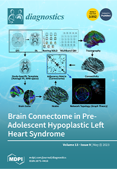
Patients with hypoplastic left heart syndrome who have been palliated with the Fontan procedure are at risk for adverse neurodevelopmental outcomes. We describe the methods and challenges of a multi-center observational study, SVRIII (Single Ventricle Reconstruction Trial) Brain Connectome. We aimed to obtain Diffusion Tensor and Resting-BOLD Imaging in 140 SVR III participants and 100 healthy controls. Brain connectome measures will be correlated with neurocognitive measures and clinical risk factors. Enrollment challenges were addressed by (1) adding study sites, (2) site coordinators' meetings, and (3) research registries and community-based groups for control recruitment. Technical challenges included harmonization and transfer of images. These hurdles were successfully overcome with frequent site visits that involved human and synthetic phantoms. View this paper
  • Issues are regarded as officially published after their release is announced to the table of contents alert mailing list.
  • You may sign up for e-mail alerts to receive table of contents of newly released issues.
  • PDF is the official format for papers published in both, html and pdf forms. To view the papers in pdf format, click on the "PDF Full-text" link, and use the free Adobe Reader to open them.
Order results
Result details
Section
Select all
Export citation of selected articles as:
19 pages, 6949 KiB  
Review
Liver Transplant in Patients with Hepatocarcinoma: Imaging Guidelines and Future Perspectives Using Artificial Intelligence
by Mihai Dan Pomohaci, Mugur Cristian Grasu, Radu Lucian Dumitru, Mihai Toma and Ioana Gabriela Lupescu
Diagnostics 2023, 13(9), 1663; https://doi.org/10.3390/diagnostics13091663 - 08 May 2023
Cited by 1 | Viewed by 2077
Abstract
Hepatocellular carcinoma is the most common primary malignant hepatic tumor and occurs most often in the setting of chronic liver disease. Liver transplantation is a curative treatment option and is an ideal solution because it solves the chronic underlying liver disorder while removing [...] Read more.
Hepatocellular carcinoma is the most common primary malignant hepatic tumor and occurs most often in the setting of chronic liver disease. Liver transplantation is a curative treatment option and is an ideal solution because it solves the chronic underlying liver disorder while removing the malignant lesion. However, due to organ shortages, this treatment can only be applied to carefully selected patients according to clinical guidelines. Artificial intelligence is an emerging technology with multiple applications in medicine with a predilection for domains that work with medical imaging, like radiology. With the help of these technologies, laborious tasks can be automated, and new lesion imaging criteria can be developed based on pixel-level analysis. Our objectives are to review the developing AI applications that could be implemented to better stratify liver transplant candidates. The papers analysed applied AI for liver segmentation, evaluation of steatosis, sarcopenia assessment, lesion detection, segmentation, and characterization. A liver transplant is an optimal treatment for patients with hepatocellular carcinoma in the setting of chronic liver disease. Furthermore, AI could provide solutions for improving the management of liver transplant candidates to improve survival. Full article
Show Figures

Figure 1

17 pages, 2673 KiB  
Article
A Novel Deep Transfer Learning-Based Approach for Automated Pes Planus Diagnosis Using X-ray Image
by Yeliz Gül, Süleyman Yaman, Derya Avcı, Atilla Hikmet Çilengir, Mehtap Balaban and Hasan Güler
Diagnostics 2023, 13(9), 1662; https://doi.org/10.3390/diagnostics13091662 - 08 May 2023
Viewed by 1894
Abstract
Pes planus, colloquially known as flatfoot, is a deformity defined as the collapse, flattening or loss of the medial longitudinal arch of the foot. The first standard radiographic examination for diagnosing pes planus involves lateral and dorsoplantar weight-bearing radiographs. Recently, many artificial intelligence-based [...] Read more.
Pes planus, colloquially known as flatfoot, is a deformity defined as the collapse, flattening or loss of the medial longitudinal arch of the foot. The first standard radiographic examination for diagnosing pes planus involves lateral and dorsoplantar weight-bearing radiographs. Recently, many artificial intelligence-based computer-aided diagnosis (CAD) systems and models have been developed for the detection of various diseases from radiological images. However, to the best of our knowledge, no model and system has been proposed in the literature for automated pes planus diagnosis using X-ray images. This study presents a novel deep learning-based model for automated pes planus diagnosis using X-ray images, a first in the literature. To perform this study, a new pes planus dataset consisting of weight-bearing X-ray images was collected and labeled by specialist radiologists. In the preprocessing stage, the number of X-ray images was augmented and then divided into 4 and 16 patches, respectively in a pyramidal fashion. Thus, a total of 21 images are obtained for each image, including 20 patches and one original image. These 21 images were then fed to the pre-trained MobileNetV2 and 21,000 features were extracted from the Logits layer. Among the extracted deep features, the most important 1312 features were selected using the proposed iterative ReliefF algorithm, and then classified with support vector machine (SVM). The proposed deep learning-based framework achieved 95.14% accuracy using 10-fold cross validation. The results demonstrate that our transfer learning-based model can be used as an auxiliary tool for diagnosing pes planus in clinical practice. Full article
(This article belongs to the Special Issue Classification of Diseases Using Machine Learning Algorithms)
Show Figures

Figure 1

12 pages, 1687 KiB  
Article
Trends in the Use of Second-Generation Androgen Receptor Axis Inhibitors for Metastatic Hormone-Sensitive Prostate Cancer and Clinical Factors Predicting Biological Recurrence
by Keita Nakane, Hiromitsu Watanabe, Taku Naiki, Kiyoshi Takahara, Takahiro Yasui, Hideaki Miyake, Ryoichi Shiroki and Takuya Koie
Diagnostics 2023, 13(9), 1661; https://doi.org/10.3390/diagnostics13091661 - 08 May 2023
Cited by 1 | Viewed by 1660
Abstract
The advent of second-generation androgen receptor axis-targeted agents (ARATs) has revolutionized the treatment of metastatic hormone-sensitive prostate cancer (mHSPC). Biochemical recurrence-free survival (BRFS) was used to compare the efficacy of each ARAT. This multicenter retrospective study included 581 patients with newly diagnosed mHSPC [...] Read more.
The advent of second-generation androgen receptor axis-targeted agents (ARATs) has revolutionized the treatment of metastatic hormone-sensitive prostate cancer (mHSPC). Biochemical recurrence-free survival (BRFS) was used to compare the efficacy of each ARAT. This multicenter retrospective study included 581 patients with newly diagnosed mHSPC who received first-line hormone therapy. The characteristics of patients treated with different ARATs were compared as well as changes in the usage of each drug over time. For BRFS, the apalutamide (Apa) and enzalutamide (Enza) groups, as well as the abiraterone acetate (Abi) and Apa/Enza groups, were compared. In addition, multivariate analysis was performed to determine predictive factors for biochemical recurrence (BCR). The use of second-generation ARATs tended to increase after May 2020. No significant difference in BRFS was found between patients receiving Apa and Enza (p = 0.490) and those receiving Abi or Apa/Enza (p = 0.906). Multivariate analysis revealed that the neutrophil-to-lymphocyte ratio (NLR) ≥ 2.76 and PSA ≥ 0.550 ng/mL were independent predictors of BCR. There were no significant differences in patient characteristics or BRFS in patients with mHSPC receiving different ARATs as first-line treatment. NLR and PSA may be prognostic factors following the first-line treatment of patients with mHSPC. Full article
(This article belongs to the Special Issue Advances in the Diagnosis and Management of Prostate Cancer)
Show Figures

Figure 1

15 pages, 3249 KiB  
Article
Microembolizations in the Arterial Cerebral Circulation in Patients with Atrial Fibrillation Ablation Using the Cryoballoon Technique—Protocol and Methodology of a Prospective Observational Study
by Damir Erkapic, Marko Aleksic, Konstantinos Roussopoulos, Kay Felix Weipert, Korkut Sözener, Karel Kostev, Jens Allendörfer, Josef Rosenbauer, Dursun Guenduez and Christian Tanislav
Diagnostics 2023, 13(9), 1660; https://doi.org/10.3390/diagnostics13091660 - 08 May 2023
Viewed by 1069
Abstract
There is considerable uncertainty regarding the impact of microembolic signals (MESs) on neuropsychological abilities in patients receiving pulmonary vein isolation and beyond using the cryoballoon technique. We conducted the largest prospective observational study on this topic, providing insights into the gradual unmasking of [...] Read more.
There is considerable uncertainty regarding the impact of microembolic signals (MESs) on neuropsychological abilities in patients receiving pulmonary vein isolation and beyond using the cryoballoon technique. We conducted the largest prospective observational study on this topic, providing insights into the gradual unmasking of procedure-related MESs and their impacts on neuropsychological outcomes. MESs were continuously detected periprocedurally using transcranial Doppler ultrasonography. Neuropsychological status was evaluated comprehensively using the CERAD Plus test battery, which consists of 11 neuropsychological subtests. Patients with atrial fibrillation were included in the study with an equal distribution (50:50) of paroxysmal or persistent presentations. Of 167 consecutive eligible patients, 100 were included within the study enrollment period from February 2021 to August 2022. The study, including the documentation of all follow-up visits, ended in November 2022. This paper focuses on describing the study protocol and methodology and presenting the baseline data. Full article
(This article belongs to the Special Issue Advances in Diagnosis and Treatment of Cardiac Arrhythmias)
Show Figures

Figure 1

11 pages, 276 KiB  
Article
Papillomavirus Infection as Potential Cause of Miscarriage in the Early Gestational Age: A Prospective Study
by Maria Teresa Bruno, Salvatore Caruso, Guido Scalia, Maria Costanzo, Salvatore Di Pasqua, Sara Boemi, Marco Marzio Panella and Marco Palumbo
Diagnostics 2023, 13(9), 1659; https://doi.org/10.3390/diagnostics13091659 - 08 May 2023
Cited by 2 | Viewed by 1360
Abstract
The possible association between human papillomavirus (HPV) infection and negative pregnancy outcomes has been debated in the literature, with conflicting results from clinical trials. While some authors support a link between HPV and miscarriage, others argue that the mere detection of the virus [...] Read more.
The possible association between human papillomavirus (HPV) infection and negative pregnancy outcomes has been debated in the literature, with conflicting results from clinical trials. While some authors support a link between HPV and miscarriage, others argue that the mere detection of the virus does not necessarily indicate a causal relationship with negative pregnancy outcomes. In this study, we conducted a prospective, controlled investigation of the potential association between HPV infection and miscarriage. Our study included 59 women who had experienced a miscarriage and 57 women who had undergone voluntary termination of pregnancy (TOP) within the 12th week of gestation. We assessed HPV prevalence, maternal age, and HPV genotype in both groups and evaluated the relationship between these factors and pregnancy outcome. Unlike previous studies that only identified HPV in cases of abortion, we also correlated the positivity of chorionic villi with gestational age in both groups. We found a close correlation between positive chorionic villi and very early gestational age, with all 13 cases of virus-positive chorionic villi in the miscarriage group occurring in gestational periods of less than 8 + 5 weeks (<60 days) (RR = 28.6). Our analysis showed no correlation between HPV infection and maternal age or viral genotypes. The results suggest that the presence of HPV alone is not enough to cause spontaneous abortion, but a high viral load in early pregnancy may increase the risk of negative outcomes. These findings have important implications for the management of HPV infection during pregnancy and may provide a rationale for the use of HPV vaccines to reduce the incidence of spontaneous abortion and infertility due to preclinical spontaneous abortions. Full article
(This article belongs to the Section Pathology and Molecular Diagnostics)
26 pages, 48566 KiB  
Article
SAA-UNet: Spatial Attention and Attention Gate UNet for COVID-19 Pneumonia Segmentation from Computed Tomography
by Shroog Alshomrani, Muhammad Arif and Mohammed A. Al Ghamdi
Diagnostics 2023, 13(9), 1658; https://doi.org/10.3390/diagnostics13091658 - 08 May 2023
Cited by 1 | Viewed by 1523
Abstract
The disaster of the COVID-19 pandemic has claimed numerous lives and wreaked havoc on the entire world due to its transmissible nature. One of the complications of COVID-19 is pneumonia. Different radiography methods, particularly computed tomography (CT), have shown outstanding performance in effectively [...] Read more.
The disaster of the COVID-19 pandemic has claimed numerous lives and wreaked havoc on the entire world due to its transmissible nature. One of the complications of COVID-19 is pneumonia. Different radiography methods, particularly computed tomography (CT), have shown outstanding performance in effectively diagnosing pneumonia. In this paper, we propose a spatial attention and attention gate UNet model (SAA-UNet) inspired by spatial attention UNet (SA-UNet) and attention UNet (Att-UNet) to deal with the problem of infection segmentation in the lungs. The proposed method was applied to the MedSeg, Radiopaedia 9P, combination of MedSeg and Radiopaedia 9P, and Zenodo 20P datasets. The proposed method showed good infection segmentation results (two classes: infection and background) with an average Dice similarity coefficient of 0.85, 0.94, 0.91, and 0.93 and a mean intersection over union (IOU) of 0.78, 0.90, 0.86, and 0.87, respectively, on the four datasets mentioned above. Moreover, it also performed well in multi-class segmentation with average Dice similarity coefficients of 0.693, 0.89, 0.87, and 0.93 and IOU scores of 0.68, 0.87, 0.78, and 0.89 on the four datasets, respectively. Classification accuracies of more than 97% were achieved for all four datasets. The F1-scores for the MedSeg, Radiopaedia P9, combination of MedSeg and Radiopaedia P9, and Zenodo 20P datasets were 0.865, 0.943, 0.917, and 0.926, respectively, for the binary classification. For multi-class classification, accuracies of more than 96% were achieved on all four datasets. The experimental results showed that the framework proposed can effectively and efficiently segment COVID-19 infection on CT images with different contrast and utilize this to aid in diagnosing and treating pneumonia caused by COVID-19. Full article
Show Figures

Figure 1

11 pages, 1569 KiB  
Article
Clinical Validation of an Artificial Intelligence Model for Detecting Distal Radius, Ulnar Styloid, and Scaphoid Fractures on Conventional Wrist Radiographs
by Kyu-Chong Lee, In Cheul Choi, Chang Ho Kang, Kyung-Sik Ahn, Heewon Yoon, Jae-Joon Lee, Baek Hyun Kim and Euddeum Shim
Diagnostics 2023, 13(9), 1657; https://doi.org/10.3390/diagnostics13091657 - 08 May 2023
Cited by 3 | Viewed by 1337
Abstract
This study aimed to assess the feasibility and performance of an artificial intelligence (AI) model for detecting three common wrist fractures: distal radius, ulnar styloid process, and scaphoid. The AI model was trained with a dataset of 4432 images containing both fractured and [...] Read more.
This study aimed to assess the feasibility and performance of an artificial intelligence (AI) model for detecting three common wrist fractures: distal radius, ulnar styloid process, and scaphoid. The AI model was trained with a dataset of 4432 images containing both fractured and non-fractured wrist images. In total, 593 subjects were included in the clinical test. Two human experts independently diagnosed and labeled the fracture sites using bounding boxes to build the ground truth. Two novice radiologists also performed the same task, both with and without model assistance. The sensitivity, specificity, accuracy, and area under the curve (AUC) were calculated for each wrist location. The AUC for detecting distal radius, ulnar styloid, and scaphoid fractures per wrist were 0.903 (95% C.I. 0.887–0.918), 0.925 (95% C.I. 0.911–0.939), and 0.808 (95% C.I. 0.748–0.967), respectively. When assisted by the AI model, the scaphoid fracture AUC of the two novice radiologists significantly increased from 0.75 (95% C.I. 0.66–0.83) to 0.85 (95% C.I. 0.77–0.93) and from 0.71 (95% C.I. 0.62–0.80) to 0.80 (95% C.I. 0.71–0.88), respectively. Overall, the developed AI model was found to be reliable for detecting wrist fractures, particularly for scaphoid fractures, which are commonly missed. Full article
(This article belongs to the Section Machine Learning and Artificial Intelligence in Diagnostics)
Show Figures

Figure 1

16 pages, 3235 KiB  
Review
Sexually Transmitted Diseases—An Update and Overview of Current Research
by Kristina Wihlfahrt, Veronika Günther, Werner Mendling, Anna Westermann, Damaris Willer, Georgios Gitas, Zino Ruchay, Nicolai Maass, Leila Allahqoli and Ibrahim Alkatout
Diagnostics 2023, 13(9), 1656; https://doi.org/10.3390/diagnostics13091656 - 08 May 2023
Cited by 1 | Viewed by 2327
Abstract
A rise in the rates of sexually transmitted diseases, both worldwide and in Germany, has been observed especially among persons between the ages of 15 and 24 years. Since many infections are devoid of symptoms or cause few symptoms, the diseases are detected [...] Read more.
A rise in the rates of sexually transmitted diseases, both worldwide and in Germany, has been observed especially among persons between the ages of 15 and 24 years. Since many infections are devoid of symptoms or cause few symptoms, the diseases are detected late, may spread unchecked, and be transmitted unwittingly. In the event of persistent infection, the effects depend on the pathogen in question. Manifestations vary widely, ranging from pelvic inflammatory disease, most often caused by Chlamydia trachomatis (in Germany nearly 30% of PID) or Neisseria gonorrhoeae (in Germany <2% of PID), to the development of genital warts or cervical dysplasia in cases of infection with the HP virus. Causal treatment does exist in most cases and should always be administered to the sexual partner(s) as well. An infection during pregnancy calls for an individual treatment approach, depending on the pathogen and the week of pregnancy. Full article
Show Figures

Figure 1

10 pages, 474 KiB  
Systematic Review
The Role of Single Positive Cultures in Presumed Aseptic Total Hip and Knee Revision Surgery—A Systematic Review of the Literature
by Jan Schwarze, Burkhard Moellenbeck, Georg Gosheger, Jan Puetzler, Niklas Deventer, Tobias Kalisch, Kristian Nikolaus Schneider, Sebastian Klingebiel and Christoph Theil
Diagnostics 2023, 13(9), 1655; https://doi.org/10.3390/diagnostics13091655 - 08 May 2023
Cited by 1 | Viewed by 1219
Abstract
(1) Background: Prior to revision hip (THA) or knee arthroplasty (TKA), periprosthetic low-grade infection (PJI) should be ruled out. Despite advances in preoperative diagnosis, unsuspected positive cultures (UPCs) may occur in initially planned aseptic revisions. Particularly, single UPCs pose a diagnostic and therapeutic [...] Read more.
(1) Background: Prior to revision hip (THA) or knee arthroplasty (TKA), periprosthetic low-grade infection (PJI) should be ruled out. Despite advances in preoperative diagnosis, unsuspected positive cultures (UPCs) may occur in initially planned aseptic revisions. Particularly, single UPCs pose a diagnostic and therapeutic dilemma, as their impact on outcome is unclear and recommendations are heterogeneous. This review investigates the frequency of single UPCs and their impact on implant survivorship. (2) Methods: In July 2022, a comprehensive literature search was performed using PubMed and Cochrane Library search. In total, 197 articles were screened. Seven retrospective studies with a total of 5821 cases were able to be included in this review. (3) Results: Based on the cases included, UPCs were found in 794/5821 cases (14%). In 530/794 cases (67%), the majority of the UPCs were single positive. The most commonly isolated pathogens were coagulase negative Staphylococci and Cutibacterium acnes. Five of seven studies reported no influence on revision- or infection-free survival following a single positive culture. In two studies, single UPCs following THA revision were correlated with subsequent re-revision for PJI. (4) Conclusions: Single UPCs of a non-virulent pathogen following presumed aseptic TKA revision may be interpreted as contaminants. A single UPC following THA revision may be a risk factor for subsequent PJI. The role of systemic antibiotic treatment remains unclear, but it should be considered if other risk factors for PJI are present. Full article
(This article belongs to the Special Issue Diagnosis and Treatment of Periprosthetic Joint Infections)
Show Figures

Figure 1

22 pages, 5825 KiB  
Article
Automatic Analysis of MRI Images for Early Prediction of Alzheimer’s Disease Stages Based on Hybrid Features of CNN and Handcrafted Features
by Ahmed Khalid, Ebrahim Mohammed Senan, Khalil Al-Wagih, Mamoun Mohammad Ali Al-Azzam and Ziad Mohammad Alkhraisha
Diagnostics 2023, 13(9), 1654; https://doi.org/10.3390/diagnostics13091654 - 08 May 2023
Cited by 9 | Viewed by 1950
Abstract
Alzheimer’s disease (AD) is considered one of the challenges facing health care in the modern century; until now, there has been no effective treatment to cure it, but there are drugs to slow its progression. Therefore, early detection of Alzheimer’s is vital to [...] Read more.
Alzheimer’s disease (AD) is considered one of the challenges facing health care in the modern century; until now, there has been no effective treatment to cure it, but there are drugs to slow its progression. Therefore, early detection of Alzheimer’s is vital to take needful measures before it develops into brain damage which cannot be treated. Magnetic resonance imaging (MRI) techniques have contributed to the diagnosis and prediction of its progression. MRI images require highly experienced doctors and radiologists, and the analysis of MRI images takes time to analyze each slice. Thus, deep learning techniques play a vital role in analyzing a huge amount of MRI images with high accuracy to detect Alzheimer’s and predict its progression. Because of the similarities in the characteristics of the early stages of Alzheimer’s, this study aimed to extract the features in several methods and integrate the features extracted from more than one method into the same features matrix. This study contributed to the development of three methodologies, each with two systems, with all systems aimed at achieving satisfactory accuracy for the detection of AD and predicting the stages of its progression. The first methodology is by Feed Forward Neural Network (FFNN) with the features of GoogLeNet and DenseNet-121 models separately. The second methodology is by FFNN network with combined features between GoogLeNet and Dense-121 models before and after high-dimensionality reduction of features using the Principal Component Analysis (PCA) algorithm. The third methodology is by FFNN network with combined features between GoogLeNet and Dense-121 models separately and features extracted by Discrete Wavelet Transform (DWT), Local Binary Pattern (LBP) and Gray Level Co-occurrence Matrix (GLCM) methods called handcrafted features. All systems yielded super results in detecting AD and predicting the stages of its progression. With the combined features of the DenseNet-121 and handcrafted, the FFNN achieved an accuracy of 99.7%, sensitivity of 99.64%, AUC of 99.56%, precision of 99.63%, and a specificity of 99.67%. Full article
(This article belongs to the Special Issue Artificial Intelligence in Alzheimer’s Disease Diagnosis)
Show Figures

Figure 1

18 pages, 3942 KiB  
Article
MMP-13, VEGF, and Disease Activity in a Cohort of Rheumatoid Arthritis Patients
by Mihail Virgil Boldeanu, Lidia Boldeanu, Oana Mariana Cristea, Dana Alexandra Ciobanu, Sabin Ioan Poenariu, Anda Lorena Dijmărescu, Andreea Lili Bărbulescu, Vlad Pădureanu, Teodor Nicuşor Sas, Ștefan Cristian Dinescu, Florentin Ananu Vreju, Horațiu Valeriu Popoviciu and Răzvan Adrian Ionescu
Diagnostics 2023, 13(9), 1653; https://doi.org/10.3390/diagnostics13091653 - 08 May 2023
Viewed by 1790
Abstract
Identifying certain serum biomarkers associated with the degree of rheumatoid arthritis (RA) activity can provide us with a more accurate view of the evolution, prognosis, and future quality of life for these patients. Our aim was to analyze the presence and clinical use [...] Read more.
Identifying certain serum biomarkers associated with the degree of rheumatoid arthritis (RA) activity can provide us with a more accurate view of the evolution, prognosis, and future quality of life for these patients. Our aim was to analyze the presence and clinical use of matrix metalloproteinase-13 (MMP-13), along with vascular endothelial growth factor (VEGF) and well-known cytokines such as tumor necrosis factor-alpha (TNF-α) and interleukin 6 (IL-6) for patients with RA. We also wanted to identify the possible correlations between MMP-13 and these serological markers, as well as their relationship with disease activity indices, quality of life, and ultrasonographic evaluation. For this purpose, we analyzed serum samples of 34 RA patients and 12 controls. In order to assess serum concentrations for MMP-13, VEGF, TNF-α, and IL-6, we used the enzyme-linked immunosorbent assay (ELISA) technique. Our results concluded that higher levels of MMP-13, VEGF, TNF-α, and IL-6 were present in the serum of RA patients compared to controls, with statistical significance. We furthermore identified moderately positive correlations between VEGF, MMP-13, and disease activity indices, as well as with the ultrasound findings. We also observed that VEGF had the best accuracy (97.80%), for differentiating patients with moderate disease activity. According to the data obtained in our study, that although MMP-13, TNF-α and C-reactive protein (CRP) have the same sensitivity (55.56%), MMP-13 has a better specificity (86.67%) in the diagnosis of patients with DAS28(4v) CRP values corresponding to moderate disease activity. Thus, MMP-13 can be used as a biomarker that can differentiate patients with moderate or low disease activity. VEGF and MMP-13 can be used as additional parameters, along with TNF-α and IL-6, that can provide the clinician a better picture of the inflammatory process, disease activity, and structural damage in patients with RA. Our data can certainly constitute a start point for future research and extended studies with multicenter involvement, to support the selection of individualized and accurate therapeutic management strategies for our patients. Full article
(This article belongs to the Special Issue Recent Advances in Diagnosis and Treatment in Rheumatology)
Show Figures

Figure 1

13 pages, 4290 KiB  
Article
An Efficient and Robust Method for Chest X-ray Rib Suppression That Improves Pulmonary Abnormality Diagnosis
by Di Xu, Qifan Xu, Kevin Nhieu, Dan Ruan and Ke Sheng
Diagnostics 2023, 13(9), 1652; https://doi.org/10.3390/diagnostics13091652 - 08 May 2023
Cited by 2 | Viewed by 1760
Abstract
Background: Suppression of thoracic bone shadows on chest X-rays (CXRs) can improve the diagnosis of pulmonary disease. Previous approaches can be categorized as either unsupervised physical models or supervised deep learning models. Physical models can remove the entire ribcage and preserve the morphological [...] Read more.
Background: Suppression of thoracic bone shadows on chest X-rays (CXRs) can improve the diagnosis of pulmonary disease. Previous approaches can be categorized as either unsupervised physical models or supervised deep learning models. Physical models can remove the entire ribcage and preserve the morphological lung details but are impractical due to the extremely long processing time. Machine learning (ML) methods are computationally efficient but are limited by the available ground truth (GT) for effective and robust training, resulting in suboptimal results. Purpose: To improve bone shadow suppression, we propose a generalizable yet efficient workflow for CXR rib suppression by combining physical and ML methods. Materials and Method: Our pipeline consists of two stages: (1) pair generation with GT bone shadows eliminated by a physical model in spatially transformed gradient fields; and (2) a fully supervised image denoising network trained on stage-one datasets for fast rib removal from incoming CXRs. For stage two, we designed a densely connected network called SADXNet, combined with a peak signal-to-noise ratio and a multi-scale structure similarity index measure as the loss function to suppress the bony structures. SADXNet organizes the spatial filters in a U shape and preserves the feature map dimension throughout the network flow. Results: Visually, SADXNet can suppress the rib edges near the lung wall/vertebra without compromising the vessel/abnormality conspicuity. Quantitively, it achieves an RMSE of ~0 compared with the physical model generated GTs, during testing with one prediction in <1 s. Downstream tasks, including lung nodule detection as well as common lung disease classification and localization, are used to provide task-specific evaluations of our rib suppression mechanism. We observed a 3.23% and 6.62% AUC increase, as well as 203 (1273 to 1070) and 385 (3029 to 2644) absolute false positive decreases for lung nodule detection and common lung disease localization, respectively. Conclusion: Through learning from image pairs generated from the physical model, the proposed SADXNet can make a robust sub-second prediction without losing fidelity. Quantitative outcomes from downstream validation further underpin the superiority of SADXNet and the training ML-based rib suppression approaches from the physical model yielded dataset. The training images and SADXNet are provided in the manuscript. Full article
(This article belongs to the Special Issue Advances in Chest Imaging Diagnostics)
Show Figures

Figure 1

12 pages, 3016 KiB  
Article
A Convolutional Neural Network Architecture for Segmentation of Lung Diseases Using Chest X-ray Images
by Adel Sulaiman, Vatsala Anand, Sheifali Gupta, Yousef Asiri, M. A. Elmagzoub, Mana Saleh Al Reshan and Asadullah Shaikh
Diagnostics 2023, 13(9), 1651; https://doi.org/10.3390/diagnostics13091651 - 08 May 2023
Cited by 8 | Viewed by 1592
Abstract
The segmentation of lungs from medical images is a critical step in the diagnosis and treatment of lung diseases. Deep learning techniques have shown great promise in automating this task, eliminating the need for manual annotation by radiologists. In this research, a convolution [...] Read more.
The segmentation of lungs from medical images is a critical step in the diagnosis and treatment of lung diseases. Deep learning techniques have shown great promise in automating this task, eliminating the need for manual annotation by radiologists. In this research, a convolution neural network architecture is proposed for lung segmentation using chest X-ray images. In the proposed model, concatenate block is embedded to learn a series of filters or features used to extract meaningful information from the image. Moreover, a transpose layer is employed in the concatenate block to improve the spatial resolution of feature maps generated by a prior convolutional layer. The proposed model is trained using k-fold validation as it is a powerful and flexible tool for evaluating the performance of deep learning models. The proposed model is evaluated on five different subsets of the data by taking the value of k as 5 to obtain the optimized model to obtain more accurate results. The performance of the proposed model is analyzed for different hyper-parameters such as the batch size as 32, optimizer as Adam and 40 epochs. The dataset used for the segmentation of disease is taken from the Kaggle repository. The various performance parameters such as accuracy, IoU, and dice coefficient are calculated, and the values obtained are 0.97, 0.93, and 0.96, respectively. Full article
(This article belongs to the Special Issue Artificial Intelligence in Lung Diseases 2.0)
Show Figures

Figure 1

16 pages, 2054 KiB  
Systematic Review
Perianal Basal Cell Carcinoma—A Systematic Review and Meta-Analysis of Real-World Data
by Tzong-Yun Tsai, Chun-Kai Liao, Bang-Yan Zhang, Yen-Lin Huang, Wen-Sy Tsai, Jeng-Fu You, Chien-Yuh Yeh and Pao-Shiu Hsieh
Diagnostics 2023, 13(9), 1650; https://doi.org/10.3390/diagnostics13091650 - 08 May 2023
Viewed by 2599
Abstract
(1) Background: BCC is a sporadic disease that develops in areas of the skin not exposed to the sun. Perianal BCC, which occurs in the anorectal region, accounts for less than 0.2% of all BCC cases. There have been only a few reported [...] Read more.
(1) Background: BCC is a sporadic disease that develops in areas of the skin not exposed to the sun. Perianal BCC, which occurs in the anorectal region, accounts for less than 0.2% of all BCC cases. There have been only a few reported cases of the disease, with fewer than 200 cases reported in total. Given the diagnostic challenges and potential for misdiagnosis, we conducted a systematic review of perianal basal cell carcinoma using real-world data to provide comprehensive and detailed information on the disease. (2) Methods: The study was reported based on the Preferred Reporting Items for Systematic Reviews and Meta-Analyses (PRISMA) guidelines, 2020. Patients’ clinical pathologic features, tumor characteristics, treatment modalities, and outcomes were presented. (3) Results: The results of 41 studies involving 140 patients were analyzed. The most common symptoms reported by patients at presentation were anorectal bleeding, pain, and pruritus. Ulceration was the most frequently observed tumor characteristic. The majority of patients underwent local excision as their primary treatment, with only eight patients experiencing a recurrence. Our analysis did not reveal any statistically significant differences in the outcomes of different treatment modalities. (4) Conclusions: Identifying perianal BCC poses a significant challenge as it closely resembles other anal diseases, thereby making it difficult to differentiate between the different conditions. However, a wide local excision with clear margins is considered an effective treatment option for most patients. Alternative treatments, such as radiotherapy, may be recommended for patients who are unable to undergo surgery. Full article
(This article belongs to the Section Pathology and Molecular Diagnostics)
Show Figures

Figure 1

9 pages, 942 KiB  
Article
Evaluation of Signal Transducer and Activator of Transcription 3 (STAT-3) Protein Expression in Non-Hodgkin Lymphoma Cases in Hospital USM
by Izyan Rifhana Muhamad, Noorul Balqis Che Ibrahim and Faezahtul Arbaeyah Hussain
Diagnostics 2023, 13(9), 1649; https://doi.org/10.3390/diagnostics13091649 - 07 May 2023
Viewed by 1144
Abstract
Background: Evolving targeted therapy on Janus Associated Kinase-Signal Transducer and Activator of Transcription (JAK-STAT) signaling pathway, especially pertaining to STAT-3 protein in non-Hodgkin lymphoma (NHL), provides new treatment strategies. STAT-3 protein also relates to the prognostication of NHL. Hence, we aimed to evaluate [...] Read more.
Background: Evolving targeted therapy on Janus Associated Kinase-Signal Transducer and Activator of Transcription (JAK-STAT) signaling pathway, especially pertaining to STAT-3 protein in non-Hodgkin lymphoma (NHL), provides new treatment strategies. STAT-3 protein also relates to the prognostication of NHL. Hence, we aimed to evaluate the expression of STAT-3 protein in NHL cases diagnosed in Hospital Universiti Sains Malaysia (USM). Methods: A retrospective cross sectional study using formalin fixed paraffin embedded (FFPE) tissue blocks of 95 NHL cases were obtained. STAT-3 immunostaining was performed and evaluated. The proportion and association between the expression of STAT-3 protein with subtypes of NHL were statistically analyzed. Results: The majority of the cases (78.9%) had positive STAT-3 protein expression. 64.2% were among aggressive B cell NHL, whilst 20.0% of them were diffuse large B cell lymphoma, a non-germinal center B subtype (DLBCL-NGCB). There is also an association between STAT-3 protein expression with DLBCL subtypes (p = 0.046). Conclusion: Our study demonstrated a remarkable expression of STAT-3 protein in NHL, in which DLBCL subtypes had significant association. A larger scale study with a combination of JAK protein evaluation should be undertaken in the future. Full article
(This article belongs to the Special Issue Haematology: Diagnosis and Management)
Show Figures

Figure 1

12 pages, 4463 KiB  
Article
Early Retinal Microvascular Alterations in Young Type 1 Diabetic Patients without Clinical Retinopathy
by Alexandra Oltea Dan, Alin Ștefănescu-Dima, Andrei Teodor Bălășoiu, Ileana Puiu, Carmen Luminița Mocanu, Mihaela Ionescu, Andreea Cornelia Tănasie, Anca Elena Târtea and Veronica Sfredel
Diagnostics 2023, 13(9), 1648; https://doi.org/10.3390/diagnostics13091648 - 07 May 2023
Cited by 4 | Viewed by 1298
Abstract
The purpose of this study is to identify and quantify preclinical changes with the help of optical coherence tomography angiography (OCTA) within the retinal microcirculation of young type 1 diabetes (T1D) patients without clinical signs of diabetic retinopathy (DR) and to compare these [...] Read more.
The purpose of this study is to identify and quantify preclinical changes with the help of optical coherence tomography angiography (OCTA) within the retinal microcirculation of young type 1 diabetes (T1D) patients without clinical signs of diabetic retinopathy (DR) and to compare these results with those obtained from healthy age-matched subjects. OCTA is currently used for monitoring diabetic retinopathy; however, there is no current consensus on which OCTA parameter alterations predict the first clinical signs of diabetic retinopathy. The main challenge that young patients with T1D face during the course of the disease is that they can rapidly progress to the development of DR, especially during adolescence. Moreover, they also present an increased risk of rapid progression toward advanced stages of DR and vision loss compared to type 2 diabetes patients, indicating the importance of early diagnosis and intervention. The limitations of the currently used screening procedures that led to the conceptualization of our study are the difficulties in performing fluorescein angiography tests for diagnosing the clinical signs of DR on young patients, namely the invasive procedure of dye injection, the risk of allergic reactions and the long duration of the examination. Moreover, given the long life expectancy of young T1D patients, it is essential to identify the preclinical changes in retinal microvasculature before reaching the first clinical signs quantifiable by FFA. The clinical study enrolled 119 subjects aged between 4 and 30 years old with a mean age of 13 years old, comprising 61 T1D patients with a mean duration of the disease of 4 years and 8 months and 58 healthy age-matched subjects for the control group. OCTA scans were performed using the RevoNX 130 OCTA device (Optopol) to evaluate the following retinal parameters: foveal avascular zone (FAZ) area, perimeter and circularity, overall foveal thickness, and superficial and deep vessel densities. Statistically significant differences between the two groups were identified for the following parameters: the FAZ area in the T1D group (0.42 ± 0.17) was larger than the control group (0.26 ± 0.080), the FAZ circularity (0.41 ± 0.11) was decreased compared to the control group (0.61 ± 0.08) and the FAZ perimeter was larger (3.63 ± 0.97) compared to the control group (2.30 ± 0.50). The overall foveal thickness was decreased in the T1D group (222.98 ± 17.33) compared to the control group (230.64 ± 20.82). The total vessel density of the superficial capillary plexus (SCP) on an investigated area of 6 X 6 mm centered around the fovea was decreased in the T1D group (37.4164 ± 2.14) compared to the control group (38.0241 ± 2.44). Our data suggest that specific imaging biomarkers such as FAZ perimeter, area and circularity, decreased overall foveal thickness and decreased vessel density in the SCP precede the clinical diagnosis of DR in young T1D patients and represent useful parameters in quantifying capillary nonperfusion in T1D patients without clinical signs of DR. Full article
(This article belongs to the Section Optical Diagnostics)
Show Figures

Figure 1

21 pages, 6709 KiB  
Review
Pitfalls of Diffusion-Weighted Imaging: Clinical Utility of T2 Shine-through and T2 Black-out for Musculoskeletal Diseases
by Yuri Kim, Seul Ki Lee, Jee-Young Kim and Jun-Ho Kim
Diagnostics 2023, 13(9), 1647; https://doi.org/10.3390/diagnostics13091647 - 07 May 2023
Cited by 5 | Viewed by 4358
Abstract
Diffusion-weighted imaging (DWI) with an apparent diffusion coefficient (ADC) value is a relatively new magnetic resonance imaging (MRI) sequence that provides functional information on the lesion by measuring the microscopic movement of water molecules. While numerous studies have evaluated the promising role of [...] Read more.
Diffusion-weighted imaging (DWI) with an apparent diffusion coefficient (ADC) value is a relatively new magnetic resonance imaging (MRI) sequence that provides functional information on the lesion by measuring the microscopic movement of water molecules. While numerous studies have evaluated the promising role of DWI in musculoskeletal radiology, most have focused on tumorous diseases related to cellularity. This review article aims to summarize DWI-acquisition techniques, considering pitfalls such as T2 shine-through and T2 black-out, and their usefulness in interpreting musculoskeletal diseases with imaging. DWI is based on the Brownian motion of water molecules within the tissue, achieved by applying diffusion-sensitizing gradients. Regardless of the cellularity of the lesion, several pitfalls must be considered when interpreting DWI with ADC values in musculoskeletal radiology. This review discusses the application of DWI in musculoskeletal diseases, including tumor and tumor mimickers, as well as non-tumorous diseases, with a focus on lesions demonstrating T2 shine-through and T2 black-out effects. Understanding these pitfalls of DWI can provide clinically useful information, increase diagnostic accuracy, and improve patient management when added to conventional MRI in musculoskeletal diseases. Full article
(This article belongs to the Special Issue Diffusion-Weighted Imaging: Technique and Medical Applications)
Show Figures

Figure 1

11 pages, 1261 KiB  
Article
Comparison between Two-Dimensional and Point Shear Wave Elastography Techniques in Evaluating Liver Fibrosis Using Histological Staging as the Reference Standard: A Prospective Pilot Study
by Sang Min Lee, Hong Il Ha, In Jae Lee, Kwanseop Lee, Jung Woo Lee, Ji Won Park, Sung-Eun Kim, Mi Jung Kwon, Ji-Young Choe, Sam-Youl Yoon, Seung-Gu Yeo and Min-Jeong Kim
Diagnostics 2023, 13(9), 1646; https://doi.org/10.3390/diagnostics13091646 - 07 May 2023
Cited by 1 | Viewed by 1807
Abstract
Evaluation of hepatic fibrosis is essential to prevent liver-related morbidity and mortality. Although various types of ultrasound shear wave elastography (SWE) have been used and validated, there are limited studies on the relatively newer technique, two-dimensional SWE (2D-SWE). Therefore, this study aimed to [...] Read more.
Evaluation of hepatic fibrosis is essential to prevent liver-related morbidity and mortality. Although various types of ultrasound shear wave elastography (SWE) have been used and validated, there are limited studies on the relatively newer technique, two-dimensional SWE (2D-SWE). Therefore, this study aimed to compare the diagnostic performances of 2D-SWE and point SWE (p-SWE) for evaluating liver fibrosis using histology as the reference standard. To measure liver stiffness (LS) values, 87 patients underwent 2D-SWE and p-SWE using the same machine. Technical failures and unreliable measurements were also evaluated. The diagnostic performances of 2D-SWE and p-SWE were compared using area under the receiver operating characteristic (AUROC) curve analysis. No technical failures were observed in either method; however, unreliable measurements were less frequent in 2D-SWE (1/87 [1.1%]) than in p-SWE (8/87 [9.2%]) (p < 0.001). The AUROC of the LS values of 2D-SWE were significantly higher than those of p-SWE for diagnosing significant fibrosis (0.965 vs. 0.872, p = 0.022) and cirrhosis (0.994 vs. 0.886, p = 0.042). In conclusion, 2D-SWE is more reliable and accurate than p-SWE for diagnosing hepatic fibrosis. Full article
(This article belongs to the Section Pathology and Molecular Diagnostics)
Show Figures

Figure 1

20 pages, 4365 KiB  
Article
Comparative Study of the Myocardium of Patients from Four COVID-19 Waves
by Lubov Borisovna Mitrofanova, Igor Aleksandrovich Makarov, Andrey Nikolaevich Gorshkov, Andrey Leonidovich Runov, Maxim Sergeevich Vonsky, Maria Mikhailovna Pisareva, Andrey Borisovich Komissarov, Taiana Alekseevna Makarova, Qingli Li, Tatiana Leonidovna Karonova, Alexandra Olegovna Konradi and Evgeny Vladimirovich Shlaykhto
Diagnostics 2023, 13(9), 1645; https://doi.org/10.3390/diagnostics13091645 - 07 May 2023
Cited by 9 | Viewed by 1741
Abstract
Background: Few studies have compared COVID-19 patients from different waves. This study aims to conduct a clinical and morphological analysis of patients who died from COVID-19 during four waves. Methods: The study involved 276 patients who died from COVID-19 during four waves, including [...] Read more.
Background: Few studies have compared COVID-19 patients from different waves. This study aims to conduct a clinical and morphological analysis of patients who died from COVID-19 during four waves. Methods: The study involved 276 patients who died from COVID-19 during four waves, including 77 patients in the first wave, 119 patients in the second wave, and 78 patients in the third wave. We performed a histological examination of myocardium samples from autopsies and additionally analyzed the samples by PCR. We conducted immunohistochemistry of the myocardium for 21 samples using antibodies against CD3, CD45, CD8, CD68, CD34, Ang1, VWF, VEGF, HLA-DR, MHC1, C1q, enteroviral VP1, and SARS-CoV-2 spike protein. We also did immunofluorescent staining of three myocardial specimens using VP1/SARS-CoV-2 antibody cocktails. Further, we ran RT-ddPCR analysis for 14 RNA samples extracted from paraffin-embedded myocardium. Electron microscopic studies of the myocardium were also performed for two samples from the fourth wave. Results: Among the 276 cases, active myocarditis was diagnosed in 5% (15/276). Of these cases, 86% of samples expressed VP1, and individual cells contained SARS-CoV-2 spike protein in 22%. Immunofluorescence confirmed the co-localization of VP1 and SARS-CoV-2 spike proteins. ddPCR did not confidently detect SARS-CoV-2 RNA in the myocardium in any myocarditis cases. However, the myocardium sample from wave IV detected a sub-threshold signal of SARS-CoV-2 by qPCR, but myocarditis in this patient was not confirmed. Electron microscopy showed several single particles similar to SARS-CoV-2 virions on the surface of the endothelium of myocardial vessels. A comparison of the cardiovascular complication incidence between three waves revealed that the incidence of hemorrhage (48 vs. 24 vs. 17%), myocardial necrosis (18 vs. 11 vs. 4%), blood clots in the intramural arteries (12 vs. 7 vs. 0%), and myocarditis (19 vs. 1 vs. 6%) decreased over time, and CD8-T-killers appeared. Immunohistochemistry confirmed the presence of endotheliitis in all 21 studied cases. Conclusions: This study compared myocardial damage in patients who died during three COVID-19 waves and showed a decrease in the incidence of endotheliitis complications (thrombosis, hemorrhage, necrosis) and myocarditis over time. However, the connection between myocarditis and SARS-CoV-2 infection remains unproven. Full article
(This article belongs to the Section Pathology and Molecular Diagnostics)
Show Figures

Figure 1

11 pages, 1891 KiB  
Article
Application of Hybridization Chain Reaction/CRISPR-Cas12a for the Detection of SARS-CoV-2 Infection
by Kate Obaayaa Sagoe, Mutinda Cleophas Kyama, Naomi Maina, Moses Kamita, Muturi Njokah, Kelvin Thiong’o, Bernard N. Kanoi, Ernest Apondi Wandera, Davies Ndegwa, Dickson Mwenda Kinyua and Jesse Gitaka
Diagnostics 2023, 13(9), 1644; https://doi.org/10.3390/diagnostics13091644 - 07 May 2023
Cited by 1 | Viewed by 2160
Abstract
Globally, the emergence of the coronavirus disease (COVID-19) has had a significant impact on life. The need for ongoing SARS-CoV-2 screening employing inexpensive and quick diagnostic approaches is undeniable, given the ongoing pandemic and variations in vaccine administration in resource-constrained regions. This study [...] Read more.
Globally, the emergence of the coronavirus disease (COVID-19) has had a significant impact on life. The need for ongoing SARS-CoV-2 screening employing inexpensive and quick diagnostic approaches is undeniable, given the ongoing pandemic and variations in vaccine administration in resource-constrained regions. This study presents results as proof of concept to use hybridization chain reaction (HCR) and clustered regularly interspaced short palindromic repeats (CRISPR)/Cas12a complex for detecting SARS-CoV-2. HCR hairpin probes were designed using the NUPACK web-based program and further used to amplify the SARS-CoV-2 N gene in archived nasopharyngeal samples. The results were visualized using agarose gels and CRISPR Cas12a-based lateral flow strips. The assay was evaluated using the gold standard, real-time polymerase chain reaction (RT-PCR), as recommended by the World Health Organization (WHO). The results show the comparative efficiency of HCR to RT-PCR. This study shows that HCR and CRISPR are viable alternatives for diagnosing SARS-CoV-2 in samples. Full article
(This article belongs to the Special Issue Point-of-Care Diagnostics (POCD) in Resource-Limited Settings)
Show Figures

Figure 1

16 pages, 1798 KiB  
Review
Diagnosis of Laryngopharyngeal Reflux: Past, Present, and Future—A Mini-Review
by Han-Chung Lien, Ping-Huan Lee and Chen-Chi Wang
Diagnostics 2023, 13(9), 1643; https://doi.org/10.3390/diagnostics13091643 - 07 May 2023
Cited by 4 | Viewed by 3325
Abstract
Laryngopharyngeal reflux (LPR) is a variant of gastroesophageal reflux disease (GERD) in which gastric refluxate irritates the lining of the aerodigestive tract and causes troublesome airway symptoms or complications. LPR is a prevalent disease that creates a significant socioeconomic burden due to its [...] Read more.
Laryngopharyngeal reflux (LPR) is a variant of gastroesophageal reflux disease (GERD) in which gastric refluxate irritates the lining of the aerodigestive tract and causes troublesome airway symptoms or complications. LPR is a prevalent disease that creates a significant socioeconomic burden due to its negative impact on quality of life, tremendous medical expense, and possible cancer risk. Although treatment modalities are similar between LPR and GERD, the diagnosis of LPR is more challenging than GERD due to its non-specific symptoms/signs. Due to the lack of pathognomonic features of endoscopy, mounting evidence focused on physiological diagnostic testing. Two decades ago, a dual pH probe was considered the gold standard for detecting pharyngeal acidic reflux episodes. Despite an association with LPR, the dual pH was unable to predict the treatment response in clinical practice, presumably due to frequently encountered artifacts. Currently, hypopharygneal multichannel intraluminal impedance-pH catheters incorporating two trans-upper esophageal sphincter impedance sensors enable to differentiate pharyngeal refluxes from swallows. The validation of pharyngeal acid reflux episodes that are relevant to anti-reflux treatment is, therefore, crucial. Given no diagnostic gold standard of LPR, this review article aimed to discuss the evolution of objective diagnostic testing and its predictive role of treatment response. Full article
Show Figures

Figure 1

10 pages, 648 KiB  
Article
Evaluation of Dental Root Development Regarding Maxillary Canine Eruption Status after Secondary Alveolar Bone Grafting in Patients with Cleft Lip and Palate
by Melissa A. Ferguson, Sercan Akyalcin, Hugo Campos, Abigail Gliksten, Kadriye Hargett, Stephanie Yang and James MacLaine
Diagnostics 2023, 13(9), 1642; https://doi.org/10.3390/diagnostics13091642 - 06 May 2023
Viewed by 1281
Abstract
In children born with cleft lip and palate, the timing of the secondary alveolar bone graft (SABG) is crucial to its success; this involves estimating the eruption of the permanent maxillary canine. Altered dental eruption in this patient group gives impetus to the [...] Read more.
In children born with cleft lip and palate, the timing of the secondary alveolar bone graft (SABG) is crucial to its success; this involves estimating the eruption of the permanent maxillary canine. Altered dental eruption in this patient group gives impetus to the identification of dental developmental factors concerning maxillary canine eruption, which may steer the clinical decision of SABG timing. Records of over nine hundred patients who received SABG with pre- and post-operative cone beam computed tomography (CBCT) scans were analyzed for inclusion and divided into two groups (erupting or non-erupting canine after SABG). Roots of the maxillary canines and premolars were segmented from the cementoenamel junction then linear and volumetric measurements were performed. The pre- and post-operative root length and volume differences were calculated and compared statistically using independent sample tests and paired t-tests. No statistically significant differences were found in the volume change (%), or reciprocal of mean root length in the erupted and unerupted groups in the canine, first premolar, or second premolar roots except for an association between the post-operative dental root length of the canine and the maxillary canine eruption status. Therefore, assessment of root development from pre-treatment CBCT scans was not deemed worthy from a diagnostic perspective. Full article
(This article belongs to the Special Issue Advances in Oral and Maxillofacial Diagnostic Imaging)
Show Figures

Figure 1

22 pages, 2294 KiB  
Article
A Hybridized Machine Learning Approach for Predicting COVID-19 Using Adaptive Neuro-Fuzzy Inference System and Reptile Search Algorithm
by Thandra Jithendra and Shaik Sharief Basha
Diagnostics 2023, 13(9), 1641; https://doi.org/10.3390/diagnostics13091641 - 06 May 2023
Cited by 5 | Viewed by 1537
Abstract
This research is aimed to escalate Adaptive Neuro-Fuzzy Inference System (ANFIS) functioning in order to ensure the veracity of existing time-series modeling. The COVID-19 pandemic has been a global threat for the past three years. Therefore, advanced forecasting of confirmed infection cases is [...] Read more.
This research is aimed to escalate Adaptive Neuro-Fuzzy Inference System (ANFIS) functioning in order to ensure the veracity of existing time-series modeling. The COVID-19 pandemic has been a global threat for the past three years. Therefore, advanced forecasting of confirmed infection cases is extremely essential to alleviate the crisis brought out by COVID-19. An adaptive neuro-fuzzy inference system-reptile search algorithm (ANFIS-RSA) is developed to effectively anticipate COVID-19 cases. The proposed model integrates a machine-learning model (ANFIS) with a nature-inspired Reptile Search Algorithm (RSA). The RSA technique is used to modulate the parameters in order to improve the ANFIS modeling. Since the performance of the ANFIS model is dependent on optimizing parameters, the statistics of infected cases in China and India were employed through data obtained from WHO reports. To ensure the accuracy of our estimations, corresponding error indicators such as RMSE, RMSRE, MAE, and MAPE were evaluated using the coefficient of determination (R2). The recommended approach employed on the China dataset was compared with other upgraded ANFIS methods to identify the best error metrics, resulting in an R2 value of 0.9775. ANFIS-CEBAS and Flower Pollination Algorithm and Salp Swarm Algorithm (FPASSA-ANFIS) attained values of 0.9645 and 0.9763, respectively. Furthermore, the ANFIS-RSA technique was used on the India dataset to examine its efficiency and acquired the best R2 value (0.98). Consequently, the suggested technique was found to be more beneficial for high-precision forecasting of COVID-19 on time-series data. Full article
(This article belongs to the Special Issue The Fuzzy Logic Approaches to Medical Diagnosis)
Show Figures

Figure 1

20 pages, 339 KiB  
Review
AI-Based Decision Support System for Traumatic Brain Injury: A Survey
by Flora Rajaei, Shuyang Cheng, Craig A. Williamson, Emily Wittrup and Kayvan Najarian
Diagnostics 2023, 13(9), 1640; https://doi.org/10.3390/diagnostics13091640 - 05 May 2023
Cited by 3 | Viewed by 1835
Abstract
Traumatic brain injury (TBI) is one of the major causes of disability and mortality worldwide. Rapid and precise clinical assessment and decision-making are essential to improve the outcome and the resulting complications. Due to the size and complexity of the data analyzed in [...] Read more.
Traumatic brain injury (TBI) is one of the major causes of disability and mortality worldwide. Rapid and precise clinical assessment and decision-making are essential to improve the outcome and the resulting complications. Due to the size and complexity of the data analyzed in TBI cases, computer-aided data processing, analysis, and decision support systems could play an important role. However, developing such systems is challenging due to the heterogeneity of symptoms, varying data quality caused by different spatio-temporal resolutions, and the inherent noise associated with image and signal acquisition. The purpose of this article is to review current advances in developing artificial intelligence-based decision support systems for the diagnosis, severity assessment, and long-term prognosis of TBI complications. Full article
(This article belongs to the Special Issue Diagnosis and Management in Trauma Surgery)
26 pages, 7185 KiB  
Article
Classification of Monkeypox Images Using LIME-Enabled Investigation of Deep Convolutional Neural Network
by M. Lakshmi and Raja Das
Diagnostics 2023, 13(9), 1639; https://doi.org/10.3390/diagnostics13091639 - 05 May 2023
Cited by 6 | Viewed by 1791
Abstract
In this research, we demonstrate a Deep Convolutional Neural Network-based classification model for the detection of monkeypox. Monkeypox can be difficult to diagnose clinically in its early stages since it resembles both chickenpox and measles in symptoms. The early diagnosis of monkeypox helps [...] Read more.
In this research, we demonstrate a Deep Convolutional Neural Network-based classification model for the detection of monkeypox. Monkeypox can be difficult to diagnose clinically in its early stages since it resembles both chickenpox and measles in symptoms. The early diagnosis of monkeypox helps doctors cure it more quickly. Therefore, pre-trained models are frequently used in the diagnosis of monkeypox, because the manual analysis of a large number of images is labor-intensive and prone to inaccuracy. Therefore, finding the monkeypox virus requires an automated process. The large layer count of convolutional neural network (CNN) architectures enables them to successfully conceptualize the features on their own, thereby contributing to better performance in image classification. The scientific community has recently articulated significant attention in employing artificial intelligence (AI) to diagnose monkeypox from digital skin images due primarily to AI’s success in COVID-19 identification. The VGG16, VGG19, ResNet50, ResNet101, DenseNet201, and AlexNet models were used in our proposed method to classify patients with monkeypox symptoms with other diseases of a similar kind (chickenpox, measles, and normal). The majority of images in our research are collected from publicly available datasets. This study suggests an adaptive k-means clustering image segmentation technique that delivers precise segmentation results with straightforward operation. Our preliminary computational findings reveal that the proposed model could accurately detect patients with monkeypox. The best overall accuracy achieved by ResNet101 is 94.25%, with an AUC of 98.59%. Additionally, we describe the categorization of our model utilizing feature extraction using Local Interpretable Model-Agnostic Explanations (LIME), which provides a more in-depth understanding of particular properties that distinguish the monkeypox virus. Full article
(This article belongs to the Special Issue Medical Image Processing and Analysis)
Show Figures

Figure 1

12 pages, 1134 KiB  
Article
Predictors of Candidemia during Febrile Episode in Lymphoreticular Malignancy Affecting Paediatric Population
by Suchita Gautam, Shukla Das, Praveen Kumar Singh, Gargi Rai, Charu Jain, Rumpa Saha, Narendra Pal Singh, Sunil Gomber, Raga Eltayeb and Sajad Ahmad Dar
Diagnostics 2023, 13(9), 1638; https://doi.org/10.3390/diagnostics13091638 - 05 May 2023
Viewed by 1144
Abstract
Limited studies on candidemia in malignancy in the paediatric population from developing countries show a high incidence, high morbidity and a unique epidemiology as compared to developed nations. Our prospective observational study aimed to explore the prevalence of invasive candidiasis, especially candidemia, in [...] Read more.
Limited studies on candidemia in malignancy in the paediatric population from developing countries show a high incidence, high morbidity and a unique epidemiology as compared to developed nations. Our prospective observational study aimed to explore the prevalence of invasive candidiasis, especially candidemia, in febrile paediatric patients with lymphoreticular malignancy. A sample size of 49 children, with 100 recorded febrile episodes was studied. The relevance of candida colonization and mannan antigen detection as indicators of impending candidemia was evaluated. Genotypic identification of the yeast isolates was followed by sequence analysis using the NCBI-BLAST program, and the generation of the phylogenetic tree using MEGA 6.0 software. We observed a 5% prevalence of candidemia among febrile paediatric patients with lymphoreticular malignancy, predominantly caused by non-albicans candida. Colonization at multiple anatomical sites decreased from day 1 to day 8 of febrile episodes. Significant candida colonization (colonization index ≥0.5) was seen in a larger proportion of candidemia patients on day 1 and day 4 (p < 0.001) displaying a definite association between the two. The receiver operator characteristic (ROC) curve analysis for mannan antigen level revealed a cut-off of ≥104.667 pg/mL, suitable for predicting candidemia with a sensitivity of 100%, specificity of 92% and area under ROC value of 0.958 (95% CI: 0.915–1; p < 0.001). A phylogenetic tree with three population groups, clade 1, 2 and 3, consisting of Candida auris (1), Candida tropicalis (2) and Candida parapsilosis (2), respectively, was generated. The diagnosis of candidemia based on mannan antigen detection gives early results and has high negative predictive values. It can be combined with other biomarkers to increase sensitivity, specificity and positive predictive value. Full article
(This article belongs to the Section Diagnostic Microbiology and Infectious Disease)
Show Figures

Figure 1

15 pages, 3491 KiB  
Article
A Critical Issue in Lung Cancer Cytology and Small Biopsies: DNA and RNA Extraction from Archival Stained Slides for Biomarker Detection through Real Time PCR and NGS—The Experience in Pathological Anatomy Unit
by Giuseppa Zannini, Ilaria Tedesco, Immacolata Cozzolino, Marco Montella, Eduardo Clery, Carminia Maria Della Corte, Floriana Morgillo, Marina Accardo, Renato Franco and Federica Zito Marino
Diagnostics 2023, 13(9), 1637; https://doi.org/10.3390/diagnostics13091637 - 05 May 2023
Viewed by 1220
Abstract
The handling of biomaterials is crucial for precision medicine in advanced-stage lung patients with only cytology or small biopsies available. The main purpose of the study was to evaluate the quantity and quality of nucleic acids extracted from mixed stained slides (MSSs), including [...] Read more.
The handling of biomaterials is crucial for precision medicine in advanced-stage lung patients with only cytology or small biopsies available. The main purpose of the study was to evaluate the quantity and quality of nucleic acids extracted from mixed stained slides (MSSs), including H&E, IHC and FISH, compared to the extraction from unstained slides (USs). A series of 35 lung adenocarcinoma surgical samples was selected to set up the method and the technical approach was validated in a series of 15 small biopsies and 38 cytological samples. DNA extracted from MSSs was adequate in all samples and the Real Time PCR was successful in 30/35 surgical samples (86%), 14/15 small biopsies (93%), and 33/38 cytological samples (87%). NGS using DNA extracted from MSSs was successful in 18/35 surgical samples (51%), 11/15 small biopsies (73%), and 26/38 cytological samples (68%). RNA extracted from MSSs was unsatisfactory in all cases showing an inadequate degree of fragmentation. Our technical approach based on the recovery of stained slides could represent a strategic way forward for DNA-based biomarker testing in lung cancer cases without biomaterials. The RNA extracted from MSSs did not represent a successful approach. Full article
Show Figures

Figure 1

11 pages, 1017 KiB  
Article
Clinical Consideration of Anatomical Variations in the Common Hepatic Arteries: An Analysis Using MDCT Angiography
by Laura Andreea Bolintineanu (Ghenciu), Sorin Lucian Bolintineanu, Nicoleta Iacob and Delia-Elena Zăhoi
Diagnostics 2023, 13(9), 1636; https://doi.org/10.3390/diagnostics13091636 - 05 May 2023
Cited by 1 | Viewed by 1314
Abstract
Purpose: The purpose of this study was to determine the prevalence of normal hepatic vascularization and variations in the common hepatic arteries using multidetector computer tomography angiography. These variants should be acknowledged before any surgery of the upper abdomen. The aim of our [...] Read more.
Purpose: The purpose of this study was to determine the prevalence of normal hepatic vascularization and variations in the common hepatic arteries using multidetector computer tomography angiography. These variants should be acknowledged before any surgery of the upper abdomen. The aim of our work was to analyze the variations in the hepatic arteries and their possible clinical and surgical implications. Materials and methods: This study was carried out on 4192 patients who underwent 64-slice MDCT angiography, from August 2015 to December 2021. We used surface and volume-rendering techniques in order to post-process images of the vascular components in the desired area. Results: We highlighted 76 cases with replaced common hepatic arteries, which are characterized by the origin of the common hepatic artery trunk located outside the classical composition of the celiac trunk. We identified three levels of origin: the abdominal aorta, the superior mesenteric artery and the left gastric artery. We observed six different aspects of the morphological variability of the celiac trunk and the superior mesenteric artery. The trajectory of the artery trunk, between the aortic origin and the hepatic pedicle portion of the hepatic portal vein, is variable and we analyzed the pancreatic trajectory accordingly. Conclusions: The prevalence of hepatic arterial variants found during this study was similar to that in other specialized studies. We came across variants that have not been described in the well-known classification of Michels and even described extremely rare variations. The study of abnormal hepatic vascularization plays an important role in the surgical planning of hepatic transplantation, liver and pancreatic resection and extrahepatic upper abdominal surgeries. Full article
(This article belongs to the Special Issue Advances in Anatomy 2.0)
Show Figures

Figure 1

11 pages, 2052 KiB  
Case Report
The Benefits and Challenges of the Multimodal Treatment in Advanced/Metastatic Malignant Melanoma
by Alexandru-Rares Stoian, Gabriela Rahnea-Nita, Anda-Natalia Ciuhu, Laurentia Gales, Rodica-Maricela Anghel, Laura-Florentina Rebegea, Roxana-Andreea Rahnea-Nita, Liliana-Florina Andronache, Ioana Soare and Gabriela Stoleriu
Diagnostics 2023, 13(9), 1635; https://doi.org/10.3390/diagnostics13091635 - 05 May 2023
Cited by 2 | Viewed by 1444
Abstract
Currently, the treatment of malignant melanoma offers the longest and the most studied experience of innovative treatments in malignant pathology. The algorithm of the therapeutic decision in advanced or metastatic melanoma must comprise: the timing of the therapeutic initiation, the sequencing of the [...] Read more.
Currently, the treatment of malignant melanoma offers the longest and the most studied experience of innovative treatments in malignant pathology. The algorithm of the therapeutic decision in advanced or metastatic melanoma must comprise: the timing of the therapeutic initiation, the sequencing of the specific oncological treatment (radiotherapy and chemotherapy still being therapeutic alternatives in selected cases), the diagnosis and the management of adverse reactions. We present the case of a patient diagnosed with metastatic malignant melanoma in November 2019, who progressed successively under new systemic treatment throughout the 3 years of treatment and experienced skin reactions of various degrees of severity. The comprehensive response to secondary hilar pulmonary lymphatic determinations under subsequent chemotherapy was specific to the presented case. The occurrence of vitiligo secondary to immunotherapy is a favorable prognostic factor, but the occurrence of secondary cerebral determinations is an extremely severe prognostic factor in malignant melanoma and a challenge in making the therapeutic decision. Previous treatment with immune checkpoint inhibitors may trigger a favorable response to systemic chemotherapy. The early and accurate diagnosis of the adverse events of the new therapies requires a multidisciplinary approach, because it can radically change the therapeutic decision. Full article
Show Figures

Figure 1

12 pages, 3025 KiB  
Article
Dark-Blood Late Gadolinium Enhancement MRI Is Noninferior to Bright-Blood LGE in Non-Ischemic Cardiomyopathies
by Jan M. Brendel, Robert J. Holtackers, Jan N. Geisel, Jens Kübler, Florian Hagen, Meinrad Gawaz, Konstantin Nikolaou, Simon Greulich and Patrick Krumm
Diagnostics 2023, 13(9), 1634; https://doi.org/10.3390/diagnostics13091634 - 05 May 2023
Cited by 4 | Viewed by 2003
Abstract
(1) Background and Objectives: Dark-blood late gadolinium enhancement has been shown to be a reliable cardiac magnetic resonance (CMR) method for assessing viability and depicting myocardial scarring in ischemic cardiomyopathy. The aim of this study was to evaluate dark-blood LGE imaging compared with [...] Read more.
(1) Background and Objectives: Dark-blood late gadolinium enhancement has been shown to be a reliable cardiac magnetic resonance (CMR) method for assessing viability and depicting myocardial scarring in ischemic cardiomyopathy. The aim of this study was to evaluate dark-blood LGE imaging compared with conventional bright-blood LGE for the detection of myocardial scarring in non-ischemic cardiomyopathies. (2) Materials and Methods: Patients with suspected non-ischemic cardiomyopathy were prospectively enrolled in this single-centre study from January 2020 to March 2023. All patients underwent 1.5 T CMR with both dark-blood and conventional bright-blood LGE imaging. Corresponding short-axis stacks of both techniques were analysed for the presence, distribution, pattern, and localisation of LGE, as well as the quantitative scar size (%). (3) Results: 343 patients (age 44 ± 17 years; 124 women) with suspected non-ischemic cardiomyopathy were examined. LGE was detected in 123 of 343 cases (36%) with excellent inter-reader agreement (κ 0.97–0.99) for both LGE techniques. Dark-blood LGE showed a sensitivity of 99% (CI 98–100), specificity of 99% (CI 98–100), and an accuracy of 99% (CI 99–100) for the detection of non-ischemic scarring. No significant difference in total scar size (%) was observed. Dark-blood imaging with mean 5.35 ± 4.32% enhanced volume of total myocardial volume, bright-blood with 5.24 ± 4.28%, p = 0.84. (4) Conclusions: Dark-blood LGE imaging is non-inferior to conventional bright-blood LGE imaging in detecting non-ischemic scarring. Therefore, dark-blood LGE imaging may become an equivalent method for the detection of both ischemic and non-ischemic scars. Full article
(This article belongs to the Special Issue Advances in Cardiovascular Magnetic Resonance)
Show Figures

Figure 1

Previous Issue
Next Issue
Back to TopTop