Review

Meta-Analysis of Cardiovascular Risk Factors in Offspring of Preeclampsia Pregnancies

1
The Second School of Clinical Medicine, Lanzhou University, Lanzhou 730000, China
2
Department of PICU, Gansu Provincial Maternity and Child-Care Hospital, Lanzhou 730000, China
3
Endoscopy Center, Gansu Provincial Maternity and Child-Care Hospital, Lanzhou 730000, China
4
Scientific Research Center, Gansu Provincial Maternity and Child-Care Hospital, Lanzhou 730000, China
5
Perinatal Medical Center, Gansu Provincial Maternity and Child-Care Hospital, Lanzhou 730000, China
6
Hypertension Center, The Second Hospital of Lanzhou University, Lanzhou 730000, China
*
Author to whom correspondence should be addressed.
These authors contributed equally to this work.
Diagnostics 2023, 13(4), 812; https://doi.org/10.3390/diagnostics13040812
Submission received: 25 November 2022 / Revised: 28 January 2023 / Accepted: 30 January 2023 / Published: 20 February 2023
(This article belongs to the Special Issue Implementing AI in Diagnosis of Cardiovascular Diseases)

Abstract

:
This study aimed to assess cardiovascular risk factors in the offspring of preeclampsia (PE) pregnancies. PubMed, Web of Science, Ovid, and other foreign language databases, as well as SinoMed, China National Knowledge Infrastructure, Wanfang, and China Science and Technology Journal Databases, were searched. The case-control studies on cardiovascular risk factors in the offspring of PE pregnancies from 1 January 2010 to 31 December 2019 were collected. A random-effects model or a fixed-effects model was used, and RevMan 5.3 software was used for meta-analysis to determine the OR value and 95%CI of each cardiovascular risk factor. A total of 16 documents were included in this research, all of which were case-control studies, with a total of 4046 cases in the experimental group and 31,505 in the control group. The meta-analysis that was conducted demonstrated that SBP [MD = 1.51, 95%CI (1.15, 1.88)] and DBP [MD = 1.90, 95%CI (1.69, 2.10)] values in the PE pregnancy offspring group presented an elevation relative to the non-PE pregnancy offspring group. The total cholesterol value in the PE pregnancy offspring group presented an elevation relative to the non-PE pregnancy offspring group [MD = 0.11, 95%CI (0.08, 0.13)]. The low-density lipoprotein cholesterol value in the PE pregnancy offspring group was comparable to that in the non-PE pregnancy offspring group [MD = 0.01, 95%CI (−0.02, 0.05)]. The high-density lipoprotein cholesterol value in the PE pregnancy offspring group presented an elevation relative to the non-PE pregnancy offspring group [MD = 0.02, 95%CI (0.01, 0.03)]. The non-HDL cholesterol value in the PE pregnancy offspring group presented an elevation relative to the non-PE pregnancy offspring group [MD = 0.16, 95%CI (0.13, 0.19)]. The triglycerides [MD = −0.02, 95%CI (−0.03, −0.01)] and glucose [MD = −0.08, 95%CI (−0.09, −0.07)] values in the PE pregnancy offspring group presented a depletion relative to the non-PE pregnancy group. The insulin value in the PE pregnancy offspring group presented a depletion relative to the non-PE pregnancy offspring group [MD = −0.21, 95%CI (−0.32, −0.09)]. The BMI value in the PE pregnancy offspring group presented an elevation relative to the non-PE pregnancy offspring group [MD = 0.42, 95%CI (0.27, 0.57)]. In conclusion, dyslipidemia, elevated blood pressure, and increased BMI occur postpartum with PE, all of which are risk factors for cardiovascular diseases.

1. Introduction

Preeclampsia (PE) is a pregnancy-specific syndrome that occurs after 20 weeks of gestation and is characterized by new-onset hypertension, proteinuria, and multiple organ dysfunction [1]. The incidence of PE is 2–8% [2]. PE seriously threatens the safety of the mother and fetus [3], it accounts for 15% of maternal deaths, and it is the third leading cause of maternal death [4]. The hazards to mother and fetus during pregnancy mainly include placental abruption, cerebrovascular accidents, pulmonary edema, heart failure, liver and kidney failure, disseminated intravascular coagulation (DIC), hemolysis, elevated liver enzymes, and low platelets (HELLP) syndrome, eclampsia complicated by adult respiratory distress syndrome, fetal growth restriction (FGR), fetal distress, stillbirth, etc. According to the Report of the American College of Obstetricians and Gynecologists’ Task Force on Hypertension in Pregnancy, the current diagnostic criteria of PE include blood pressure ≥140 mmHg systolic or ≥90 mmHg diastolic on two occasions at least 4 h apart after 20 weeks of pregnancy in a woman with a previously normal blood pressure and proteinuria ≥300 mg/24 h of urine collection [5].
PE is not just a pathological pregnancy problem that accompanies pregnancy and delivery, and its postpartum risks to the mother still exist [6], including an increased risk of postpartum cardiovascular diseases and metabolic diseases [7]. Cardiovascular disease is the leading cause of morbidity and mortality in the whole world. In 2015, the World Health Organization (WHO) estimated that cardiovascular disease accounted for more than 17.7 million deaths, representing a total 31% of global deaths [8]. Cardiovascular metabolic risk (CMR) refers to a set of risk factors for cardiovascular diseases and diabetes risks, including age, race, gender, family history, overweight and obesity, abnormal glucose metabolism, abnormal lipid metabolism, elevated blood pressure, metabolic syndrome, smoking, physical inactivity, inflammation, hypercoagulability, etc. [9]. In 2011, the guidelines for reducing the risk of cardiovascular diseases in women issued by the American Heart Association (AHA) clearly listed PE as a risk factor for cardiovascular disease in women for the first time and clearly evaluated women’s cardiovascular risk from a metabolic perspective [10]. Several studies have confirmed that women who develop PE are at an increased risk of cardiovascular diseases later in life. It is reported that women with PE have about twice the risk of cardiovascular diseases, ischemic heart disease, and stroke as other women. Compared with women with normal pregnancy, women with PE are also more likely to have the risk factors of cardiovascular disease [11]. Importantly, recent emerging research also shows that the risk of cardiovascular diseases in the offspring of PE pregnancies also increases in adulthood. A research report shows that, compared with the offspring of normal pregnancy, the risk of stroke in the offspring of PE pregnancy is about twice as high [12]. Another study investigated the risk factors of cardiovascular diseases in the offspring of PE pregnancy and found that the young offspring of PE pregnancy had higher blood pressure and body mass index (BMI) [13]. To date, the cardiometabolic risks of PE include postpartum hypertension, diabetes and abnormal glucose metabolism, dyslipidemia, and metabolic syndrome. Identifying cardiovascular risk factors in the offspring of PE pregnancies and implementing prevention and early intervention is of great significance for reducing cardiovascular morbidity and improving perinatal outcomes in offspring of PE pregnancies.
This study aimed to identify which cardiovascular risk factors the offspring of PE pregnancies exhibit and may be utilized for screening for primary prevention of cardiovascular disease in the offspring of PE pregnancies. We focused on the most common cardiovascular risk factors, such as glucose, insulin, triglycerides, total cholesterol, BMI, low-density lipoprotein cholesterol, high-density lipoprotein (HDL) cholesterol, non-HDL cholesterol, and blood pressure. This research conducted a systematic review and meta-analysis of independent studies published from 1 January 2010 to 31 December 2019, with the aim of screening out the cardiovascular risk factors and association strengths of offspring of PE pregnancies, providing evidence for the prevention and intervention of cardiovascular diseases in the offspring of PE pregnancies.

2. Materials and Methods

2.1. Literature Retrieval Strategies

Foreign language databases such as PubMed, Web of Science, Ovid, etc., as well as SinoMed, China National Knowledge Infrastructure (CNKI), Wanfang, and China Science and Technology Journal Database (CQVIP), were searched. From the English literature, case-control studies on cardiovascular risk factors in offspring of PE pregnancies from 1 January 2010 to 31 December 2019 were collected. The English retrieval terms were “Cardiovascular”, “risk factor”, “influence factor”, and “preeclampsia pregnancy”. The method of combining subject headings and keywords was used for retrieval under the language limited to English. For a more comprehensive retrieval of the desired literature, after the databases was searched, the references of the included literature were manually searched. The literature screening process is shown in Figure 1.

2.2. Inclusion and Exclusion Criteria

Inclusion criteria: (i) The type of study was a case-control study. (ii) The definitions and quantification of risk-factor variables were basically the same in all studies. (iii) The risk indicators (OR value and 95%CI) of the research factors could be provided or further calculated from the data. (iv) When calculating the risk indicators of related factors, univariate and multivariate methods were used for the analysis. (v) At least one risk factor was included.
Exclusion criteria: (i) An evaluation was performed according to the Cochrane-recommended non-randomized controlled study systematic review tool—NOS—including the selection of study subjects, comparability between groups, and outcome measurement or exposure factor measurement, with a total score of 9 points. Works in from the literature that were duplicate reports, were of low quality, had too little reported information, and were unusable articles were excluded. (ii) The risk factors studied were laboratory-related indicators.

2.3. Literature Inclusion and Data Extraction

Methods of literature inclusion: The literature works were included by one researcher according to the inclusion criteria and reviewed by another researcher. When the researchers disagreed, the decision was made through discussion.
Data extraction: Data were extracted by one researcher according to a predesigned table, including general characteristics of research, type of research, research subjects, research factors, and research results, and they were reviewed by another researcher. When the two researchers disagreed, the decision was made through discussion.

2.4. Statistical Analysis

All processes of the meta-analysis were carried out by using RevMan5.3 software, the final-effects indicators were measured by mean ± standard deviation (mean ± SD), and the heterogeneity was assessed by using Q test and I2. If p ≥ 0.05 and I2 ≤ 50%, it indicated no statistical heterogeneity or small heterogeneity among the study results, and a fixed-effects model could be used for the meta-analysis; if p < 0.05 and I2 > 50%, it indicated statistical heterogeneity among the study results, and a random-effects model could be used for the meta-analysis. Evidence-based medicine suggested the greater stability of the random-effects model than the fixed-effects model. If the 95%CI of the combined OR value did not include 1.0, the OR value presented statistical significance at 0.05; if the 95%CI of the combined MD value included 0, the OR value presented no statistical difference at 0.05.

3. Results

3.1. Basic Characteristics of the Literature

A total of 16 articles were included in this research, all of which were case-control studies, with a total of 4046 cases in the experimental group and 31,505 in the control group. The quality evaluation of the included literature is shown in Table 1, and basic information is given in Table 2.

3.2. Results of Meta-Analysis

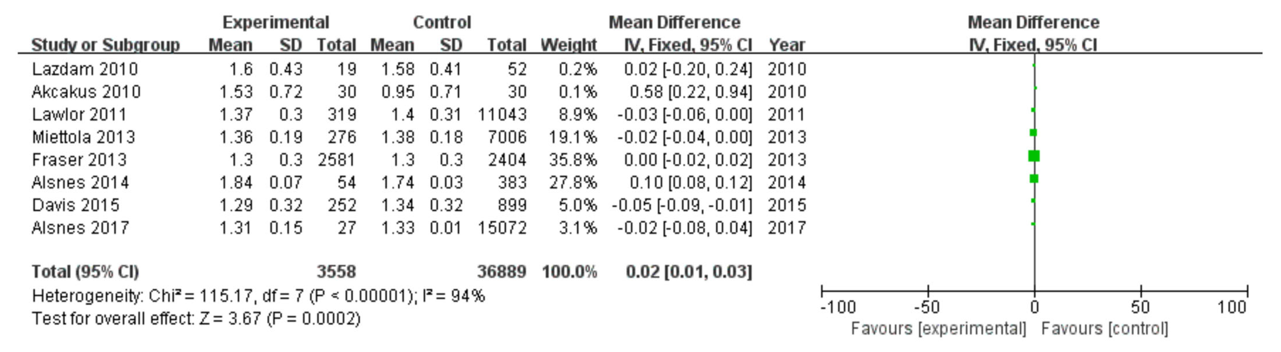
3.2.1. Systolic Blood Pressure

Among 16 included studies, 13 studies investigated SBP data of patients, including 3952 patients in the experimental group and 42,416 in the control group, with data heterogeneity (I2 = 97%) (Figure 2). The analysis showed that the SBP value in the PE pregnancy offspring group presented an elevation relative to the non-PE pregnancy offspring group [MD = 1.51, 95%CI (1.15, 1.88), p < 0.00001], with statistical significance (Figure 2).

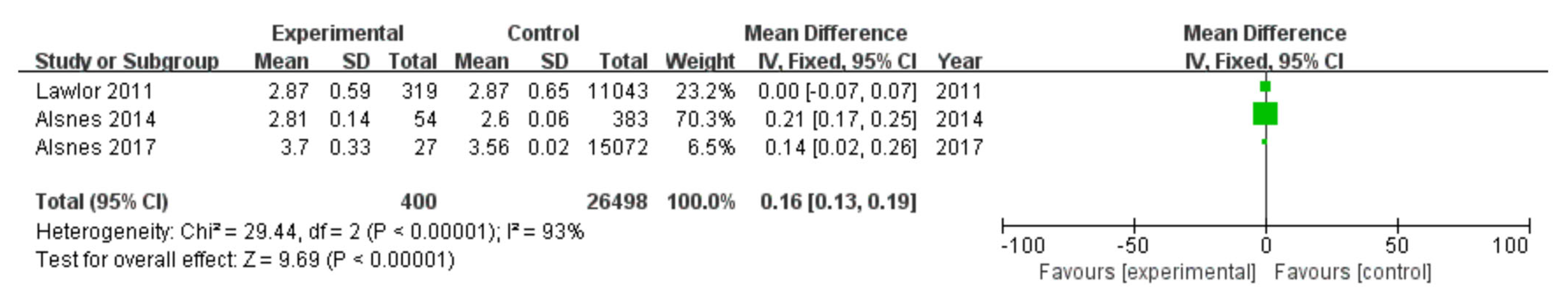
3.2.2. Diastolic Blood Pressure

Among 16 included studies, 13 studies investigated the DBP data of patients, including 3952 patients in the experimental group and 42,416 in the control group, with data heterogeneity (I2 = 98%) (Figure 3). The analysis showed that the DBP value in the PE pregnancy offspring group presented an elevation relative to the non-PE pregnancy offspring group [MD = 1.90, 95%CI (1.69, 2.10), p < 0.00001], with statistical significance (Figure 3).

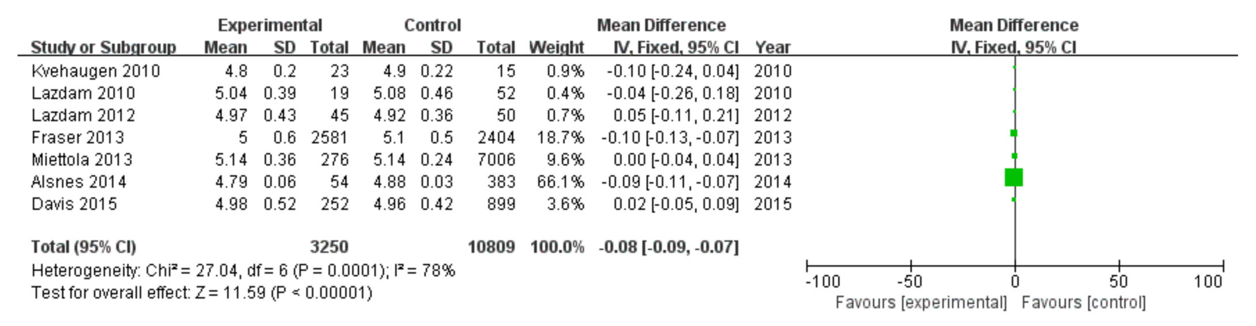
3.2.3. Total Cholesterol

Among 16 included studies, 7 studies evaluated the total cholesterol data of patients, including 3257 patients in the experimental group and 10,824 in the control group, with data heterogeneity (I2 = 96%) (Figure 4). The analysis showed that the total cholesterol value in the PE pregnancy offspring group presented an elevation relative to the non-PE pregnancy offspring group [MD = 0.11, 95%CI (0.08, 0.13), p < 0.00001], with statistical significance (Figure 4).

3.2.4. Low-Density Lipoprotein Cholesterol

Among 16 included studies, 6 studies investigated low-density lipoprotein cholesterol data of patients, including 3203 patients in the experimental group and 10,441 in the control group, without data heterogeneity (I2 = 16%) (Figure 5). The analysis showed that the low-density lipoprotein cholesterol value in the PE pregnancy offspring group was comparable to that in the non-PE pregnancy offspring group [MD = 0.01, 95%CI (−0.02, 0.05), p = 0.48], without statistical significance (Figure 5).

3.2.5. High-Density Lipoprotein Cholesterol

Among 16 included studies, 8 studies investigated high-density lipoprotein cholesterol data of patients, including 3558 patients in the experimental group and 36,889 in the control group, with data heterogeneity (I2 = 94%) (Figure 6). The analysis showed that the high-density lipoprotein cholesterol value in the PE pregnancy offspring group presented an elevation relative to the non-PE pregnancy offspring group [MD = 0.02, 95%CI (0.01, 0.03), p = 0.0002], with statistical significance (Figure 6).

3.2.6. Non-HDL Cholesterol

Among 16 included studies, 3 studies investigated non-HDL cholesterol data of patients, including 400 patients in the experimental group and 26,498 in the control group, with data heterogeneity (I2 = 93%) (Figure 7). The analysis showed that the non-HDL cholesterol value in the PE pregnancy offspring group presented an elevation relative to the non-PE pregnancy offspring group [MD = 0.16, 95%CI (0.13, 0.19), p < 0.00001], with statistical significance (Figure 7).

3.2.7. Triglycerides

Among 16 included studies, 8 studies investigated the triglycerides data of patients, including 3549 patients in the experimental group and 36,556 in the control group, with data heterogeneity (I2 = 88%) (Figure 8). The analysis showed that the triglycerides value in the PE pregnancy offspring group presented a depletion relative to the non-PE pregnancy offspring group [MD = −0.02, 95%CI (−0.03, −0.01), p < 0.00001], with statistical significance (Figure 8).

3.2.8. Glucose

Among 16 included studies, 7 studies investigated the glucose data of patients, including 3250 patients in the experimental group and 10,809 in the control group, with data heterogeneity (I2 = 78%) (Figure 9). The analysis showed that the glucose value in the PE pregnancy offspring group presented a depletion relative to the non-PE pregnancy offspring group [MD = −0.08, 95%CI (−0.09, −0.07), p < 0.00001], with statistical significance (Figure 9).

3.2.9. Insulin

Among 16 included studies, 5 studies investigated the insulin data of patients, including 3173 patients in the experimental group and 10,411 in the control group, with data heterogeneity (I2 = 79%) (Figure 10). The analysis showed that the insulin value in the PE pregnancy offspring group presented a depletion relative to the non-PE pregnancy offspring group [MD = −0.21, 95%CI (−0.32, −0.09), p = 0.0004], with statistical significance (Figure 10).

3.2.10. BMI

Among 16 included studies, 14 studies investigated the BMI data of patients, including 3920 patients in the case group and 42,082 in the control group, with data heterogeneity (I2 = 83%) (Figure 11). The analysis showed that the BMI value in the PE pregnancy offspring group presented an elevation relative to the non-PE pregnancy offspring group [MD = 0.42, 95%CI (0.27, 0.57), p < 0.00001], with statistical significance (Figure 11).

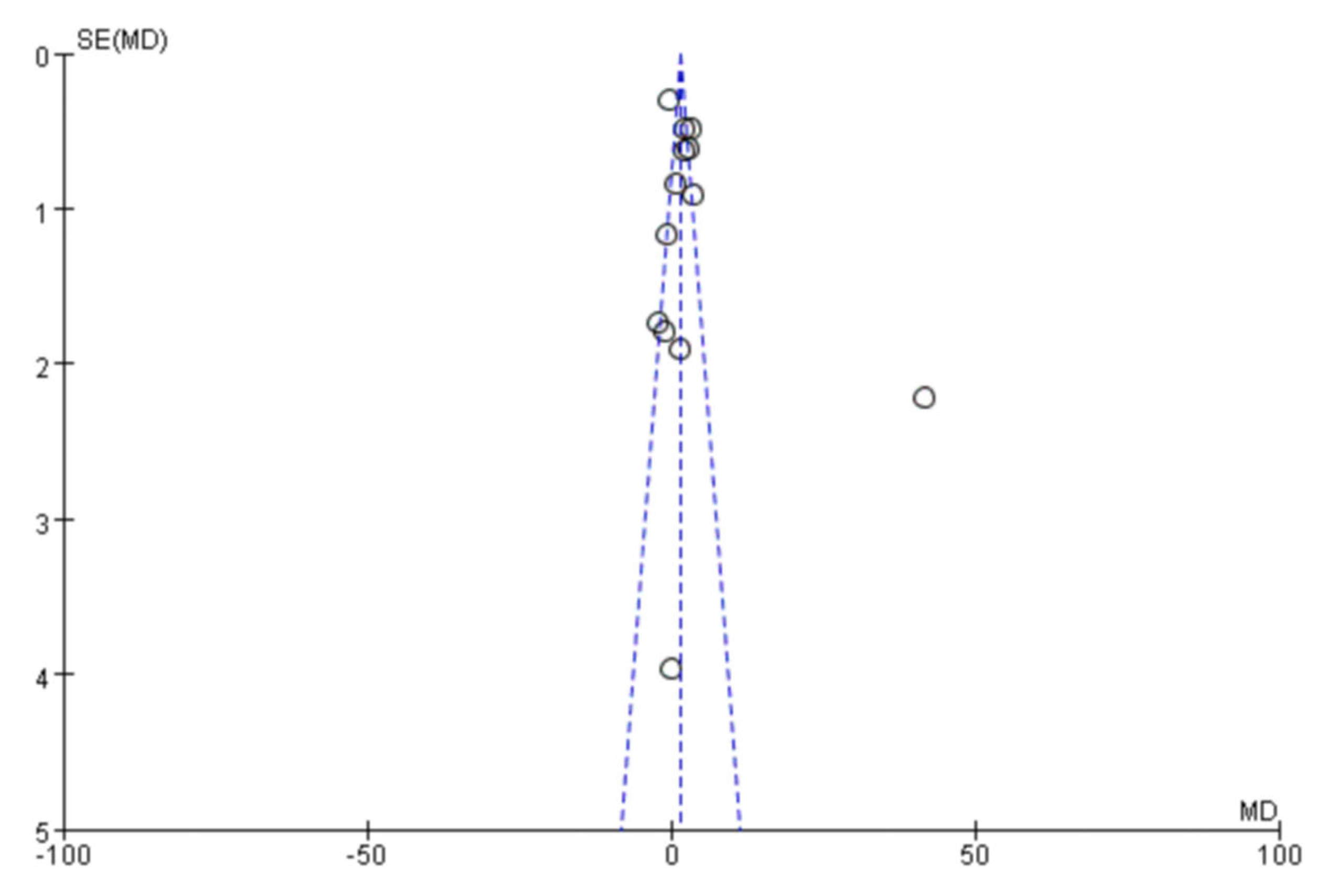
3.3. Analysis of Publication Bias

The accuracy of the conclusions of the meta-analysis largely depends on the completeness of included studies, which can be measured by reporting bias. Publication bias is one of the reporting biases that has received the most attention. By drawing a funnel plot, we found that one of the included works from the literature was outside the confidence interval, and the rest were within the confidence interval. Moreover, the distribution was relatively clustered, indicating that there was a certain publication bias, and the publication bias was not large (Figure 12).

4. Discussion

PE is a pregnancy-specific syndrome that occurs after 20 weeks of gestation and is characterized by new-onset hypertension, proteinuria, and multiple organ dysfunction [30]. The incidence of PE ranges from 2% to 8% [31]. PE seriously threatens the safety of the mother and fetus [32], and it is the third leading cause of maternal death [33]. The hazards to the mother and fetus during pregnancy mainly include placental abruption, cerebrovascular accidents, pulmonary edema, heart failure, liver and kidney failure, DIC, HELLP syndrome, eclampsia complicated by adult respiratory distress syndrome, FGR, fetal distress, stillbirth, etc. Furthermore, PE is not just a pathological pregnancy problem that accompanies pregnancy and delivery; its postpartum risks to the mother still exist, including an increased risk of postpartum cardiovascular diseases and metabolic diseases [34]. CMR refers to a set of risk factors for cardiovascular diseases and diabetes risks, including age, race, gender, family history, overweight and obesity, abnormal glucose metabolism, abnormal lipid metabolism, elevated blood pressure, metabolic syndrome, smoking, physical inactivity, inflammation, hypercoagulability, etc. [33]. In 2011, the guidelines for reducing the risk of cardiovascular diseases in women issued by the AHA clearly listed PE as a risk factor for cardiovascular disease in women for the first time. National guidance in England suggests that women who suffer from PE should be informed of the elevated risk of high blood pressure and some other related complications [35]. Moreover, in the US, the AHA suggests that women with a history of pregnancy-induced hypertension should be referred to estimate cardiovascular risk factors [36]. A systematic review and meta-analysis showed that, in comparison to women with single PE and subsequent uncomplicated pregnancy, women with recurrent PE had a three-fold increase in heart-failure risk, a two- to three-fold increase in hypertension risk, and an almost two-fold increase in overall cardiovascular disease risk [37]. The only long-term mortality follow-up study reported so far showed a 1.9-fold increased risk of stroke mortality in the offspring of PE pregnancies [12]. The occurrence and development of PE are inextricably linked with metabolism. A large number of studies have revealed that the metabolic disorder of PE persists until postpartum [38], and metabolic factors may accompany the whole process of disease development in PE patients. Metabolic factors associated with postpartum CMR will provide evidence-based medical proof for the prevention and blockade of cardiovascular disease and metabolic diseases in women.
Herein, the meta-analysis demonstrated that the SBP value in the PE pregnancy offspring group presented an elevation relative to the non-PE pregnancy offspring group [MD = 1.51, 95%CI (1.15, 1.88), p < 0.00001]. The DBP value in the PE pregnancy offspring group presented an elevation relative to the non-PE pregnancy offspring group [MD = 1.90, 95%CI (1.69, 2.10), p < 0.00001]. The total cholesterol value in the PE pregnancy offspring group presented an elevation relative to the non-PE pregnancy offspring group [MD = 0.11, 95%CI (0.08, 0.13), p < 0.00001]. The low-density lipoprotein cholesterol value in the PE pregnancy offspring group was comparable to that in the non-PE pregnancy offspring group [MD = 0.01, 95%CI (−0.02, 0.05), p = 0.48]. The high-density lipoprotein cholesterol value in the PE pregnancy offspring group presented an elevation relative to the non-PE pregnancy offspring group [MD = 0.02, 95%CI (0.01, 0.03), p = 0.0002]. The non-HDL cholesterol value in the PE pregnancy offspring group presented an elevation relative to the non-PE pregnancy offspring group [MD = 0.16, 95%CI (0.13, 0.19), p < 0.00001]. The triglycerides value in the PE pregnancy offspring group presented a depletion relative to the non-PE pregnancy group [MD = −0.02, 95%CI (−0.03, −0.01), p < 0.00001]. The glucose value in the PE pregnancy offspring group presented a depletion relative to the non-PE pregnancy offspring group [MD = −0.08, 95%CI (−0.09, −0.07), p < 0.00001]. The insulin value in the PE pregnancy offspring group presented a depletion relative to the non-PE pregnancy offspring group [MD = −0.21, 95%CI (−0.32, −0.09), p = 0.0004]. The BMI value in the PE pregnancy offspring group presented an elevation relative to the non-PE pregnancy offspring group [MD = 0.42, 95 %CI (0.27, 0.57), p < 0.00001]. It was suggested that postpartum PE patients have persistently elevated blood pressure; elevated total cholesterol; decreased fasting blood glucose, triglycerides, and insulin; elevated high-density lipoprotein; and decreased low-density lipoprotein and still have obvious insulin resistance and metabolic abnormalities.
Previously, metabolic changes amplified during pregnancy with PE persisted from 24 to 48 h postpartum [39] to 3 months postpartum [40]. Thus, postpartum metabolic abnormalities are very likely to be a continuation of metabolic abnormalities during pregnancy. An elevated insulin level is an indirect sign of insulin resistance. Insulin resistance is closely related to metabolic syndrome and leads to vascular endothelial dysfunction, dyslipidemia, hypertension, and vascular inflammation, all of which promote the development of cardiovascular disease [41]. PE patients had significant postpartum insulin resistance relative to normal controls [42], similar to our findings. Metabolic syndrome has been revealed to exert the crucial function in the pathophysiology of gestational hypertension and PE and might be the latent mechanism linking PE in pregnancy and cardiovascular disease. Girouard J et al. focused on evaluating postpartum-related metabolic changes and found that, relative to the normal pregnancy group, the postpartum body weight, LDL, lipoprotein B/lipoprotein A1, homocysteine, leptin, and insulin levels in the hypertensive disorder complicating pregnancy (HDCP) group presented a marked elevation, suggesting that the levels of various metabolic indicators of HDCP in the first 10 years after delivery were still very high (p < 0.004) [42], and this supported our findings. A prospective cohort study revealed that postpartum PE patients develop dyslipidemia, elevated blood pressure, and elevated BMI, which are risk factors for cardiovascular diseases. They also noted that postpartum blood pressure elevations were more pronounced in patients with recurrent PE than in patients with first PE. Postpartum systolic blood pressure was, on average, 27 mm Hg higher (95%CI: 18–37 mm Hg), and diastolic blood pressure was, on average, 12 mm Hg higher (95%CI: 5–19 mm Hg) in patients with hypertension in three consecutive pregnancies than in patients with hypertension in only one pregnancy [43]. Furthermore, a meta-analysis of 53,029 individuals of whom 1599 were exposed to PE in utero indicates a 5.17 mm Hg greater systolic blood pressure among those exposed to PE in comparison of controls, and the meta-analyses of 52,993 individuals, of whom 1583 were exposed to PE in utero, indicates a 4.06 mm Hg greater diastolic blood pressure among those exposed to PE in comparison to the controls [44]. The BMI is an anthropometric index that is utilized as a surrogate marker for fat mass and for classifying obesity. BMI and total adiposity are positively correlated with cardiometabolic disease risk at the population level. A study reported a 0.62 kg/m2 increase in BMI among offspring of PE pregnancies compared with controls (39,473 individuals; 1062 exposed to PE) [13].
Studies have confirmed some cardioprotective strategies of PE patients during pregnancy. SGLT2 inhibitors are reported to reduce blood pressure and proteinuria, the typical clinical manifestations of PE [45,46]. Different gliflozins (SGLT2 inhibitors) can play a cardioprotective role, reduce cardiovascular death, and treat heart failure [47]. Since November 2020, dapagliflozin has been approved for the first time as an SGLT2 inhibitor for the treatment of patients with heart failure. Empagliflozin lowered by 32%, 38%, and 35% the risk of all-cause mortality, cardiovascular mortality, and hospitalization due to worsening heart failure in Type 2 Diabetes Mellitus patients [48]. The CANVAS program has indicated that canagliflozin lowers the risk of cardiovascular death by 14% and the risk of heart failure hospitalization by 33% [49]. Ruonan Zhai et al. revealed that oral empagliflozin can reduce high systolic blood pressure and proteinuria and improve kidney histopathology, thereby improving PE without affecting fetal outcomes [50]. Statins are used for the treatment and prevention of cardiovascular diseases through lipid-lowering therapy [51]. The effect of pravastatin during pregnancy has been demonstrated in many rodent models of PE [52]. Studies have shown that pravastatin treatment can significantly reduce the maternal sFlt-1 level, lower blood pressure, and improve vascular conditions [53,54]. Aspirin is the most commonly used treatment to prevent cardiovascular complications [55]. Studies have shown that aspirin therapy initiated at ≥16 weeks of gestation is associated with a 50% reduction in PE in preterm infants with a dose-dependent effect [56]. A clinical trial involving 1776 patients with low early placental growth factor level reported that a daily dose of 150 mg aspirin could reduce PE by 62% [57]. On the other hand, nutraceuticals have proved to be of great benefit in combating the progress of cardiovascular disease [58]. The nano drug delivery system has shown remarkable results in delivering nutraceuticals in various diseases, including cancer, neurodegenerative diseases, cardiovascular diseases, etc. [59]. Common nutraceuticals in cardiovascular diseases include resveratrol, vitamin D, quercetin, curcumin, flavanol, etc. Resveratrol is a polyphenolic compound from the stilbene group and is mainly a component of red wines. It has been shown to have cardioprotective effects due to its anti-inflammatory and antioxidant properties [60]. Reports have indicated that resveratrol exerts a protective function in animals with dyslipidemia and insulin resistance, and it can reduce cardiac hypertrophy and systolic dysfunction [61]. Quercetin is a flavonoid that improves lipid metabolism, vascular function, blood pressure, and glucose metabolism, and it is thought to reduce or prevent the progression of cardiovascular disease [62]. One study showed that 730 mg of quercetin given daily for four weeks reduces systolic and diastolic blood pressure in patients with stage 1 hypertension [63]. Vitamin D deficiency has been associated with the development of cardiovascular disease, microbial infections, or tumor development [64]. In a cohort study performed on 13,806 pregnant women, maternal vitamin D deficiency was intensely related to an increased risk for PE [65]. Studies have shown that low maternal vitamin D levels are associated with a roughly twofold increase in the prevalence of congenital heart defects in offspring [66]. Vitamin D supplementation has been proved to enhance the effect of nifedipine in the treatment of PE [67]. Nanoparticles can be absorbed on the plasma membrane through endocytosis and non-endocytosis, and they can be transferred from passive diffusion to active transport [68]. Nanocarriers involved in drug delivery can increase the water solubility of insoluble drugs, thus preventing the degradation and inactivation of active ingredients [69]. Hyaluronic acid (HA) is a natural mucopolysaccharide and is the main constituent of the extracellular matrix, which exerts a crucial role in cell growth and in maintaining the structural stability of tissue [70]. Importantly, because HA is biocompatible, non-immunogenic, non-toxic, biodegradable, chemically modifiable, highly hydrophilic, and can absorb water to produce viscoelastic gel, it has been widely studied as a drug delivery system [71]. The hydrophilic shell of hyaluronic acid granules extends its circulation time in the blood, which can increase the probability of reaching the treatment site after systemic administration [72]. The development of hyaluronic-acid-based nanomedicines can help improve the oral bioavailability of cardioprotective natural molecules such as quercetin, resveratrol, and vitamin D. This may be an effective treatment strategy to reduce cardiovascular risk factors in the offspring of PE patients.
Herein, the risk factors of postpartum cardiovascular risk in PE patients were mainly metabolic factors in the fully developed stage of the disease. Though prenatal basic BMI, blood pressure and other factors were also considered, prenatal metabolic factors and other possible factors were not considered, and the risk factors for postpartum risk of PE were not fully understood. In our research, relevant documents were collected as comprehensively as possible, and the literature was screened strictly according to the inclusion and exclusion criteria, as well as literature quality, to avoid retrieval bias to the greatest extent. Nevertheless, some documents can only be discarded due to different effect indicators or the inability to calculate the original data. Additionally, language bias, publication bias, and reporting bias may all have an impact on the results of this research. Due to the limitation of the type of study, this study only searched the published literature, and the existence of unpublished studies that may affect the results of this study cannot be excluded.

5. Conclusions

PE, as a critical illness in obstetrics, can achieve early prevention, early diagnosis, and early intervention for high-risk groups by recognizing high-risk factors, screening high-risk groups, and avoiding exposure to some cardiovascular risk factors in early pregnancy, thereby reducing the incidence of PE in pregnancy and improving the perinatal outcomes of mothers and babies. There is a need to improve obstetricians’ awareness of the risk and risk factors for PE and to strengthen pre-pregnancy and pregnancy care.

Author Contributions

Conceptualization, W.W. and J.Y.; literature collection, R.L. and X.X.; formal analysis, L.Y. and Y.W.; writing—original draft preparation, W.W., B.M. and X.X.; writing—review and editing, R.L., L.Y. and J.Y.; supervision, J.Y; project administration, W.W. and J.Y. All authors have read and agreed to the published version of the manuscript.

Funding

This work was supported by the National Key R&D Program of China (2018YFC1002504) and Natural Science Foundation of Gansu Province (20JR10RA428).

Institutional Review Board Statement

Not applicable.

Informed Consent Statement

Not applicable.

Data Availability Statement

Not applicable.

Conflicts of Interest

The authors declare that the research was conducted in the absence of any commercial or financial relationships that could be construed as a potential conflict of interest.

References

  1. Kuehn, B.M. Rising Prepregnancy Hypertension, Preeclampsia Disparities Raise Alarm. Circulation 2021, 143, 753–754. [Google Scholar] [CrossRef] [PubMed]
  2. Sutton, E.F.; Gemmel, M.; Powers, R.W. Nitric oxide signaling in pregnancy and preeclampsia. Nitric Oxide 2020, 95, 55–62. [Google Scholar] [CrossRef] [PubMed]
  3. Barr, L.; Pudwell, J.; Herr, J.; Johri, A.; Smith, G. Microvascular outcomes of a pregnancy complicated by preeclampsia. Can. J. Cardiol. 2021, 37, e12. [Google Scholar] [CrossRef]
  4. Le, Q.; Akhter, R.; Coulton, K.; Vo, N.; Duong, L.; Nong, H.; Yaacoub, A.; Condous, G.; Eberhard, J.; Nanan, R. Periodontitis and preeclampsia in pregnancy: A systematic review and meta-analysis. Matern. Child Health J. 2022, 26, 2419–2443. [Google Scholar] [CrossRef] [PubMed]
  5. Karatza, A.A.; Dimitriou, G. Preeclampsia Emerging as a Novel Risk Factor for Cardiovascular Disease in the Offspring. Curr. Pediatr. Rev. 2020, 16, 194–199. [Google Scholar] [CrossRef]
  6. Wang, Y.; Wu, N.; Shen, H. A Review of Research Progress of Pregnancy with Twins with Preeclampsia. Risk Manag. Healthc. Policy 2021, 14, 1999–2010. [Google Scholar] [CrossRef]
  7. Qu, H.; Khalil, R.A. Vascular mechanisms and molecular targets in hypertensive pregnancy and preeclampsia. Am. J. Physiol.-Heart Circ. Physiol. 2020, 319, H661–H681. [Google Scholar] [CrossRef]
  8. Roth, G.A.; Johnson, C.; Abajobir, A.; Abd-Allah, F.; Abera, S.F.; Abyu, G.; Ahmed, M.; Aksut, B.; Alam, T.; Alam, K.; et al. Global, Regional, and National Burden of Cardiovascular Diseases for 10 Causes, 1990 to 2015. J. Am. Coll. Cardiol. 2017, 70, 1–25. [Google Scholar] [CrossRef]
  9. Ha, M.; Park, J. Shiftwork and metabolic risk factors of cardiovascular disease. J. Occup. Health 2005, 47, 89–95. [Google Scholar] [CrossRef] [Green Version]
  10. Smith, G.N.; Walker, M.C.; Liu, A.; Wen, S.W.; Swansburg, M.; Ramshaw, H.; White, R.R.; Roddy, M.; Hladunewich, M. A history of preeclampsia identifies women who have underlying cardiovascular risk factors. Am. J. Obstet. Gynecol. 2009, 200, 58.E1–58.E8. [Google Scholar] [CrossRef]
  11. Heida, K.Y.; Bots, M.L.; de Groot, C.J.; van Dunné, F.M.; Hammoud, N.M.; Hoek, A.; Laven, J.S.; Maas, A.H.; Roeters van Lennep, J.E.; Velthuis, B.K.; et al. Cardiovascular risk management after reproductive and pregnancy-related disorders: A Dutch multidisciplinary evidence-based guideline. Eur. J. Prev. Cardiol. 2016, 23, 1863–1879. [Google Scholar] [CrossRef]
  12. Kajantie, E.; Eriksson, J.G.; Osmond, C.; Thornburg, K.; Barker, D.J. Pre-eclampsia is associated with increased risk of stroke in the adult offspring: The Helsinki birth cohort study. Stroke 2009, 40, 1176–1180. [Google Scholar] [CrossRef] [Green Version]
  13. Davis, E.F.; Lazdam, M.; Lewandowski, A.J.; Worton, S.A.; Kelly, B.; Kenworthy, Y.; Adwani, S.; Wilkinson, A.R.; McCormick, K.; Sargent, I.; et al. Cardiovascular risk factors in children and young adults born to preeclamptic pregnancies: A systematic review. Pediatrics 2012, 129, e1552–e1561. [Google Scholar] [CrossRef]
  14. Akcakus, M.; Altunay, L.; Yikilmaz, A.; Yazici, C.; Koklu, E. The relationship between abdominal aortic intima-media thickness and lipid profile in neonates born to mothers with preeclampsia. J. Pediatr. Endocrinol. Metab. 2010, 23, 1143–1149. [Google Scholar] [CrossRef] [PubMed]
  15. Lazdam, M.; de la Horra, A.; Pitcher, A.; Mannie, Z.; Diesch, J.; Trevitt, C.; Kylintireas, I.; Contractor, H.; Singhal, A.; Lucas, A.; et al. Elevated blood pressure in offspring born premature to hypertensive pregnancy: Is endothelial dysfunction the underlying vascular mechanism? Hypertension 2010, 56, 159–165. [Google Scholar] [CrossRef] [PubMed] [Green Version]
  16. Kvehaugen, A.S.; Andersen, L.F.; Staff, A.C. Anthropometry and cardiovascular risk factors in women and offspring after pregnancies complicated by preeclampsia or diabetes mellitus. Acta Obstet. Gynecol. Scand. 2010, 89, 1478–1485. [Google Scholar] [CrossRef]
  17. Geelhoed, J.J.; Fraser, A.; Tilling, K.; Benfield, L.; Davey Smith, G.; Sattar, N.; Nelson, S.M.; Lawlor, D.A. Preeclampsia and gestational hypertension are associated with childhood blood pressure independently of family adiposity measures: The Avon Longitudinal Study of Parents and Children. Circulation 2010, 122, 1192–1199. [Google Scholar] [CrossRef] [PubMed] [Green Version]
  18. Jayet, P.Y.; Rimoldi, S.F.; Stuber, T.; Salmòn, C.S.; Hutter, D.; Rexhaj, E.; Thalmann, S.; Schwab, M.; Turini, P.; Sartori-Cucchia, C.; et al. Pulmonary and systemic vascular dysfunction in young offspring of mothers with preeclampsia. Circulation 2010, 122, 488–494. [Google Scholar] [CrossRef] [Green Version]
  19. Fugelseth, D.; Ramstad, H.B.; Kvehaugen, A.S.; Nestaas, E.; Støylen, A.; Staff, A.C. Myocardial function in offspring 5-8years after pregnancy complicated by preeclampsia. Early Hum. Dev. 2011, 87, 531–535. [Google Scholar] [CrossRef]
  20. Lawlor, D.A.; Macdonald-Wallis, C.; Fraser, A.; Nelson, S.M.; Hingorani, A.; Davey Smith, G.; Sattar, N.; Deanfield, J. Cardiovascular biomarkers and vascular function during childhood in the offspring of mothers with hypertensive disorders of pregnancy: Findings from the Avon Longitudinal Study of Parents and Children. Eur. Heart J. 2012, 33, 335–345. [Google Scholar] [CrossRef] [PubMed] [Green Version]
  21. Kvehaugen, A.S.; Dechend, R.; Ramstad, H.B.; Troisi, R.; Fugelseth, D.; Staff, A.C. Endothelial function and circulating biomarkers are disturbed in women and children after preeclampsia. Hypertension 2011, 58, 63–69. [Google Scholar] [CrossRef] [PubMed] [Green Version]
  22. Lazdam, M.; de la Horra, A.; Diesch, J.; Kenworthy, Y.; Davis, E.; Lewandowski, A.J.; Szmigielski, C.; Shore, A.; Mackillop, L.; Kharbanda, R.; et al. Unique blood pressure characteristics in mother and offspring after early onset preeclampsia. Hypertension 2012, 60, 1338–1345. [Google Scholar] [CrossRef] [PubMed] [Green Version]
  23. Miettola, S.; Hartikainen, A.L.; Vääräsmäki, M.; Bloigu, A.; Ruokonen, A.; Järvelin, M.R.; Pouta, A. Offspring’s blood pressure and metabolic phenotype after exposure to gestational hypertension in utero. Eur. J. Epidemiol. 2013, 28, 87–98. [Google Scholar] [CrossRef] [PubMed]
  24. Fraser, A.; Nelson, S.M.; Macdonald-Wallis, C.; Sattar, N.; Lawlor, D.A. Hypertensive disorders of pregnancy and cardiometabolic health in adolescent offspring. Hypertension 2013, 62, 614–620. [Google Scholar] [CrossRef] [Green Version]
  25. Alsnes, I.V.; Janszky, I.; Forman, M.R.; Vatten, L.J.; Økland, I. A population-based study of associations between preeclampsia and later cardiovascular risk factors. Am. J. Obstet. Gynecol. 2014, 211, e651–e657. [Google Scholar] [CrossRef]
  26. Davis, E.F.; Lewandowski, A.J.; Aye, C.; Williamson, W.; Boardman, H.; Huang, R.C.; Mori, T.A.; Newnham, J.; Beilin, L.J.; Leeson, P. Clinical cardiovascular risk during young adulthood in offspring of hypertensive pregnancies: Insights from a 20-year prospective follow-up birth cohort. BMJ Open 2015, 5, e008136. [Google Scholar] [CrossRef] [Green Version]
  27. Reveret, M.; Boivin, A.; Guigonnis, V.; Audibert, F.; Nuyt, A.M. Preeclampsia: Effect on newborn blood pressure in the 3 days following preterm birth: A cohort study. J. Hum. Hypertens. 2015, 29, 115–121. [Google Scholar] [CrossRef] [PubMed]
  28. Alsnes, I.V.; Vatten, L.J.; Fraser, A.; Bjørngaard, J.H.; Rich-Edwards, J.; Romundstad, P.R.; Åsvold, B.O. Hypertension in Pregnancy and Offspring Cardiovascular Risk in Young Adulthood: Prospective and Sibling Studies in the HUNT Study (Nord-Trøndelag Health Study) in Norway. Hypertension 2017, 69, 591–598. [Google Scholar] [CrossRef] [Green Version]
  29. Gürlek, B.; Yılmaz, A.; Durakoğlugil, M.E.; Karakaş, S.; Kazaz, İ.M.; Önal, Ö.; Şatıroğlu, Ö. Evaluation of serum apelin-13 and apelin-36 concentrations in preeclamptic pregnancies. J. Obstet. Gynaecol. Res. 2020, 46, 58–65. [Google Scholar] [CrossRef]
  30. Wolf, M.; Hubel, C.A.; Lam, C.; Sampson, M.; Ecker, J.L.; Ness, R.B.; Rajakumar, A.; Daftary, A.; Shakir, A.S.; Seely, E.W.; et al. Preeclampsia and future cardiovascular disease: Potential role of altered angiogenesis and insulin resistance. J. Clin. Endocrinol. Metab. 2004, 89, 6239–6243. [Google Scholar] [CrossRef] [PubMed]
  31. McDonald, S.D.; Malinowski, A.; Zhou, Q.; Yusuf, S.; Devereaux, P.J. Cardiovascular sequelae of preeclampsia/eclampsia: A systematic review and meta-analyses. Am. Heart J. 2008, 156, 918–930. [Google Scholar] [CrossRef] [PubMed]
  32. Craici, I.; Wagner, S.; Garovic, V.D. Preeclampsia and future cardiovascular risk: Formal risk factor or failed stress test? Ther. Adv. Cardiovasc. Dis. 2008, 2, 249–259. [Google Scholar] [CrossRef] [Green Version]
  33. Melchiorre, K.; Sharma, R.; Thilaganathan, B. Cardiovascular implications in preeclampsia: An overview. Circulation 2014, 130, 703–714. [Google Scholar] [CrossRef] [PubMed]
  34. Hibbard, J.U.; Shroff, S.G.; Lang, R.M. Cardiovascular changes in preeclampsia. Semin. Nephrol. 2004, 24, 580–587. [Google Scholar] [CrossRef] [PubMed]
  35. Kattah, A.G.; Garovic, V.D. The management of hypertension in pregnancy. Adv. Chronic Kidney Dis. 2013, 20, 229–239. [Google Scholar] [CrossRef] [PubMed] [Green Version]
  36. Mosca, L.; Benjamin, E.J.; Berra, K.; Bezanson, J.L.; Dolor, R.J.; Lloyd-Jones, D.M.; Newby, L.K.; Piña, I.L.; Roger, V.L.; Shaw, L.J.; et al. Effectiveness-based guidelines for the prevention of cardiovascular disease in women--2011 update: A guideline from the american heart association. Circulation 2011, 123, 1243–1262. [Google Scholar] [CrossRef] [PubMed] [Green Version]
  37. Brouwers, L.; van der Meiden-van Roest, A.J.; Savelkoul, C.; Vogelvang, T.E.; Lely, A.T.; Franx, A.; van Rijn, B.B. Recurrence of pre-eclampsia and the risk of future hypertension and cardiovascular disease: A systematic review and meta-analysis. BJOG 2018, 125, 1642–1654. [Google Scholar] [CrossRef] [Green Version]
  38. Wellmann, S.; Benzing, J.; Fleischlin, S.; Morgenthaler, N.; Fouzas, S.; Bührer, C.A.; Szinnai, G.; Burkhardt, T.; Lapaire, O. Cardiovascular biomarkers in preeclampsia at triage. Fetal. Diagn. Ther. 2014, 36, 202–207. [Google Scholar] [CrossRef] [PubMed]
  39. Lei, Q.; Lv, L.J.; Zhang, B.Y.; Wen, J.Y.; Liu, G.C.; Lin, X.H.; Niu, J.M. Ante-partum and post-partum markers of metabolic syndrome in pre-eclampsia. J. Hum. Hypertens. 2011, 25, 11–17. [Google Scholar] [CrossRef] [PubMed] [Green Version]
  40. Carty, D.M.; Delles, C.; Dominiczak, A.F. Preeclampsia and future maternal health. J. Hypertens. 2010, 28, 1349–1355. [Google Scholar] [CrossRef]
  41. DeFronzo, R.A.; Ferrannini, E. Insulin resistance. A multifaceted syndrome responsible for NIDDM, obesity, hypertension, dyslipidemia, and atherosclerotic cardiovascular disease. Diabetes Care 1991, 14, 173–194. [Google Scholar] [CrossRef]
  42. Girouard, J.; Giguère, Y.; Moutquin, J.M.; Forest, J.C. Previous hypertensive disease of pregnancy is associated with alterations of markers of insulin resistance. Hypertension 2007, 49, 1056–1062. [Google Scholar] [CrossRef] [Green Version]
  43. Magnussen, E.B.; Vatten, L.J.; Smith, G.D.; Romundstad, P.R. Hypertensive disorders in pregnancy and subsequently measured cardiovascular risk factors. Obstet. Gynecol. 2009, 114, 961–970. [Google Scholar] [CrossRef] [PubMed]
  44. Andraweera, P.H.; Lassi, Z.S. Cardiovascular Risk Factors in Offspring of Preeclamptic Pregnancies-Systematic Review and Meta-Analysis. J. Pediatr. 2019, 208, 104–113.e106. [Google Scholar] [CrossRef] [PubMed]
  45. Georgianos, P.I.; Agarwal, R. Ambulatory Blood Pressure Reduction with SGLT-2 Inhibitors: Dose-Response Meta-analysis and Comparative Evaluation with Low-Dose Hydrochlorothiazide. Diabetes Care 2019, 42, 693–700. [Google Scholar] [CrossRef] [PubMed] [Green Version]
  46. Mosenzon, O.; Wiviott, S.D.; Cahn, A.; Rozenberg, A.; Yanuv, I.; Goodrich, E.L.; Murphy, S.A.; Heerspink, H.J.L.; Zelniker, T.A.; Dwyer, J.P.; et al. Effects of dapagliflozin on development and progression of kidney disease in patients with type 2 diabetes: An analysis from the DECLARE-TIMI 58 randomised trial. Lancet Diabetes Endocrinol. 2019, 7, 606–617. [Google Scholar] [CrossRef]
  47. Rauch-Kröhnert, U.; Landmesser, U. Gliflozins-in future cardioprotective drugs? Internist 2021, 62, 786–795. [Google Scholar] [CrossRef] [PubMed]
  48. Zinman, B.; Wanner, C.; Lachin, J.M.; Fitchett, D.; Bluhmki, E.; Hantel, S.; Mattheus, M.; Devins, T.; Johansen, O.E.; Woerle, H.J.; et al. Empagliflozin, Cardiovascular Outcomes, and Mortality in Type 2 Diabetes. N. Engl. J. Med. 2015, 373, 2117–2128. [Google Scholar] [CrossRef] [PubMed] [Green Version]
  49. Neal, B.; Perkovic, V.; Mahaffey, K.W.; de Zeeuw, D.; Fulcher, G.; Erondu, N.; Shaw, W.; Law, G.; Desai, M.; Matthews, D.R. Canagliflozin and Cardiovascular and Renal Events in Type 2 Diabetes. N. Engl. J. Med. 2017, 377, 644–657. [Google Scholar] [CrossRef]
  50. Zhai, R.; Liu, Y.; Tong, J.; Yu, Y.; Yang, L.; Gu, Y.; Niu, J. Empagliflozin Ameliorates Preeclampsia and Reduces Postpartum Susceptibility to Adriamycin in a Mouse Model Induced by Angiotensin Receptor Agonistic Autoantibodies. Front. Pharmacol. 2022, 13, 826792. [Google Scholar] [CrossRef] [PubMed]
  51. Oesterle, A.; Laufs, U.; Liao, J.K. Pleiotropic Effects of Statins on the Cardiovascular System. Circ. Res. 2017, 120, 229–243. [Google Scholar] [CrossRef] [PubMed] [Green Version]
  52. Smith, D.D.; Costantine, M.M. The role of statins in the prevention of preeclampsia. Am. J. Obstet. Gynecol. 2022, 226, S1171–S1181. [Google Scholar] [CrossRef] [PubMed]
  53. Ahmed, A.; Singh, J.; Khan, Y.; Seshan, S.V.; Girardi, G. A new mouse model to explore therapies for preeclampsia. PLoS ONE 2010, 5, e13663. [Google Scholar] [CrossRef] [PubMed] [Green Version]
  54. Costantine, M.M.; Tamayo, E.; Lu, F.; Bytautiene, E.; Longo, M.; Hankins, G.D.V.; Saade, G.R. Using pravastatin to improve the vascular reactivity in a mouse model of soluble fms-like tyrosine kinase-1-induced preeclampsia. Obstet. Gynecol. 2010, 116, 114–120. [Google Scholar] [CrossRef] [PubMed]
  55. Soodi, D.; VanWormer, J.J.; Rezkalla, S.H. Aspirin in Primary Prevention of Cardiovascular Events. Clin. Med. Res. 2020, 18, 89–94. [Google Scholar] [CrossRef]
  56. Roberge, S.; Bujold, E.; Nicolaides, K.H. Aspirin for the prevention of preterm and term preeclampsia: Systematic review and metaanalysis. Am. J. Obstet. Gynecol. 2018, 218, 287–293.e281. [Google Scholar] [CrossRef] [Green Version]
  57. Rolnik, D.L.; Wright, D.; Poon, L.C.; O’Gorman, N.; Syngelaki, A.; de Paco Matallana, C.; Akolekar, R.; Cicero, S.; Janga, D.; Singh, M.; et al. Aspirin versus Placebo in Pregnancies at High Risk for Preterm Preeclampsia. N. Engl. J. Med. 2017, 377, 613–622. [Google Scholar] [CrossRef]
  58. Russell, C.; Keshavamurthy, S.; Saha, S. Nutraceuticals in the Management of Cardiovascular Risk Factors: Where is the Evidence? Cardiovasc. Hematol. Disord. Drug Targets 2021, 21, 150–161. [Google Scholar] [CrossRef]
  59. Jahangir, M.A.; Jain, P.; Verma, R.; Taleuzzaman, M.; Ahsan, M.J.; Chettupalli, A.K.; Muheem, A.; Mirza, M.A. Transdermal Nutraceuticals Delivery System for CNS Disease. CNS Neurol. Disord. Drug Targets 2022, 21, 977–993. [Google Scholar] [CrossRef]
  60. Breuss, J.M.; Atanasov, A.G.; Uhrin, P. Resveratrol and Its Effects on the Vascular System. Int. J. Mol. Sci. 2019, 20, 1523. [Google Scholar] [CrossRef] [Green Version]
  61. Rivera, L.; Morón, R.; Zarzuelo, A.; Galisteo, M. Long-term resveratrol administration reduces metabolic disturbances and lowers blood pressure in obese Zucker rats. Biochem. Pharmacol. 2009, 77, 1053–1063. [Google Scholar] [CrossRef] [PubMed] [Green Version]
  62. Kozłowska, A.; Szostak-Węgierek, D. Targeting Cardiovascular Diseases by Flavonols: An Update. Nutrients 2022, 14, 1439. [Google Scholar] [CrossRef] [PubMed]
  63. Edwards, R.L.; Lyon, T.; Litwin, S.E.; Rabovsky, A.; Symons, J.D.; Jalili, T. Quercetin reduces blood pressure in hypertensive subjects. J. Nutr. 2007, 137, 2405–2411. [Google Scholar] [CrossRef] [PubMed] [Green Version]
  64. Manson, J.E.; Cook, N.R.; Lee, I.M.; Christen, W.; Bassuk, S.S.; Mora, S.; Gibson, H.; Gordon, D.; Copeland, T.; D’Agostino, D.; et al. Vitamin D Supplements and Prevention of Cancer and Cardiovascular Disease. N. Engl. J. Med. 2019, 380, 33–44. [Google Scholar] [CrossRef]
  65. Zhao, X.; Fang, R.; Yu, R.; Chen, D.; Zhao, J.; Xiao, J. Maternal Vitamin D Status in the Late Second Trimester and the Risk of Severe Preeclampsia in Southeastern China. Nutrients 2017, 9, 138. [Google Scholar] [CrossRef] [PubMed] [Green Version]
  66. Heyden, E.L.; Wimalawansa, S.J. Vitamin D: Effects on human reproduction, pregnancy, and fetal well-being. J. Steroid. Biochem. Mol. Biol. 2018, 180, 41–50. [Google Scholar] [CrossRef] [PubMed]
  67. Shi, D.D.; Wang, Y.; Guo, J.J.; Zhou, L.; Wang, N. Vitamin D Enhances Efficacy of Oral Nifedipine in Treating Preeclampsia with Severe Features: A Double Blinded, Placebo-Controlled and Randomized Clinical Trial. Front. Pharmacol. 2017, 8, 865. [Google Scholar] [CrossRef] [Green Version]
  68. Eilers, A.; Witt, S.; Walter, J. Aptamer-Modified Nanoparticles in Medical Applications. Adv. Biochem. Eng. Biotechnol. 2020, 174, 161–193. [Google Scholar] [CrossRef] [PubMed]
  69. Kumari, P.; Ghosh, B.; Biswas, S. Nanocarriers for cancer-targeted drug delivery. J. Drug Target. 2016, 24, 179–191. [Google Scholar] [CrossRef]
  70. Pereira, H.; Sousa, D.A.; Cunha, A.; Andrade, R.; Espregueira-Mendes, J.; Oliveira, J.M.; Reis, R.L. Hyaluronic Acid. Adv. Exp. Med. Biol. 2018, 1059, 137–153. [Google Scholar] [CrossRef] [PubMed]
  71. Vasvani, S.; Kulkarni, P.; Rawtani, D. Hyaluronic acid: A review on its biology, aspects of drug delivery, route of administrations and a special emphasis on its approved marketed products and recent clinical studies. Int. J. Biol. Macromol. 2020, 151, 1012–1029. [Google Scholar] [CrossRef] [PubMed]
  72. Rosso, F.; Quagliariello, V.; Tortora, C.; Di Lazzaro, A.; Barbarisi, A.; Iaffaioli, R.V. Cross-linked hyaluronic acid sub-micron particles: In vitro and in vivo biodistribution study in cancer xenograft model. J. Mater. Sci. Mater. Med. 2013, 24, 1473–1481. [Google Scholar] [CrossRef] [PubMed]
Figure 1. The flowchart of the literature retrieval and screening.
Diagnostics 13 00812 g001
Figure 2. Meta-analysis forest map for systolic blood pressure of offspring of PE pregnancies. Geelhoed 2010, [17]; Pierre 2010, [18]; Kvehaugen 2010, [16]; Fugelseth 2011, [19]; Lawlor 2011, [20]; Kvehaugen 2011, [21]; Fraser 2013, [24]; Miettola 2013, [23]; Alsnes 2014, [25]; Davis 2015, [26]; Reveret 2015, [27]; Alsnes 2017, [25]; Gürlek 2019 [29].
Diagnostics 13 00812 g002
Figure 3. Meta-analysis forest map for diastolic blood pressure of offspring of PE pregnancies. Pierre 2010, [18]; Geelhoed 2010, [17]; Kvehaugen 2010, [16]; Lawlor 2011, [20]; Kvehaugen 2011, [21]; Fugelseth 2011, [19]; Miettola 2013, [23]; Fraser 2013, [24]; Alsnes 2014, [25]; Reveret 2015, [27]; Davis 2015, [26]; Alsnes 2017, [25]; Gürlek 2019 [29].
Diagnostics 13 00812 g003
Figure 4. Meta-analysis forest map for total cholesterol of offspring of PE pregnancies. Akcakus 2010 [14]; Lazdam 2010 [15]; Lazdam 2012 [22]; Fraser 2013, [24]; Miettola 2013, [23]; Alsnes 2014, [25]; Davis 2015, [26].
Diagnostics 13 00812 g004
Figure 5. Meta-analysis forest map for low-density lipoprotein cholesterol of offspring of PE pregnancies. Akcakus 2010 [14]; Lazdam 2010 [15]; Lazdam 2012 [22]; Fraser 2013, [24]; Miettola 2013, [23]; Davis 2015, [26].
Diagnostics 13 00812 g005
Figure 6. Meta-analysis forest map for high-density lipoprotein cholesterol of offspring of PE pregnancies. Lazdam 2010 [15]; Akcakus 2010 [14]; Lawlor 2011, [20]; Miettola 2013, [23]; Fraser 2013, [24]; Alsnes 2014, [25]; Davis 2015, [26]; Alsnes 2017, [28].
Diagnostics 13 00812 g006
Figure 7. Meta-analysis forest map for non-HDL cholesterol of offspring of PE pregnancies. Lawlor 2011, [20]; Alsnes 2014, [25]; Alsnes 2017, [28].
Diagnostics 13 00812 g007
Figure 8. Meta-analysis forest map for triglycerides of offspring of PE pregnancies. Lazdam 2010 [15]; Akcakus 2010 [14]; Lawlor 2011, [20]; Lazdam 2012 [22]; Fraser 2013 [24]; Miettola 2013, [23]; Davis 2015, [26]; Alsnes 2017, [28].
Diagnostics 13 00812 g008
Figure 9. Meta-analysis forest map for glucose of offspring of PE pregnancies. Kvehaugen 2010, [16]; Lazdam 2010 [15]; Lazdam 2012 [22]; Fraser 2013 [24]; Miettola 2013, [23]; Alsnes 2014, [25]; Davis 2015, [26].
Diagnostics 13 00812 g009
Figure 10. Meta-analysis forest map for insulin of offspring of PE pregnancies. Lazdam 2010 [15]; Lazdam 2012 [22]; Fraser 2013 [24]; Miettola 2013, [23]; Davis 2015, [26].
Diagnostics 13 00812 g010
Figure 11. Meta-analysis forest map for BMI of offspring of PE pregnancies. Kvehaugen 2010, [16]; Pierre 2010, [18]; Lazdam 2010 [15]; Akcakus 2010 [14]; Geelhoed 2010, [17]; Kvehaugen 2011, [21]; Lawlor 2011, [20]; Fugelseth 2011, [19]; Lazdam 2012 [22]; Miettola 2013, [23]; Fraser 2013, [24]; Davis 2015, [26]; Alsnes 2017, [25]; Gürlek 2019 [29].
Diagnostics 13 00812 g011
Figure 12. Funnel plot.
Diagnostics 13 00812 g012
Table 1. Quality evaluation of included studies.
AuthorYear of
Publication
Selection of
Research Subjects (4 Points)
Comparability
between Groups (2 Points)
Outcome
Measurement/Exposure Factor Measurement (3 Points)
Total Scores
Mustafa Akcakus [14]20103238
Merzaka Lazdam [15]20103227
ANNE STINE Kvehaugen [16]20104239
JJ Miranda Geelhoed [17]20103137
Pierre-Yves Jayet, MD [18]20104138
Drude Fugelseth [19]20114239
Debbie Anne Lawlor [20]20113126
Anne Stine Kvehaugen [21]20114138
Merzaka Lazdam [22]20124239
Satu Miettola [23]20134116
Abigail Fraser [24]20134228
Ingvild V. Alsnes [25]20144138
Esther F Davis [26]20153238
M Reveret [27]20153137
Ingvild V. Alsnes [28]20174127
Beril Gürlek [29]20194239
Table 2. Basic information of included studies.
AuthorYearType of StudyNThe Studied Risk Factors
Experimental GroupControl Group
Mustafa Akcakus [14]2010Case-control3030c,d,e,g,i
Merzaka Lazdam [15]2010Case-control1952c,d,e,f,g,h,i,j
ANNE STINE Kvehaugen [16]2010Case-control2315a,b,h,j
JJ Miranda Geelhoed [17]2010Case-control2055345a,b,j
Pierre-Yves Jayet, MD [18]2010Case-control4890a,b,j
Drude Fugelseth [19]2011Case-control2515a,b,j
Debbie Anne Lawlor [20]2011Case-control31911,043a,b,e,f,g,j
Anne Stine Kvehaugen [21]2011Case-control2617a,b,j
Merzaka Lazdam [22]2012Case-control4550c,d,g,h,i,j
Satu Miettola [23]2013Case-control2767006a,b,c,d,e,g,h,i,j
Abigail Fraser [24]2013Case-control25812404a,b,c,d,e,f,g,i,j
Ingvild V. Alsnes [25]2014Case-control54383a,b,c,e,f,h
Esther F Davis [26]2015Case-control252899a,b,c,d,e,g,h,i,j
M Reveret [27]2015Case-control7283a,b
Ingvild V. Alsnes [28]2017Case-control2715,072a,b,c,e,f,h
Beril Gürlek [29]2019Case-control4444a,b,j
Note: a, systolic blood pressure; b, diastolic blood pressure; c, total cholesterol; d, low-density lipoprotein cholesterol; e, high-density lipoprotein cholesterol; f, non-HDL cholesterol; g, triglycerides; h, glucose; i, insulin; j, BMI.
Disclaimer/Publisher’s Note: The statements, opinions and data contained in all publications are solely those of the individual author(s) and contributor(s) and not of MDPI and/or the editor(s). MDPI and/or the editor(s) disclaim responsibility for any injury to people or property resulting from any ideas, methods, instructions or products referred to in the content.

