Article

A Nanoplex PCR Assay for the Simultaneous Detection of Vancomycin- and Linezolid-Resistant Genes in Enterococcus

1
Department of Medical Microbiology and Parasitology, School of Medical Sciences, Universiti Sains Malaysia, Kubang Kerian 16150, Malaysia
2
Department of Zoology, Faculty of Life Sciences, Ahmadu Bello University, Zaria 810211, Nigeria
3
Hospital Universiti Sains Malaysia, Universiti Sains Malaysia, Kubang Kerian 16150, Malaysia
*
Author to whom correspondence should be addressed.
Diagnostics 2023, 13(4), 722; https://doi.org/10.3390/diagnostics13040722
Submission received: 22 December 2022 / Revised: 2 February 2023 / Accepted: 13 February 2023 / Published: 14 February 2023
(This article belongs to the Special Issue Molecular Diagnostics of Infectious Diseases)

Abstract

:
Background: Enterococci are Gram-positive cocci found in the guts of humans and animals. The goal of this research is to develop a multiplex PCR assay that can detect the Enterococcus genus, four VRE genes, and three LZRE genes simultaneously. Methods: Primers used in this study were specifically designed for the detection of 16S rRNA of Enterococcus genus, vanA—vanB—vanC—vanD for vancomycin, cfr methyltransferase, and optrA, and poxtA, as well as an adenosine triphosphate-binding cassette (ABC) transporter for linezolid. A Vibrio cholerae ctxA (internal amplification control) was included. Optimization of primer concentrations and PCR components was also done. This was followed by evaluating the sensitivity and specificity of the optimized multiplex PCR. Results: Final Primer concentrations were optimized as follows: 16S rRNA is 1.0 pmol/μL, vanA is 1.0 pmol/μL, optrA is 1.0 pmol/μL, cfr is 1.0 pmol/μL, poxtA is 0.1 pmol/μL, vanB is 0.08 pmol/μL, ctxA is 0.07 pmol/μL, vanC is 0.8 pmol/μL, and vanD is 0.1 pmol/μL. Further, the optimized concentrations for MgCl2, dNTPs and Taq DNA polymerase were 2.5 mM, 0.16 mM, and 0.75 units respectively, and an annealing temperature of 64.5 °C. Conclusions: The developed multiplex PCR is sensitive and species-specific. The development of a multiplex PCR assay that will take into account all known VRE genes and linezolid mutation is highly recommended.

1. Introduction

Enterococci, particularly E. faecalis and E. faecium, were once considered harmless commensals of the gastrointestinal tract; however, they are now playing a progressively critical role in hospital infections [1].
Vancomycin-Resistant Enterococcus (VRE) was reported in several countries around the world, and its prevalence is still on the rise. Vancomycin used to be the last treatment option for enterococcal infection until resistance began to emerge. Linezolid, a synthetic oxazolidinone group of antibiotics was designed to treat VRE. Being a synthetic antibiotic, resistance was not expected to be seen; however, linezolid-resistant Enterococcus (LZRE) and other bacteria species have been reported worldwide, although their prevalence is low.
A global concern regarding development and health is antimicrobial resistance (AMR). To attain the Sustainable Development Goals, urgent multisectoral action is needed (SDGs) [2].
One of the top 10 worldwide public health hazards to humanity, according to the WHO, is AMR [2]. Drug-resistant infections are mostly brought on by the improper use and overuse of antibiotics. The proliferation of bacteria, some of which may be resistant to antibiotic therapy, is encouraged by a lack of clean water, proper sanitation, and effective infection prevention and control [2]. The economic burden of AMR is substantial. Long-term disease not only increases the risk of mortality and incapacity, but also lengthens hospital stays, necessitates the use of more expensive medications, and puts a strain on the finances of those affected [2]. Without efficient antimicrobials, infections would be more difficult to treat in modern medicine, notably during major surgery and cancer chemotherapy [3].
According to research published in The Lancet, 4.95 million people worldwide passed away in 2019 as a result of diseases in which bacterial AMR had a role. AMR was directly responsible for 1.27 million of those deaths, which means that drug-resistant bacteria killed more people than HIV/AIDS (864,000 deaths) or malaria (643,000 deaths) combined [4].
Thus far, no multiplex PCR (mPCR) has been developed for the simultaneous detection of both vancomycin- and linezolid-resistant genes in Enterococcus, making the one developed in this study the first, to the best of our knowledge. Yean et al. [5] developed a nanoplex PCR that simultaneously detects a bifunctional aminoglycoside- and vancomycin-resistant gene in Enterococcus. Further, Bender et al. [6] also developed a multiplex PCR for the detection of linezolid-resistant genes in Enterococcus.
The culture method is still regarded as the gold standard for the identification and detection of bacteria; however, the drawback is that the culture method often has low sensitivities, especially in clinical samples. Monoplex PCR, on the other hand, is laborious, especially when dealing with large samples and, consequently, they are not cost-effective and require multiple pipetting steps.
There is also a major worry that the genes that lead to vancomycin and linezolid resistance may be transmitted from enterococci to other bacteria, such as Staphylococcus aureus, for which vancomycin is one of the last-resort antibiotics, leaving few or no therapeutic interventions except for linezolid, to which it has also developed a resistance.
Although using multiplex PCR to detect more than two targets simultaneously in a sample can save money and time, multiplex PCR is difficult to develop and is generally less sensitive than a monoplex PCR. Multiplex PCR has the benefit of being able to employ a set of primers as an internal amplification control. False negatives owing to botch reaction or false positives as a result of contamination are two potential issues with a basic PCR. Multiplex tests frequently indicate false negatives, as each amplicon serves as an internal check for other amplicons. Multiplex PCR uses less reagents and takes far less time to prepare than methods that require multiple tubes of monoplex PCRs [6,7,8]. A multiplex approach is best suited for sparing expensive polymerase and limited templates. In a multiplex PCR reaction, the quality of the template can be estimated more precisely than in a basic PCR reaction. Multiplex PCR’s rapid amplification and internal controls can be utilized to quantify the amount of a certain target in a sample. The quantity of standard template, reaction cycles, and the minimum restriction of the potential replication of product for each cycle must all be taken into account when using multiplex PCR to accurately quantify templates.
Therefore, this study was designed to develop an mPCR assay for the simultaneous detection of the Enterococcus genus, an internal amplification control (IAC) gene, four VRE genes, and three LZRE genes.

2. Materials and Methods

2.1. Bacterial Reference Strains and Clinical Isolates

2.1.1. Reference Strains

For this study, reference strains were obtained from ATCC, BCCM, and the Institute of Medical Research (IMR). These reference strains were utilized in the evaluation of monoplex and developed multiplex PCR assays. The list of the reference strains utilized in this study is shown in Table 1.

2.1.2. Clinical Isolates

A total of 26 clinical isolates comprising 16 Gram-negative and 10 Gram-positive bacteria were utilized in this study for specificity evaluation. These isolates were obtained from the stock culture at the Medical Microbiology and Parasitology Department of Universiti Sains Malaysia, and are shown in Table 2.

2.1.3. Synthetic dsDNA

Synthetic dsDNA utilized is the gBlocks™ (Integrated DNA Technologies, Coralville, IA, USA), which is a double-stranded gene fragment. Synthetic DNA were utilized in this study as positive amplification control (PAC) (n = 8) and internal amplification control (IAC) (n = 1). The characteristics of the synthetic DNA are shown in Table 3.
Synthetic dsDNA preparation was also carried out according to the manufacturer’s instructions (Integrated DNA Technologies, Coralville, IA, USA). Briefly, the tube was centrifuged for 3 min at 5000× g, ensuring that the DNA is at the bottom of the tube. Next, nuclease-free dH2O was added to achieve a 10 ng/μL concentration. The tube was then vortexed to ensure proper mixing and incubated at 50 °C for 20 min. Finally, it was briefly vortexed, centrifuged, and stored at −20 °C.
In preparing 100 μL of the synthetic dsDNA working solution with 1 ng/μL concentration, a 10 μL of synthetic dsDNA stock was suspended in 90 μL of nuclease-free dH2O and stored at −20 °C for further use.

2.2. Primers

Primers were designed to amplify the genes of interest. These genes were: 16S rRNA of Enterococcus genus, vanA—vanB—vanC—vanD for vancomycin, cfr methyltransferase, and optrA and poxtA; an adenosine triphosphate-binding cassette (ABC) transporter for linezolid. A Vibrio cholerae ctxA primer was adopted from the study of Yean et al. [5]. Primers for resistant genes were designed from the synthetic dsDNA utilized as the PAC. The clustalW program in Vector NTI version 9.0 software was used to design the primers. The primers were designed in such a way that their sizes were different from one another in the range of 50–150 bp (Table 4). The specificity of the designed primers was checked using the NCBI-BLAST program.

2.3. Preparation of DNA Templates from Clinical Isolates

The boiling procedure was used to prepare a bacterial cell lysate that included chromosomal and plasmid DNA for PCR amplification. In this approach, a colony of bacteria from an overnight agar plate was inserted in a 1.5 mL centrifuge tube with 50 μL distilled water. Following that, the bacteria suspension was boiled at 100 °C for 10 min after thorough mixing. After boiling, the tube was then centrifuged at 13,000× g (gravitational force). The supernatant was transferred into a sterile tube, either utilized right away for PCR amplification or kept for at least 4 weeks at 4 °C, and the pellets were discarded.

2.4. Development of Nanoplex PCR
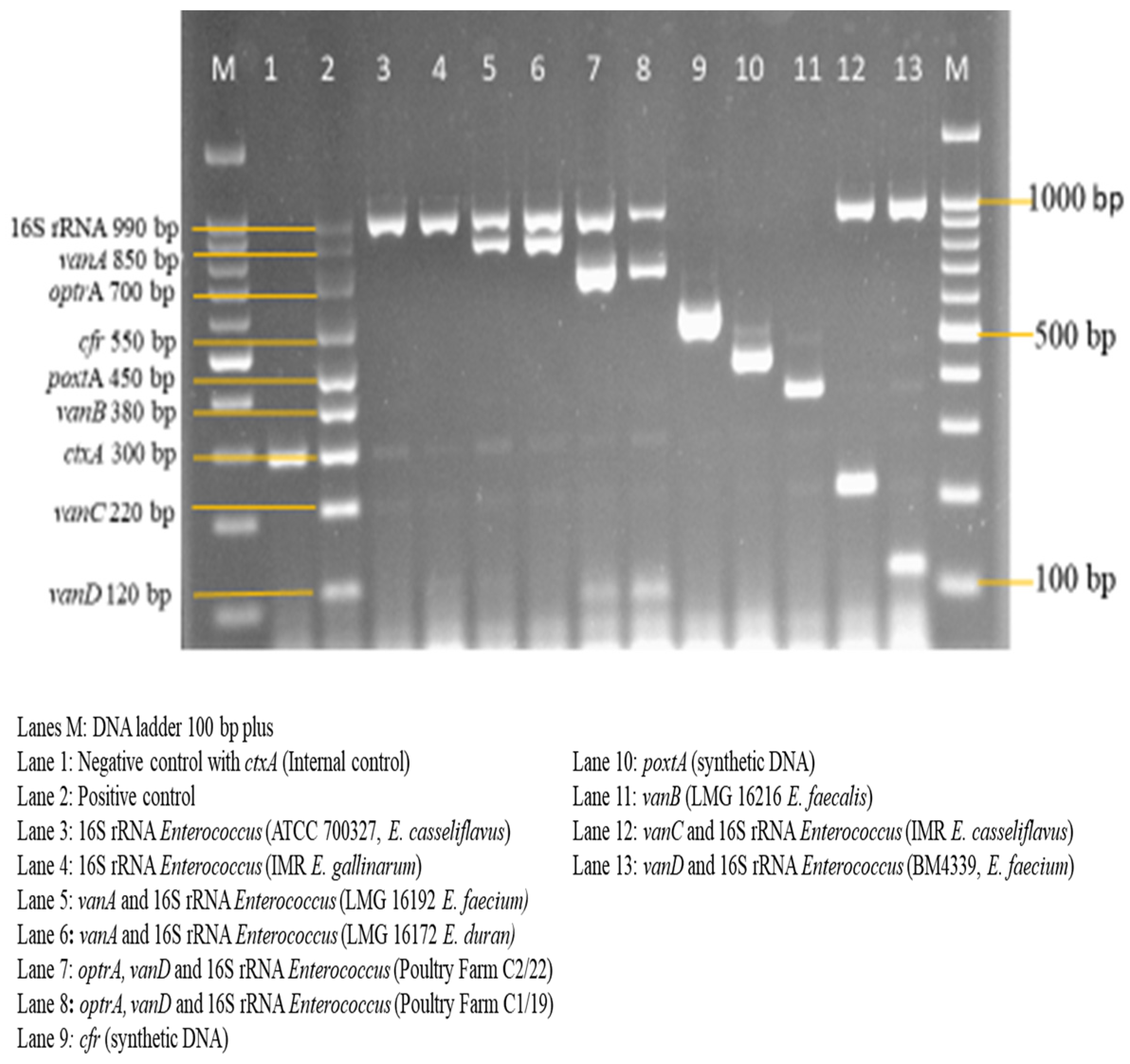
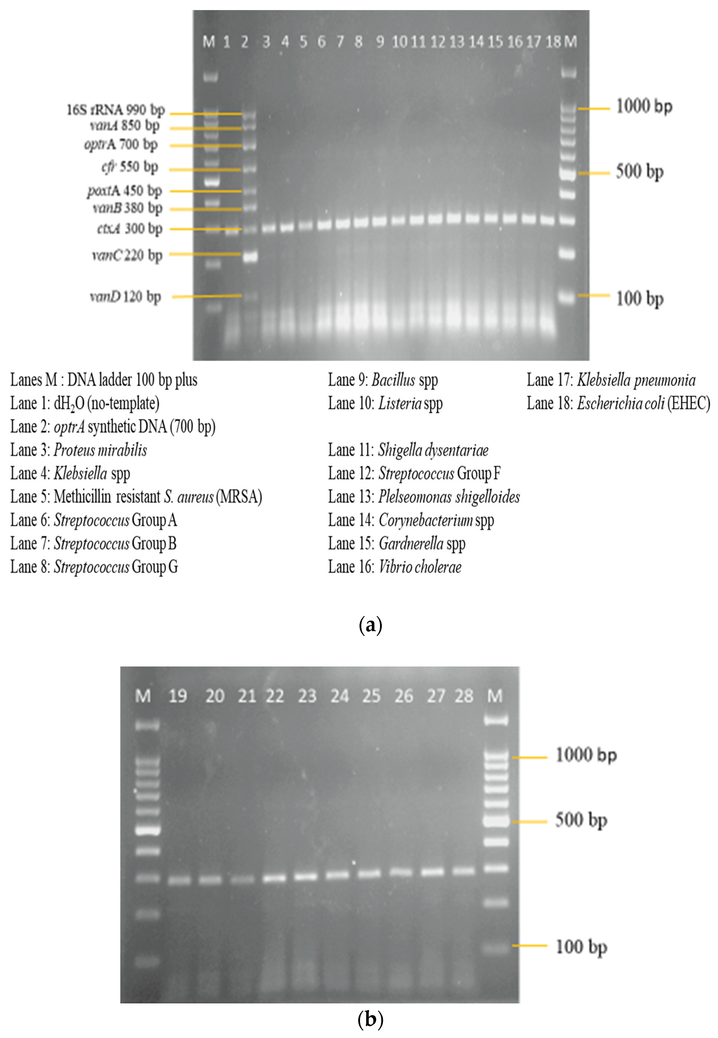
In this study, an internal amplification control (IAC) was incorporated in addition to utilizing a set of primers tailored for the simultaneous detection of vancomycin- and linezolid-resistant genes in Enterococcus in a multiplex PCR. These primers were then analyzed for their sensitivity utilizing the same species and specificity, utilizing different bacteria species that are Gram-positive and Gram-negative. A standard monoplex PCR was performed in a total volume of 20 μL containing 1 X PCR buffer (Apical Scientific Sdn.Bhd., Selangor, Malaysia), 2.5 mM magnesium chloride (MgCl2) (Apical Scientific Sdn.Bhd., Selangor, Malaysia), 0.2 mM deoxynucleotides (dNTPs) (Apical Scientific Sdn.Bhd., Selangor Malaysia), 1 μM of each sense and antisense primer, and 0.75 units of Taq DNA polymerase enzyme (Apical Scientific Sdn.Bhd., Selangor, Malaysia).
The cycling conditions used in this study consisted of initial denaturation at 95 °C (5 min), 30 cycles of denaturation at 95 °C (30 s), annealing at 64 °C (30 s), elongation at 72 °C (30 s), and a final elongation at 72 °C (5 min).
Then, to boost throughput and reliability, and reduce non-specific amplification, PCR components and conditions of the nanoplex were optimized based on the conditions of the monoplex PCR. Different concentrations of primer mixture ranging from 1.0 to 0.07 μM were prepared and tested on the PAC template. Following the primer optimization, optimization of dNTP concentration was done in the range of 0.08–0.24 mM. Different concentrations of MgCl2 were optimized in the range of 1.5–3.5 mM. Subsequently, Taq DNA polymerase enzyme was optimized in a range of 0.5–1.5 units. The IAC template concentration was optimized in the multiplex PCR in a range of 1 ng/μL–1 pg/μL. A set of Ta was utilized within 5 °C above and below the calculated Ta by using a gradient PCR thermal cycler.
PCR products were separated by electrophoresis at 100 volts for 90 min on 2.0% agarose gel and stained with FloroSafe DNA stain (1ST BASE, Singapore Science Park II, Singapore). Lastly, the multiplex PCR was evaluated for its sensitivity and specificity after the optimization.

3. Results

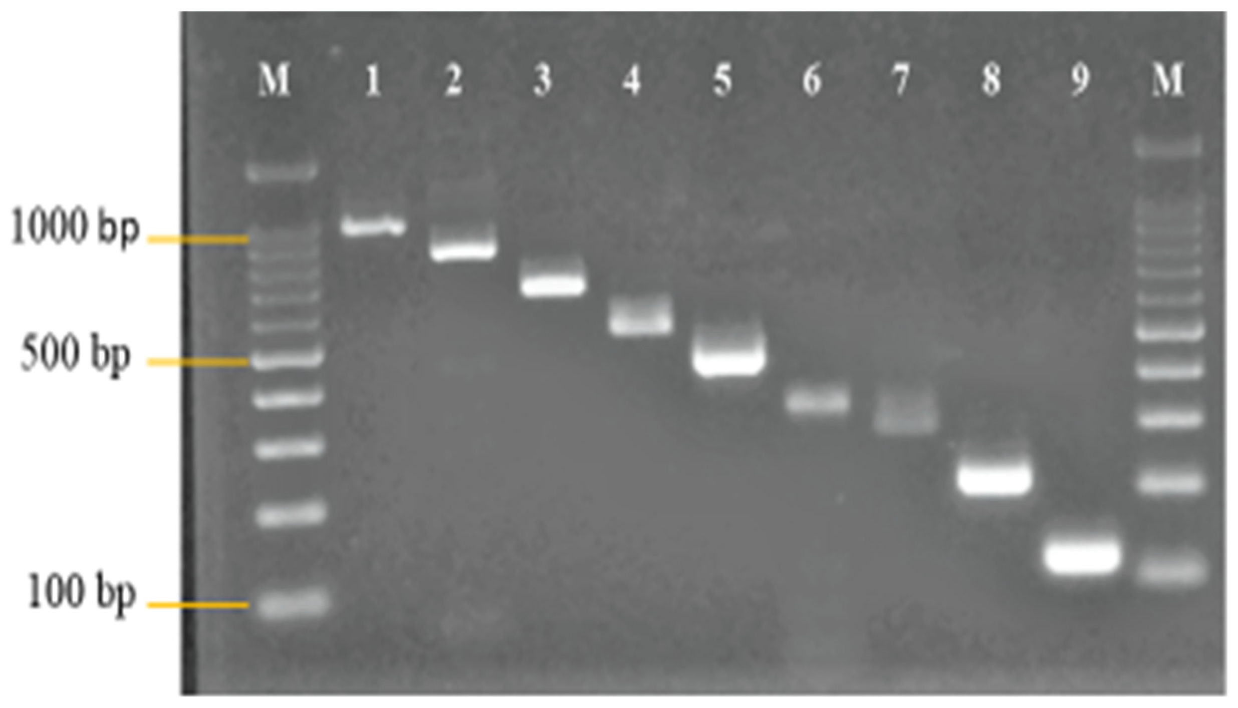
3.1. Primer Design and Analysis

Primers were designed to amplify the genes of interest. These genes are: 16S rRNA of Enterococcus genus, vanA—vanB—vanC—vanD for vancomycin, cfr methyltransferase, and optrA and poxtA, as well as an adenosine triphosphate-binding cassette (ABC) transporter for linezolid. A Vibrio cholerae ctxA primer was adopted from the study of Yean et al. [5] (Figure 1). The primers for the resistant genes were designed from the synthetic dsDNA utilized as the PAC. The clustalW program in Vector NTI version 9.0 software was used to design the primers. The primers were designed in such a way that their sizes were different from one another in the range of 50–150 bp. The specificity of the designed primers was checked using the NCBI- BLAST program.
Following the analysis of the designed primers in silico, in vitro analysis was also carried out. Standardized monoplex PCRs of the designed primers were performed to confirm the accuracy of the primers. Figure 1 shows the monoplex PCR of the designed primers at different sizes.
Subsequently, each of the primers were tested by evaluating their sensitivity to reference strains and specificity to the non-intended clinical isolates. All of the primers designed in this study were sensitive and specific. Synthetic dsDNA of the resistant genes was utilized as the positive control in the evaluation of the primers sensitivity and specificity.

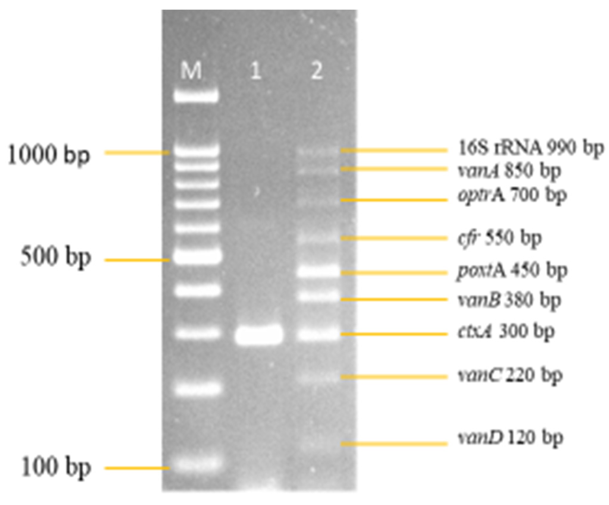
3.2. Development and Optimization of the Nanoplex PCR

Following the series of optimizations, all nine of the primer concentrations were finally optimized and are shown in Figure 2. The final primer concentration for 16S rRNA was 1.0 pmol/μL, vanA was 1.0 pmol/μL, optrA was 1.0 pmol/μL, cfr was 1.0 pmol/μL, poxtA was 0.1 pmol/μL, vanB was 0.08 pmol/μL, ctxA was 0.07 pmol/μL, vanC was 0.8 pmol/μL, and vanD was 0.1 pmol/μL.
A series of optimizations was carried out for the eight primers, including ctxA (IAC), to be amplified in the multiplex PCR assay. This occurred in a single-tube reaction and the optimal concentrations of the primers, MgCl2, dNTPs, and Taq DNA polymerase, were obtained. To amplify all nine genes simultaneously, the annealing temperature of the assay was also optimized.
Therefore, 2.5 mM of MgCl2 on lane 3 was selected for further optimization (Figure 3). The optimum concentration of dNTPs selected in this study was 0.16 mM on lane 4 (Figure 4), as well, 0.75 units of Taq DNA polymerase on lane 3 was selected (Figure 5), synthetic dsDNA template concentration of 1 ng/μL on lane 1 was selected (Figure 6), the optimum annealing temperature selected for this study was 64.5 °C on lane 8 (Figure 7), and an IAC template concentration of 10 pg/μL was selected on lanes 5 and 6 (Figure 8).

Final Optimised Parameters of the Nanoplex PCR Assay

Following the series of optimizations performed in this multiplex PCR, the final optimized parameters of the multiplex PCR are outlined in Table 5.

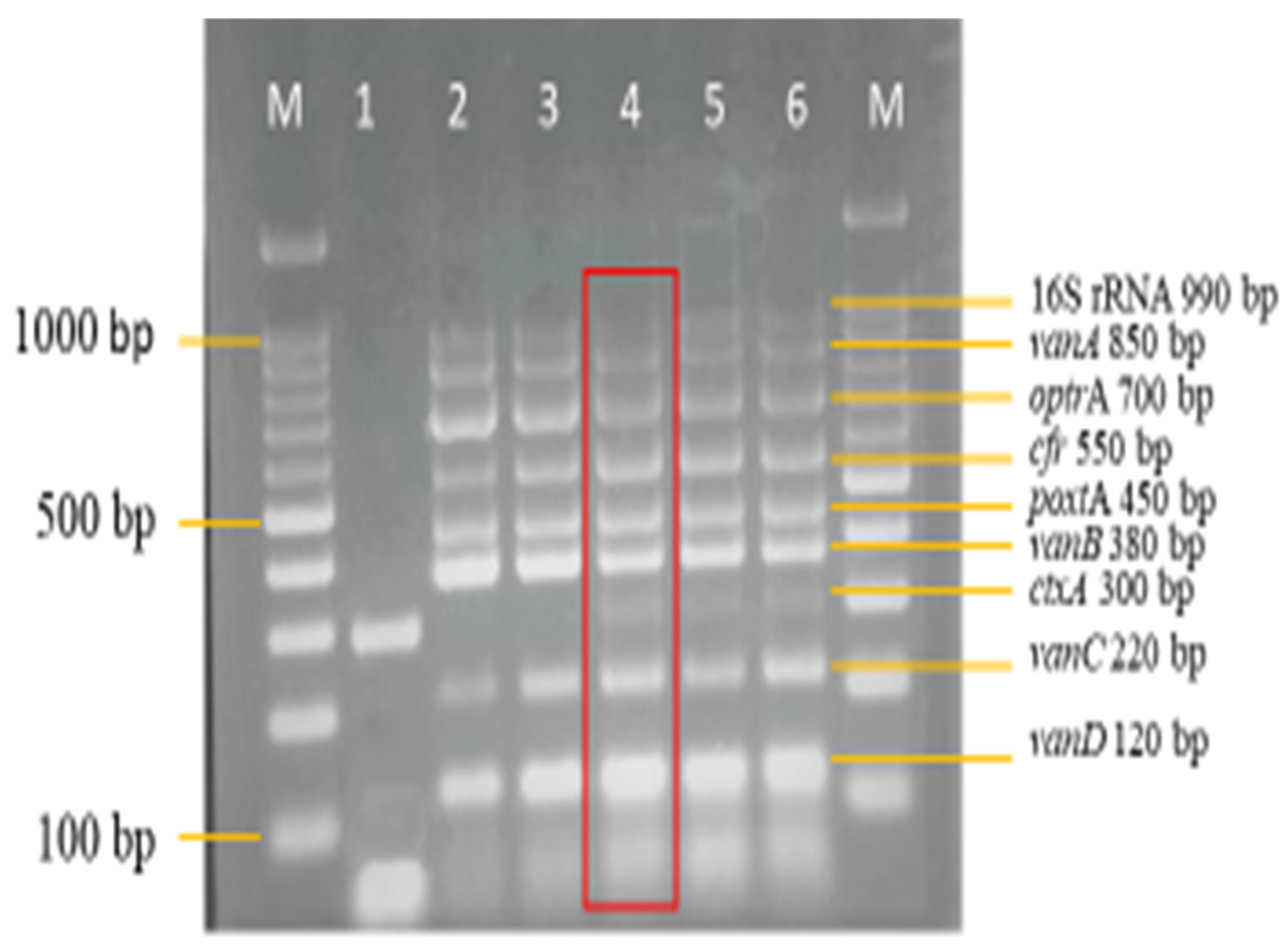
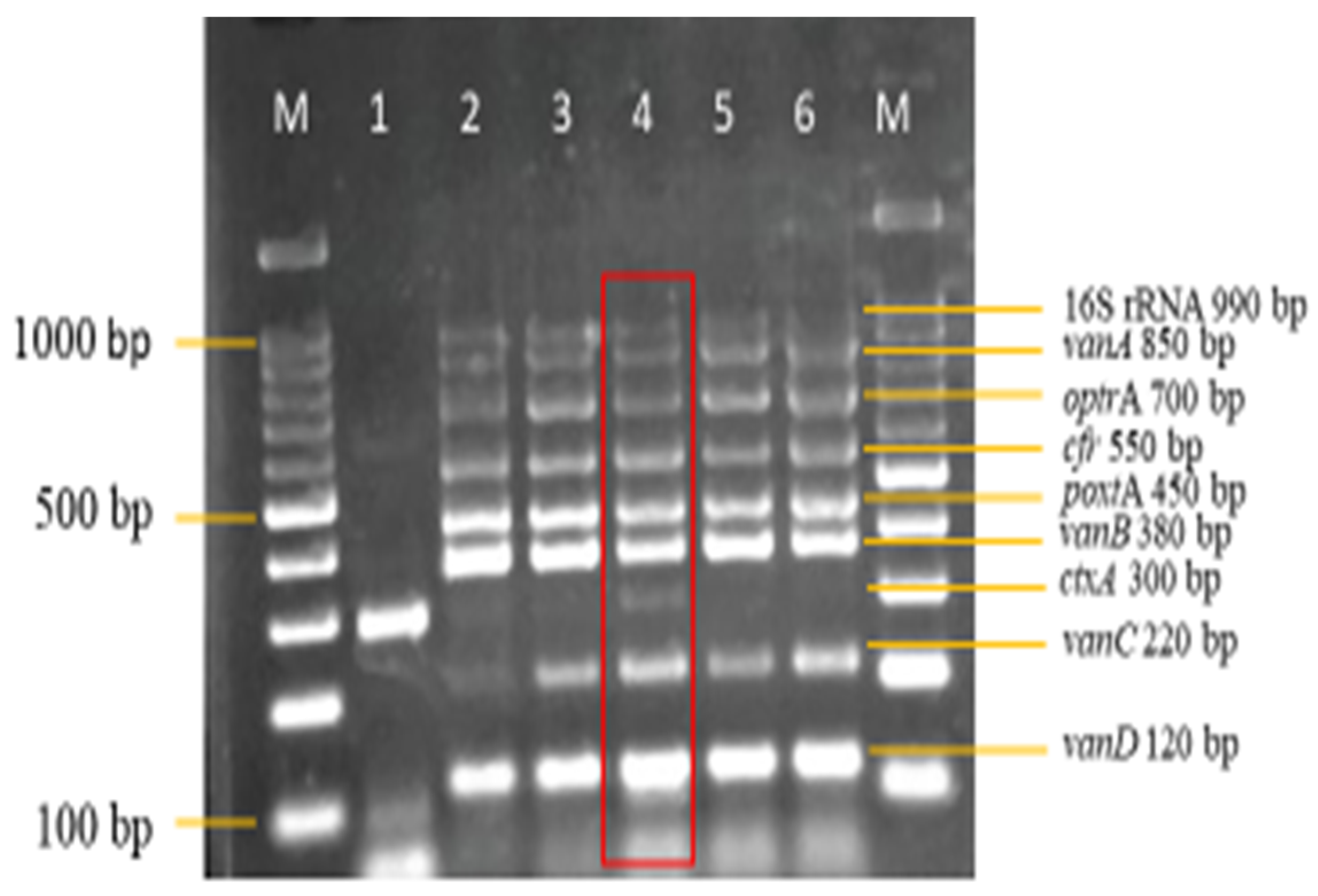
3.3. Sensitivity and Specificity Evaluation of the Nanoplex PCR Assay

A sensitivity evaluation of the optimized multiplex PCR assay was carried out using 2 μL of extracted genomic DNA of the 11 target reference strains. These target strains included 550 bp cfr and 450 bp poxtA synthetic dsDNA. Synthetic dsDNA was utilized because of the unavailability of the cfr and poxtA reference strains. The result shown in Figure 9 shows the amplification of all 11 target reference strains, which were as expected. The optimized multiplex PCR assay was also evaluated for its specificity against 26 non-intended targets, which are clinical isolates. Here, also, 2 μL of DNA template of the isolates was utilized. The results in Figure 10a,b show the non-amplification of the non-intended targets with clear amplification of the IAC on all lanes.

4. Discussion

This study was designed to develop a nanoplex PCR assay to simultaneously detect vancomycin- and linezolid-resistant genes in Enterococcus. Vancomycin used to be the last treatment option for an enterococcal infection until resistance began to emerge. This resistance was thought to be a result of the overuse of the growth promoter Avoparcin in Europe, where the resistance was first seen. Subsequently, VRE has been reported in several countries around the world and its prevalence is still on the rise. Linezolid, a synthetic oxazolidinone group of antibiotics, was designed to treat VRE. Being synthetic antibiotics, resistance was not expected to be seen. However, linezolid resistance in Enterococcus and other bacteria species has been reported worldwide, although their prevalence is low.
Thus far, no multiplex PCR has been developed for the simultaneous detection of both vancomycin- and linezolid-resistant genes in Enterococcus, meaning that this is the first, to the best of our knowledge. Yean et al. [5] developed a nanoplex PCR that simultaneously detect a bifunctional aminoglycoside and vancomycin-resistant genes in Enterococcus. Further, Bender et al. [6] also developed a multiplex PCR for the detection of linezolid-resistant genes in Enterococcus.
The multiplex PCR developed in this study includes four vancomycin-resistant genes (vanA, vanB, vanC, and vanD). These vancomycin-resistant genes were used in this study because they are the most commonly detected genes in clinical or environmental isolates. Similarly, three linezolid resistant genes (cfr, optrA, and poxtA) were also included in the multiplex PCR, as they are common and known resistant genes. This study is centered on Enterococcus; therefore, a 16S rRNA Enterococcus gene was included. The 16S rRNA Enterococcus gene is present in all Enterococcus species and highly conserved; therefore, this study did not take various Enterococcus species into consideration. Finally, a ctxA Vibrio cholerae gene was also included to function as the internal amplification control. This gene, from this bacterium, was used because it is Gram-negative, a non-target DNA, highly conserved, and not expected to amplify unless deliberately included in the reaction. This ctxA gene helps in ruling out the occurrence of PCR inhibitors and false-negative results in the multiplex PCR assay. In total, nine genes were included in the development of this multiplex PCR assay.
The primer concentrations were optimized in this study. Enterococcus 16S rRNA, vanA, optrA, and cfr all had a primer concentration of 1.0 pmol/μL. In the multiplex PCR of Yean et al. [5], an Enterococcus 16S rRNA primer concentration of 0.2 pmol/μL was reported, which was lower than that reported in this study, and a vanA primer concentration of 0.8 pmol/μL was also reported, which was closer to that reported in this study. Further, a primer concentration of 0.08 pmol/μL vanB, 0.8 pmol/μL vanC, 0.1 pmol/μL vanD, and 0.07 pmol/μL ctxA was obtained in this study. This was also close to the primer concentration reported by Yean et al. [5] in their study. They reported a primer concentration of 0.05 pmol/μL vanB, 0.7 pmol/μL vanC, higher than that reported in this study, 0.4 pmol/μL vanD closer to that reported in this study and 0.2 pmol/μL ctxA higher than that reported in this study. The ctxA Vibrio cholerae primer was adopted from the study of Yean et al. [5], and it is expected that their primer concentrations would be the same; however, they were not. The composition of other PCR parameters and the concentrations of different PCR primers could be responsible for this.
Similarly, the primer concentrations for linezolid-resistant genes were in contrast with those reported by Bender et al. [6] in their multiplex PCR. A primer concentration of 0.1 pmol/μL poxtA was reported in this study, which is the same poxtA primer concentration reported by Bender et al., 2019. However, Bender et al. [6] reported a primer concentration of 0.1 pmol/μL for both optrA and cfr, which varies from the primer concentration reported in this study. The differences in this primer concentration as observed in these studies could be a result of different concentrations of other PCR reagents and parameters. Further, the primers utilized in all of these studies were specifically designed to amplify their target genes and, as such, possess unique parameters, resulting in their varying concentrations in the multiplex PCR.
The optimization of MgCl2, dNTPs, and Taq DNA polymerase were also carried out. Magnesium chloride ions bind to the enzyme’s active site and increase its ability to perform the reaction. As a result, Taq DNA polymerase’s ability to add dNTPs to growing DNA strands is improved. Furthermore, MgCl2 enhances the reaction’s Tm. Mg2+ ions in MgCl2 attach to the PO3 and reduce the electrostatic barrier between DNA strands, temporarily protecting the negatively charged phosphate. The primer cannot connect to its exact site due to the electrostatic barrier between two DNA strands. The inclusion of MgCl2 facilitates the primer’s proper binding to its complementary bases. The right amount of MgCl2 improves the PCR specificity, whereas too much MgCl2 causes nonspecific binding, which reduces the accuracy and yield of the reaction. An MgCl2 concentration of 2.5 mM was observed in this study, which varies from the concentration (4.0 mM) reported by Yean et al. [5].
The function of deoxynucleotide triphosphates (dNTPs) in PCR is to help Taq DNA polymerase increase the growing DNA strand. They form hydrogen bonds with the complementary DNA strand. The amplification of all of the target genes in this multiplex PCR was dependent upon the successful optimization of the concentration of dNTPs.
Taq DNA polymerase is derived from Thermus aquaticus, a bacterium that plays a thermophilic function in a PCR reaction to amplify the DNA in order to produce a myriad of DNA. It can also function at high temperatures because it is thermostable. It is useful in the last step of PCR, extension, in which Taq DNA polymerase synthesises the DNA region between the primers utilising dNTPs (denoxynucleoside triphosphates) and Mg2+. Subsequently, concentrations of 0.16 mM and 0.75 units were observed for dNTPs and Taq DNA polymerase, respectively, in this study, which also varies from those reported by Yean et al. [5] in their study (300 μM dNTPS and two units Taq DNA polymerase). Bender et al. [6] did not report concentrations for any of the above reagents, as their multiplex PCR optimization was done using a master mix. The annealing temperature, varying concentrations of primers, and other PCR reagent, could be accountable for the variances observed in the concentration of the aforementioned reagents.
The importance of annealing temperature in a PCR cannot be overemphasized. An optimal annealing temperature is essential in a PCR because it determines the specificity of the PCR products. During the annealing phase of PCR, the reaction temperature must be low enough to allow both forward and reverse primers to bind to the template, but not low enough to allow the formation of unwanted, non-specific duplexes or intramolecular hairpins, both of which reduce reaction performance. The optimization of annealing temperature was carried out in a range of temperatures from 58 to 65.8 °C using a gradient program. In this study, an annealing temperature of 64.5 °C was utilized. However, this is close to the annealing temperature (65 °C) used by Yean et al. [5], and in contrast to that (50 °C) used by Bender et al. [6] in their multiplex PCR. An annealing temperature with a higher or lower degree could result in the impairment or inhibition of one or more of the target genes. The annealing temperature is dependent upon the composition of the nucleotide that makes up a primer. A primer with more guanine and cytosine nucleotide bases will most likely have a higher annealing temperature than a primer with adenine and thymine bases. This could be responsible for the variations observed in the annealing temperatures of these studies and, in addition, the concentration of other PCR reagents and parameters could also be responsible.
The optimized nanoplex PCR assay was further tested for its sensitivity and specificity. Trevethan [10] defined sensitivity as a screening test’s ability to detect a true positive, while specificity is defined as a screening test’s ability to detect a true negative. A test was considered positive when all of the target amplicons were observed even though the IAC might be absent, while a negative test was affirmed negative only when an IAC was amplified. This is indicative that a test could be declared invalid when an IAC is not amplified in negative samples. Nik Zuraina et al. [11] developed a heptaplex PCR assay for the detection of six respiratory bacteria pathogens. Their developed heptaplex PCR, similar to our developed nanoplex, was sensitive and specific. Although the heptaplex PCR of Nik Zuraina et al. [11] was thermostabilised, it was able to detect true positive and true negatives samples.
Timely and accurate detection of vancomycin and linezolid resistant genes could enable appropriate treatment and minimize the spread of antibiotic resistance pathogens. This study utilized an end-point detection method for reading reaction of a PCR. Although this approach provides reliable results, it can be prone to cross-over contamination if not performed within a controlled environment. In addition, the nanoplex PCR assay which was developed in this study was not thermostabilised. A thermostabilised mPCR is known for its rapidity, simplicity, accuracy, and does not involve the use of cold-chain.

5. Conclusions

To the best of our knowledge, this is the first study to develop a multiplex PCR for the simultaneous detection of a 16S rRNA Enterococcus gene, four vancomycin-resistant genes, three linezolid resistant genes, and a ctxA Vibrio cholerae gene functioning as an IAC. This developed multiplex PCR is sensitive, species-specific, rapid, and capable of detecting vancomycin- and linezolid-resistant genes in all types of settings (clinical, environmental, and farm) because the resistant genes that were utilized have been reported in all of these settings. The ctxA IAC will also ensure that false-negative results and inhibitors are taken care of. The development of a multiplex PCR assay that will take into account all known VRE genes and linezolid mutations is highly recommended. This would ensure that no resistant genes are missed during routine laboratory diagnosis. Future research directions may include the development of a thermostabilised nanoplex PCR assay, taking into cognizance all known vancomycin- and linezolid-resistant genes.

Author Contributions

Conceptualization, Y.W., A.R.Z., C.Y.Y. and A.H.; methodology, Y.W. and C.Y.Y.; validation, A.R.Z., C.Y.Y. and A.H.; formal analysis, Y.W. and C.Y.Y.; writing—original draft preparation, Y.W.; writing—review and editing, Y.W., A.R.Z., C.Y.Y. and A.H.; supervision, A.R.Z., C.Y.Y. and A.H. All authors have read and agreed to the published version of the manuscript.

Funding

This research was supported by a Universiti Sains Malaysia Research University Grant (RUI): 1001.PPSP.8012259 and Y.W. is supported by the USM Fellowship.

Institutional Review Board Statement

This study was approved by the Institutional Review Board of the Human Research Ethics Committee, USM (USM/JEPeM/19050308).

Informed Consent Statement

Not applicable.

Data Availability Statement

Dataset used and/or analyzed during the correct study are included in the manuscript.

Acknowledgments

The authors would like to thank the Department of Medical Microbiology and Parasitology, School of Medical Sciences, Universiti Sains Malaysia, for providing the bacterial strains and facilities used in this study.

Conflicts of Interest

The authors declare no conflict of interest. The funders had no role in the design of the study; in the collection, analyses, or interpretation of data; in the writing of the manuscript, or in the decision to publish the results.

References

  1. Wada, Y.; Harun, A.B.; Yean, C.Y.; Zaidah, A.R. Vancomycin-Resistant Enterococcus: Issues in Human Health, Animal Health, Resistant Mechanisms and the Malaysian Paradox. Adv. Anim. Vet. Sci. 2019, 7, 1021–1034. [Google Scholar] [CrossRef]
  2. Antimicrobial Resistance. Available online: https://www.who.int/news-room/fact-sheets/detail/antimicrobial-resistance (accessed on 19 December 2022).
  3. Browne, A.J.; Chipeta, M.G.; Haines-Woodhouse, G.; Kumaran, E.P.A.; Hamadani, B.H.K.; Zaraa, S.; Henry, N.J.; Deshpande, A.; Reiner, R.C.; Day, N.P.J.; et al. Global Antibiotic Consumption and Usage in Humans, 2000–2018: A Spatial Modelling Study. Lancet Planet. Health 2021, 5, e893–e904. [Google Scholar] [CrossRef] [PubMed]
  4. Murray, C.J.; Ikuta, K.S.; Sharara, F.; Swetschinski, L.; Robles Aguilar, G.; Gray, A.; Han, C.; Bisignano, C.; Rao, P.; Wool, E.; et al. Global Burden of Bacterial Antimicrobial Resistance in 2019: A Systematic Analysis. Lancet 2022, 399, 629–655. [Google Scholar] [CrossRef] [PubMed]
  5. Yean, C.Y.; Yin, L.S.; Lalitha, P.; Ravichandran, M. A Nanoplex PCR Assay for the Rapid Detection of Vancomycin and Bifunctional Aminoglycoside Resistance Genes in Enterococcus Species. BMC Microbiol. 2007, 7, 112. [Google Scholar] [CrossRef] [PubMed]
  6. Bender, J.K.; Fleige, C.; Klare, I.; Werner, G. Development of a Multiplex-PCR to Simultaneously Detect Acquired Linezolid Resistance Genes Cfr, OptrA and PoxtA in Enterococci of Clinical Origin. J. Microbiol. Methods 2019, 160, 101–103. [Google Scholar] [CrossRef] [PubMed]
  7. Zangenberg, G.; Saiki, R.K.; Reynolds, R. Multiplex PCR: Optimization Guidelines. In PCR Applications; Academic Press: Cambridge, MA, USA, 1999; pp. 73–94. [Google Scholar] [CrossRef]
  8. Song, H.; Bae, Y.; Jeon, E.; Kwon, Y.; Joh, S. Multiplex PCR Analysis of Virulence Genes and Their Influence on Antibiotic Resistance in Enterococcus Spp. Isolated from Broiler Chicken. J. Vet. Sci. 2019, 20, e26. [Google Scholar] [CrossRef] [PubMed]
  9. Mohamad Nasir, N.S.; Chan, Y.Y.; Harun, A.; Husin, A.; Kamaruzzaman, N.F.; Wada, Y.; Abdul-Rahman, Z. Malaysian Journal of Microbiology Linezolid-resistant Enterococcus casseliflavus and Enterococcus gallinarum isolated from poultry farms in Kelantan, Malaysia. Malays. J. Microbiol. 2021, 17, 361–368. [Google Scholar] [CrossRef]
  10. Trevethan, R. Sensitivity, Specificity, and Predictive Values: Foundations, Pliabilities, and Pitfalls in Research and Practice. Front. Public Health 2017, 5, 307. [Google Scholar] [CrossRef] [PubMed]
  11. Nik Zuraina, N.M.N.; Goni, M.D.; Amalina, K.N.; Hasan, H.; Mohamad, S.; Suraiya, S. Thermostable Heptaplex PCR Assay for the Detection of Six Respiratory Bacterial Pathogens. Diagnostics 2021, 11, 753. [Google Scholar] [CrossRef] [PubMed]
Figure 1. Monoplex PCR of 16S rRNA Enterococcus, ctxA (IAC), vancomycin and linezolid resistant genes primers. The differences in the sizes of the primers ranged from 50–150 bp. The designed primers were all amplified at their expected sizes. Lanes M: DNA ladder 100 bp plus. Lane 1: 16S rRNA Enterococcus (990 bp). Lane 2: vanA (850 bp). Lane 3: optrA (700 bp). Lane 4: cfr (550 bp). Lane 5: poxtA (450 bp). Lane 6: vanB (380 bp). Lane 7: ctxA (300 bp). Lane 8: vanC (220 bp). Lane 9: vanD (120 bp).
Diagnostics 13 00722 g001
Figure 2. Final optimized multiplex PCR primer concentration. All other PCR parameters remained unchanged. Lanes M: DNA ladder 100 bp plus. Lane 1: Negative control with ctxA (Internal control). Lane 2: Optimized multiplex primer concentration. 16S rRNA = 1.0 pmol/μL. vanA = 1.0 pmol/μL. optrA = 1.0 pmol/μL. cfr = 1.0 pmol/μL. poxtA = 0.1 pmol/μL. vanB = 0.08 pmol/μL. ctxA = 0.07 pmol/μL. vanC = 0.8 pmol/μL. vanD = 0.1 pmol/μL.
Diagnostics 13 00722 g002
Figure 3. MgCl2 concentration optimization in the multiplex PCR. A concentration of 2.5 mM on lane 4 from a range of concentration (1.5–3.5 mM) was selected as the optimum concentration. Lane M: DNA ladder 100 bp plus. Lane 1: Negative control with ctxA (Internal control). Lane 2: 1.5 mM of MgCl2. Lane 3: 2.0 mM of MgCl2. Lane 4: 2.5 mM of MgCl2. Lane 5: 3.0 mM of MgCl2. Lane 6: 3.5 mM of MgCl2.
Diagnostics 13 00722 g003
Figure 4. dNTPs concentration optimization in the multiplex PCR. A concentration of 0.16 mM on lane 4 from a range of concentrations (0.08–0.24 mM) was selected as the optimum concentration. Lane M: DNA ladder 100 bp plus. Lane 1: Negative control with ctxA (Internal control). Lane 2: 0.08 mM of dNTPs. Lane 3: 0.12 mM of dNTPs. Lane 4: 0.16 mM of dNTPs. Lane 5: 0.20 mM of dNTPs. Lane 6: 0.24 mM of dNTPs.
Diagnostics 13 00722 g004
Figure 5. Taq DNA polymerase concentration optimization in the multiplex PCR. A concentration of 0.75 units on lane 3 from a range of concentrations (0.5–1.50 units) was selected as the optimum concentration. Lane M: DNA ladder 100 bp plus. Lane 1: Negative control with ctxA (Internal control). Lane 2: 0.50 Units. Lane 3: 0.75 Units. Lane 4: 1.00 Units. Lane 5: 1.25 Units. Lane 6: 1.50 Units.
Diagnostics 13 00722 g005
Figure 6. Synthetic dsDNA template concentration optimization in the multiplex PCR. A concentration of 1 ng/μL on lane 1 from a range of template concentrations (1 ng/μL–1 pg/μL) was selected as the optimum template concentration. Lane M: DNA ladder 100 bp plus. Lane 1: 1 ng/μL cocktail synthetic DNA template. Lane 2: 100 pg/μL cocktail synthetic DNA template. Lane 3: 10 pg/μL cocktail synthetic DNA template. Lane 4: 1 pg/μL cocktail synthetic DNA template.
Diagnostics 13 00722 g006
Figure 7. Annealing temperature optimization in the multiplex PCR. An annealing temperature of 64.5 °C on lane 8 from a range of temperature set (58–65.8 °C) was selected as the optimum annealing temperature. Lane M: DNA ladder 100 bp plus. Lane 1: 58 °C. Lane 2: 58.7 °C. Lane 3: 59.5 °C. Lane 4: 60.5 °C. Lane 5: 61.5 °C. Lane 6: 62.5 °C. Lane 7: 63.5 °C. Lane 8: 64.5 °C. Lane 9: 65.3 °C. Lane 10: 65.8 °C.
Diagnostics 13 00722 g007
Figure 8. Internal amplification control template concentration optimization in the multiplex PCR Assay. A concentration of 10 pg/μL on lanes 5 and 6 from a range of template concentrations (1 ng/μL–1 pg/μL) was selected as the optimum template concentration. Lane M: DNA ladder 100 bp plus. Lane 1: 1 ng ctxA (Internal control). Lane 2: 1 ng ctxA (Internal control) with other targets. Lane 3: 100 pg ctxA (Internal control). Lane 4: 100 pg ctxA (Internal control) with other targets. Lane 5: 10 pg ctxA (Internal control). Lane 6: 10 pg ctxA (Internal control) with other targets. Lane 7: 1 pg ctxA (Internal control). Lane 8: 1 pg ctxA (Internal control) with other targets.
Diagnostics 13 00722 g008
Figure 9. Sensitivity evaluation of multiplex PCR assay on intended reference bacterial strains (n = 11); 300 bp ctxA doubles as the negative control and IAC on lane 1 while lane 2, with all targets, was designated as the positive control.
Diagnostics 13 00722 g009
Figure 10. (a) Specificity evaluation of multiplex PCR assay on non-intended clinical isolates (n = 16). Amplification is absent except for the IAC, indicating the specificity of the multiplex PCR assay; 300 bp ctxA doubles as the negative control and IAC on lane 1, while lane 2, with all targets, was designated as the positive control. (b) Specificity evaluation of multiplex PCR assay on non-intended clinical isolates (n = 10) continued. Lane M: DNA ladder 100 bp plus. Lane 19: Escherichia coli (EPEC). Lane 20: Vibrio parahaemolyticus. Lane 21: Shigella sonnei. Lane 22: Shigella boydii. Lane 23: Citrobacter freundii. Lane 24: Yersinia enterocolitica. Lane 25: Acinetobacter baumannii. Lane 26: Acinetobacter spp. Lane 27: Pseudomonas aeruginosa. Lane 28: Staphylococcus aureus.
Diagnostics 13 00722 g010
Table 1. Reference strains utilized for the sensitivity evaluation of multiplex and monoplex PCR assays.
SpeciesReferenceTarget Gene
Enterococcus casseliflavus aATCC 70032716S rRNA Enterococcus
Enterococcus gallinarum bIMR16S rRNA Enterococcus
Enterococcus raffinosus cLMG 1217216S rRNA Enterococcus
Enterococcus mundti cLMG 1230816S rRNA Enterococcus
Enterococcus faecium cLMG 1619216S rRNA Enterococcus, vanA
Enterococcus durans cLMG 1617216S rRNA Enterococcus, vanA,
Enterococcus faecalis cLMG 16216vanB
Enterococcus casseliflavus bIMR16S rRNA Enterococcus, vanC
Enterococcus faecium dBM433916S rRNA Enterococcus, vanD
Enterococcus casseliflavus ePoultry Farm C2/2216S rRNA Enterococcus, optrA, poxtA
Enterococcus casseliflavus ePoultry Farm C1/1916S rRNA Enterococcus, optrA, poxtA
a ATCC strain was obtained from the Department of Medical Microbiology and Parasitology, USM. b Obtained from the Institute of Medical Research, Malaysia. c Obtained from BCCM, Ghent Belgium. d Provided by Professor Patrice Courvalin and Dr Bruno Perichon, Institut Pasteur, Paris, France. e Obtained from Mohamad Nasir, N.S [9] and confirmed by sequencing.
Table 2. Clinical isolates utilized for evaluation of multiplex and monoplex PCR assays.
S/NoBacteria Strains
Gram-positive
1Methicillin-resistant Staphylococcus aureus (MRSA)
2Streptococcus Group A
3Streptococcus Group B
4Streptococcus Group G
5Streptococcus Group F
6Bacillus species
7Listeria species
8Corynebacterium species
9Staphylococcus aureus
10Gardnerella species
Gram-negative
1Proteus mirabilis
2Klebsiella species
3Shigella dysentariae
4Plesiomonas shigelloides
5Vibrio cholerae
6Klebsiella pneumoniae
7Escherichia coli (Enterohemorrhagic EHEC)
8Escherichia coli (Enteropathogenic EPEC)
9Vibrio parahaemolyticus
10Shigella sonnei
11Shigella boydii
12Citrobacter freundii
13Yersinia enterocolitica
14Acinetobacter baumannii
15Acinetobacter species
16Pseudomonas aeruginosa
Table 3. Synthetic DNA utilized as positive and internal amplification control.
Synthetic dsDNASize (bp)
16S rRNA Enterococcus993
vanA1032
optrA880
cfr725
poxtA600
vanB1029
ctxA *615
vanC699
vanD1032
* Internal Amplification Control.
Table 4. Characteristics of the primers used in this study.
Target GenePrimer Sequence (5′-3′)Product Length (bp)
16S rRNA EnterococcusF-5′-TTC CAC CGG AGC TTG CTC C-3′
R-5′-TTT GCC CCC GAA GGG GAA G-3′
990
vanAF-5′-TTT GGG GGT TGC TCA GAG G-3′
R-5′-CAC ACG GGC TAG ACC TCT A-3′
850
optrAF-5′-TGG AAA AAC AAC CTT GCT AAA AGC-3′
R-5′-CAA GCG TGT AAT CCT TTC AAT TTC-3′
700
cfrF-5′-CAA AGA ATT AGT CGA TTT GAG GA-3′
R-5′-GTT CCT CAC TAT AAG GTG AGT-3′
550
poxtAF-5′-TGC TTT TTC TCC AGG GGA CA-3′
R-5′-GTG GAG AGC TGC AAA AGA GA-3′
450
vanBF-5′-AAA ACG GCG TAT GGA AGC TAT G-3′
R-5′-CGG CTT CAC AAA GAC AGG GTA G-3′
380
ctxA *F-5′-AAC TCA GAC GGG ATT TGT TAG GC-3′
R-5′-TCT CTG TAG CCC CTA TTA CGA TGT-3′
300 [5]
vanCF-5′-CAG CAG CCA TTG GCG TAC A-3′
R-5′-TGT AGG AGC ACT GCG GAA C-3′
220
vanDF-5′-AAG CTC CGT GAT CTG CAT GG-3′
R-5′-AAA TCC TCC GTT TCC AGG C-3′
120
*—Internal Amplification Control. F—Forward or Sense sequence. R—Reverse or Antisense sequence. bp—Base pair.
Table 5. Final optimized parameters of the nanoplex PCR assay for the detection of vancomycin- and linezolid-resistant genes in Enterococcus.
ComponentsInitial ConcentrationPer Reaction (μL)Final Concentration
PCR-Grade dH2O-2.37-
10× Reaction Buffer10×2.0
MgCl225 mM2.02.5 mM
dNTPs10 mM0.320.16 mM
Primers (Sense and Anti-sense)
16S rRNA Enterococcus20 μM1.01 μM
vanA20 μM1.01 μM
optrA20 μM1.01 μM
cfr20 μM1.01 μM
poxtA20 μM0.10.1 μM
vanB20 μM0.080.08 μM
ctxA (IAC)20 μM0.070.07 μM
vanC20 μM0.80.8 μM
vanD20 μM0.10.1 μM
Taq DNA Polymerase5 units0.150.75 units
ctxA Template (IAC)10 ng1.010 pg
Target DNA Cocktail mix (Synthetic dsDNA)10 ng/μL of each target2.01 ng/μL of each target
Final Volume (μL) 20
Disclaimer/Publisher’s Note: The statements, opinions and data contained in all publications are solely those of the individual author(s) and contributor(s) and not of MDPI and/or the editor(s). MDPI and/or the editor(s) disclaim responsibility for any injury to people or property resulting from any ideas, methods, instructions or products referred to in the content.

