

Potential of Unenhanced Ultra-Low-Dose Abdominal Photon-Counting CT with Tin Filtration: A Cadaveric Study
 , , , ,
Abstract
:1. Introduction
2. Methods
2.1. Scan and Image Reconstruction Parameters
2.2. Objective Image Analysis
2.3. Subjective Image Analysis
2.4. Statistics
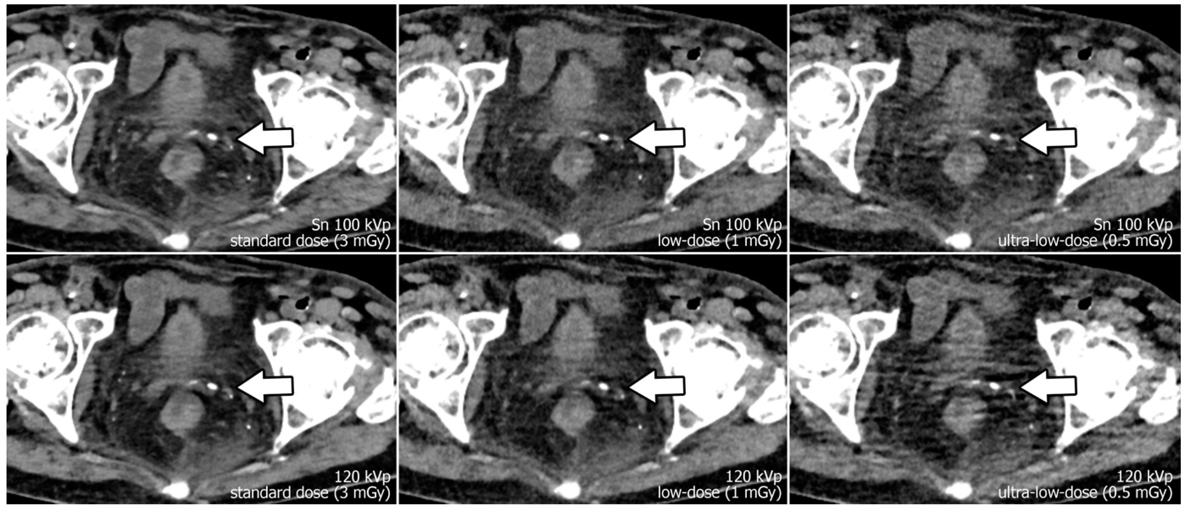
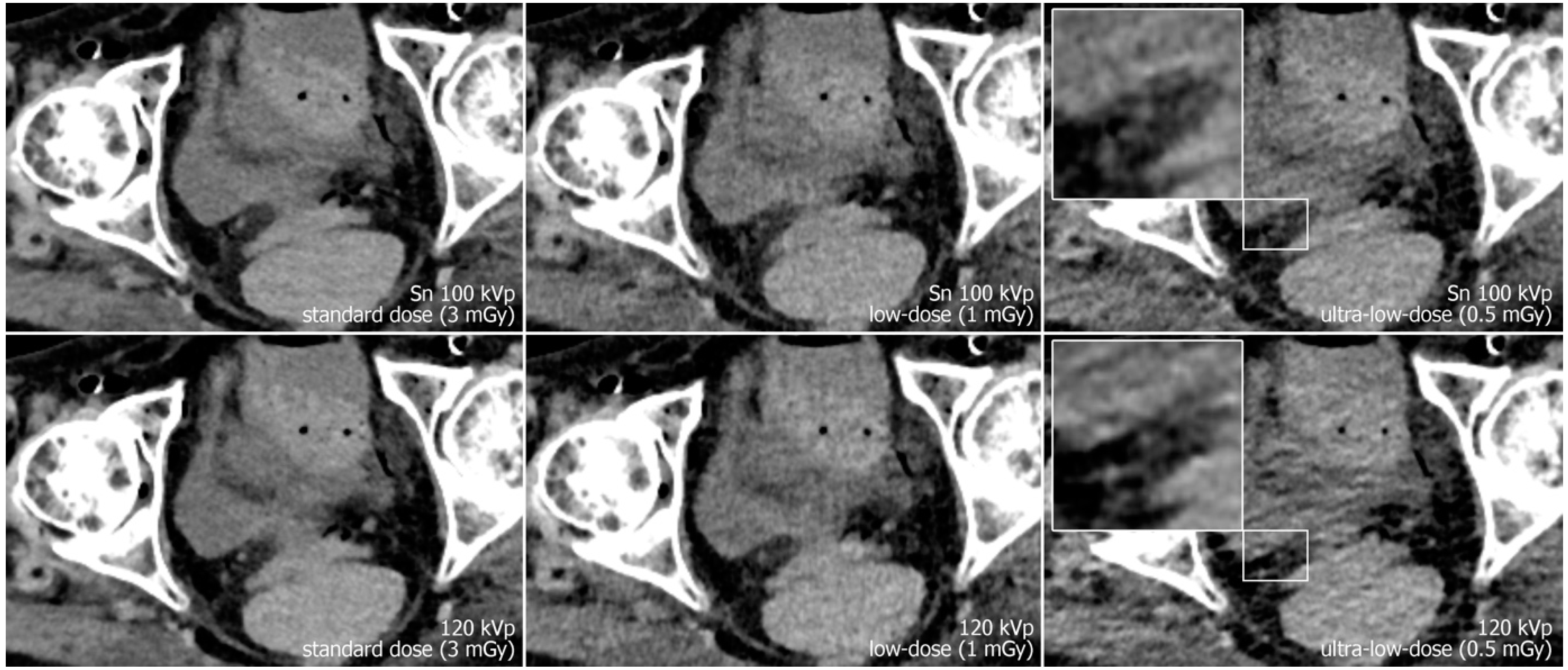
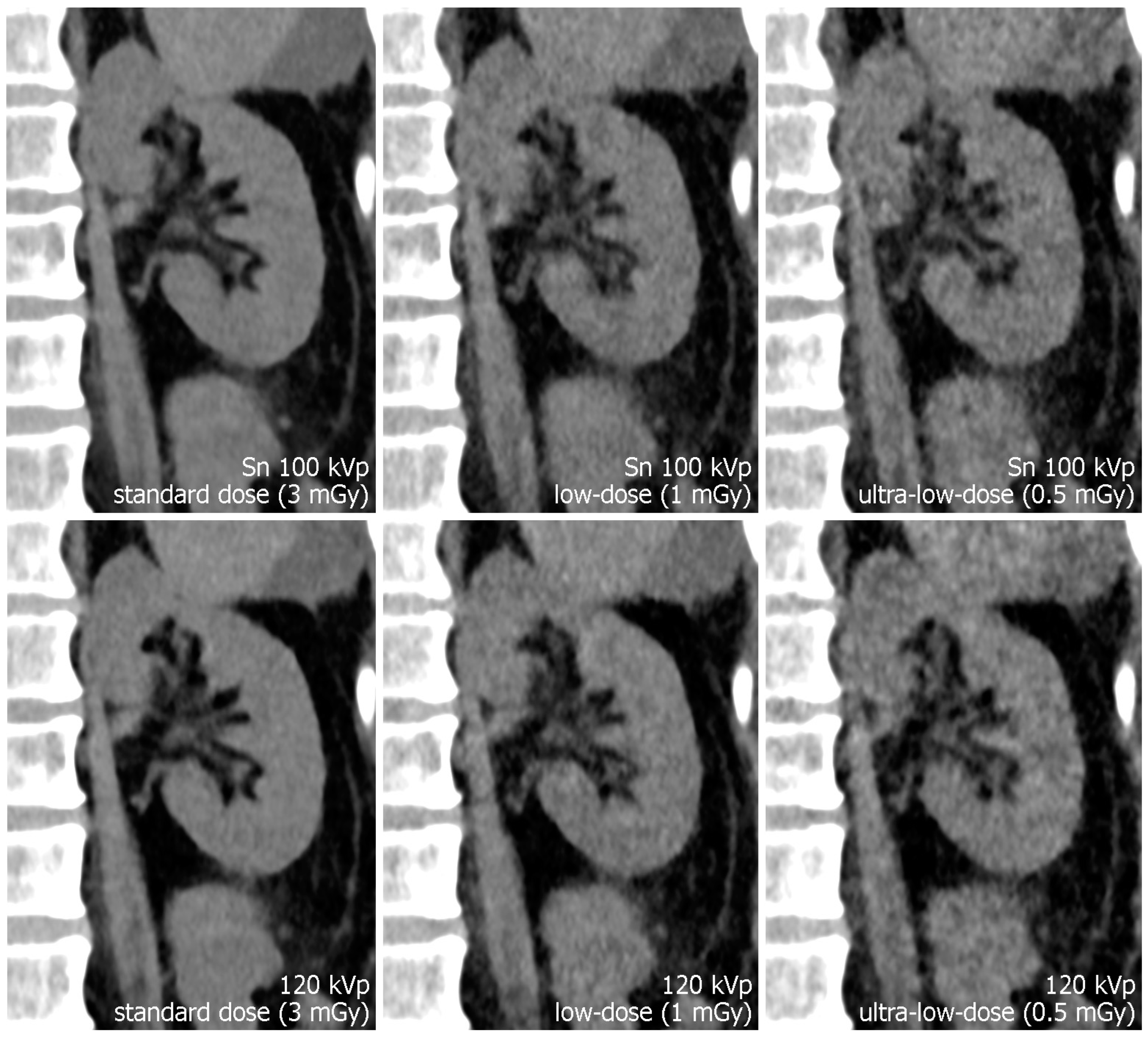
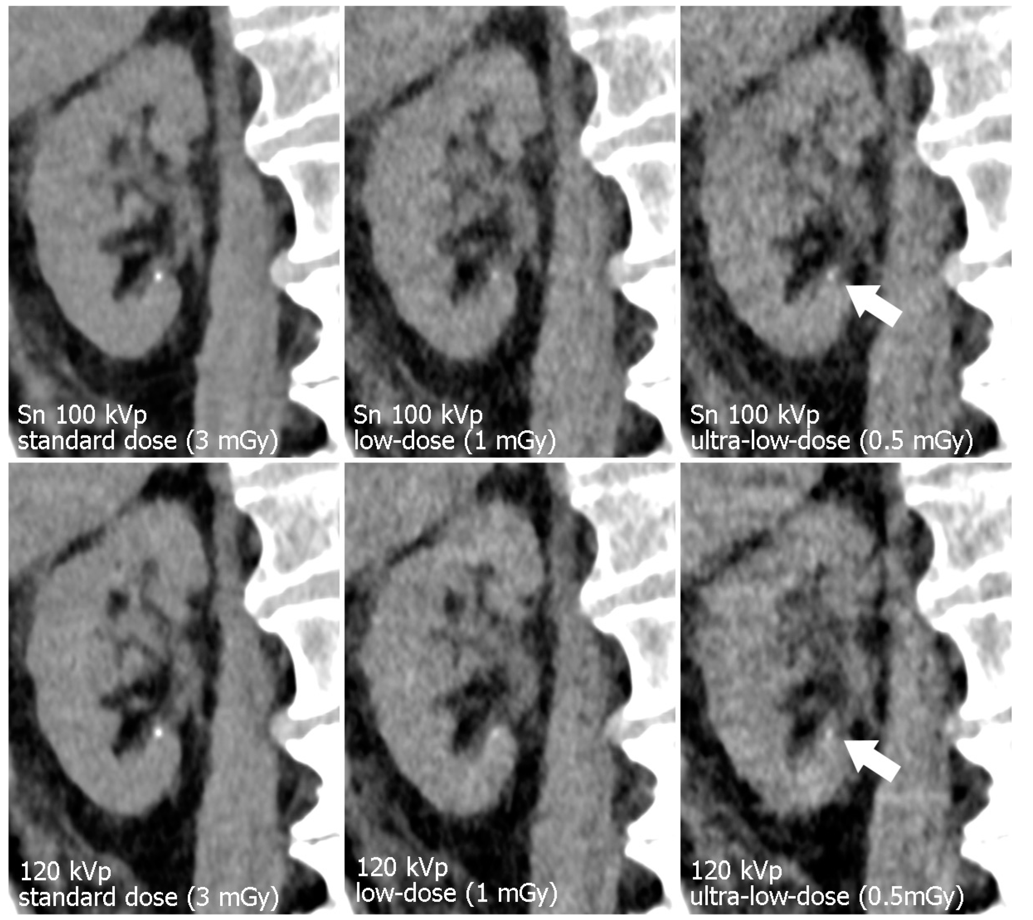
3. Results
3.1. Radiation Doses
3.2. Objective Image Quality
3.3. Subjective Image Quality
4. Discussion
5. Conclusions
Author Contributions
Funding
Institutional Review Board Statement
Informed Consent Statement
Data Availability Statement
Conflicts of Interest
Abbreviations and Acronyms
| CNR | contrast-to-noise ratio |
| CTDIvol | volume computed tomography dose index |
| IQR | interquartile range |
| PCD-CT | photon-counting detector computed tomography |
References
- Greffier, J.; Pereira, F.; Hamard, A.; Addala, T.; Beregi, J.P.; Frandon, J. Effect of tin filter-based spectral shaping CT on image quality and radiation dose for routine use on ultralow-dose CT protocols: A phantom study. Diagn. Interv. Imaging 2020, 101, 373–381. [Google Scholar] [CrossRef]
- Petritsch, B.; Kosmala, A.; Weng, A.M.; Bley, T.A. Tin-filtered 100 kV ultra-low-dose CT of the paranasal sinus: Initial clinical results. Zeng L, ed. PLoS ONE 2019, 14, e0216295. [Google Scholar] [CrossRef]
- Grunz, J.-P.; Petritsch, B.; Luetkens, K.S.; Kunz, A.S.; Lennartz, S.; Ergün, S.; Bley, T.A.; Huflage, H. Ultra-Low-Dose Photon-Counting CT Imaging of the Paranasal Sinus With Tin Prefiltration. Investig. Radiol. 2022, 57, 728–733. [Google Scholar] [CrossRef]
- Baldi, D.; Tramontano, L.; Alfano, V.; Punzo, B.; Cavaliere, C.; Salvatore, M. Whole Body Low Dose Computed Tomography Using Third-Generation Dual-Source Multidetector With Spectral Shaping: Protocol Optimization and Literature Review. Dose-Response 2020, 18, 1–7. [Google Scholar] [CrossRef]
- Stern, C.; Sommer, S.; Germann, C.; Galley, J.; Pfirrmann, C.W.A.; Fritz, B.; Sutter, R. Pelvic bone CT: Can tin-filtered ultra-low-dose CT and virtual radiographs be used as alternative for standard CT and digital radiographs? Eur. Radiol. 2021, 31, 6793–6801. [Google Scholar] [CrossRef]
- Hackenbroch, C.; Schnaidt, S.; Halt, D.; Wilde, F.; Beer, M.; Wunderlich, A. Dose Reduction in Dental CT: A Phantom Study With Special Focus on Tin Filter Technique. Am. J. Roentgenol. 2020, 215, 945–953. [Google Scholar] [CrossRef]
- Hackenbroch, C.; Feilhuber, M.; Halt, D.; Riesner, H.J.; Beer, M.; Wunderlich, A. Low-dose CT in pelvic imaging: Comparing dose and image quality in relation to clinical value in a phantom study. Am. J. Roentgenol. 2021, 216, 453–463. [Google Scholar] [CrossRef]
- Grunz, J.-P.; Halt, D.; Schüle, S.; Beer, M.; Hackenbroch, C. Thermoluminescence Dosimetry in Abdominal CT for Urinary Stone Detection. Investig. Radiol. 2022. [Google Scholar] [CrossRef]
- Kunz, A.S.; Grunz, J.; Halt, D.; Kalogirou, C.; Luetkens, K.S.; Patzer, T.S.; Christner, S.A.; Sauer, S.T.; Bley, T.A.; Huflage, H. Tin-filtered 100 kV Ultra-low-dose Abdominal CT for Calculi Detection in the Urinary Tract: A Comparative Study of 510 Cases. Acad. Radiol. 2022, 29. [Google Scholar] [CrossRef]
- Zhang, G.-M.-Y.; Shi, B.; Sun, H.; Xue, H.-D.; Wang, Y.; Liang, J.-X.; Xu, K.; Wang, M.; Wang, M.; Xu, M.; et al. High-pitch low-dose abdominopelvic CT with tin-filtration technique for detecting urinary stones. Abdom. Radiol. 2017, 42, 2127–2134. [Google Scholar] [CrossRef]
- Grunz, J.-P.; Heidenreich, J.F.; Lennartz, S.; Weighardt, J.P.; Bley, T.A.; Ergün, S.; Petritsch, B.; Huflage, H. Spectral Shaping Via Tin Prefiltration in Ultra-High-Resolution Photon-Counting and Energy-Integrating Detector CT of the Temporal Bone. Investig. Radiol. 2022, 57, 819–825. [Google Scholar] [CrossRef] [PubMed]
- Hagen, F.; Hofmann, J.; Wrazidlo, R.; Gutjahr, R.; Schmidt, B.; Faby, S.; Nikolaou, K.; Horger, M. Image quality and dose exposure of contrast-enhanced abdominal CT on a 1st generation clinical dual-source photon-counting detector CT in obese patients vs. a 2nd generation dual-source dual energy integrating detector CT. Eur. J. Radiol. 2022, 151, 110325. [Google Scholar] [CrossRef] [PubMed]
- Wrazidlo, R.; Walder, L.; Estler, A.; Gutjahr, R.; Schmidt, B.; Faby, S.; Fritz, J.; Nikolaou, K.; Horger, M.; Hagen, F. Radiation Dose Reduction in Contrast-Enhanced Abdominal CT: Comparison of Photon-Counting Detector CT with 2nd Generation Dual-Source Dual-Energy CT in an oncologic cohort. Acad. Radiol. 2022. [Google Scholar] [CrossRef] [PubMed]
- Graafen, D.; Emrich, T.; Halfmann, M.C.; Mildenberger, P.; Düber, C.; Yang, Y.; Othman, A.E.; Doherty, J.O.; Müller, L.; Kloeckner, R. Dose Reduction and Image Quality in Photon-counting Detector High-resolution Computed Tomography of the Chest. J. Thorac. Imaging 2022, 37, 315–322. [Google Scholar] [CrossRef] [PubMed]
- Grunz, J.P.; Huflage, H.; Heidenreich, J.F.; Ergün, S.; Petersilka, M.; Allmendinger, T.; Bley, T.A.; Petritsch, B. Image Quality Assessment for Clinical Cadmium Telluride-Based Photon-Counting Computed Tomography Detector in Cadaveric Wrist Imaging. Investig. Radiol. 2021, 56, 785–790. [Google Scholar] [CrossRef] [PubMed]
- Benson, D.A.; Maxwell, R.M.; Poeter, E.; Ibrahim, H.; Dean, A.; Revielle, J.; Dogan, M.; Major, E. EAU guidelines on urolithiasis. Eur. Assoc. Urol. 2019, 49, 1–88. [Google Scholar]
- Lee, J.Y.; Andonian, S.; Bhojani, N.; Bjazevic, J.; Chew, B.H.; De, S.; Elmansy, H.; Lantz-Powers, A.G.; Pace, K.T.; Schuler, T.D.; et al. Canadian Urological Association guideline: Management of ureteral calculi—Abridged version. Can. Urol. Assoc. J. 2021, 15, 383–393. [Google Scholar] [CrossRef]
- Coursey, C.A.; Casalino, D.D.; Remer, E.M.; Arellano, R.S.; Bishoff, J.T.; Dighe, M.; Fulgham, P.; Goldfarb, S.; Israel, G.M.; Lazarus, E.; et al. ACR Appropriateness Criteria® Acute Onset Flank Pain–Suspicion of Stone Disease. Ultrasound Q. 2012, 28, 227–233. [Google Scholar] [CrossRef]
- Ulusan, S.; Koc, Z.; Tokmak, N. Accuracy of sonography for detecting renal stone: Comparison with CT. J. Clin. Ultrasound 2007, 35, 256–261. [Google Scholar] [CrossRef]
- Fowler, K.A.B.; Locken, J.A.; Duchesne, J.H.; Williamson, M.R. US for Detecting Renal Calculi with Nonenhanced CT as a Reference Standard. Radiology 2002, 222, 109–113. [Google Scholar] [CrossRef]
- Niemann, T.; Kollmann, T.; Bongartz, G. Diagnostic Performance of Low-Dose CT for the Detection of Urolithiasis: A Meta-Analysis. Am. J. Roentgenol. 2008, 191, 396–401. [Google Scholar] [CrossRef]
- Chewcharat, A.; Curhan, G. Trends in the prevalence of kidney stones in the United States from 2007 to 2016. Urolithiasis 2021, 49, 27–39. [Google Scholar] [CrossRef] [PubMed]
- Paraboschi, I.; Gnech, M.; De Marco, E.A.; Minoli, D.G.; Bebi, C.; Zanetti, S.P.; Manzoni, G.; Montanari, E.; Berrettini, A. Pediatric Urolithiasis: Current Surgical Strategies and Future Perspectives. Front. Pediatr. 2022, 10, 1–7. [Google Scholar] [CrossRef] [PubMed]
- Saigal, C.S.; Joyce, G.; Timilsina, A.R. Direct and indirect costs of nephrolithiasis in an employed population: Opportunity for disease management? Kidney Int. 2005, 68, 1808–1814. [Google Scholar] [CrossRef]
- Hendee, W.R.; O’Connor, M.K. Radiation risks of medical imaging: Separating fact from fantasy. Radiology 2012, 264, 312–321. [Google Scholar] [CrossRef]
- Berrington de González, A. Projected Cancer Risks From Computed Tomographic Scans Performed in the United States in 2007. Arch. Intern. Med. 2009, 169, 2071. [Google Scholar] [CrossRef]
- Marcus, R.P.; Fletcher, J.G.; Ferrero, A.; Leng, S.; Halaweish, A.F.; Gutjahr, R.; Vrtiska, T.J.; Wells, M.L.; Enders, F.T.; McCollough, C.H. Detection and Characterization of Renal Stones by Using Photon-Counting–based CT. Radiology 2018, 289, 436–442. [Google Scholar] [CrossRef] [PubMed]
- Greffier, J.; Villani, N.; Defez, D.; Dabli, D.; Si-Mohamed, S. Spectral CT imaging: Technical principles of dual-energy CT and multi-energy photon-counting CT. Diagn. Interv. Imaging. 2022. [Google Scholar] [CrossRef] [PubMed]
- Rajendran, K.; Petersilka, M.; Henning, A.; Shanblatt, E.R.; Schmidt, B.; Flohr, T.G.; Ferrero, A.; Baffour, F.; Diehn, F.E.; Yu, L.; et al. First Clinical Photon-counting Detector CT System: Technical Evaluation. Radiology 2022, 303, 130–138. [Google Scholar] [CrossRef]
- Yel, I.; Booz, C.; Albrecht, M.H.; Gruber-Rouh, T.; Polkowski, C.; Jacobi, M.; Lenga, L.; Schultz, M.; Frank, J.; Marzi, I.; et al. Optimization of image quality and radiation dose using different cone-beam CT exposure parameters. Eur. J. Radiol. 2019, 116, 68–75. [Google Scholar] [CrossRef]
- Koo, T.K.; Li, M.Y. A Guideline of Selecting and Reporting Intraclass Correlation Coefficients for Reliability Research. J. Chiropr. Med. 2016, 15, 155–163. [Google Scholar] [CrossRef] [Green Version]
- Liu, L.P.; Shapira, N.; Chen, A.A.; Shinohara, R.T.; Sahbaee, P.; Schnall, M.; Litt, H.I.; Noël, P.B. First-generation clinical dual-source photon-counting CT: Ultra-low-dose quantitative spectral imaging. Eur. Radiol. 2022, 32, 8579–8587. [Google Scholar] [CrossRef] [PubMed]
- Sartoretti, T.; Landsmann, A.; Nakhostin, D.; Eberhard, M.; Roeren, C.; Mergen, V.; Higashigaito, K.; Raupach, R.; Alkadhi, H.; Euler, A. Quantum Iterative Reconstruction for Abdominal Photon-counting Detector CT Improves Image Quality. Radiology 2022, 303, 339–348. [Google Scholar] [CrossRef] [PubMed]
- Decker, J.A.; Bette, S.; Lubina, N.; Rippel, K.; Braun, F.; Risch, F.; Woźnicki, P.; Wollny, C.; Scheurig-Muenkler, C.; Kroencke, T.J.; et al. Low-dose CT of the abdomen: Initial experience on a novel photon-counting detector CT and comparison with energy-integrating detector CT. Eur. J. Radiol. 2022, 148, 110181. [Google Scholar] [CrossRef] [PubMed]


| Protocol Parameters | Tin Prefiltration | Polychromatic Mode |
|---|---|---|
| Tube voltage | Sn 100 kVp | 120 kVp |
| Targeted CTDIvol [mGy] | 3; 1; 0.5 | |
| Tube current-time product [mAs] | Automatic | |
| Rotation time [s] | 0.5 | |
| Pitch factor | 1.0 | |
| Convolution kernel | Br36 | |
| Iterative reconstruction strength | QIR Level 4 | |
| Slice thickness [mm] | 3 | |
| Increment [mm] | 3 | |
| Mode | Tin Prefiltration (Sn 100 kVp) | Polychromatic Mode (120 kVp) | ||||
|---|---|---|---|---|---|---|
| Protocol | Standard Dose | Low Dose | Ultra-Low Dose | Standard Dose | Low Dose | Ultra-Low Dose |
| CTDIvol [mGy] | CTDIvol [mGy] | |||||
| Specimen 1 | 2.73 | 0.9 | 0.44 | 2.75 | 0.94 | 0.44 |
| Specimen 2 | 2.61 | 0.87 | 0.43 | 2.63 | 0.91 | 0.44 |
| Specimen 3 | 3.22 | 1.07 | 0.53 | 3.35 | 1.13 | 0.53 |
| Specimen 4 | 3.83 | 1.28 | 0.63 | 4.15 | 1.43 | 0.64 |
| Specimen 5 | 3.00 | 0.98 | 0.49 | 3.07 | 1.07 | 0.5 |
| Specimen 6 | 3.78 | 1.27 | 0.62 | 4.07 | 1.36 | 0.63 |
| Specimen 7 | 3.78 | 1.23 | 0.62 | 3.97 | 1.36 | 0.62 |
| Specimen 8 | 4.1 | 1.35 | 0.67 | 4.29 | 1.47 | 0.68 |
| Mean CTDIvol ± SD [mGy] | 3.38 ± 0.53 | 1.12 ± 0.18 | 0.55 ± 0.09 | 3.53 ± 0.62 | 1.21 ± 0.21 | 0.56 ± 0.09 |
| Mode | Tin Prefiltration (Sn 100 kVp) | Polychromatic Mode (120 kVp) | ||||
|---|---|---|---|---|---|---|
| Protocol | Standard Dose | Low Dose | Ultra-Low Dose | Standard Dose | Low Dose | Ultra-Low Dose |
| Mean CNR ± SD | 17.75 ± 3.51 | 13.99 ± 2.6 | 11.06 ± 1.74 | 14.13 ± 4.02 | 10.68 ± 2.17 | 8.88 ± 2.01 |
| Median rating [IQR] | 5 [5; 5] | 4 [4; 4] | 3 [3; 4] | 5 [5; 5] | 4 [4; 4] | 3 [2; 3] |
Disclaimer/Publisher’s Note: The statements, opinions and data contained in all publications are solely those of the individual author(s) and contributor(s) and not of MDPI and/or the editor(s). MDPI and/or the editor(s) disclaim responsibility for any injury to people or property resulting from any ideas, methods, instructions or products referred to in the content. |
© 2023 by the authors. Licensee MDPI, Basel, Switzerland. This article is an open access article distributed under the terms and conditions of the Creative Commons Attribution (CC BY) license (https://creativecommons.org/licenses/by/4.0/).
Share and Cite
Huflage, H.; Grunz, J.-P.; Patzer, T.S.; Pannenbecker, P.; Feldle, P.; Sauer, S.T.; Petritsch, B.; Ergün, S.; Bley, T.A.; Kunz, A.S. Potential of Unenhanced Ultra-Low-Dose Abdominal Photon-Counting CT with Tin Filtration: A Cadaveric Study. Diagnostics 2023, 13, 603. https://doi.org/10.3390/diagnostics13040603
Huflage H, Grunz J-P, Patzer TS, Pannenbecker P, Feldle P, Sauer ST, Petritsch B, Ergün S, Bley TA, Kunz AS. Potential of Unenhanced Ultra-Low-Dose Abdominal Photon-Counting CT with Tin Filtration: A Cadaveric Study. Diagnostics. 2023; 13(4):603. https://doi.org/10.3390/diagnostics13040603
Chicago/Turabian StyleHuflage, Henner, Jan-Peter Grunz, Theresa Sophie Patzer, Pauline Pannenbecker, Philipp Feldle, Stephanie Tina Sauer, Bernhard Petritsch, Süleyman Ergün, Thorsten Alexander Bley, and Andreas Steven Kunz. 2023. "Potential of Unenhanced Ultra-Low-Dose Abdominal Photon-Counting CT with Tin Filtration: A Cadaveric Study" Diagnostics 13, no. 4: 603. https://doi.org/10.3390/diagnostics13040603