
Diagnostic Methods in Forensic Pathology: A New Sign in Death from Hanging
 ,
,  , , ,
, , ,  , , , and
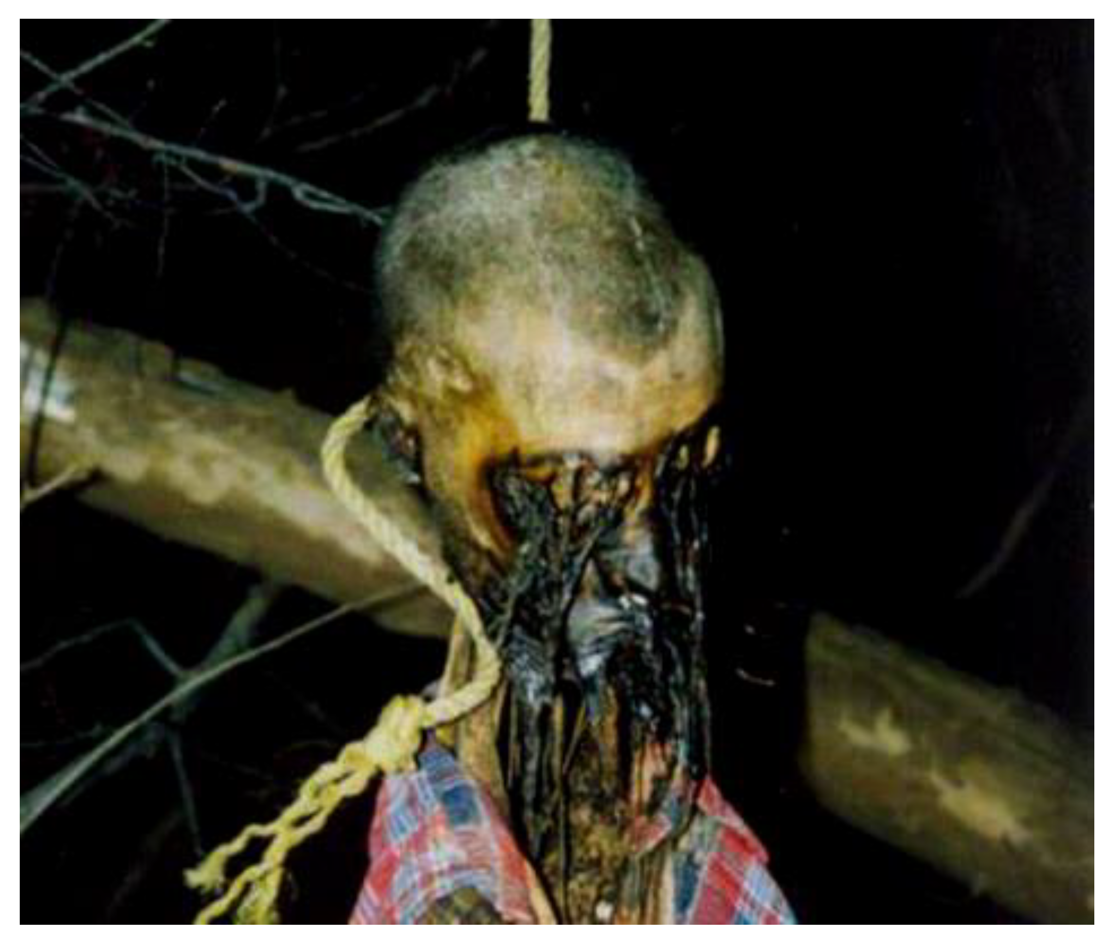
Abstract
:1. Introduction
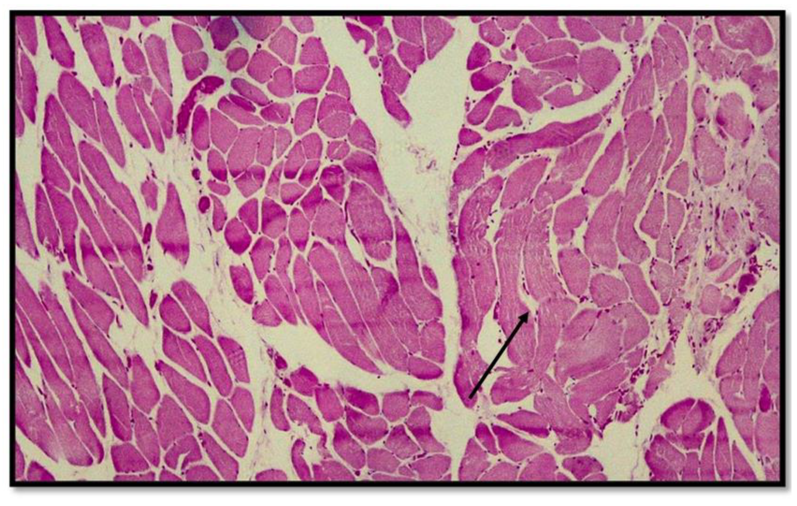
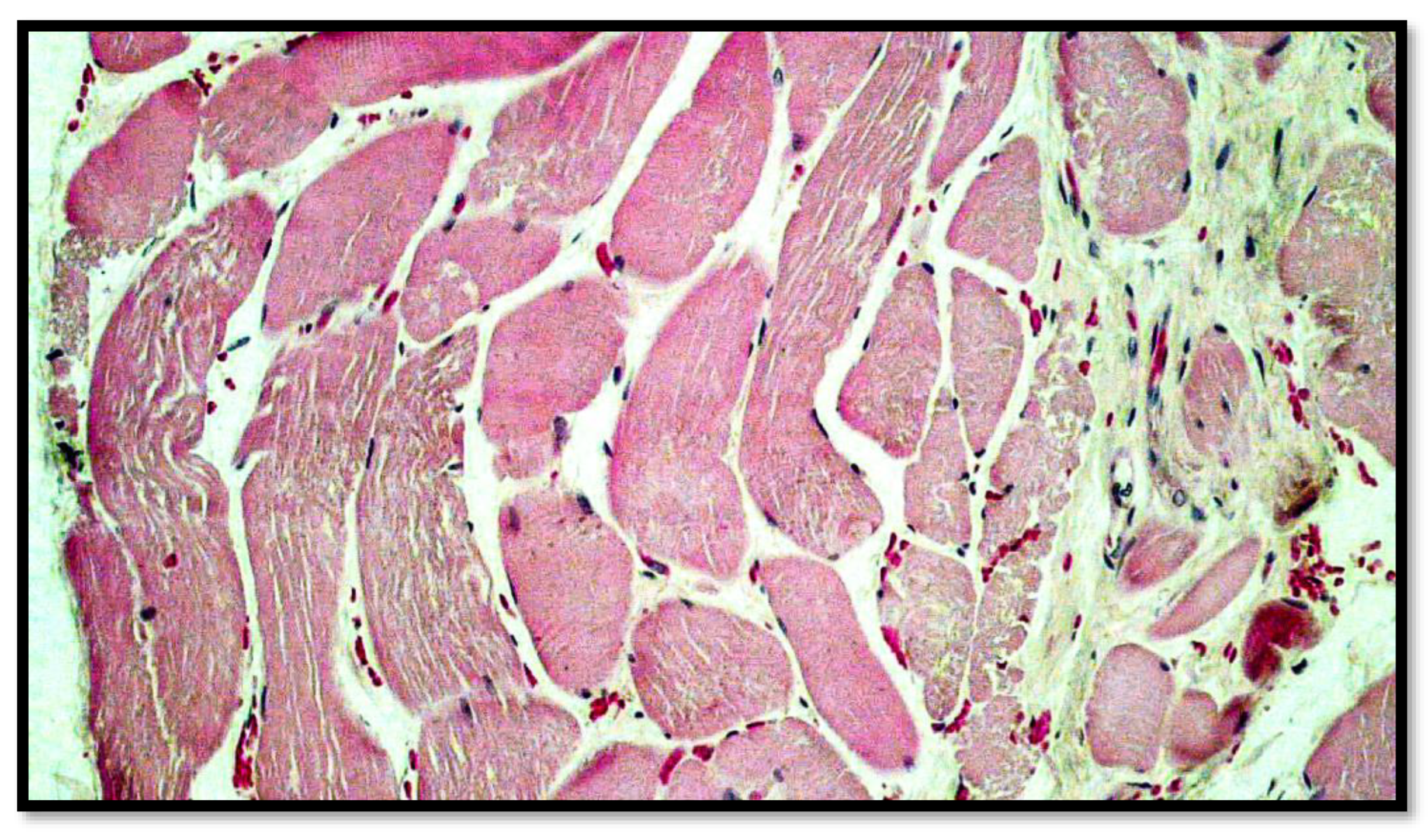
- (1)
- Excoriations and septal hemorrhages at the level of the sulcus, with hemorrhagic extravasations into the dermis, subcutaneous, and interstitial tissues of the neck;
- (2)
- Lesions of the carotid intima (Amussat’s sign);
- (3)
- Minute hemorrhages of the adventitia of the common carotid artery (Friedberg’s sign);
- (4)
- Fragmentation of the myelinated fibers of the vagus nerve (the sign of Dotto);
- (5)
- Hemorrhages in the cervical lymph nodes (Jankovich and Incze’s sign);
- (6)
- Retropharyngeal and paravertebral ecchymosis produced by compression of the base of the tongue against the pharynx (Brouardel’s sign);
- (7)
- The presence of subconjunctival and endocardial hemorrhagic petechiae, indicative of overinsufflation;
- (8)
- Hemorrhages below the anterior longitudinal ligament in the dorso-lumbar segment;
- (9)
- Tumidity of the penis and spermatorrhea [7].
2. Materials and Methods
Case Report
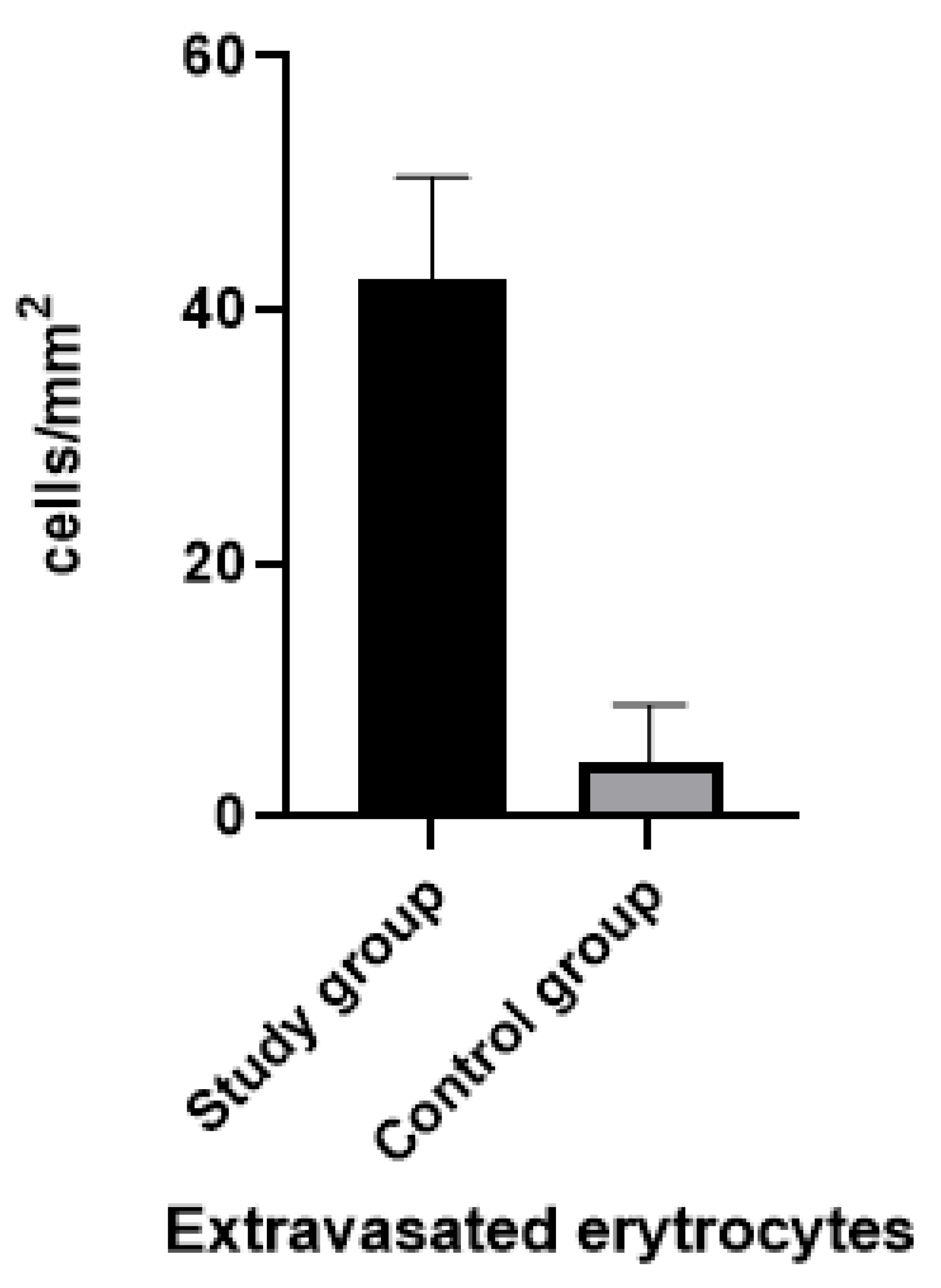
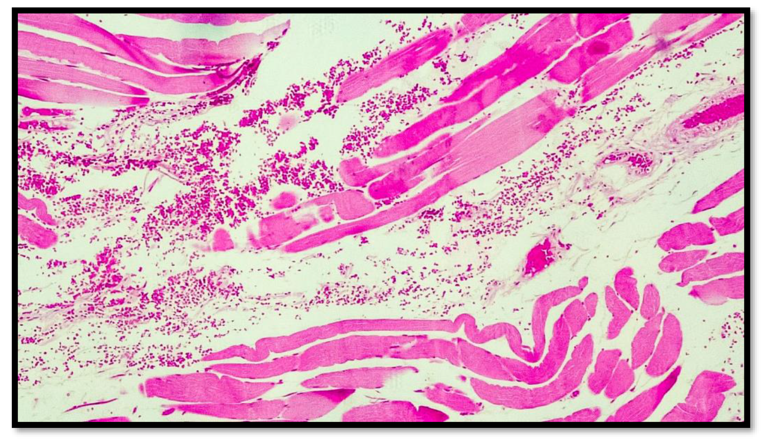
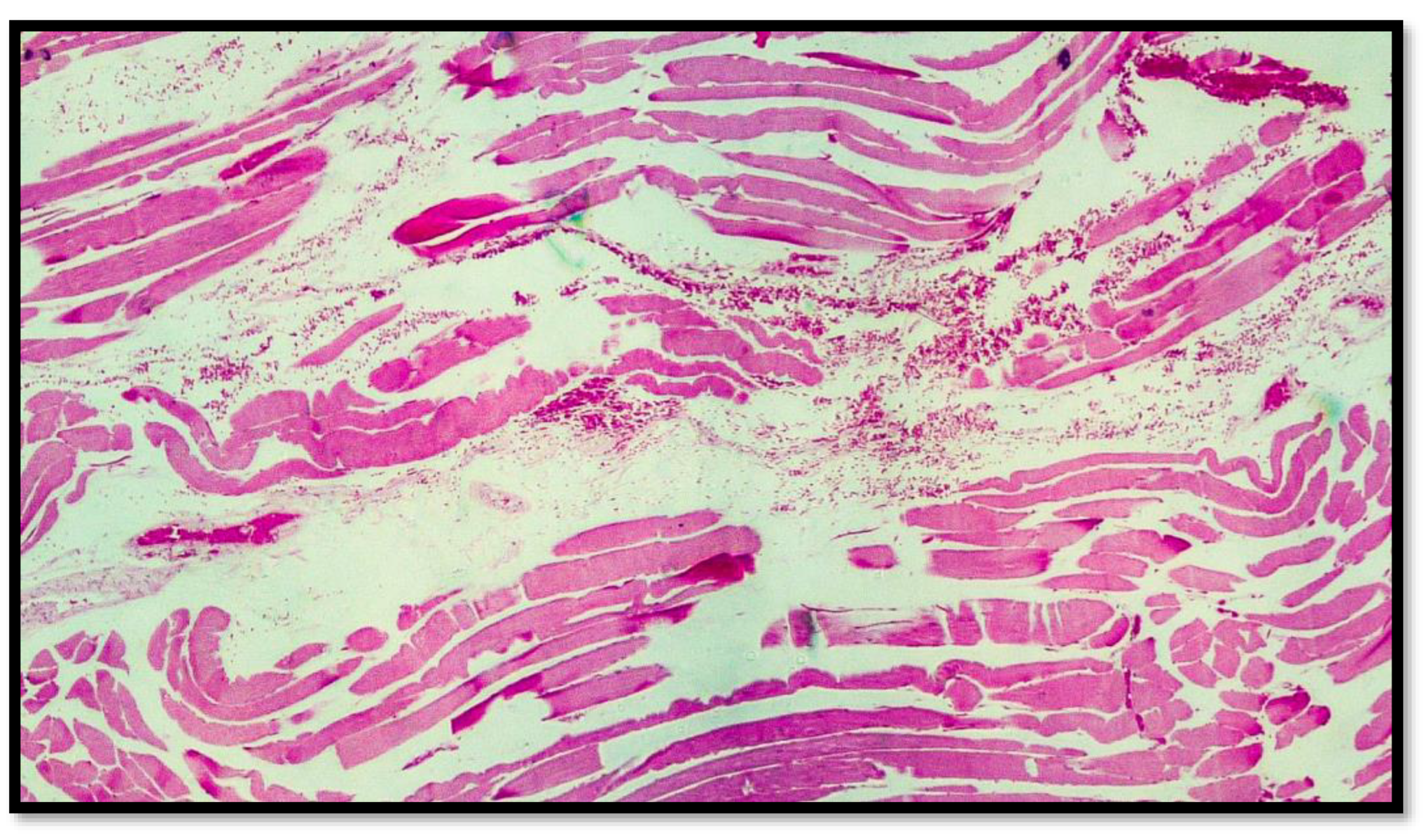
3. Results
4. Discussion
5. Conclusions
Author Contributions
Funding
Institutional Review Board Statement
Informed Consent Statement
Data Availability Statement
Conflicts of Interest
References
- Sabrinskas, R.; Hamilton, B.; Daniel, C.; Oliffe, J. Suicide by hanging: A scoping review. Int. J. Ment. Health Nurs. 2022, 31, 278–294. [Google Scholar] [CrossRef] [PubMed]
- Marrone, M.; Buongiorno, L.; Strisciullo, G.; Cappelli, G.; Dugheri, S.; Vinci, F.; Stellacci, A. Solve the Cube and Breathe: Case Report of. J. Leg. Ethical Regul. Issues 2022, 25, 1–8. [Google Scholar]
- Marrone, M.; Tarantino, F.; Stellacci, A.; Lonero Baldassarra, S.; Cazzato, G.; Vinci, F.; Dell’erba, A. Forensic Analysis and Identification Processes in Mass Disasters: Explosion of Gun Powder in the Fireworks Factory. Molecules 2022, 27, 244. [Google Scholar] [CrossRef] [PubMed]
- Buitendag, J.; Ras, A.; Kong, V.Y.; Weale, R.D.; Blodgett, J.Q.; Bruce, J.L.; Clarke, D.L.; Laing, G. Hanging-related injury in Pietermaritzburg, South Africa. S. Afr. Med. J. 2020, 110, 400–402. [Google Scholar] [CrossRef] [PubMed]
- Geisenberger, D.; Pollak, S.; Thierauf-Emberger, A. Homicidal strangulation and subsequent hanging of the victim to simulate suicide: Delayed elucidation based on reassessment of the autopsy findings. Forensic Sci. Int. 2019, 298, 419–423. [Google Scholar] [CrossRef]
- Vinci, F.; De Donno, A.; Marrone, M. Sinossi di Medicina Legale Ad Uso Degli Stuzdenti Dei Corsi di Laurea Per le Professioni Sanitarie e di Giurisprudenza; Medicina legale, criminalistica e scienze sociali; Aracne: Rome, Italy, 2022; ISBN 9791259946928. [Google Scholar]
- Suárez-Peñaranda, J.M.; Alvarez, T.; Miguéns, X.; Rodríguez-Calvo, M.S.; de Abajo, B.L.; Cortesão, M.; Cordeiro, C.; Vieira, D.N.; Muñoz, J.I. Characterization of lesions in hanging deaths. J. Forensic Sci. 2008, 53, 720–723. [Google Scholar] [CrossRef] [PubMed]
- Li, J.; Jadin, K.; Masuda, K.; Sah, R.; Muehleman, C. Characterization of lesions of the talus and description of tram-track lesions. Foot ankle Int. 2006, 27, 344–355. [Google Scholar] [CrossRef] [PubMed]
- Zátopková, L.; Janík, M.; Urbanová, P.; Mottlová, J.; Hejna, P. Laryngohyoid fractures in suicidal hanging: A prospective autopsy study with an updated review and critical appraisal. Forensic Sci. Int. 2018, 290, 70–84. [Google Scholar] [CrossRef] [PubMed]
- Polacco, M.; d’Alessio, P.; Ausania, F.; Beomonte Zobel, B.; Pascali, V.; d’Aloja, E.; Miscusi, M.; De Giorgio, F. Virtual Autopsy in Hanging. Am. J. Forensic Med. Pathol. 2013, 34, 107–109. [Google Scholar] [CrossRef]
- Nikolic, S.; Micic, J.; Atanasijevic, T.; Djokic, V.; Djonic, D. Analysis of neck injuries in hanging. Am. J. Forensic Med. Pathol. 2003, 24, 179–182. [Google Scholar] [CrossRef]
- Sharma, B.R.; Harish, D.; Sharma, A.; Sharma, S.; Singh, H. Injuries to neck structures in deaths due to constriction of neck, with a special reference to hanging. J. Forensic Leg. Med. 2008, 15, 298–305. [Google Scholar] [CrossRef] [PubMed]
- Nikolić, S.; Zivković, V.; Babić, D.; Juković, F.; Atanasijević, T.; Popović, V. Hyoid-laryngeal fractures in hanging: Where was the knot in the noose? Med. Sci. Law 2011, 51, 21–25. [Google Scholar] [CrossRef] [PubMed]
- Anders, S.; Schulz, F.; Tsokos, M. Ausgeprägte intramuskuläre Hämorrhagien vitaler Genese bei letaler suizidaler Strombeibringung. Rechtsmedizin 2012, 10, 105–109. [Google Scholar] [CrossRef]
- McEwen, B.J. Nondrowning Asphyxia in Veterinary Forensic Pathology: Suffocation, Strangulation, and Mechanical Asphyxia. Vet. Pathol. 2016, 53, 1037–1048. [Google Scholar] [CrossRef] [Green Version]
- Vanezis, P. Pathology of Neck Injury; Butterworth Scientific Ltd.: Guildford, UK, 1989; 133p. [Google Scholar]
- Shkrum, M.; Ramsay, D. Forensic Pathology of Trauma: Common Problems for the Pathologist; Humana: London, UK, 2007; ISBN 978-1-58829-458-6. [Google Scholar]
- Canuto, T. Medicina Legale e Delle Assicurazioni, 12th ed.; Malatesta: Rimini, Italy, 2009; ISBN 8829912611. [Google Scholar]
- Mansueto, G.; Feola, A.; Zangani, P.; Porzio, A.; Carfora, A.; Campobasso, C. Pietro A Clue on the Skin: A Systematic Review on Immunohistochemical Analyses of the Ligature Mark. Int. J. Environ. Res. Public Health 2022, 19, 2035. [Google Scholar] [CrossRef]
- Nitkowski, D.; Petermann, F. Non-suicidal self-injury and suicide attempts: Grounding of differential diagnosis. Fortschr. Neurol. Psychiatr. 2010, 78, 9–17. [Google Scholar] [CrossRef]
- Maghin, F.; Andreola, S.A.; Boracchi, M.; Gentile, G.; Maciocco, F.; Muccino, E.; Zoja, R. Technical note: A histochemical approach in diagnosing hanging mechanical asphyxia on cadavers undergoing advanced putrefactive phenomena. Med. Leg. J. 2018, 86, 208–213. [Google Scholar] [CrossRef] [PubMed]
- Ilic, M.; Ilic, I. Trends in suicide by hanging, strangulation, and suffocation in Serbia, 1991–2020: A joinpoint regression and age-period-cohort analysis. World J. Psychiatry 2022, 12, 505–520. [Google Scholar] [CrossRef]
- Helm, K.; Matzenauer, C.; Neuhuber, F.; Monticelli, F.; Meyer, H.; Pittner, S.; Gotsmy, W. Suitability of specific soft tissue swabs for the forensic identification of highly decomposed bodies. Int. J. Legal Med. 2021, 135, 1319–1327. [Google Scholar] [CrossRef]
- Clément, R.; Guay, J.-P.; Redpath, M.; Sauvageau, A. Petechiae in hanging: A retrospective study of contributing variables. Am. J. Forensic Med. Pathol. 2011, 32, 378–382. [Google Scholar] [CrossRef] [PubMed]
- Zou, J.; Bauler, L.D.; Brown, T.T. An Illustrative Case of Suicidal Hanging Versus Accidental Hanging Associated With Autoerotic Activity: Necessary Medicolegal Death Investigation and Role of Cognitive Bias. Am. J. Forensic Med. Pathol. 2020, 41, 220–222. [Google Scholar] [CrossRef] [PubMed]
- Marrone, M.; Ferorelli, D.; Stellacci, A.; Vinci, F. A fatal drowning filmed in a private pool: Analysis of the sequences of submersion. Forensic Sci. Int. Reports 2021, 3, 100189. [Google Scholar] [CrossRef]
- Marrone, M.; Tarantino, F.; Baldassarra, S.L.; Cazzato, G.; Santobuono, V.E.; Vinci, F.; Stellacci, A. Value of the Medical-Legal Investigation. Correspondence of the Findings With Legally Established Reality. EuroMediterranean Biomed. J. 2021, 16, 148–152. [Google Scholar] [CrossRef]
- Romanelli, M.C.; Marrone, M.; Veneziani, A.; Gianciotta, R.; Leonardi, S.; Beltempo, P.; Vinci, F. Hypostasis and time since death: State of the art in Italy and a novel approach for an operative instrumental protocol. Am. J. Forensic Med. Pathol. 2015, 36, 99–103. [Google Scholar] [CrossRef] [PubMed]


Disclaimer/Publisher’s Note: The statements, opinions and data contained in all publications are solely those of the individual author(s) and contributor(s) and not of MDPI and/or the editor(s). MDPI and/or the editor(s) disclaim responsibility for any injury to people or property resulting from any ideas, methods, instructions or products referred to in the content. |
© 2023 by the authors. Licensee MDPI, Basel, Switzerland. This article is an open access article distributed under the terms and conditions of the Creative Commons Attribution (CC BY) license (https://creativecommons.org/licenses/by/4.0/).
Share and Cite
Marrone, M.; Cazzato, G.; Caricato, P.; Angeletti, C.; Ingravallo, G.; Casatta, N.; Lupo, C.; Vinci, F.; Agazzino, G.; Stellacci, A.; et al. Diagnostic Methods in Forensic Pathology: A New Sign in Death from Hanging. Diagnostics 2023, 13, 510. https://doi.org/10.3390/diagnostics13030510
Marrone M, Cazzato G, Caricato P, Angeletti C, Ingravallo G, Casatta N, Lupo C, Vinci F, Agazzino G, Stellacci A, et al. Diagnostic Methods in Forensic Pathology: A New Sign in Death from Hanging. Diagnostics. 2023; 13(3):510. https://doi.org/10.3390/diagnostics13030510
Chicago/Turabian StyleMarrone, Maricla, Gerardo Cazzato, Pierluigi Caricato, Carlo Angeletti, Giuseppe Ingravallo, Nadia Casatta, Carmelo Lupo, Francesco Vinci, Gisella Agazzino, Alessandra Stellacci, and et al. 2023. "Diagnostic Methods in Forensic Pathology: A New Sign in Death from Hanging" Diagnostics 13, no. 3: 510. https://doi.org/10.3390/diagnostics13030510