
Porcelain Aorta in a Young Person Living with HIV Who Presented with Angina
 ,
, 
Abstract
:1. Introduction
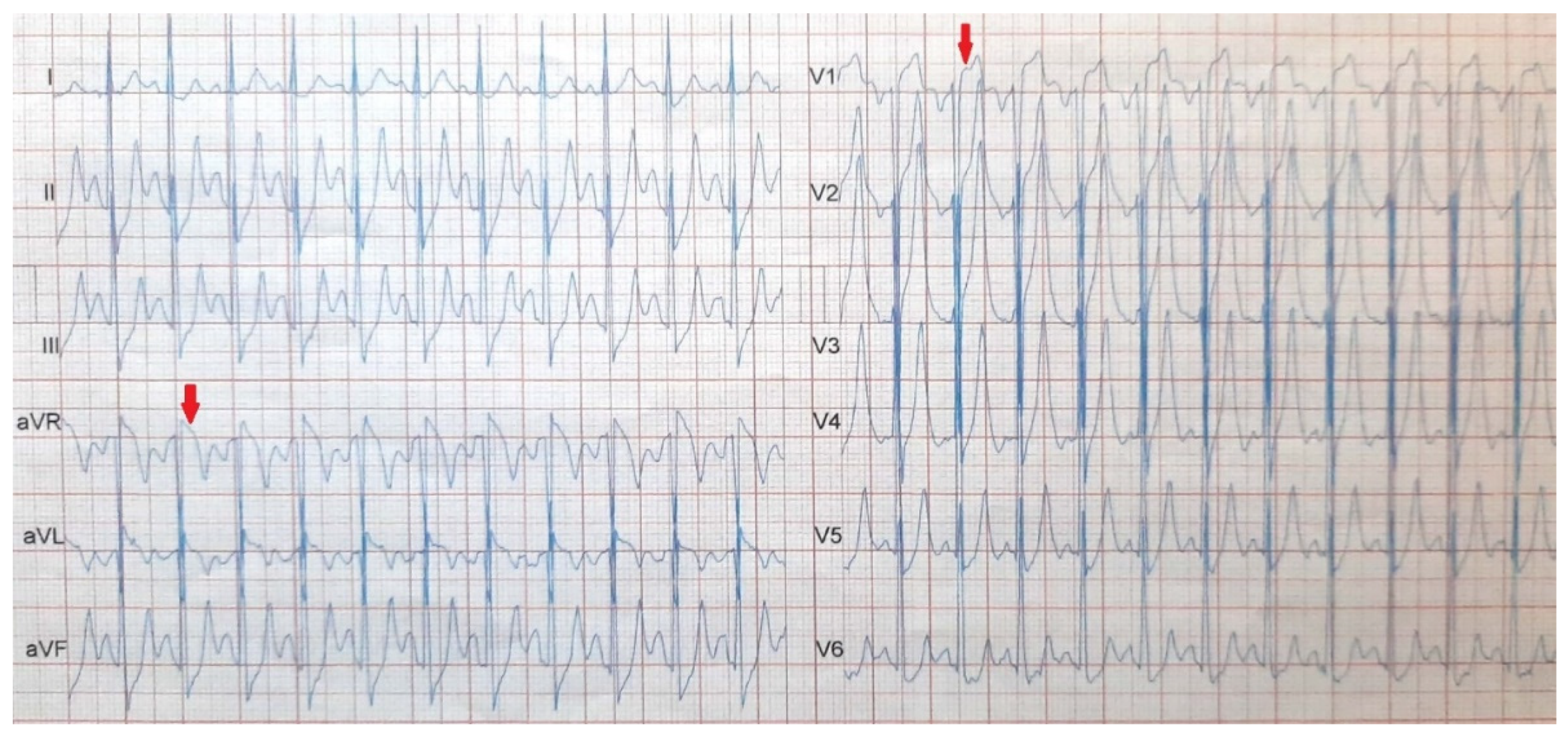
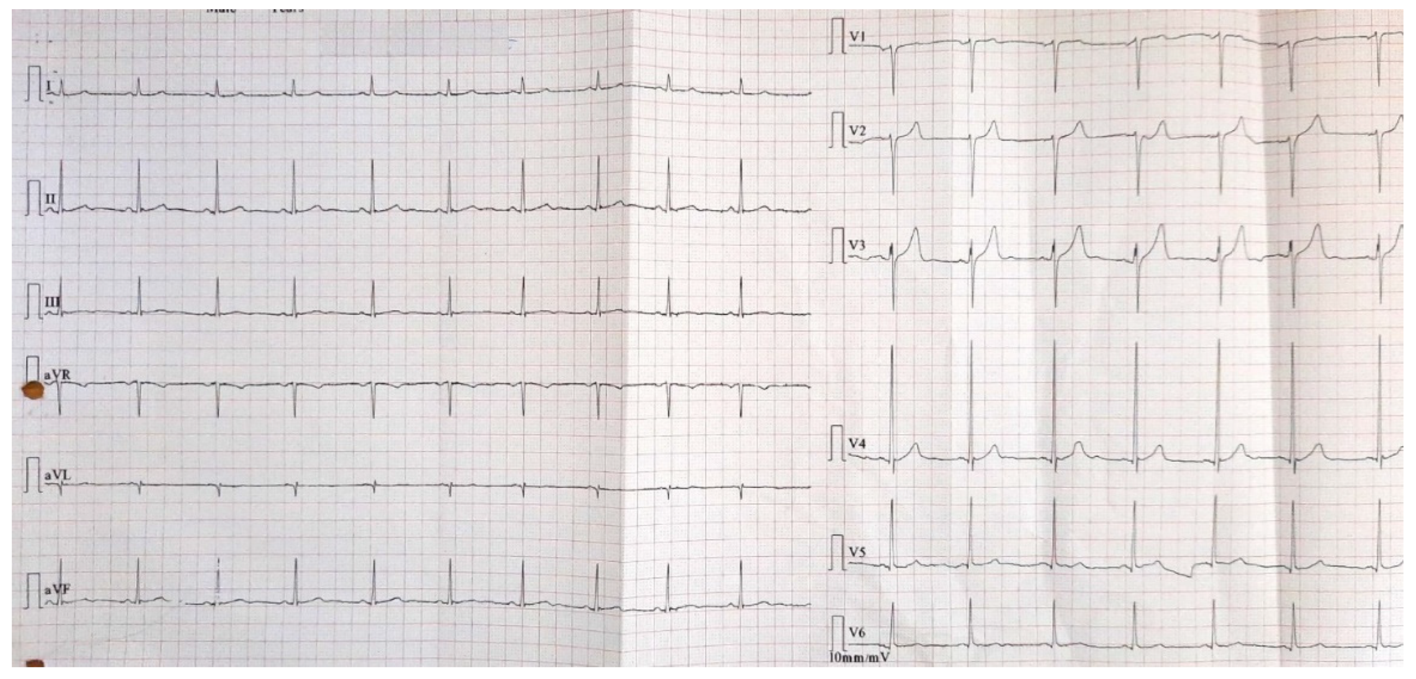
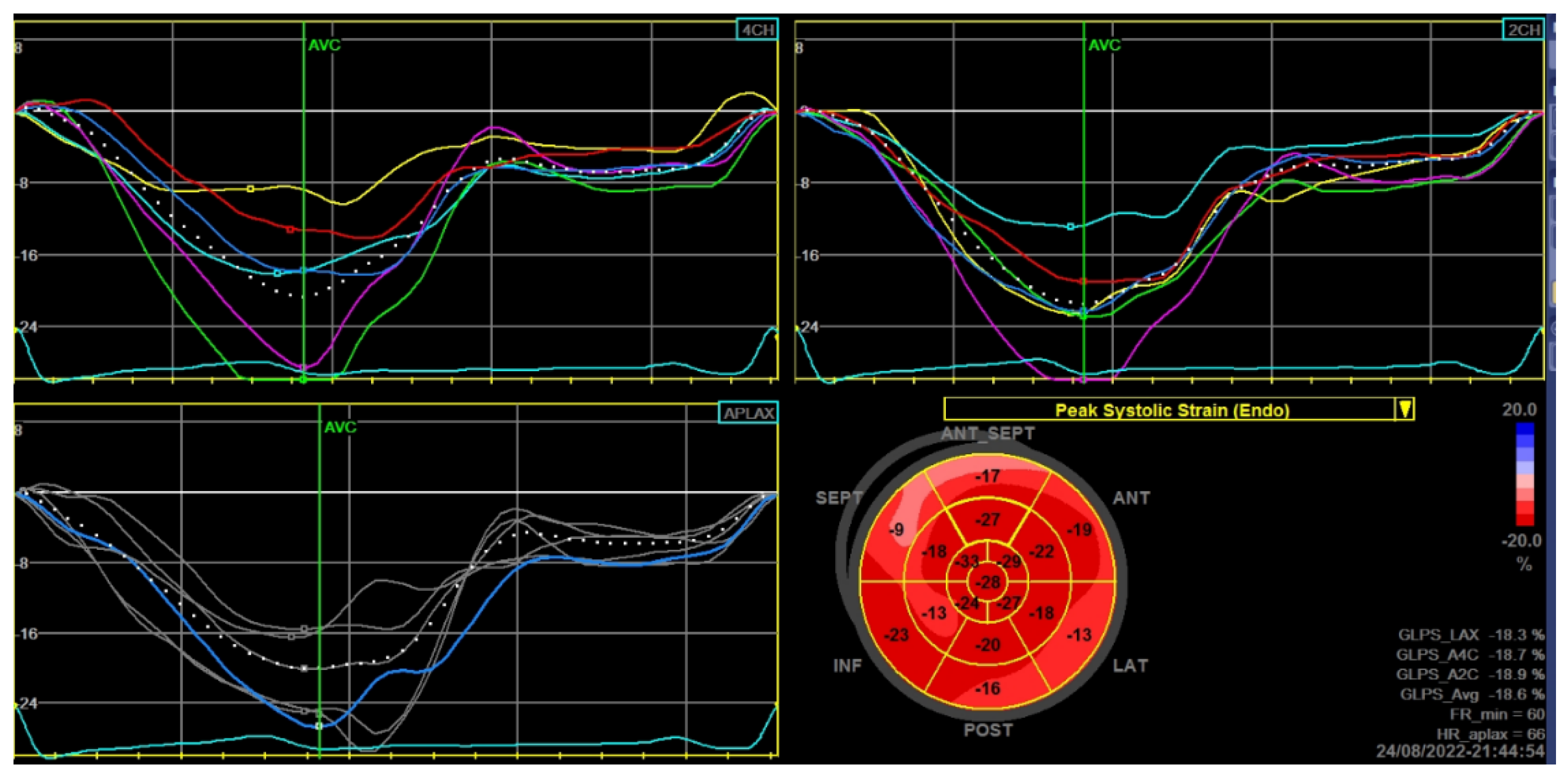
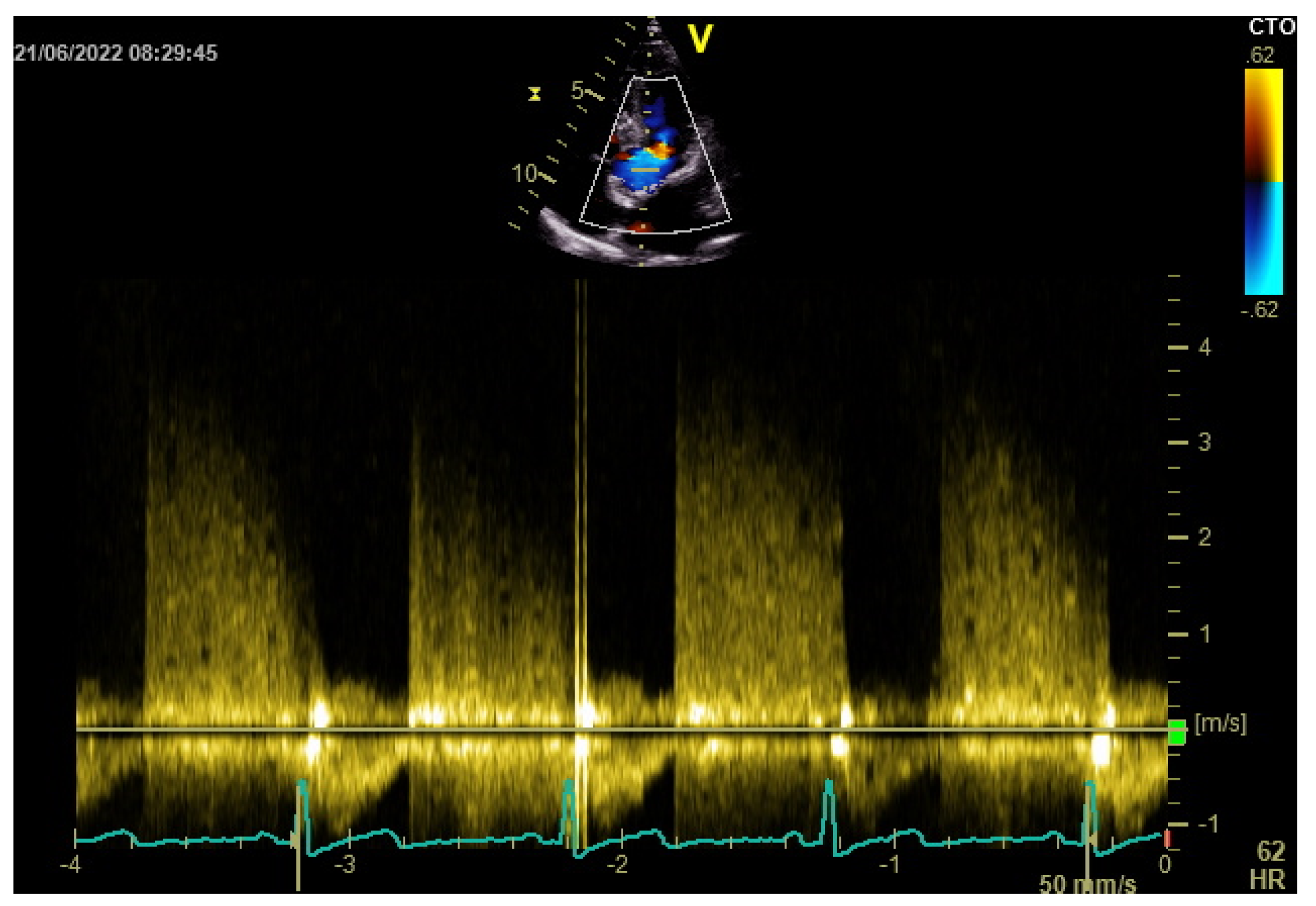
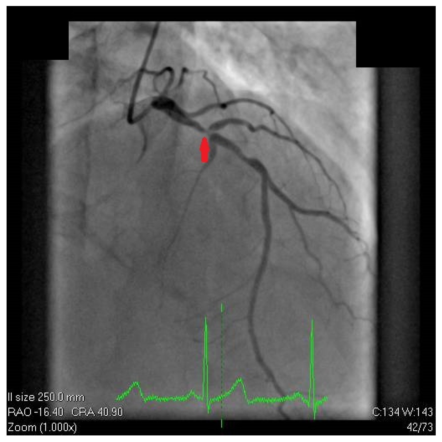
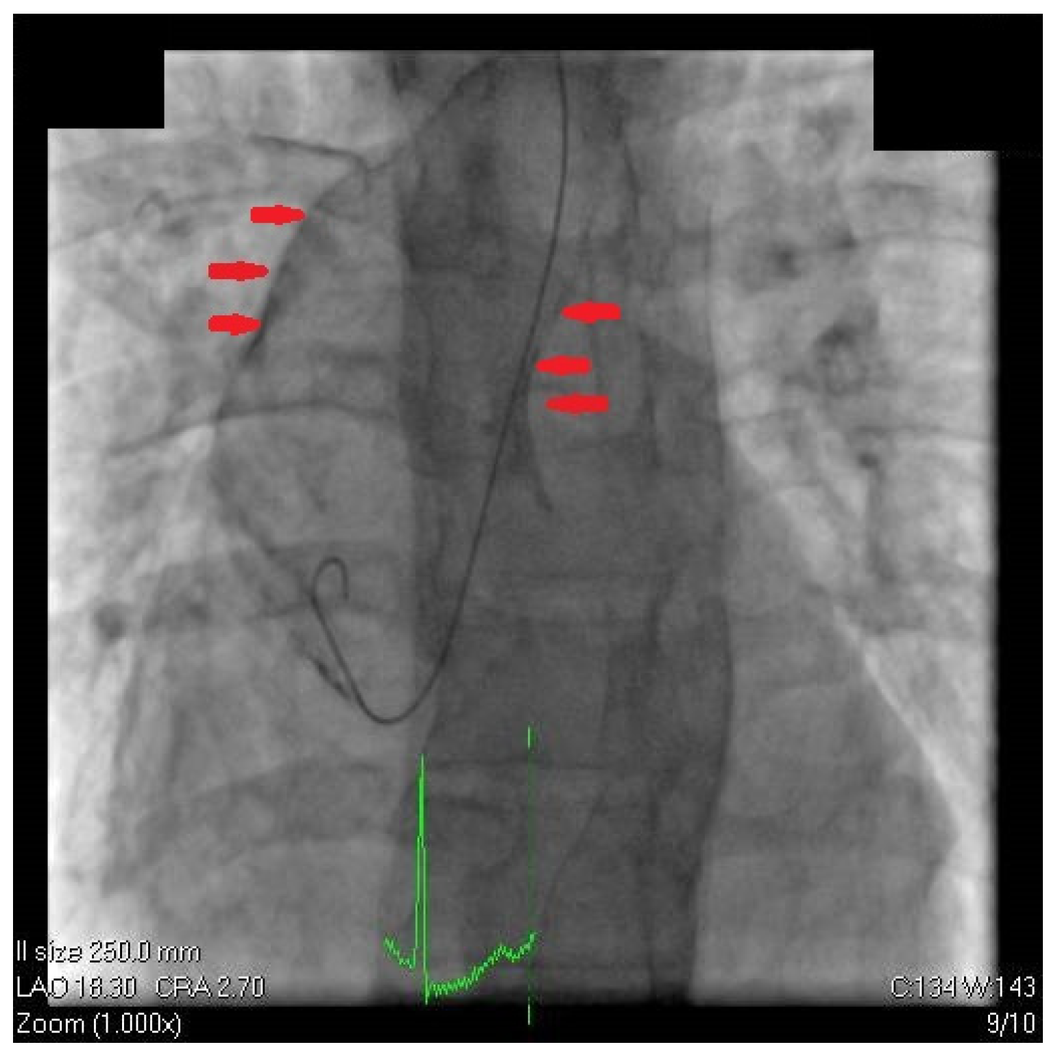
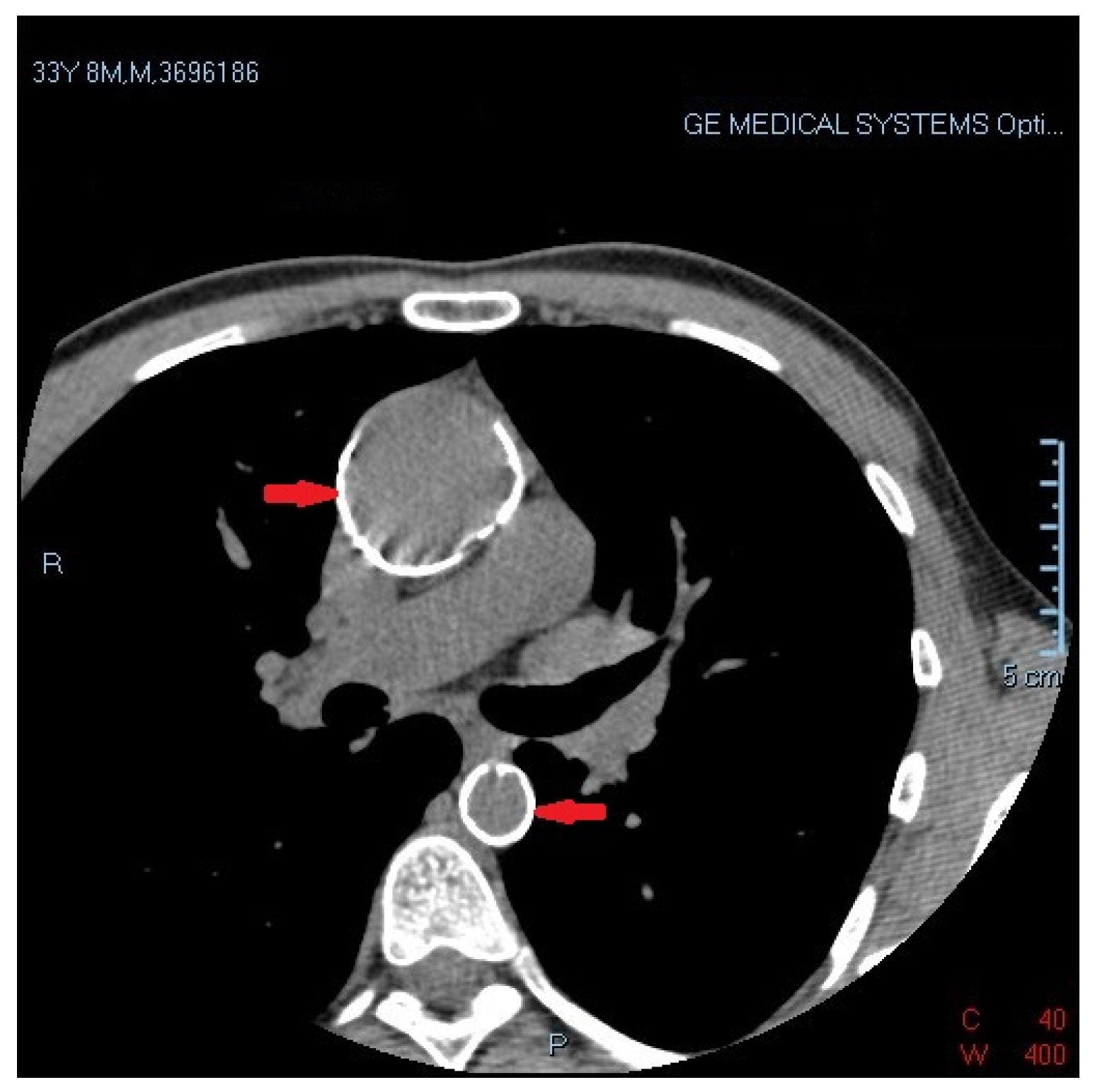
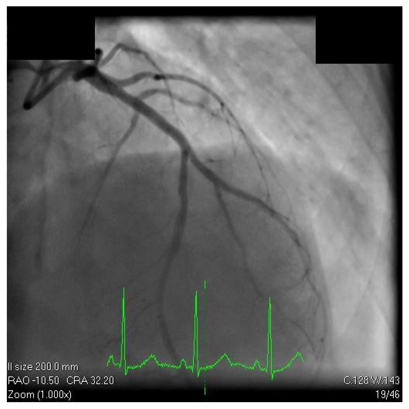
2. Case Report
3. Discussion
4. Conclusions
Author Contributions
Funding
Institutional Review Board Statement
Informed Consent Statement
Data Availability Statement
Acknowledgments
Conflicts of Interest
References
- Boccara, F.; Lang, S.; Meuleman, C.; Ederhy, S.; Mary-Krause, M.; Costagliola, D.; Capeau, J.; Cohen, A. HIV and coronary heart disease: Time for a better understanding. J. Am. Coll. Cardiol. 2013, 61, 511–523. [Google Scholar] [CrossRef] [PubMed] [Green Version]
- Bavinger, C.; Bendavid, E.; Niehaus, K.; Olshen, R.A.; Olkin, I.; Sundaram, V.; Wein, N.; Holodniy, M.; Hou, N.; Owens, D.K.; et al. Risk of Cardiovascular Disease from Antiretroviral Therapy for HIV: A Systematic Review. PLoS ONE 2013, 8, e59551. [Google Scholar] [CrossRef] [PubMed]
- Triant, V.A.; Lee, H.; Hadigan, C.; Grinspoon, S.K. Increased acute myocardial infarction rates and cardiovascular risk factors among patients with human immunodeficiency virus disease. J. Clin. Endocrinol. Metab. 2007, 92, 2506–2512. [Google Scholar] [CrossRef] [PubMed]
- Smit, M.; Brinkman, K.; Geerlings, S.; Smit, C.; Thyagarajan, K.; van Sighem, A.V.; de Wolf, F.; Hallett, T.B. Future challenges for clinical care of an ageing population infected with HIV: A modelling study. Lancet Infect. Dis. 2015, 15, 810–818. [Google Scholar] [CrossRef] [PubMed] [Green Version]
- Abramowitz, Y.; Jilaihawi, H.; Chakravarty, T.; Mack, M.J.; Makkar, R.R. Porcelain aorta: A comprehensive review. Circulation 2015, 131, 827–836. [Google Scholar] [CrossRef] [Green Version]
- Leyh, R.G.; Bartels, C.; Nötzold, A.; Sievers, H.H. Management of porcelain aorta during coronary artery bypass grafting. Ann. Thorac. Surg. 1999, 67, 986–988. [Google Scholar] [CrossRef]
- Gillinov, A.M.; Lytle, B.W.; Hoang, V.; Cosgrove, D.M.; Banbury, M.K.; McCarthy, P.M.; Sabik, J.F.; Pettersson, G.B.; Smedira, N.G.; Blackstone, E.H. The atherosclerotic aorta at aortic valve replacement: Surgical strategies and results. J. Thorac. Cardiovasc. Surg. 2000, 120, 957–963. [Google Scholar] [CrossRef] [Green Version]
- Wareing, T.H.; Davila-Roman, V.G.; Barzilai, B.; Murphy, S.F.; Kouchoukos, N.T. Management of the severely atherosclerotic ascending aorta during cardiac operations. J. Thorac. Cardiovasc. Surg. 1992, 103, 453–462. [Google Scholar] [CrossRef]
- Snow, T.; Semple, T.; Duncan, A.; Barker, S.; Rubens, M.; DiMario, C.; Davies, S.; Moat, N.; Nicol, E.D. “Porcelain aorta”: A proposed definition and classification of ascending aortic calcification. Open Heart 2018, 5, e000703. [Google Scholar] [CrossRef] [Green Version]
- Faggiano, P.; Frattini, S.; Zilioli, V.; Rossi, A.; Nistri, S.; Dini, F.L.; Lorusso, R.; Tomasi, C.; Cas, L.D. Prevalence of comorbidities and associated cardiac diseases in patients with valve aortic stenosis. Potential implications for the decision-making process. Int. J. Cardiol. 2012, 159, 94–99. [Google Scholar] [CrossRef]
- Anan, A.; Rmilah, A.; Srikanth Yandrapalli, F.B.B. Porcelain Aorta. In Treasure Isl; StatPearls Publ.: Tampa, FL, USA, 2022. [Google Scholar]
- Seyahi, E.; Ucgul, A.; Cebi Olgun, D.; Ugurlu, S.; Akman, C.; Tutar, O.; Yurdakul, S.; Yazici, H. Aortic and coronary calcifications in Takayasu arteritis. Semin. Arthritis Rheum. 2013, 43, 96–104. [Google Scholar] [CrossRef] [PubMed]
- Smith, C.J.; Ryom, L.; Weber, R.; Morlat, P.; Pradier, C.; Reiss, P.; Kowalska, J.D.; De Wit, S.; Law, M.; Sadr, W.; et al. Trends in underlying causes of death in people with HIV from 1999 to 2011 (D:A:D): A multicohort collaboration. Lancet 2014, 384, 241–248. [Google Scholar] [CrossRef] [PubMed] [Green Version]
- Freiberg, M.S.; Chang, C.C.H.; Skanderson, M.; Patterson, O.V.; DuVall, S.L.; Brandt, C.A.; So-Armah, K.A.; Vasan, R.S.; Oursler, K.A.; Gottdiener, J.; et al. Association between HIV infection and the risk of heart failure with reduced ejection fraction and preserved ejection fraction in the antiretroviral therapy era: Results from the veterans aging cohort study. JAMA Cardiol. 2017, 2, 536–546. [Google Scholar] [CrossRef] [PubMed] [Green Version]
- May, M.; Gill, J.; Lewden, C.; Saag, M.; Mugavero, M.; Reiss, P.; Ledergerber, B.; Mocroft, A.; Harris, R.; Fux, C.A.; et al. Causes of death in HIV-1-Infected patients treated with antiretroviral therapy, 1996-2006: Collaborative analysis of 13 HIV cohort studies. Clin. Infect. Dis. 2010, 50, 1387–1396. [Google Scholar] [CrossRef] [Green Version]
- Triant, V.A. Cardiovascular disease and HIV infection. Curr. HIV/AIDS Rep. 2013, 10, 199–206. [Google Scholar] [CrossRef]
- Maggi, P.; Bellacosa, C.; Leone, A.; Volpe, A.; Ricci, E.D.; Ladisa, N.; Cicalini, S.; Grilli, E.; Viglietti, R.; Chirianni, A.; et al. Cardiovascular risk in advanced naïve HIV-infected patients starting antiretroviral therapy: Comparison of three different regimens - PREVALEAT II cohort. Atherosclerosis 2017, 263, 398–404. [Google Scholar] [CrossRef]
- Durand, M.; Sheehy, O.; Baril, J.G.; Lelorier, J.; Tremblay, C.L. Association between HIV infection, antiretroviral therapy, and risk of acute myocardial infarction: A cohort and nested case-control study using Québec’s Public Health Insurance database. J. Acquir. Immune Defic. Syndr. 2011, 57, 245–253. [Google Scholar] [CrossRef]
- Worm, S.W.; Sabin, C.; Weber, R.; Reiss, P.; El-Sadr, W.; Dabis, F.; De Wit, S.; Law, M.; Monforte, A.D.A.; Friis-Møller, N.; et al. Risk of Myocardial Infarction in Patients with HIV Infection Exposed to Specific Individual Antiretroviral Drugs from the 3 Major Drug Classes: The Data Collection on Adverse Events of Anti-HIV Drugs (D:A:D) Study. J. Infect. Dis. 2010, 201, 218–330. [Google Scholar] [CrossRef]
- Obel, N.; Farkas, D.K.; Kronborg, G.; Larsen, C.S.; Pedersen, G.; Riis, A.; Pedersen, C.; Gerstoft, J.; Sørensen, H.T. Abacavir and risk of myocardial infarction in HIV-infected patients on highly active antiretroviral therapy: A population-based nationwide cohort study. HIV Med. 2010, 11, 130–136. [Google Scholar] [CrossRef]
- Collado-Diaz, V.; Andujar, I.; Sanchez-Lopez, A.; Orden, S.; Blanch-Ruiz, M.A.; Martinez-Cuesta, M.A.; Blas-García, A.; Esplugues, J.V.; Álvarez, Á. Abacavir induces arterial thrombosis in a murine model. J. Infect. Dis. 2018, 218, 228–233. [Google Scholar] [CrossRef]
- Bedimo, R.J.; Westfall, A.O.; Drechsler, H.; Vidiella, G.; Tebas, P. Abacavir use and risk of acute myocardial infarction and cerebrovascular events in the highly active antiretroviral therapy era. Clin. Infect. Dis. 2011, 53, 84–91. [Google Scholar] [CrossRef] [PubMed] [Green Version]
- Martínez, E.; Larrousse, M.; Podzamczer, D.; Pérez, I.; Gutiérrez, F.; Loncá, M.; Barragán, P.; Deulofeu, R.; Casamitjana, R.; Mallolas, J.; et al. Abacavir-based therapy does not affect biological mechanisms associated with cardiovascular dysfunction. AIDS 2010, 24, F1–F9. [Google Scholar] [CrossRef]
- Ryom, L.; Cotter, A.; De Miguel, R.; Béguelin, C.; Podlekareva, D.; Arribas, J.R.; Marzolini, C.; Mallon, P.G.M.; Rauch, A.; Kirk, O.; et al. 2019 update of the European AIDS Clinical Society Guidelines for treatment of people living with HIV version 10.0. HIV Med. 2020, 10, 617–624. [Google Scholar] [CrossRef] [PubMed]
- Boccara, F.; Mary-Krause, M.; Potard, V.; Teiger, E.; Lang, S.; Hammoudi, N.; Chauvet, M.; Ederhy, S.; Dufour-Soulat, L.; Ancedy, Y.; et al. Hiv infection and long-term residual cardiovascular risk after acute coronary syndrome. J. Am. Heart Assoc. 2020, 9, e017578. [Google Scholar] [CrossRef]
- Shah, A.S.V.; Stelzle, D.; Lee, K.K.; Beck, E.J.; Alam, S.; Clifford, S.; Longenecker, C.T.; Strachan, F.; Bagchi, S.; Whiteley, W.; et al. Global Burden of Atherosclerotic Cardiovascular Disease in People Living with HIV. Circulation 2018, 138, 1100–1112. [Google Scholar] [CrossRef] [PubMed]
- Hsue, P.Y.; Waters, D.D. HIV infection and coronary heart disease: Mechanisms and management. Nat. Rev. Cardiol. 2019, 16, 745–759. [Google Scholar] [CrossRef] [Green Version]
- Bajdechi, M.; Mihai, C.; Scafa-Udriste, A.; Cherry, A.; Zamfir, D.; Dumitru, I.; Cernat, R.; Rugina, S. Severe coronary artery disease in a person living with hiv. Medicina 2021, 57, 595. [Google Scholar] [CrossRef]
- Poznyak, A.V.; Bezsonov, E.E.; Borisov, E.E.; Grechko, A.V.; Kartuesov, A.G.; Orekhov, A.N. Atherosclerosis in HIV Patients: What Do We Know so Far? Int. J. Mol. Sci. 2022, 23, 2504. [Google Scholar] [CrossRef]
- Anand, A.R.; Rachel, G.; Parthasarathy, D. HIV Proteins and Endothelial Dysfunction: Implications in Cardiovascular Disease. Front. Cardiovasc. Med. 2018, 5, 1–10. [Google Scholar] [CrossRef]
- Eisen, A.; Tenenbaum, A.; Koren-Morag, N.; Tanne, D.; Shemesh, J.; Imazio, M.; Fisman, E.Z.; Motro, M.; Schwammenthal, E.; Adler, Y. Calcification of the thoracic aorta as detected by spiral computed tomography among stable angina pectoris patients: Association with cardiovascular events and death. Circulation 2008, 118, 1328–1334. [Google Scholar] [CrossRef]


Publisher’s Note: MDPI stays neutral with regard to jurisdictional claims in published maps and institutional affiliations. |
© 2022 by the authors. Licensee MDPI, Basel, Switzerland. This article is an open access article distributed under the terms and conditions of the Creative Commons Attribution (CC BY) license (https://creativecommons.org/licenses/by/4.0/).
Share and Cite
Bajdechi, M.; Scafa-Udriste, A.; Ploscaru, V.; Calmac, L.; Bajeu, T.; Gurghean, A.; Rugina, S. Porcelain Aorta in a Young Person Living with HIV Who Presented with Angina. Diagnostics 2022, 12, 3147. https://doi.org/10.3390/diagnostics12123147
Bajdechi M, Scafa-Udriste A, Ploscaru V, Calmac L, Bajeu T, Gurghean A, Rugina S. Porcelain Aorta in a Young Person Living with HIV Who Presented with Angina. Diagnostics. 2022; 12(12):3147. https://doi.org/10.3390/diagnostics12123147
Chicago/Turabian StyleBajdechi, Mircea, Alexandru Scafa-Udriste, Vlad Ploscaru, Lucian Calmac, Teodor Bajeu, Adriana Gurghean, and Sorin Rugina. 2022. "Porcelain Aorta in a Young Person Living with HIV Who Presented with Angina" Diagnostics 12, no. 12: 3147. https://doi.org/10.3390/diagnostics12123147