Next Issue
Volume 13, January-1
Previous Issue
Volume 12, November


Diagnostics, Volume 12, Issue 12 (December 2022) – 329 articles

Cover Story (view full-size image): 
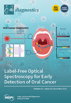
Oral cancer is the 16th most common cancer worldwide, frequently first detected at a late stage. Early diagnosis is critical, both for optimizing survival and for quality of life after treatment. There is thus a need for non-invasive screening and diagnostics to detect and differentiate early-stage malignant lesions. Optical spectroscopy may provide an appropriate solution to facilitate the early detection of such lesions. This review summarizes the current state of the art and progress in oral cancer diagnostics in vivo and in body fluids and discusses the importance of optical techniques in oral cancer diagnostics. In particular, major developments in label-free optical spectroscopy, including Raman spectroscopy, fluorescence spectroscopy, and diffuse reflectance spectroscopy are included. View this paper 
  • Issues are regarded as officially published after their release is announced to the table of contents alert mailing list.
  • You may sign up for e-mail alerts to receive table of contents of newly released issues.
  • PDF is the official format for papers published in both, html and pdf forms. To view the papers in pdf format, click on the "PDF Full-text" link, and use the free Adobe Reader to open them.
Order results
Result details
Section
Select all
Export citation of selected articles as:
10 pages, 1743 KiB  
Case Report
Orthopedic Device-Related Infections Due to Emerging Pathogens Diagnosed by a Combination of Microbiological Approaches: Case Series and Literature Review
by Angela Quirino, Nadia Marascio, Giuseppe Guido Maria Scarlata, Claudia Cicino, Grazia Pavia, Marta Pantanella, Giovanni Carlisi, Michele Mercurio, Filippo Familiari, Salvatore Rotundo, Vincenzo Olivadese, Valentina La Gamba, Francesca Serapide, Giorgio Gasparini and Giovanni Matera
Diagnostics 2022, 12(12), 3224; https://doi.org/10.3390/diagnostics12123224 - 19 Dec 2022
Cited by 2 | Viewed by 1702
Abstract
Orthopedic and trauma device-related infections (ODRI) due to high virulence microorganisms are a devastating complication after orthopedic surgery. Coagulase-negative Staphylococci (CoNS) are mainly involved but commensal bacteria, located in human mucous membranes, are emerging pathogens in ODRI. Currently, bacterial culture is the gold [...] Read more.
Orthopedic and trauma device-related infections (ODRI) due to high virulence microorganisms are a devastating complication after orthopedic surgery. Coagulase-negative Staphylococci (CoNS) are mainly involved but commensal bacteria, located in human mucous membranes, are emerging pathogens in ODRI. Currently, bacterial culture is the gold standard for ODRI but the diagnostic process remains time consuming and laborious. We evaluated a combination of microbiological approaches in the diagnosis of emerging pathogens involved in ODRI. We analyzed two synovial fluids, five tissue samples and five surgical wound swabs from two different patients with ODRI, attending the Department of Orthopedic and Trauma Surgery of Mater Domini Teaching Hospital, Catanzaro, Italy. Identification was carried out with a combination of microbiological approaches (culture, mass spectrometry and 16s rRNA gene sequencing). We demonstrated the importance of a combination of microbiological approaches for the diagnosis of emerging pathogens in ODRI, because the low number of cases in the literature makes it very difficult to formulate guidelines for the management of patients. Full article
(This article belongs to the Section Diagnostic Microbiology and Infectious Disease)
Show Figures

Figure 1

20 pages, 708 KiB  
Review
Artificial Intelligence in Emergency Radiology: Where Are We Going?
by Michaela Cellina, Maurizio Cè, Giovanni Irmici, Velio Ascenti, Elena Caloro, Lorenzo Bianchi, Giuseppe Pellegrino, Natascha D’Amico, Sergio Papa and Gianpaolo Carrafiello
Diagnostics 2022, 12(12), 3223; https://doi.org/10.3390/diagnostics12123223 - 19 Dec 2022
Cited by 14 | Viewed by 3597
Abstract
Emergency Radiology is a unique branch of imaging, as rapidity in the diagnosis and management of different pathologies is essential to saving patients’ lives. Artificial Intelligence (AI) has many potential applications in emergency radiology: firstly, image acquisition can be facilitated by reducing acquisition [...] Read more.
Emergency Radiology is a unique branch of imaging, as rapidity in the diagnosis and management of different pathologies is essential to saving patients’ lives. Artificial Intelligence (AI) has many potential applications in emergency radiology: firstly, image acquisition can be facilitated by reducing acquisition times through automatic positioning and minimizing artifacts with AI-based reconstruction systems to optimize image quality, even in critical patients; secondly, it enables an efficient workflow (AI algorithms integrated with RIS–PACS workflow), by analyzing the characteristics and images of patients, detecting high-priority examinations and patients with emergent critical findings. Different machine and deep learning algorithms have been trained for the automated detection of different types of emergency disorders (e.g., intracranial hemorrhage, bone fractures, pneumonia), to help radiologists to detect relevant findings. AI-based smart reporting, summarizing patients’ clinical data, and analyzing the grading of the imaging abnormalities, can provide an objective indicator of the disease’s severity, resulting in quick and optimized treatment planning. In this review, we provide an overview of the different AI tools available in emergency radiology, to keep radiologists up to date on the current technological evolution in this field. Full article
(This article belongs to the Special Issue Artificial Intelligence in Radiology 2.0)
Show Figures

Figure 1

12 pages, 728 KiB  
Article
Mapping HPV 16 Sub-Lineages in Anal Cancer and Implications for Disease Outcomes
by Daniel Guerendiain, Laila Sara Arroyo Mühr, Raluca Grigorescu, Matthew T. G. Holden and Kate Cuschieri
Diagnostics 2022, 12(12), 3222; https://doi.org/10.3390/diagnostics12123222 - 19 Dec 2022
Cited by 2 | Viewed by 1684
Abstract
The incidence of anal cancer is rising worldwide. As identified in cervical cancer management, an improvement in the early detection and management of anal pre-cancer is essential. In other cancers associated with human papillomavirus (HPV), HPV 16 sub-lineages have been shown to be [...] Read more.
The incidence of anal cancer is rising worldwide. As identified in cervical cancer management, an improvement in the early detection and management of anal pre-cancer is essential. In other cancers associated with human papillomavirus (HPV), HPV 16 sub-lineages have been shown to be associated with disease status and prognosis. However, in anal cancer, they have been under-explored. A total of 119 HPV 16-positive anal cancer lesions diagnosed between 2009 and 2018 in Scotland and 134 HPV 16-positive residual rectal swabs from asymptomatic men collected in 2016/7 were whole genome sequenced. The association of HPV 16 sub-lineages with underlying disease status (cancer vs. asymptomatic) and overall survival in anal cancer samples was assessed (comparing A1 vs non-A1 sub-lineages). A1 was the dominant sub-lineage present in the anal cancer (76.5%) and the asymptomatic (76.1%) cohorts. A2 was the second most dominant sub-lineage in both groups (16.8% and 17.2%, respectively). We did not observe significant associations of sub-lineage with demographics, clinical variables or survival (A1 vs. non-A1 sub-lineages (HR 0.83, 0.28–2.46 p = 0.743)). HPV 16 sub-lineages do to not appear to cluster with disease vs asymptomatic carriage or be independently associated with outcomes in anal cancer patients. Further international studies on anal HPV sub-lineage mapping will help to determine whether this is a consistent observation. Full article
(This article belongs to the Special Issue Diagnosis of Lower Genital Tract Disease)
Show Figures

Figure 1

8 pages, 12562 KiB  
Interesting Images
The Pitfall of Ganglioneuroblastoma-Nodular Diagnosis: Clinical and Imaging Considerations over a Rare Bifocal Sporadic Case
by Claudio Montante, Francesco Fabozzi, Maria Felicia Villani, Maria Luisa D’Andrea, Alessandra Stracuzzi, Gian Luigi Natali, Giada Del Baldo, Francesca Del Bufalo, Maria Carmen Garganese, Annalisa Serra, Paolo Tomà, Rita Alaggio, Sabina Vennarini, Giovanna Stefania Colafati, Angela Mastronuzzi and Maria Antonietta De Ioris
Diagnostics 2022, 12(12), 3221; https://doi.org/10.3390/diagnostics12123221 - 19 Dec 2022
Cited by 4 | Viewed by 1734
Abstract
Neuroblastic tumors (NTs) represent the most common extracranial neoplasm occurring in childhood. Although ganglioneuroblastoma intermixed (GNBI) and ganglioneuroma (GN) are classified as very low-risk tumors, neuroblastoma (NB) and ganglioneuroblastoma-nodular (GNBN) may represent a serious risk to survival. Unfortunately, areas of GNBI and GNBN [...] Read more.
Neuroblastic tumors (NTs) represent the most common extracranial neoplasm occurring in childhood. Although ganglioneuroblastoma intermixed (GNBI) and ganglioneuroma (GN) are classified as very low-risk tumors, neuroblastoma (NB) and ganglioneuroblastoma-nodular (GNBN) may represent a serious risk to survival. Unfortunately, areas of GNBI and GNBN can coexist in the same mass, leading to incorrect risk staging when only biopsy is performed. Herein, we describe a case of multifocal NT (thoracic and abdominal localization) occurring in a 4-year-old male. Different histological subtypes, namely GNBI and GNBN, were revealed in the two lesions. We focus on the difficulties of proper diagnosis and risk stratification, underlining the usefulness of several diagnostic tools for appropriate management and therapeutic choices. Full article
(This article belongs to the Section Medical Imaging and Theranostics)
Show Figures

Figure 1

15 pages, 262 KiB  
Review
Chronic Active T-Cell Mediated Kidney Rejection as a Clinically Significant Type of Allograft Loss?
by Jakub Mizera, Justyna Pilch, Dorota Kamińska, Magdalena Krajewska, Piotr Donizy and Mirosław Banasik
Diagnostics 2022, 12(12), 3220; https://doi.org/10.3390/diagnostics12123220 - 19 Dec 2022
Cited by 2 | Viewed by 2137
Abstract
The purpose of this article is to assess the present knowledge about chronic active (CA) T-cell mediated rejection (TCMR) of a kidney. In the research authors review current Banff diagnostic criteria used in kidney rejection, focus on their possible future evolution, and investigate [...] Read more.
The purpose of this article is to assess the present knowledge about chronic active (CA) T-cell mediated rejection (TCMR) of a kidney. In the research authors review current Banff diagnostic criteria used in kidney rejection, focus on their possible future evolution, and investigate the role of currently available molecular methods that could be implemented into the diagnostic scheme. Research also points out previously and currently available treatment methods applied to CA TCMR and takes into account possible side effects consequent upon the therapy. Moreover, attention is being paid to the CA TCMR coincidence with other kidney rejection types such as antibody-mediated rejection (ABMR) and its influence on the treatment approach. Authors also mark the possibility of non-HLA antibodies coexistence in patients with CA TCMR and describe its possible resonance on kidney allograft function. Nonetheless, it seems that current knowledge about CA TCMR is not sufficient and requires further investigation. Full article
(This article belongs to the Section Pathology and Molecular Diagnostics)
12 pages, 6731 KiB  
Case Report
An Extremely Rare Case of Disseminated Peritoneal Leiomyomatosis with a Pelvic Leiomyosarcoma and Omental Metastasis after Laparoscopic Morcellation: Systematic Review of the Literature
by Antonella Vimercati, Carla Mariaflavia Santarsiero, Angela Esposito, Carmela Putino, Antonio Malvasi, Gianluca Raffaello Damiani, Antonio Simone Laganà, Amerigo Vitagliano, Marco Marinaccio, Leonardo Resta, Ettore Cicinelli, Gerardo Cazzato, Eliano Cascardi and Miriam Dellino
Diagnostics 2022, 12(12), 3219; https://doi.org/10.3390/diagnostics12123219 - 19 Dec 2022
Cited by 5 | Viewed by 2103
Abstract
Minimally invasive treatment of uterine fibroids usually requires a power morcellation, which could be associated with several complications. A rare sequela is disseminated peritoneal leiomyomatosis. Indeed, recurrence or metastasis in these cases could be attributed to iatrogenic or under-evaluation of primary tumors, although [...] Read more.
Minimally invasive treatment of uterine fibroids usually requires a power morcellation, which could be associated with several complications. A rare sequela is disseminated peritoneal leiomyomatosis. Indeed, recurrence or metastasis in these cases could be attributed to iatrogenic or under-evaluation of primary tumors, although a subset of cases is a sporadic sample of biological progression. We present an extremely rare case of a patient who underwent laparoscopic morcellation and after 12 years developed a pelvic leiomyosarcoma with two omental metastases, disseminated peritoneal leiomyomatosis with a parasite leiomyoma with bizarre nuclei and a parasite cellular leiomyoma simultaneously. The diagnosis was predicted preoperatively by an expert sonographer who recognized the ultrasound characteristics of uterine sarcoma and the localization of some of the masses, so the patient was referred to the gynaecological oncologists who could appropriately treat her. We present here a case report and a systematic review that could be a useful tool for further discussion and future clinical practice guidelines. Full article
(This article belongs to the Special Issue Diagnosis and Management of Gynecological Cancers: Volume 2)
Show Figures

Figure 1

11 pages, 711 KiB  
Review
Image Fusion Involving Real-Time Transabdominal or Endoscopic Ultrasound for Gastrointestinal Malignancies: Review of Current and Future Applications
by Ben S. Singh, Irina M. Cazacu, Carlos A. Deza, Bastien S. Rigaud, Adrian Saftoiu, Gabriel Gruionu, Lucian Guionu, Kristy K. Brock, Eugene J. Koay, Joseph M. Herman and Manoop S. Bhutani
Diagnostics 2022, 12(12), 3218; https://doi.org/10.3390/diagnostics12123218 - 19 Dec 2022
Cited by 2 | Viewed by 1632
Abstract
Image fusion of CT, MRI, and PET with endoscopic ultrasound and transabdominal ultrasound can be promising for GI malignancies as it has the potential to allow for a more precise lesion characterization with higher accuracy in tumor detection, staging, and interventional/image guidance. We [...] Read more.
Image fusion of CT, MRI, and PET with endoscopic ultrasound and transabdominal ultrasound can be promising for GI malignancies as it has the potential to allow for a more precise lesion characterization with higher accuracy in tumor detection, staging, and interventional/image guidance. We conducted a literature review to identify the current possibilities of real-time image fusion involving US with a focus on clinical applications in the management of GI malignancies. Liver applications have been the most extensively investigated, either in experimental or commercially available systems. Real-time US fusion imaging of the liver is gaining more acceptance as it enables further diagnosis and interventional therapy of focal liver lesions that are difficult to visualize using conventional B-mode ultrasound. Clinical studies on EUS guided image fusion, to date, are limited. EUS–CT image fusion allowed for easier navigation and profiling of the target tumor and/or surrounding anatomical structure. Image fusion techniques encompassing multiple imaging modalities appear to be feasible and have been observed to increase visualization accuracy during interventional and diagnostic applications. Full article
(This article belongs to the Section Medical Imaging and Theranostics)
Show Figures

Figure 1

13 pages, 4909 KiB  
Review
Pulmonary Fibrosis Related to Amiodarone—Is It a Standard Pathophysiological Pattern? A Case-Based Literature Review
by Corina Eugenia Budin, Iuliu Gabriel Cocuz, Adrian Horațiu Sabău, Raluca Niculescu, Ingrid Renata Ianosi, Vladimir Ioan and Ovidiu Simion Cotoi
Diagnostics 2022, 12(12), 3217; https://doi.org/10.3390/diagnostics12123217 - 19 Dec 2022
Cited by 8 | Viewed by 3612
Abstract
Amiodarone hydrochloride is an antiarrhythmic drug, with proven efficacy in prevention and treatment of numerous arrhythmias, atrial fibrillation especially, or ventricular arrhythmias, with a long half-life (55–60 days). The increased risk of developing amiodarone-induced pulmonary fibrosis is directly related to the dose and [...] Read more.
Amiodarone hydrochloride is an antiarrhythmic drug, with proven efficacy in prevention and treatment of numerous arrhythmias, atrial fibrillation especially, or ventricular arrhythmias, with a long half-life (55–60 days). The increased risk of developing amiodarone-induced pulmonary fibrosis is directly related to the dose and the duration of the intake. Amiodarone-induced pulmonary toxicity is conditioned by dose, patient’s age, and pre-existent pulmonary pathologies. The pattern for drug-induced lung injury may vary in many forms, but the amiodarone can cause polymorphous injuries such as diffuse alveolar damage, chronical interstitial pneumonia, organizing pneumonia, pulmonary hemorrhage, lung nodules or pleural disease. The pathological mechanism of pulmonary injury induced by amiodarone consists of the accumulation of phospholipid complexes in histocytes and type II pneumocytes. Differential diagnosis of pulmonary fibrosis induced by amiodarone is made mainly with idiopathic pulmonary fibrosis, left ventricular failure or infectious disease. Before starting treatment with amiodarone, patients should be informed of potential adverse effects and any new respiratory symptoms should promptly be reported to their family physician or attending physician. The assessment carried out at the initiation of amiodarone treatment should include at least chest X-ray and respiratory function tests and extrapulmonary evaluation. Full article
(This article belongs to the Special Issue Advances in Cardiopulmonary Imaging)
Show Figures

Figure 1

12 pages, 5485 KiB  
Article
Deep Learning Classifies Low- and High-Grade Glioma Patients with High Accuracy, Sensitivity, and Specificity Based on Their Brain White Matter Networks Derived from Diffusion Tensor Imaging
by Sreejith Vidyadharan, Budhiraju Veera Venkata Satya Naga Prabhakar Rao, Yogeeswari Perumal, Kesavadas Chandrasekharan and Venkateswaran Rajagopalan
Diagnostics 2022, 12(12), 3216; https://doi.org/10.3390/diagnostics12123216 - 19 Dec 2022
Cited by 7 | Viewed by 1968
Abstract
Classifying low-grade glioma (LGG) patients from high-grade glioma (HGG) is one of the most challenging tasks in planning treatment strategies for brain tumor patients. Previous studies derived several handcrafted features based on the tumor’s texture and volume from magnetic resonance images (MRI) to [...] Read more.
Classifying low-grade glioma (LGG) patients from high-grade glioma (HGG) is one of the most challenging tasks in planning treatment strategies for brain tumor patients. Previous studies derived several handcrafted features based on the tumor’s texture and volume from magnetic resonance images (MRI) to classify LGG and HGG patients. The accuracy of classification was moderate. We aimed to classify LGG from HGG with high accuracy using the brain white matter (WM) network connectivity matrix constructed using diffusion tensor tractography. We obtained diffusion tensor images (DTI) of 44 LGG and 48 HGG patients using routine clinical imaging. Fiber tractography and brain parcellation were performed for each patient to obtain the fractional anisotropy, axial diffusivity, radial diffusivity, and mean diffusivity weighted connectivity matrices. We used a deep convolutional neural network (DNN) for classification and the gradient class activation map (GRAD-CAM) technique to identify the neural connectivity features focused on by the DNN. DNN could classify both LGG and HGG with 98% accuracy. The sensitivity and specificity values were above 0.98. GRAD-CAM analysis revealed a distinct WM network pattern between LGG and HGG patients in the frontal, temporal, and parietal lobes. Our results demonstrate that glioma affects the WM network in LGG and HGG patients differently. Full article
(This article belongs to the Special Issue Deep Learning for Early Detection of Cancer)
Show Figures

Graphical abstract

17 pages, 430 KiB  
Article
Ensemble Learning Based on Hybrid Deep Learning Model for Heart Disease Early Prediction
by Ahmed Almulihi, Hager Saleh, Ali Mohamed Hussien, Sherif Mostafa, Shaker El-Sappagh, Khaled Alnowaiser, Abdelmgeid A. Ali and Moatamad Refaat Hassan
Diagnostics 2022, 12(12), 3215; https://doi.org/10.3390/diagnostics12123215 - 18 Dec 2022
Cited by 17 | Viewed by 3601
Abstract
Many epidemics have afflicted humanity throughout history, claiming many lives. It has been noted in our time that heart disease is one of the deadliest diseases that humanity has confronted in the contemporary period. The proliferation of poor habits such as smoking, overeating, [...] Read more.
Many epidemics have afflicted humanity throughout history, claiming many lives. It has been noted in our time that heart disease is one of the deadliest diseases that humanity has confronted in the contemporary period. The proliferation of poor habits such as smoking, overeating, and lack of physical activity has contributed to the rise in heart disease. The killing feature of heart disease, which has earned it the moniker the “silent killer,” is that it frequently has no apparent signs in advance. As a result, research is required to develop a promising model for the early identification of heart disease using simple data and symptoms. The paper’s aim is to propose a deep stacking ensemble model to enhance the performance of the prediction of heart disease. The proposed ensemble model integrates two optimized and pre-trained hybrid deep learning models with the Support Vector Machine (SVM) as the meta-learner model. The first hybrid model is Convolutional Neural Network (CNN)-Long Short-Term Memory (LSTM) (CNN-LSTM), which integrates CNN and LSTM. The second hybrid model is CNN-GRU, which integrates CNN with a Gated Recurrent Unit (GRU). Recursive Feature Elimination (RFE) is also used for the feature selection optimization process. The proposed model has been optimized and tested using two different heart disease datasets. The proposed ensemble is compared with five machine learning models including Logistic Regression (LR), Random Forest (RF), K-Nearest Neighbors (K-NN), Decision Tree (DT), Naïve Bayes (NB), and hybrid models. In addition, optimization techniques are used to optimize ML, DL, and the proposed models. The results obtained by the proposed model achieved the highest performance using the full feature set. Full article
(This article belongs to the Special Issue Medical Data Processing and Analysis)
Show Figures

Figure 1

10 pages, 1668 KiB  
Article
Artificial Intelligence (AI) for Detection and Localization of Unobturated Second Mesial Buccal (MB2) Canals in Cone-Beam Computed Tomography (CBCT)
by Lina Albitar, Tianyun Zhao, Chuan Huang and Mina Mahdian
Diagnostics 2022, 12(12), 3214; https://doi.org/10.3390/diagnostics12123214 - 18 Dec 2022
Cited by 7 | Viewed by 2542
Abstract
The aim of this study was to develop a deep learning model to automatically detect and segment unobturated mesial buccal 2 (MB2) canals on endodontically obturated maxillary molars depicted in CBCT studies. Fifty-seven deidentified CBCT studies of maxillary molars with clinically confirmed unobturated [...] Read more.
The aim of this study was to develop a deep learning model to automatically detect and segment unobturated mesial buccal 2 (MB2) canals on endodontically obturated maxillary molars depicted in CBCT studies. Fifty-seven deidentified CBCT studies of maxillary molars with clinically confirmed unobturated MB2 canals were retrieved from a dental institution radiology database. One-hundred and two maxillary molar roots with and without unobturated MB2 canals were segmented using ITK-SNAP. The data were split into training and testing samples designated to train and evaluate the performance, respectively, of a convolutional neural network (CNN), U-Net. The detection performance revealed a sensitivity of 0.8, a specificity of 1, a high PPV of 1, and a NPV of 0.83 for the testing set, along with an accuracy of 0.9. The segmentation performance of unobturated MB2 canals, assessed using the custom metric, rendered a mean value of 0.3018 for the testing set. The current AI algorithm has the potential to identify obturated and unobturated canals in endodontically treated teeth. However, the AI algorithm is still somewhat affected by metallic artifacts, variations in canal calcifications, and the applied configuration. Thus, further development is needed to improve the algorithm and validate the accuracy using external validation data sets. Full article
(This article belongs to the Special Issue Advances in Oral Imaging)
Show Figures

Figure 1

15 pages, 3070 KiB  
Article
HaTU-Net: Harmonic Attention Network for Automated Ovarian Ultrasound Quantification in Assisted Pregnancy
by Vivek Kumar Singh, Elham Yousef Kalafi, Eugene Cheah, Shuhang Wang, Jingchao Wang, Arinc Ozturk, Qian Li, Yonina C. Eldar, Anthony E. Samir and Viksit Kumar
Diagnostics 2022, 12(12), 3213; https://doi.org/10.3390/diagnostics12123213 - 18 Dec 2022
Cited by 1 | Viewed by 3749
Abstract
Antral follicle Count (AFC) is a non-invasive biomarker used to assess ovarian reserves through transvaginal ultrasound (TVUS) imaging. Antral follicles’ diameter is usually in the range of 2–10 mm. The primary aim of ovarian reserve monitoring is to measure the size of ovarian [...] Read more.
Antral follicle Count (AFC) is a non-invasive biomarker used to assess ovarian reserves through transvaginal ultrasound (TVUS) imaging. Antral follicles’ diameter is usually in the range of 2–10 mm. The primary aim of ovarian reserve monitoring is to measure the size of ovarian follicles and the number of antral follicles. Manual follicle measurement is inhibited by operator time, expertise and the subjectivity of delineating the two axes of the follicles. This necessitates an automated framework capable of quantifying follicle size and count in a clinical setting. This paper proposes a novel Harmonic Attention-based U-Net network, HaTU-Net, to precisely segment the ovary and follicles in ultrasound images. We replace the standard convolution operation with a harmonic block that convolves the features with a window-based discrete cosine transform (DCT). Additionally, we proposed a harmonic attention mechanism that helps to promote the extraction of rich features. The suggested technique allows for capturing the most relevant features, such as boundaries, shape, and textural patterns, in the presence of various noise sources (i.e., shadows, poor contrast between tissues, and speckle noise). We evaluated the proposed model on our in-house private dataset of 197 patients undergoing TransVaginal UltraSound (TVUS) exam. The experimental results on an independent test set confirm that HaTU-Net achieved a Dice coefficient score of 90% for ovaries and 81% for antral follicles, an improvement of 2% and 10%, respectively, when compared to a standard U-Net. Further, we accurately measure the follicle size, yielding the recall, and precision rates of 91.01% and 76.49%, respectively. Full article
Show Figures

Figure 1

9 pages, 1549 KiB  
Article
Computer-Aided Assessment of Three-Dimensional Standard Bone Morphology of the Distal Radius
by Akira Ikumi, Yuichi Yoshii, Yusuke Eda and Tomoo Ishii
Diagnostics 2022, 12(12), 3212; https://doi.org/10.3390/diagnostics12123212 - 17 Dec 2022
Cited by 1 | Viewed by 1070
Abstract
The present study attempted to define the three-dimensional (3D) locations of reference points and standard measures of the distal radius of a normal wrist joint. One hundred wrists from 50 males and 50 females who matched the age distribution (19–95 years old, mean: [...] Read more.
The present study attempted to define the three-dimensional (3D) locations of reference points and standard measures of the distal radius of a normal wrist joint. One hundred wrists from 50 males and 50 females who matched the age distribution (19–95 years old, mean: 56.0 years old) were evaluated. Computed tomography (CT) images of normal wrist joints acquired for comparison with the affected side were used. The absence of a previous history and complaints in the unaffected wrist was confirmed in an interview and with medical records. Three-dimensional images of the distal radius were reconstructed using the data obtained from CT scans. The site at which the major axis of the radial diaphysis contacted the distal radius joint surface was defined as the origin. The 3D coordinates of reference points for the radial styloid process (1), sigmoid notch volar edge (2), and sigmoid notch dorsal edge (3) as well as the barycenter for the joint surface and joint surface area were evaluated. A slope of the line connecting coordinates 1–2 in the coronal plane was evaluated as the 3D radial inclination (3DRI) and that connecting coordinates 2–3 in the sagittal plane as the 3D palmar tilt (3DPT). Each measurement value was compared between males and females. The positions of each reference point from the origin were as follows: (1) 14.2 ± 1.3/12.6 ± 1.1 mm for the distal-palmar-radial position; (2) 19.3 ± 1.3/16.9 ± 1.3 mm for the proximal-palmar-ulnar position; (3) 15.6 ± 1.4/14.1 ± 0.9 mm for the proximal-dorsal-ulnar position; and (barycenter) 4.1 ± 0.7/3.7 ± 0.7 mm for the proximal-volar-ulnar position for males and females, respectively. The areas of the radius articular surface were 429.0 ± 67.9/347.6 ± 44.6 mm2 for males and females, respectively. The 3DRI and 3DPT were 24.2 ± 4.0/25.7 ± 3.1° and 10.9 ± 5.1/13.2 ± 4.4° for males and females, respectively. Significant differences were observed in all measurement values between males and females (p < 0.01). The reference points and measured values obtained in the present study will serve as criteria for identifying the dislocation direction and reduction conditions of distal radius fractures in 3D images. Full article
(This article belongs to the Special Issue Computer Aided Diagnosis in Orthopedics Volume II)
Show Figures

Figure 1

16 pages, 6138 KiB  
Review
The Benign Side of the Abdominal Wall: A Pictorial Review of Non-Neoplastic Diseases
by Giorgia Porrello, Federica Vernuccio, Eduardo Alvarez-Hornia Pérez, Giuseppe Brancatelli and Roberto Cannella
Diagnostics 2022, 12(12), 3211; https://doi.org/10.3390/diagnostics12123211 - 17 Dec 2022
Cited by 1 | Viewed by 5412
Abstract
The abdominal wall is the location of a wide spectrum of pathological conditions, from benign to malignant ones. Imaging is often recommended for the evaluation of known palpable abdominal masses. However, abdominal wall pathologies are often incidentally discovered and represent a clinical and [...] Read more.
The abdominal wall is the location of a wide spectrum of pathological conditions, from benign to malignant ones. Imaging is often recommended for the evaluation of known palpable abdominal masses. However, abdominal wall pathologies are often incidentally discovered and represent a clinical and diagnostic challenge. Knowledge of the possible etiologies and complications, combined with clinical history and laboratory findings, is crucial for the correct management of these conditions. Specific imaging clues can help the radiologist narrow the differential diagnosis and distinguish between malignant and benign processes. In this pictorial review, we will focus on the non-neoplastic benign masses and processes that can be encountered on the abdominal wall on cross-sectional imaging, with a particular focus on their management. Distinctive sonographic imaging clues, compared with computed tomography (CT) and magnetic resonance (MR) findings will be highlighted, together with clinical and practical tips for reaching the diagnosis and guiding patient management, to provide a complete diagnostic guide for the radiologist. Full article
Show Figures

Figure 1

9 pages, 2550 KiB  
Communication
Superpixel-Based Optic Nerve Head Segmentation Method of Fundus Images for Glaucoma Assessment
by Francisco J. Ávila, Juan M. Bueno and Laura Remón
Diagnostics 2022, 12(12), 3210; https://doi.org/10.3390/diagnostics12123210 - 17 Dec 2022
Cited by 2 | Viewed by 2220
Abstract
Glaucoma disease is the second leading cause of blindness in the world. This progressive ocular neuropathy is mainly caused by uncontrolled high intraocular pressure. Although there is still no cure, early detection and appropriate treatment can stop the disease progression to low vision [...] Read more.
Glaucoma disease is the second leading cause of blindness in the world. This progressive ocular neuropathy is mainly caused by uncontrolled high intraocular pressure. Although there is still no cure, early detection and appropriate treatment can stop the disease progression to low vision and blindness. In the clinical practice, the gold standard used by ophthalmologists for glaucoma diagnosis is fundus retinal imaging, in particular optic nerve head (ONH) subjective/manual examination. In this work, we propose an unsupervised superpixel-based method for the optic nerve head (ONH) segmentation. An automatic algorithm based on linear iterative clustering is used to compute an ellipse fitting for the automatic detection of the ONH contour. The tool has been tested using a public retinal fundus images dataset with medical expert ground truths of the ONH contour and validated with a classified (control vs. glaucoma eyes) database. Results showed that the automatic segmentation method provides similar results in ellipse fitting of the ONH that those obtained from the ground truth experts within the statistical range of inter-observation variability. Our method is a user-friendly available program that provides fast and reliable results for clinicians working on glaucoma screening using retinal fundus images. Full article
(This article belongs to the Special Issue Diagnosis and Management of Glaucoma)
Show Figures

Figure 1

15 pages, 1462 KiB  
Article
Expression Analysis of Five Different Long Non-Coding Ribonucleic Acids in Nonsmall-Cell Lung Carcinoma Tumor and Tumor-Derived Exosomes
by Samaneh Talebi, Asal Jalal Abadi, Golnesa Kazemioula, Nayyerehalsadat Hosseini, Forough Taheri, Saba Pourali, Touba Mahdloo, Marzieh Rezaei, Mohammadreza Mirinezhad, Naser Ajami and Arash Salmaninejad
Diagnostics 2022, 12(12), 3209; https://doi.org/10.3390/diagnostics12123209 - 17 Dec 2022
Cited by 2 | Viewed by 1729
Abstract
Long non-coding ribonucleic acids (LncRNAs) are recently known for their role in regulating gene expression and the development of cancer. Controversial results indicate a correlation between the tissue expression of LncRNA and LncRNA content of extracellular vesicles. The present study aimed to evaluate [...] Read more.
Long non-coding ribonucleic acids (LncRNAs) are recently known for their role in regulating gene expression and the development of cancer. Controversial results indicate a correlation between the tissue expression of LncRNA and LncRNA content of extracellular vesicles. The present study aimed to evaluate the expression of different LncRNAs in non-small cell lung cancer (NSCLC) patients in tumor tissue, adjacent non-cancerous tissue (ANCT), and exosome-mediated lncRNA. Tumor and ANCT, as well as serum samples of 168 patient with NSCLC, were collected. The GHSROS, HNF1A-AS1, HOTAIR, HMlincRNA717, and LINCRNA-p21 relative expressions in tumor tissue, ANCT, and serum exosomes were evaluated in NSCLC patients. Among 168 NSCLC samples, the expressions of GHSROS (REx = 3.64, p = 0.028), HNF1A-AS1 (REx = 2.97, p = 0.041), and HOTAIR (REx = 2.9, p = 0.0389) were upregulated, and the expressions of HMlincRNA717 (REx = −4.56, p = 0.0012) and LINCRNA-p21 (REx = −5.14, p = 0.00334) were downregulated in tumor tissue in contrast to ANCT. Moreover, similar statistical differences were seen in the exosome-derived RNA of tumor tissues in contrast to ANCT samples. A panel of the five lncRNAs demonstrated that the area under the curve (AUC) for exosome and tumor was 0.937 (standard error: 0.012, p value < 0.0001). LncRNAs GHSROS, HNF1A-AS1, and HOTAIR showed high expression in tumor tissue and exosome content in NSCLC, and a panel that consisted of all five lncRNAs improved diagnosis of NSCLC. Full article
(This article belongs to the Special Issue Diagnosing Rare Diseases: Advances and Challenges)
Show Figures

Figure 1

19 pages, 24308 KiB  
Article
Smart Visualization of Medical Images as a Tool in the Function of Education in Neuroradiology
by Aleksandar Simović, Maja Lutovac-Banduka, Snežana Lekić and Valentin Kuleto
Diagnostics 2022, 12(12), 3208; https://doi.org/10.3390/diagnostics12123208 - 17 Dec 2022
Viewed by 1573
Abstract
The smart visualization of medical images (SVMI) model is based on multi-detector computed tomography (MDCT) data sets and can provide a clearer view of changes in the brain, such as tumors (expansive changes), bleeding, and ischemia on native imaging (i.e., a non-contrast MDCT [...] Read more.
The smart visualization of medical images (SVMI) model is based on multi-detector computed tomography (MDCT) data sets and can provide a clearer view of changes in the brain, such as tumors (expansive changes), bleeding, and ischemia on native imaging (i.e., a non-contrast MDCT scan). The new SVMI method provides a more precise representation of the brain image by hiding pixels that are not carrying information and rescaling and coloring the range of pixels essential for detecting and visualizing the disease. In addition, SVMI can be used to avoid the additional exposure of patients to ionizing radiation, which can lead to the occurrence of allergic reactions due to the contrast media administration. Results of the SVMI model were compared with the final diagnosis of the disease after additional diagnostics and confirmation by neuroradiologists, who are highly trained physicians with many years of experience. The application of the realized and presented SVMI model can optimize the engagement of material, medical, and human resources and has the potential for general application in medical training, education, and clinical research. Full article
(This article belongs to the Special Issue Quantitative and Intelligent Analysis of Medical Imaging)
Show Figures

Figure 1

11 pages, 1397 KiB  
Communication
Antibiotic Misuse in Wound Care: Can Bacterial Localization through Fluorescence Imaging Help?
by Wayne J. Caputo, Patricia Monterosa and Donald Beggs
Diagnostics 2022, 12(12), 3207; https://doi.org/10.3390/diagnostics12123207 - 17 Dec 2022
Cited by 7 | Viewed by 1598
Abstract
(1) Background: Systemic antibiotic use in chronic wounds is alarmingly high worldwide. Between 53% to 71% of patients are prescribed at least one course per chronic wound. Systemic antibiotic use should follow antibiotic stewardship guidelines and ought to be reserved for situations where [...] Read more.
(1) Background: Systemic antibiotic use in chronic wounds is alarmingly high worldwide. Between 53% to 71% of patients are prescribed at least one course per chronic wound. Systemic antibiotic use should follow antibiotic stewardship guidelines and ought to be reserved for situations where their use is deemed supported by clinical indications. Unfortunately, in the field of wound care, indiscriminate and often inadequate use of systemic antibiotics is leading to both patient complications and worsening antibiotic resistance rates. Implementing novel tools that help clinicians prevent misuse or objectively determine the true need for systemic antibiotics is essential to reduce prescribing rates. (2) Methods: We present a compendium of available systemic antibiotic prescription rates in chronic wounds. The impact of various strategies used to improve these rates, as well as preliminary data on the impact of implementing fluorescence imaging technology to finesse wound status diagnosis, are presented. (3) Results: Interventions including feedback from wound care surveillance and treatment data registries as well as better diagnostic strategies can ameliorate antibiotic misuse. (4) Conclusions: Interventions that mitigate unnecessary antibiotic use are needed. Effective strategies include those that raise awareness of antibiotic overprescribing and those that enhance diagnosis of infection, such as fluorescence imaging. Full article
(This article belongs to the Special Issue The Rise of Diagnostics in the Treatment of Chronic Wounds 2.0)
Show Figures

Figure 1

14 pages, 3146 KiB  
Article
The Effect of Primary Aldosteronism on Carotid Artery Texture in Ultrasound Images
by Sumit Kaushik, Bohumil Majtan, Robert Holaj, Denis Baručić, Barbora Kološová, Jiří Widimský, Jr. and Jan Kybic
Diagnostics 2022, 12(12), 3206; https://doi.org/10.3390/diagnostics12123206 - 17 Dec 2022
Viewed by 1270
Abstract
Primary aldosteronism (PA) is the most frequent cause of secondary hypertension. Early diagnoses of PA are essential to avoid the long-term negative effects of elevated aldosterone concentration on the cardiovascular and renal system. In this work, we study the texture of the carotid [...] Read more.
Primary aldosteronism (PA) is the most frequent cause of secondary hypertension. Early diagnoses of PA are essential to avoid the long-term negative effects of elevated aldosterone concentration on the cardiovascular and renal system. In this work, we study the texture of the carotid artery vessel wall from longitudinal ultrasound images in order to automatically distinguish between PA and essential hypertension (EH). The texture is characterized using 140 Haralick and 10 wavelet features evaluated in a region of interest in the vessel wall, followed by the XGBoost classifier. Carotid ultrasound studies were carried out on 33 patients aged 42–72 years with PA, 52 patients with EH, and 33 normotensive controls. For the most clinically relevant task of distinguishing PA and EH classes, we achieved a classification accuracy of 73% as assessed by a leave-one-out procedure. This result is promising even compared to the 57% prediction accuracy using clinical characteristics alone or 63% accuracy using a combination of clinical characteristics and intima-media thickness (IMT) parameters. If the accuracy is improved and the method incorporated into standard clinical procedures, this could eventually lead to an improvement in the early diagnosis of PA and consequently improve the clinical outcome for these patients in future. Full article
(This article belongs to the Special Issue Advances in Vascular Imaging)
Show Figures

Figure 1

12 pages, 3279 KiB  
Article
Superficial and Deep Capillary Plexuses: Potential Biomarkers of Focal Retinal Defects in Eyes Affected by Macular Idiopatic Epiretinal Membranes? A Pilot Study
by Andrea Maria Coppe, Giuliana Lapucci, Luca Buzzonetti, Guido Ripandelli and Giancarlo Iarossi
Diagnostics 2022, 12(12), 3205; https://doi.org/10.3390/diagnostics12123205 - 17 Dec 2022
Viewed by 1018
Abstract
Inner macular layers are the most involved in the retinal distortion caused by idiopathic epiretinal membrane (iERM). They represent the anatomical structures in which the superficial (SCP) and deep (DCP) capillary plexus are embedded. We quantified flow signal (FS) in these capillary plexuses [...] Read more.
Inner macular layers are the most involved in the retinal distortion caused by idiopathic epiretinal membrane (iERM). They represent the anatomical structures in which the superficial (SCP) and deep (DCP) capillary plexus are embedded. We quantified flow signal (FS) in these capillary plexuses using Swept Source OCT angiography to identify possible markers for postoperative outcome. The software ImageJ was used to quantify the FS in a 150 µm radius area around each point analyzed by MAIA microperimeter. In 16 patients with unilateral iERM, focal light sensitivity (FLS) in the para- and perimacular areas was measured to evaluate macular function in 24 points overlapping macular plexuses and compared with normal fellow eyes (FEs). t-Test for independent samples iERM eyes (iERMEs) vs. fellow eyes (FEs) and Pearson correlation coefficient of FS vs. FLS in each point were calculated. A level of p < 0.05 was accepted as statistically significant. As a whole, FLS was significantly higher in FEs vs. ERMEs (p < 0.001); FS in both SCP and DCP was not significantly different between ERMEs and FEs (p = 0.827, p = 0.791). Correlation in focal retinal areas between FLS and FS in ERMEs was significant in SCP (p = 0.002) and not significant in DCP (p = 0.205); in FEs was significant in both SCP (p < 0.001) and DCP (p = 0.022). As previously described, these defective areas were located mainly in sites of distortion of retinal layers; therefore, it can be hypothesized that a focal change in FS, occurring mostly in SCP, could be involved in the onset of the functional defect. Full article
(This article belongs to the Special Issue Structure-Function Relationship in Retinal Diseases)
Show Figures

Figure 1

20 pages, 18838 KiB  
Article
Automated Detection of Corneal Ulcer Using Combination Image Processing and Deep Learning
by Isam Abu Qasmieh, Hiam Alquran, Ala’a Zyout, Yazan Al-Issa, Wan Azani Mustafa and Mohammed Alsalatie
Diagnostics 2022, 12(12), 3204; https://doi.org/10.3390/diagnostics12123204 - 17 Dec 2022
Cited by 3 | Viewed by 1873
Abstract
A corneal ulcers are one of the most common eye diseases. They come from various infections, such as bacteria, viruses, or parasites. They may lead to ocular morbidity and visual disability. Therefore, early detection can reduce the probability of reaching the visually impaired. [...] Read more.
A corneal ulcers are one of the most common eye diseases. They come from various infections, such as bacteria, viruses, or parasites. They may lead to ocular morbidity and visual disability. Therefore, early detection can reduce the probability of reaching the visually impaired. One of the most common techniques exploited for corneal ulcer screening is slit-lamp images. This paper proposes two highly accurate automated systems to localize the corneal ulcer region. The designed approaches are image processing techniques with Hough transform and deep learning approaches. The two methods are validated and tested on the publicly available SUSTech-SYSU database. The accuracy is evaluated and compared between both systems. Both systems achieve an accuracy of more than 90%. However, the deep learning approach is more accurate than the traditional image processing techniques. It reaches 98.9% accuracy and Dice similarity 99.3%. However, the first method does not require parameters to optimize an explicit training model. The two approaches can perform well in the medical field. Moreover, the first model has more leverage than the deep learning model because the last one needs a large training dataset to build reliable software in clinics. Both proposed methods help physicians in corneal ulcer level assessment and improve treatment efficiency. Full article
(This article belongs to the Section Machine Learning and Artificial Intelligence in Diagnostics)
Show Figures

Figure 1

16 pages, 2268 KiB  
Article
Utility of CK8, CK10, CK13, and CK17 in Differential Diagnostics of Benign Lesions, Laryngeal Dysplasia, and Laryngeal Squamous Cell Carcinoma
by Novica Boricic, Ivan Boricic, Ivan Soldatovic, Jovica Milovanovic, Aleksandar Trivic and Tatjana Terzic
Diagnostics 2022, 12(12), 3203; https://doi.org/10.3390/diagnostics12123203 - 16 Dec 2022
Cited by 1 | Viewed by 1305
Abstract
There are no reliable immunohistochemical markers for diagnosing laryngeal squamous cell carcinoma (SCC) or diagnosing and grading laryngeal dysplasia. We aimed to evaluate the diagnostic utility of CK8, CK10, CK13, and CK17 in benign laryngeal lesions, laryngeal dysplasia, and laryngeal SCC. This retrospective [...] Read more.
There are no reliable immunohistochemical markers for diagnosing laryngeal squamous cell carcinoma (SCC) or diagnosing and grading laryngeal dysplasia. We aimed to evaluate the diagnostic utility of CK8, CK10, CK13, and CK17 in benign laryngeal lesions, laryngeal dysplasia, and laryngeal SCC. This retrospective study included 151 patients diagnosed with laryngeal papilloma, laryngeal polyps, laryngeal dysplasia, and laryngeal SCC who underwent surgical treatment between 2010 and 2020. Immunohistochemistry (IHC) was carried out using specific monoclonal antibodies against CK8, CK10, CK13, and CK17. Two experienced pathologists performed semi-quantitative scoring of IHC positivity. The diagnostic significance of the markers was analyzed. CK13 showed a sensitivity of 100% and a specificity of 82.5% for distinguishing between laryngeal SCC and laryngeal dysplasia and benign lesions. CK17 showed a sensitivity of 78.3% and specificity of 57.1% for the detection of laryngeal SCC vs. laryngeal dysplasia. CK10 showed a sensitivity of 80.0% for discriminating between low-grade and high-grade dysplasia, and a specificity of 61.1%. Loss of CK13 expression is a reliable diagnostic tool for diagnosing laryngeal lesions with malignant potential and determining resection lines. In lesions with diminished CK13 expression, CK17 could be used as an auxiliary immunohistochemical marker in diagnosing laryngeal SCC. In CK13-negative and CK17-positive lesions, CK10 positivity could be used to determine low-grade dysplasia. CK8 is not a useful IHC marker in differentiating between benign laryngeal lesions, laryngeal dysplasia, and laryngeal SCC. Full article
(This article belongs to the Special Issue Head and Neck Cancers: Diagnosis and Management)
Show Figures

Figure 1

18 pages, 2690 KiB  
Review
Endoscopic Diagnosis of Eosinophilic Esophagitis: Basics and Recent Advances
by Yasuhiko Abe, Yu Sasaki, Makoto Yagi, Naoko Mizumoto, Yusuke Onozato, Matsuki Umehara and Yoshiyuki Ueno
Diagnostics 2022, 12(12), 3202; https://doi.org/10.3390/diagnostics12123202 - 16 Dec 2022
Cited by 2 | Viewed by 5444
Abstract
Eosinophilic esophagitis (EoE) is a chronic, immune-mediated inflammatory disease, characterized by esophageal dysfunction and intense eosinophil infiltration localized in the esophagus. In recent decades, EoE has become a growing concern as a major cause of dysphagia and food impaction in adolescents and adults. [...] Read more.
Eosinophilic esophagitis (EoE) is a chronic, immune-mediated inflammatory disease, characterized by esophageal dysfunction and intense eosinophil infiltration localized in the esophagus. In recent decades, EoE has become a growing concern as a major cause of dysphagia and food impaction in adolescents and adults. EoE is a clinicopathological disease for which the histological demonstration of esophageal eosinophilia is essential for diagnosis. Therefore, the recognition of the characteristic endoscopic features with subsequent biopsy are critical for early definitive diagnosis and treatment, in order to prevent complications. Accumulating reports have revealed that EoE has several non-specific characteristic endoscopic findings, such as rings, furrows, white exudates, stricture/narrowing, edema, and crepe-paper esophagus. These findings were recently unified under the EoE endoscopic reference score (EREFS), which has been widely used as an objective, standard measurement for endoscopic EoE assessment. However, the diagnostic consistency of those findings among endoscopists is still inadequate, leading to underdiagnosis or misdiagnosis. Some endoscopic findings suggestive of EoE, such as multiple polypoid lesions, caterpillar sign, ankylosaurus back sign, and tug sign/pull sign, will aid the diagnosis. In addition, image-enhanced endoscopy represented by narrow band imaging, endocytoscopy, and artificial intelligence are expected to render endoscopic diagnosis more efficient and less invasive. This review focuses on suggestions for endoscopic assessment and biopsy, including recent advances in optical technology which may improve the diagnosis of EoE. Full article
(This article belongs to the Special Issue Advanced Endoscopic Imaging in Gastrointestinal Diseases)
Show Figures

Figure 1

11 pages, 617 KiB  
Article
Clinical Performance Evaluation of the NeuMoDx Flu A-B/RSV/SARS-CoV-2 Vantage Assay
by Georgios Meletis, Areti Tychala, Ioanna Gkeka, Athanasia Gkotzia, Aikaterini Triantafyllou, Styliani Pappa, Maria Exindari, Georgia Gioula, Anna Papa and Lemonia Skoura
Diagnostics 2022, 12(12), 3201; https://doi.org/10.3390/diagnostics12123201 - 16 Dec 2022
Cited by 1 | Viewed by 1651
Abstract
SARS-CoV-2 infections may present with various symptoms that are similar to those of other respiratory diseases. For this reason, the need for simultaneous detection of at least RSV and influenza viruses together with SARS-CoV-2 was evident from the early stages of the pandemic. [...] Read more.
SARS-CoV-2 infections may present with various symptoms that are similar to those of other respiratory diseases. For this reason, the need for simultaneous detection of at least RSV and influenza viruses together with SARS-CoV-2 was evident from the early stages of the pandemic. In the present study, we evaluated the clinical performance of the NeuMoDx™ Flu A-B/RSV/SARS-CoV-2 Vantage Assay against the conventional low-plex PCR utilized to detect influenza A-B, RSV, and SARS-CoV-2. There were 115 known positive clinical samples and 35 negative controls obtained from asymptomatic health-care workers included in the study; 25 samples were positive for influenza viruses, 46 for RSV, and 44 for SARS-CoV-2. The sensitivity, specificity, positive predictive value, and negative predictive value of the evaluated method for influenza and SARS-CoV-2 were 100%. The Spearman correlation coefficient was 0.586 (p < 0.05) for influenza and 0.893 (p < 0.05) for SARS-CoV-2. The sensitivity of the aforementioned assay for RSV was 93.47%; the specificity and the positive predictive value were 100%, and the negative predictive value was 92.10%, while the Spearman correlation coefficient was not applicable for the RSV. Overall, the assay under evaluation was shown to be a reliable alternative for the simultaneous detection of influenza viruses, RSV and SARS-CoV-2. Full article
(This article belongs to the Special Issue Diagnosis of Viral Respiratory Infections)
Show Figures

Figure 1

8 pages, 1254 KiB  
Article
Additional Diffusion-Weighted Imaging with Background Body Signal Suppression (DWIBS) Improves Pre-Therapeutical Detection of Early-Stage (pT1a) Glottic Cancer: A Feasibility and Interobserver Reliability Study
by Stephan Schleder, Matthias May, Werner Habicher, Johannes Dinkel, Andreas G. Schreyer, Antoniu-Oreste Gostian and Andreas Schicho
Diagnostics 2022, 12(12), 3200; https://doi.org/10.3390/diagnostics12123200 - 16 Dec 2022
Cited by 1 | Viewed by 1104
Abstract
(1) Background: Early-stage glottic cancer is easily missed on magnetic resonance imaging (MRI). Diffusion-weighted imaging (DWI) may improve diagnostic accuracy. Therefore, our aim was to assess the value of adding diffusion-weighted imaging with background body signal suppression (DWIBS) to pre-therapeutic MRI staging. (2) [...] Read more.
(1) Background: Early-stage glottic cancer is easily missed on magnetic resonance imaging (MRI). Diffusion-weighted imaging (DWI) may improve diagnostic accuracy. Therefore, our aim was to assess the value of adding diffusion-weighted imaging with background body signal suppression (DWIBS) to pre-therapeutic MRI staging. (2) Methods: Two radiologists with 8 and 13 years of experience, blinded to each other’s findings, initially interpreted only standard MRI, later DWIBS alone, and afterward, standard MRI + DWIBS in 41 patients with histopathologically proven pT1a laryngeal cancer of the glottis. (3) Results: Detectability rates with standard MRI, DWIBS only, and standard MRI + DWIBS were 68–71%, 63–66%, and 73–76%, respectively. Moreover, interobserver reliability was calculated as good (κ = 0.712), very good (κ = 0.84), and good (κ = 0.69) for standard MRI, DWIBS only, and standard MRI + DWIBS, respectively. (4) Conclusions: Standard MRI, DWIBS alone, and standard MRI + DWIBS showed an encouraging detection rate, as well as distinct interobserver reliability in the diagnosis of early-stage laryngeal cancer when compared to the definitive histopathologic report. Full article
(This article belongs to the Special Issue Advances in Diagnostic Imaging of Head and Neck Tumors)
Show Figures

Figure 1

16 pages, 518 KiB  
Article
Prevalence and Predictors of Renal Disease in a National Representative Sample of the Romanian Adult Population: Data from the SEPHAR IV Survey
by Călin Pop, Oana Florentina Gheorghe Fronea, Ioana Antonia Branea, Lucian Mihai Itu, Roxana Darabont, Irinel Parepa, Theodora Benedek and Maria Dorobantu
Diagnostics 2022, 12(12), 3199; https://doi.org/10.3390/diagnostics12123199 - 16 Dec 2022
Cited by 5 | Viewed by 1510
Abstract
Background: The prevalence of chronic kidney disease (CKD) correlates with the prevalence of hypertension (HT). We studied the prevalence and predictors of CKD in a representative sample of the Romanian adult population. Methods: A sample of 1470 subjects were enrolled in the SEPHAR [...] Read more.
Background: The prevalence of chronic kidney disease (CKD) correlates with the prevalence of hypertension (HT). We studied the prevalence and predictors of CKD in a representative sample of the Romanian adult population. Methods: A sample of 1470 subjects were enrolled in the SEPHAR IV (Study for the Evaluation of Prevalence of Hypertension and Cardiovascular Risk) survey. All subjects were evaluated for blood pressure (BP) and extensive evaluations of target organ damage, blood, and urine samples were undertaken. Results: A total of 883 subjects were included in the statistical analysis. Those experiencing CKD with an eGFR < 60 mL/min/1.73 m2 were older at 71.94 ± 7.4 years (n = 19, 2.15%) compared with those without renal impairment at 50.3 ± 16.21 years (n = 864, 97.85%), p < 0.0001. The prevalence of CKD among hypertensives (379 from 883) was 4.49% (17/379), while 17 out of 19 subjects with CKD had HT (89.47%). After adjusting for age, sex, and diabetic status, only serum uric acid (SUR) > 6.9 mg/dL (OR: 6.61; 95% CI: 2.063, 10.83; p = 0.004) was an independent risk factor and a predictor of CKD. Conclusions: The prevalence of CKD in hypertensive Romanian adults was more than ten times higher than in the normotensive population. Levels of SUR > 6.9 mg/dL were predictors of CKD. Full article
(This article belongs to the Special Issue Advances in the Diagnosis and Management of Kidney Diseases)
Show Figures

Figure 1

17 pages, 1691 KiB  
Article
Somatosensory and Gustatory Profiling in the Orofacial Region
by Amely Hartmann, Claudia Welte-Jzyk, Irene Schmidtmann, Christian Geber, Bilal Al-Nawas and Monika Daubländer
Diagnostics 2022, 12(12), 3198; https://doi.org/10.3390/diagnostics12123198 - 16 Dec 2022
Cited by 1 | Viewed by 1092
Abstract
Quantitative sensory testing (QST) is a standard procedure in medicine to describe sensory patterns in various pathologies. The aim of this prospective clinical study was to define reference values of the trigeminal nerve (V3), including taste qualities, to create a compatibility for sensory [...] Read more.
Quantitative sensory testing (QST) is a standard procedure in medicine to describe sensory patterns in various pathologies. The aim of this prospective clinical study was to define reference values of the trigeminal nerve (V3), including taste qualities, to create a compatibility for sensory loss or gain in pathologies. Fifty-one patients were included, and a standardized testing battery with 11 QST parameters according to the German Research Network on Neuropathic Pain (DFNS) was applied complemented by quantitative gustatory assessments. Significant somatosensory differences were found between the test sites (MDT at the chin, WDT at the lower lip) but no effect was detected for gender, age, and between body types. Taste sensitivity was dependent on concentration, gender (females being more sensitive) and increasing age (for bitter and sour taste). We provide reference values for somatosensory and gustatory testing of the facial area. Our data facilitate the detection of neurosensory abnormalities in the orofacial region. This might also serve as a control setting for COVID-19. Full article
(This article belongs to the Section Pathology and Molecular Diagnostics)
Show Figures

Figure 1

16 pages, 2617 KiB  
Systematic Review
Diagnostic Accuracy of AI for Opportunistic Screening of Abdominal Aortic Aneurysm in CT: A Systematic Review and Narrative Synthesis
by Maria R. Kodenko, Yuriy A. Vasilev, Anton V. Vladzymyrskyy, Olga V. Omelyanskaya, Denis V. Leonov, Ivan A. Blokhin, Vladimir P. Novik, Nicholas S. Kulberg, Andrey V. Samorodov, Olesya A. Mokienko and Roman V. Reshetnikov
Diagnostics 2022, 12(12), 3197; https://doi.org/10.3390/diagnostics12123197 - 16 Dec 2022
Cited by 5 | Viewed by 1948
Abstract
In this review, we focused on the applicability of artificial intelligence (AI) for opportunistic abdominal aortic aneurysm (AAA) detection in computed tomography (CT). We used the academic search system PubMed as the primary source for the literature search and Google Scholar as a [...] Read more.
In this review, we focused on the applicability of artificial intelligence (AI) for opportunistic abdominal aortic aneurysm (AAA) detection in computed tomography (CT). We used the academic search system PubMed as the primary source for the literature search and Google Scholar as a supplementary source of evidence. We searched through 2 February 2022. All studies on automated AAA detection or segmentation in noncontrast abdominal CT were included. For bias assessment, we developed and used an adapted version of the QUADAS-2 checklist. We included eight studies with 355 cases, of which 273 (77%) contained AAA. The highest risk of bias and level of applicability concerns were observed for the “patient selection” domain, due to the 100% pathology rate in the majority (75%) of the studies. The mean sensitivity value was 95% (95% CI 100–87%), the mean specificity value was 96.6% (95% CI 100–75.7%), and the mean accuracy value was 95.2% (95% CI 100–54.5%). Half of the included studies performed diagnostic accuracy estimation, with only one study having data on all diagnostic accuracy metrics. Therefore, we conducted a narrative synthesis. Our findings indicate high study heterogeneity, requiring further research with balanced noncontrast CT datasets and adherence to reporting standards in order to validate the high sensitivity value obtained. Full article
(This article belongs to the Special Issue Artificial Intelligence in Clinical Medical Imaging Analysis)
Show Figures

Figure 1

14 pages, 312 KiB  
Article
The Impact of Clostridioides Difficile Infection in Hospitalized Patients: What Changed during the Pandemic?
by Alina Boeriu, Adina Roman, Daniela Dobru, Mircea Stoian, Septimiu Voidăzan and Crina Fofiu
Diagnostics 2022, 12(12), 3196; https://doi.org/10.3390/diagnostics12123196 - 16 Dec 2022
Cited by 7 | Viewed by 1275
Abstract
(1) Background: Clostridioides difficile (C. difficile) and SARS-CoV-2 coronavirus represent significant health threats. Our study focused on the impact of concurrent infections on patient outcomes against the backdrop of changes imposed by the pandemic. (2) Materials and methods. We performed a [...] Read more.
(1) Background: Clostridioides difficile (C. difficile) and SARS-CoV-2 coronavirus represent significant health threats. Our study focused on the impact of concurrent infections on patient outcomes against the backdrop of changes imposed by the pandemic. (2) Materials and methods. We performed a retrospective analysis and included patients diagnosed with CDI who were admitted in our hospital before and during the pandemic. We compared patient exposure to risk factors for CDI in both groups and patient negative outcomes: need for ICU care, prolonged hospitalization, organ failure, toxic megacolon, and death. (3) Results. Overall, 188 patients were included, of which 100 had CDI (the pre-pandemic group), and 88 patients presented both CDI and COVID-19 (the pandemic group). Patients in the pandemic group were significantly older, with a higher Charlson Comorbidity Index (CCI) and a greater exposure to antibiotics and corticosteroids, and were more likely to develop organ dysfunction, to require ICU care and have prolonged hospitalization. The severity of COVID-19, leukocytosis and increased D-dimer levels were indicators of poor prognosis in the pandemic group. Higher CCI scores and leukocytosis increased the risk for negative outcomes in CDI alone patients. (4) Conclusions. The study highlights the negative impact of associated infections on patient outcome. The severity of COVID-19 directly influences the prognosis of patients with concurrent infections Full article
(This article belongs to the Section Diagnostic Microbiology and Infectious Disease)
13 pages, 3783 KiB  
Article
Automated Classification of Idiopathic Pulmonary Fibrosis in Pathological Images Using Convolutional Neural Network and Generative Adversarial Networks
by Atsushi Teramoto, Tetsuya Tsukamoto, Ayano Michiba, Yuka Kiriyama, Eiko Sakurai, Kazuyoshi Imaizumi, Kuniaki Saito and Hiroshi Fujita
Diagnostics 2022, 12(12), 3195; https://doi.org/10.3390/diagnostics12123195 - 16 Dec 2022
Cited by 2 | Viewed by 1254
Abstract
Interstitial pneumonia of uncertain cause is referred to as idiopathic interstitial pneumonia (IIP). Among the various types of IIPs, the prognosis of cases of idiopathic pulmonary fibrosis (IPF) is extremely poor, and accurate differentiation between IPF and non-IPF pneumonia is critical. In this [...] Read more.
Interstitial pneumonia of uncertain cause is referred to as idiopathic interstitial pneumonia (IIP). Among the various types of IIPs, the prognosis of cases of idiopathic pulmonary fibrosis (IPF) is extremely poor, and accurate differentiation between IPF and non-IPF pneumonia is critical. In this study, we consider deep learning (DL) methods owing to their excellent image classification capabilities. Although DL models require large quantities of training data, collecting a large number of pathological specimens is difficult for rare diseases. In this study, we propose an end-to-end scheme to automatically classify IIPs using a convolutional neural network (CNN) model. To compensate for the lack of data on rare diseases, we introduce a two-step training method to generate pathological images of IIPs using a generative adversarial network (GAN). Tissue specimens from 24 patients with IIPs were scanned using a whole slide scanner, and the resulting images were divided into patch images with a size of 224 × 224 pixels. A progressive growth GAN (PGGAN) model was trained using 23,142 IPF images and 7817 non-IPF images to generate 10,000 images for each of the two categories. The images generated by the PGGAN were used along with real images to train the CNN model. An evaluation of the images generated by the PGGAN showed that cells and their locations were well-expressed. We also obtained the best classification performance with a detection sensitivity of 97.2% and a specificity of 69.4% for IPF using DenseNet. The classification performance was also improved by using PGGAN-generated images. These results indicate that the proposed method may be considered effective for the diagnosis of IPF. Full article
Show Figures

Figure 1

Previous Issue
Back to TopTop