Article

Upfront Screening by Quantitative Real-Time PCR Assay Identifies NUP98::NSD1 Fusion Transcript in Indian AML Patients

by
Arunim Shah
1,†,
Akhilesh Sharma
2,†,
Shobhita Katiyar
1,
Anshul Gupta
2 and
Chandra Prakash Chaturvedi
1,*
1
Stem Cell Research Center, Department of Hematology, Sanjay Gandhi Postgraduate Institute of Medical Sciences, Raebareli Road, Lucknow 226014, India
2
Department of Hematology, Sanjay Gandhi Postgraduate Institute of Medical Sciences, Raebareli Road, Lucknow 226014, India
*
Author to whom correspondence should be addressed.
These authors contributed equally to this work.
Diagnostics 2022, 12(12), 3001; https://doi.org/10.3390/diagnostics12123001
Submission received: 12 October 2022 / Revised: 8 November 2022 / Accepted: 15 November 2022 / Published: 1 December 2022
(This article belongs to the Special Issue Advances in the Diagnosis and Management of Tumors/Cancers)

Abstract

:
NUP98::NSD1 fusion, a cryptic translocation of t(5;11)(q35;p15.5), occurs predominantly in pediatric AML, having a poor prognostic outcome. There are limited studies on the diagnosis of NUP98::NSD1 fusion in a clinical setting, and most of the data are from Western countries. No study on the detection of this translocation has been reported from the Indian subcontinent to date. One possible reason could be the lack of availability of a potential tool to detect the fusion transcript. We have developed a real-time quantitative PCR (qRT-PCR)-based assay to detect NUP98::NSD1 fusion transcript with high sensitivity and specificity. Screening 150 AML patients (38 pediatric and 112 adults) using the assay showed the presence of fusion transcript in six patients including 03 pediatric, and 03 adult patients. We observed a prevalence rate of 7.89% (3/38) and 2.67% (3/112) fusion transcript in pediatric and adult patients, respectively. Sanger sequencing further validated the occurrence of NUP98::NSD1 fusion in all six patients. Molecular characterization of these patients revealed a co-occurrence of FLT3-ITD mutation, accompanied by altered expression of the HOX and other genes associated with AML. All six patients responded poorly to induction therapy. Overall, this is the first study to show the presence of the NUP98::NSD1 fusion transcript in Indian AML patients. Further, we demonstrate that our in-house developed qRT-PCR assay can be used to screen NUP98::NSD1 fusion in clinical settings.

1. Introduction

NSD1 (nuclear receptor binding SET domain-containing protein 1) is a histone methyltransferase that mediates gene activation [1]. NSD1 is vital for normal growth and development, and any alteration in the expression of NSD1 can lead to a developmental defect such as SOTOS syndrome [2] and cancers, including neuroblastoma, glioblastoma [3], head and neck squamous cell carcinoma (HNSCC) [4], lung carcinoma [5], renal carcinoma [6], and hematological cancers such as acute myeloid leukemia (AML) (6). In AML, NSD1 on chromosome 11 undergoes a chromosomal translocation with Nucleoporin 98 (NUP98) on chromosome 5 to form a fusion transcript NUP98::NSD1 [t(5;11)(q35;p15.5)] [7]. The oncogenic fusion transcript is generated by the fusion of amino-terminal FG repeat domains of NUP98 to the carboxyl-terminal region of NSD1 that contains a 5-PHD finger repeat, a PHD finger-like Cys-His rich domain, a PWWP domain, and a HMT domain [8,9]. The breakpoint in the fusion gene consistently corresponds to exon 12 of NUP98 (1407 nucleotide from transcription start site) and exon 6 of NSD1 (3935 positions from ATG start codon up to the end (stop codon) of the gene) [9]. The fusion protein has an overall prevalence of 4–5% in pediatric AML and 1.2–3% in adult AML, with a poor prognostic outcome [9,10,11,12]. NUP98::NSD1 translocation is found to co-occur with other types of mutation, namely, FLT3 internal tandem duplication (FLT3-ITD), Wilms’ tumor suppressor gene1 (WT1), neuroblastoma RAS (NRAS), CCAAT enhancer binding protein alpha (CEBPA) and MYC, of which the most frequent co-occurrence is observed with FLT3-ITD, which accounts for greater than 80% of the total NUP98::NSD1 positive cases [10]. The patients showing co-occurrence of FLT3-ITD mutation with NUP98::NSD1 have a poorer outcome than those expressing either NUP98::NSD1 or FLT3-ITD mutations. It was observed that four-year event-free survival was less than 10% in pediatric and adult cases with NUP98-NSD1 fusion, which turned even worse when co-occurring with FLT3-ITD [10]. Though there are few studies from Western countries [11,13,14] and other parts of the world [15] on the prediction and prevalence of NUP98::NSD1 in AML patients, nevertheless, the prevalence of this fusion protein remains unknown in Indian AML patients, possibly due to the non-availability of reliable screening methods.
Therefore, the present study was designed with the following objectives: (i) to develop a RT-qPCR-based assay to detect and determine the copy number of NUP98::NSD1 [t(5;11)(q35;p15.5)] transcript with high sensitivity and specificity; (ii) to use this assay for routine screening to find out the presence of this fusion transcript in AML patients.

2. Materials and Methods

2.1. Sample Collection

Peripheral blood (3 mL) was collected after taking written informed consent from adult AML patients, legally accepted guardians of pediatric AML patients, and five healthy donors. The study was approved by the Institutional Ethics Committee (IEC code: 2021-12-SRF-118). The clinical information of the patients was collected from the hospital information system (HIS) of the Institute.

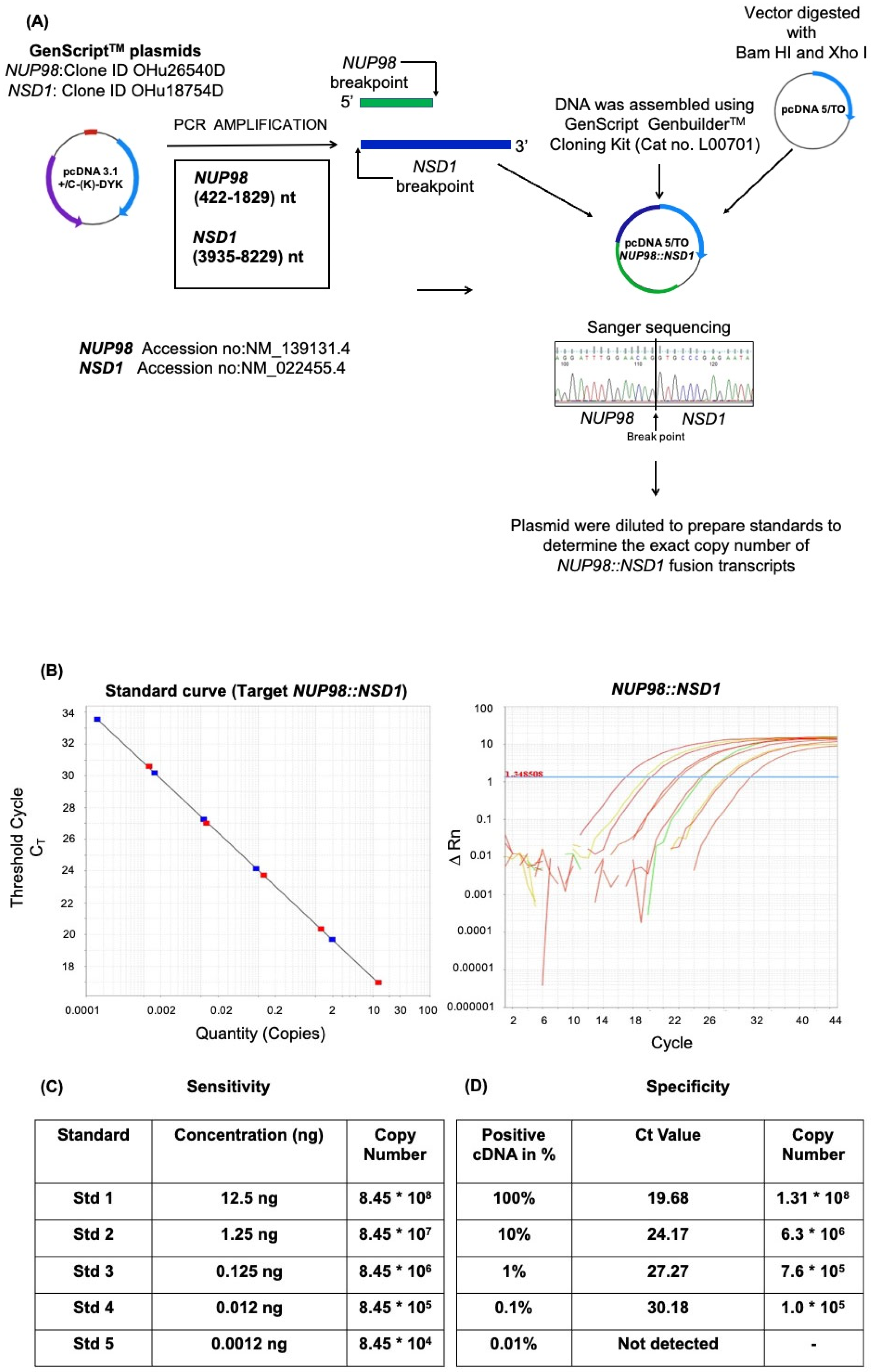
2.2. Plasmid Construction

NUP98 breakpoint region (1408 bp) and NSD1 breakpoint region (4295 bp) were PCR amplified using NUP98::NSD1 forward and NUP98::NSD1 reverse primers (Table 1) using GenScript®, Piscataway, NJ, USA NUP98 (Clone ID OHu26540D) and NSD1 (Clone ID OHu18754D) plasmids as PCR templates. The NUP98::NSD1 fusion fragment was ligated in pcDNA5/TO vector using GenScript®, Piscataway, NJ, USA GenBuilderTM cloning kit (Cat no. L00701-10), as per the manufacturer’s instructions. The cloning of the fusion gene in the pcDNA5/TO was confirmed by restriction–digestion analysis and sanger sequencing.

2.3. Designing of Primers and Probes

Primers and probes were designed using ABI Primer Express 3 software to detect the breakpoint fusion region of NUP98::NSD1 and Abelson tyrosine-protein kinase 1 (ABL1) as an internal control. These primers and FAM/VIC- labelled probes (Table 1) were used for Taqman quantitative RT-PCR in a single multiplex reaction.

2.4. Sensitivity of Assay

The assay’s sensitivity was determined by qRT-PCR of serially diluted plasmids from 12.5 ng with 10 folds of serial dilution until no detectable relative fluorescence was observed in qRT PCR [16].

2.5. Specificity of the Assay

The specificity of the NUP98::NSD1 qRT-PCR assay was determined by qRT-PCR, as previously described [17]. Briefly, a known positive sample for NUP98::NSD1 fusion was serially diluted 10-fold from 100 ng to 0.01 ng. The total concentration of cDNA was maintained at 100 ng in each diluted fraction by healthy control cDNA.

2.6. RNA Extraction and cDNA Preparation

Total RNA was extracted using the PurelinkTM RNA extraction kit (Thermo Fisher Scientific, Waltham, MA, USA) as per the manufacturer’s protocol. Two micrograms of RNA were used to prepare cDNA using a high-capacity cDNA synthesis kit (Applied BiosystemsTM, Waltham, MA, USA), as per the manufacturer’s instructions. The cDNA was used for Sanger sequencing, and qRT-PCR expression analysis of HOX genes and other genes involved in NUP98::NSD1 mediated leukemogenesis.

2.7. Quantitative qRT-PCR

Real-time quantitative PCR was conducted on BioRad CFX 96 real-time instrument using Taq-Man probes (Applied BiosystemsTM, Waltham, MA, USA) and gene-specific primers to detect NUP98::NSD1 and ABL1 transcripts, whereas minor groove binder EVA-green for HOX and other genes transcript estimation was also considered. The ABL1 gene was used for normalization in the NUP98-NSD1 quantitation assay, while GAPDH was used as an internal control for the relative quantification of HOX genes and other gene transcripts. The relative quantification was conducted, as described previously [18]. The primers and probes used for qRT-PCR are mentioned in Table 1.

2.8. Statistical Data Analysis

All the data are expressed as the mean ± standard deviation (SD). Statistical significance was determined using a Student t-test. Three independent biological replicates were used for statistics, and standard deviation was calculated to generate error bars.

2.9. Sanger’s Sequencing for Validation of NUP98-NSD1 Fusion

NUP98::NSD1 transcripts were PCR amplified using specific primers (Table 1). PCR product was purified using the QIAquick PCR purification kit (Qiagen, Hamburg, Germany). The cycle sequencing of purified PCR products was conducted using 1 pmol primer and Big Dye terminator cycle sequencing kit v3 (Applied BiosystemsTM, Waltham, MA, USA). Cycle sequencing products were purified using the Big Dye X-terminator kit (Applied BiosystemsTM, Waltham, MA, USA), and the purified products were run on 3500 Genetic Analyzer ABI 3500 (Applied BiosystemsTM, Waltham, MA, USA) for capillary electrophoresis. The data obtained were analyzed for the presence of fusion transcript in patients’ samples by aligning the sequence data using the NCBI blast tool.

2.10. FLT3-ITD Fragment Analysis

Genomic DNA was extracted from peripheral blood using the QIAamp DNA mini kit (Qiagen, Hamburg, Germany). The catalytic domain of the FLT3 gene was PCR amplified for fragment analysis to detect ITD mutation using a 5′ labelled FAM forward primer and a reverse primer (Table 1). Fragment analysis of PCR products was conducted by sizing-standard LIZ 600 using 3500 Genetic Analyzer ABI 3500 (Applied BiosystemsTM, Waltham, MA, USA). The data obtained were analyzed using Gene Mapper v5.0 (Applied BiosystemsTM, Waltham, MA, USA). The allelic ratio (AR) ratio was calculated, as previously described [19].

3. Results

3.1. Development of a qRT-PCR Assay to Determine NUP98::NSD1 Fusion Transcript

The cloning of the fusion gene in the pcDNA/TO5 expression vector and the validation were conducted, as presented in Figure 1A. For copy number detection, the plasmid containing the NUP98::NSD1 fusion gene was serially diluted to obtain five standards corresponding to the copy number of NUP98::NSD1 fusion depicted using qRT-PCR (Figure 1B). The copy number was mathematically calculated by the calculator for determining the number of copies of a template, as per the formula [number of copies = (amount of DNA in ng × 6.022 × 10²³)/(length of templet (bps) × 1 × 109 × 650)] mentioned on the website http://cels.uri.edu/gsc/cndna.html (accessed on 11 August 2020) by URI Genomics & Sequencing Center. To determine the sensitivity of our qRT-PCR assay for the detection of NUP98::NSD1 fusion, the standard curve was plotted against five standards of variable concentration ranging from 12.5 to 0.00125 ng (Figure 1C). We found that the lower limit of detection of standard control in our assay was 0.00125 ng, which corresponds to log10−4. As per the CLSI/NCCLS EP17-a guideline, the minimum required sensitivity of a qRT-PCR assay should be log10−3. Thus, our in-house developed assay had a sensitivity as per international standards.
To assess the specificity of our assay, a known positive cDNA sample for NUP98::NSD1 fusion was serially diluted 10-fold from 100 ng to 0.01 ng with a healthy control cDNA, such that the final concentration of total cDNA was 100 ng in all diluted samples. 100% cDNA corresponds to 100 ng of NUP98::NSD1 of the positive patient; likewise, 10% cDNA corresponds to 10 ng of NUP98::NSD1 of the positive cDNA plus 90 ng of healthy individual cDNA. Similarly, the dilution series was established for other fractions. On the log scale, 100% cDNA corresponds to log 10 of the transcript, while 0.01% cDNA corresponds to log 10−4 of the transcript. Our qRT-PCR assay for NUP98::NSD1 fusion can detect up to log 10−3 of the transcripts, corresponding to 0.1 ng of positive cDNA of the sample (Figure 1D). Thus, the specificity of the limit of detection of NUP98::NSD1 fusion transcript in our assay is 0.1 ng or log10−3 of the transcript.
Further, the amplification efficiency of our assay was calculated using a serial dilution of our target cDNA samples followed by qRT-PCR. The Ct values were plotted on the logarithmic scale along with the corresponding concentrations, and a linear regression curve was plotted to calculate a slope line. The efficiency of the assay was calculated using the formula E = −1 + 10(−1/slope) and was 97.25%.

3.2. Screening of Patients for the Presence of NUP98::NSD1 Fusion in the Indian Cohort

To demonstrate the utility of our in-house developed qRT-PCR assay to detect the presence of NUP98::NSD1 oncogenic fusion in patients, we screened 150 acute myeloid leukemia (AML) patients. Among 150 AML patients, six AML patients showed the expression of NUP98::NSD1 fusion transcript (Figure 2A). Three out of 112 adults and three out of 38 pediatric patients were positive for the fusion transcript, indicating a prevalence rate of the fusion transcript of 2.67% and 7.89% in adult and pediatric AML cases, respectively. All the positive patients had a blast count of more than 50%.

3.3. Sanger Sequencing Confirmation of NUP98::NSD1 Breakpoint Fusion

To further confirm and validate the NUP98::NSD1 fusion transcript identified by qRT-PCR assay in patients, we sequenced the fusion region of NUP98::NSD1 in all six positive patients using Sanger sequencing. All the patients had the common breakpoint of exon 12 of NUP98 (1407 nucleotide from transcription start site) and exon 6 of NSD1 (3935 positions from ATG start codon till 8229 position), as shown in Figure 2B. This breakpoint was comparable to our positive control and previously reported literature [13,14,20].

3.4. FLT3-ITD Status in NUP98::NSD1 Fusion Positive Patients

Internal tandem duplication of FLT3 has been reported to co-occur in 85–90% of NUP98::NSD1+ive cases [19,21]. Therefore, fragment analysis of FLT3-ITD with Nup98-NSD1 in six positive patients was performed to determine the co-occurrence of FLT3. Fragment analysis revealed that all six patients with NUP98::NSD1 fusion had co-occurrence of FLT3-ITD mutation. Adult patients 1 and 2 had 27 bp and 30 bp FLT3-ITD, respectively. Adult patient 3 had 54 bp FLT3-ITD. In the case of pediatric patients, pediatric patient 1 had 63 bp FLT3-ITD, pediatric patient 2 had 66 bp FLT3-ITD, and pediatric patient 3 had 42 bp FLT3-ITD (Figure 2C).

3.5. NUP98::NSD1 Fusion Patients Have Altered the Expression of Genes Associated with Self-Renewal and Differentiation

NUP98::NSD1 translocation is associated with the deregulation of the HOX gene clusters [9,22]. To assess if our patients with NUP98::NSD1 fusion also have altered expression of HOX genes, we studied the expression of HOX gene clusters in these patients. Of the eight of ten HOX genes tested—HOXA1 (fold increase = 5), HOXA3 (fold increase = 14), HOXA5 (fold increase = 10), HOXA6 (fold increase = 6), HOXA7 (fold increase = 6), HOXA9 (fold increase = 13), HOXA10 (fold increase = 6), and HOXB6 (fold increase = 3)—were upregulated (Figure 3). No significant change was observed for HOXA4 and HOX A11 genes. In addition to HOX genes, we also tested for gene expression profiles of the other representative genes of acute myeloid leukemia (AML). We found that PRDM16 (fold increase = 13) and MECOM (fold increase = 5), an alternatively spliced variant of MDS1/EVI1, highly homologous to PRDM16, were highly upregulated. We also found a five-fold increase in the expression of the VENTX gene in NUP98::NSD1-positive patients, while UTF and NKX2-3 were also upregulated more than three-fold in these patients (Figure 3). The fold increase in upregulated genes was plotted as average mean values of all patients with standard deviation.

3.6. NUP98::NSD1 Patients Show Poor Responses to Induction Therapy

The NUP98::NSD1 patients are reported to show poor outcomes to induction therapy and/or hematopoietic stem cell transplantation (HSCT) [10,23]. In the current study, all six NUP98-NSD1 patients had a high WBC count (Average mean WBC count 18.06 × 10⁹/L), with % of blast cells greater than 50% at diagnosis. All six patients have FLT3-ITD co-occurrence with an AR (allelic ratio) less than 0.4 (Table 2) and were treated as per the standard treatment guidelines [24]. Five patients were treated with 3 + 7 (daunorubicin + cytosine arabinoside (ara-C)) combination induction chemotherapy, while one patient received azacitidine + venetoclax in view of poor performance status at presentation. Of the six NUP98::NSD1 positive patients, only one patient achieved complete remission to induction chemotherapy, while five patients failed induction chemotherapy and had to be administered salvage chemotherapy. All five patients who failed induction chemotherapy succumbed to sepsis due to severe febrile neutropenia. Further, one patient who responded to induction chemotherapy underwent an allogenic stem cell transplant but relapsed seven months post-transplant (Table 2).

4. Discussion

AML patients with NUP98::NSD1 fusion have a poor prognosis, high induction failure, and poor survival [10,19]. Since the fusion is cytogenetically cryptic, it is not detected in the karyogram during conventional karyotyping [25]. Therefore, a robust and reliable method is required to screen and identify these patients during diagnosis. Fewer studies have used qRT-PCR-based techniques using SYBR Green/TaqMan assay to screen this fusion [26]; however, the key limitation with these assays was the lack of a quantitative method to determine the copy number of the fusion transcript. We have developed a reliable qRT-PCR based multiplex assay with high sensitivity and specificity for detecting NUP98::NSD1 transcripts in patient samples. The assay has a log 10−3 sensitivity corresponding to the detection of 0.1 ng of the fusion transcript. Furthermore, the assay is highly specific to detect only the fusion transcript, as no NUP98::NSD1 fusion transcript was detected up to 100 ng of negative cDNA sample. The assay’s limit of quantification (LOQ) and limit of detection (LOD) was at par with the commercially available kits, as previously described [16,17]. Thus the assay can be used for the routine investigation to see the presence of the NUP98::NSD1 fusion transcript for newly admitted patients, specifically AML patients showing the FLT3-ITD and not responding to induction therapy. Additionally, it can be used as a tool for MRD detection in the future.
After successfully developing the qRT-PCR assay for NUP98::NSD1 fusion detection, our goal was to use it to detect fusion transcripts in the Indian cohort of AML patients. The screening of 150 AML patients using the assay identified six patients with NUP98::NSD1 translocation, which included three adults and three pediatric patients. While the worldwide data shows a 4–5% prevalence rate of NUP98::NSD1 in pediatric AML and 1.3–3% in adult AML [9,10,11,12], we found that the percentage prevalence of NUP98::NSD1 fusion transcript in our pediatric was 7.89% (3/38), which was comparatively higher than reported previously. However, this difference could be because of the smaller sample size in our case. Studies on a larger cohort of pediatric patients will be helpful in the evaluation of NUP98::NSD1 prevalence in pediatric patients. The rate of NUP98::NSD1 positivity in adult patients was 2.67% (3/112), which concords with the published studies [10,11,12]. However, this is the first study from the Indian subcontinent to demonstrate the presence and prevalence of NUP98::NSD1 transcripts in AML patients.
NUP98::NSD1 translocation is reported to associate with the co-occurrence of FLT3-ITD, with more than 85% of NUP98::NSD1 patients having FLT3-ITD [10,19]. To see if our NUP98::NSD1 patients also have the co-occurrence of FLT3-ITD, we studied the presence of this rearrangement using fragment analysis. We found that all 06 NUP98::NSD1 positive patients have co-occurrence of FLT3-ITD, which was in line with published studies [12,19,27].
To develop effective therapeutic strategies for NUP98::NSD1 leukemogenesis, a clear understanding of the mechanistic basis of NUP98::NSD1 leukemogenesis needs to be well characterized. The gene expression profile of NUP98::NSD1 patients has revealed an alteration in the genes associated with HOX gene clusters [9,22]. We also found that HOX genes were highly upregulated in all the NUP98::NSD1 patients, specifically HOX A1, HOXA3, HOXA5, HOXA6, HOX A7, HOX A9, HOX A10, and HOXB6 expression, suggesting that the alteration is conserved in our AML patients with NUP98::NSD1. The increased expression of HOX genes has been reported to promote stem cell self-renewal and to block terminal differentiation, thus leading to NUP98::NSD1-mediated leukemogenesis [9].
We also found that several other genes associated with AML were significantly altered in NUP98::NSD1 patients. The high-level expression of PRMD16 is a key clinical feature of AML [28,29]. We found that PRDM16, along with homologous protein MECOM, is upregulated in NUP98::NSD1 patients. Functionally, PRDM16 is essential for hematopoietic stem cell maintenance, and deregulation of PRDM16 induces leukemogenesis [28]. Several reports conclude that high PRDM16 expression is a crucial marker for poor prognosis in AML patients [28]. Similarly, VENTX, an oncogenic transcription factor that promotes myeloid differentiation, was also upregulated in NUP98::NSD1 patients, suggesting its role in leukemogenesis [30]. The UTF gene that is expressed during embryonic development, along with NKX2-3, were also upregulated in NUP98 AML patients [30]. These results suggest that our AML patients with NUP98::NSD1 transcript showed altered genomic profile, deregulated expression of HOX cluster, and other leukemic genes, as previously described [9,22].
It has been reported that NUP98::NSD1 is an independent predictor of poor prognosis [9]; however, frequent co-occurrence of FLT3-ITD and NUP98::NSD1 have a poorer prognosis, thus raising the concern about whether this poor prognostic outcome is because of NUP98::NSD1 or determined by the co-occurrence of FLT3-ITD. The AR (allelic ratio) of FLT3-ITD also has a prognostic significance in AML [19]. Patients with FLT-ITD AR greater than 0.4 are categorized as high-risk patients with high chances of induction failure [19]. In the current study, all six patients were detected with FLT3-ITD AR of less than 0.4; still, these patients showed poor responses to the induction therapy and/or to HSCT. These results suggest that the co-occurrence of NUP98::NSD1 fusion in low-risk FLT3-ITD patients with AR less than 0.4 may transform these patients into a high-risk category showing poor clinical outcome and induction therapy failure, thus speculating that the presence of NUP98-NSD1 may cause disease severity, poor drug response, and a high rate of induction failure. Therefore, these patients should be robustly screened and categorized as a new subset of AML patients. Furthermore, more in-depth molecular characterization of these patients is necessary to understand the disease’s pathobiology and develop novel treatment protocols. However, our findings should be validated in larger prospective cohort studies. Overall, the current study, for the first time, reports the identification and prevalence of NUP98::NSD1 fusion transcript in Indian AML patients.

5. Conclusions

In summary, we have developed a qRT-PCR assay with high sensitivity and specificity to determine the copy number of the NUP98::NSD1 fusion transcript. The assay can robustly screen the NUP98::NSD1-positive AML patients with absolute copy number detection. Furthermore, our data suggest that the NUP98::NSD1 fusion co-occurs with FLT3-ITD and worsens the prognosis of FLT3-ITD positive patients independent of FLT3-ITD allelic ratio; hence it should be routinely screened in all FLT3-ITD-positive AML patients, as it has important treatment ramifications.

Author Contributions

Conceptualization, C.P.C.; Data curation, A.S. (Arunim Shah) and A.S. (Akhilesh Sharma); Formal analysis, A.S. (Arunim Shah), A.S. (Akhilesh Sharma), S.K. and A.G.; Funding acquisition, C.P.C.; Investigation, A.S. (Akhilesh Sharma), S.K., A.G. and C.P.C.; Methodology, A.S. (Arunim Shah) and A.S. (Akhilesh Sharma); Project administration, A.S. (Arunim Shah) and C.P.C.; Resources, A.S. (Arunim Shah), A.S. (Akhilesh Sharma) and C.P.C.; Supervision, C.P.C.; Validation, A.S. (Arunim Shah), S.K. and C.P.C.; Writing—original draft, A.S. (Arunim Shah); Writing—review and editing, A.S. (Akhilesh Sharma), A.S. (Arunim Shah) and C.P.C. All authors have read and agreed to the published version of the manuscript.

Funding

This work was supported by the Wellcome Trust DBT India Alliance Fellowship Grant (IA/I/16/1/502374) sanctioned to CPC. Arunim Shah and Shobhita Katiyar are recipients of the Senior Research Fellowship 2020-6607 and 2020-7639 respectively from the Indian Council of Medical Research (ICMR), New Delhi.

Institutional Review Board Statement

The study was conducted following the Declaration of Helsinki and approved by the Institutional Review Board (or Ethics Committee) of Sanjay Gandhi Post Graduate Institute of Medical Sciences (IEC code: 2021-12-SRF-118 dated 15 February 2021).

Informed Consent Statement

Written Informed consent was obtained from all subjects involved in the study.

Data Availability Statement

Not applicable.

Acknowledgments

The authors thank the patients and healthy donors who participated in the study.

Conflicts of Interest

The authors declare no conflict of interest. The funders had no role in the design of the study; in the collection, analyses, or interpretation of data; in the writing of the manuscript; or in the decision to publish the results.

References

  1. Morishita, M.; Mevius, D.; Di Luccio, E. In vitro histone lysine methylation by NSD1, NSD2/MMSET/WHSC1 and NSD3/WHSC1L. BMC Struct. Biol. 2014, 14, 25. [Google Scholar] [CrossRef] [Green Version]
  2. Faravelli, F. NSD1 mutations in Sotos syndrome. Am. J. Med. Genet. Part C Semin. Med. Genet. 2005, 137C, 24–31. [Google Scholar] [CrossRef] [PubMed]
  3. Berdasco, M.; Ropero, S.; Setien, F.; Fraga, M.F.; Lapunzina, P.; Losson, R.; Alaminos, M.; Cheung, N.-K.; Rahman, N.; Esteller, M. Epigenetic inactivation of the Sotos overgrowth syndrome gene histone methyltransferase NSD1 in human neuroblastoma and glioma. Proc. Natl. Acad. Sci. USA 2009, 106, 21830–21835. [Google Scholar] [CrossRef] [PubMed] [Green Version]
  4. Farhangdoost, N.; Horth, C.; Hu, B.; Bareke, E.; Chen, X.; Li, Y.; Coradin, M.; Garcia, B.A.; Lu, C.; Majewski, J. Chromatin dysregulation associated with NSD1 mutation in head and neck squamous cell carcinoma. Cell Rep. 2021, 34, 108769. [Google Scholar] [CrossRef] [PubMed]
  5. Yang, C.; Wang, K.; Liang, Q.; Tian, T.-T.; Zhong, Z. Role of NSD1 as potential therapeutic target in tumor. Pharmacol. Res. 2021, 173, 105888. [Google Scholar] [CrossRef]
  6. Su, X.; Zhang, J.; Mouawad, R.; Compérat, E.; Rouprêt, M.; Allanic, F.; Parra, J.; Bitker, M.-O.; Thompson, E.J.; Gowrishankar, B.; et al. NSD1 Inactivation and SETD2 Mutation Drive a Convergence toward Loss of Function of H3K36 Writers in Clear Cell Renal Cell Carcinomas. Cancer Res. 2017, 77, 4835–4845. [Google Scholar] [CrossRef] [Green Version]
  7. Taketani, T.; Taki, T.; Nakamura, H.; Taniwaki, M.; Masuda, J.; Hayashi, Y. NUP98–NSD3 fusion gene in radiation-associated myelodysplastic syndrome with t(8;11)(p11;p15) and expression pattern of NSD family genes. Cancer Genet. Cytogenet. 2009, 190, 108–112. [Google Scholar] [CrossRef]
  8. Tauchmann, S.; Schwaller, J. NSD1: A Lysine Methyltransferase between Developmental Disorders and Cancer. Life 2021, 11, 877. [Google Scholar] [CrossRef]
  9. Hollink, I.H.I.M.; Heuvel-Eibrink, M.M.V.D.; Arentsen-Peters, S.T.C.J.M.; Pratcorona, M.; Abbas, S.; Kuipers, J.E.; Van Galen, J.F.; Beverloo, H.B.; Sonneveld, E.; Kaspers, G.-J.J.L.; et al. NUP98/NSD1 characterizes a novel poor prognostic group in acute myeloid leukemia with a distinct HOX gene expression pattern. Blood 2011, 118, 3645–3656. [Google Scholar] [CrossRef] [Green Version]
  10. Ostronoff, F.; Othus, M.; Gerbing, R.B.; Loken, M.R.; Raimondi, S.C.; Hirsch, B.A.; Lange, B.J.; Petersdorf, S.; Radich, J.; Appelbaum, F.R.; et al. NUP98/NSD1 and FLT3/ITD coexpression is more prevalent in younger AML patients and leads to induction failure: A COG and SWOG report. Blood J. Am. Soc. Hematol. 2014, 124, 2400–2407. [Google Scholar] [CrossRef]
  11. Cerveira, N.; Correia, C.; Doria, S.; Bizarro, S.; Rocha, P.; Gomes, P.; Torres, L.; Norton, L.; Borges, B.S.; Castedo, S.; et al. Frequency of NUP98-NSD1 fusion transcript in childhood acute myeloid leukaemia. Leukemia 2003, 17, 2244–2247. [Google Scholar] [CrossRef] [PubMed] [Green Version]
  12. Shimada, A.; Iijima-Yamashita, Y.; Tawa, A.; Tomizawa, D.; Yamada, M.; Norio, S.; Watanabe, T.; Taga, T.; Iwamoto, S.; Terui, K.; et al. Risk-stratified therapy for children with FLT3-ITD-positive acute myeloid leukemia: Results from the JPLSG AML-05 study. Int. J. Hematol. 2018, 107, 586–595. [Google Scholar] [CrossRef] [PubMed]
  13. Brown, J.; Jawad, M.; Twigg, S.R.F.; Saracoglu, K.; Sauerbrey, A.; Thomas, A.E.; Eils, R.; Harbott, J.; Kearney, L. A cryptic t(5;11)(q35;p15.5) in 2 children with acute myeloid leukemia with apparently normal karyotypes, identified by a multiplex fluorescence in situ hybridization telomere assay. Blood 2002, 99, 2526–2531. [Google Scholar] [CrossRef] [Green Version]
  14. Jaju, R.J. A novel gene, NSD1, is fused to NUP98 in the t(5;11)(q35;p15.5) in de novo childhood acute myeloid leukemia. Blood 2001, 98, 1264–1267. [Google Scholar] [CrossRef] [PubMed] [Green Version]
  15. Lim, H.J.; Lee, J.H.; Lee, Y.E.; Baek, H.J.; Kook, H.; Park, J.H.; Lee, S.Y.; Choi, H.W.; Choi, H.J.; Kee, S.J.; et al. The First Korean Case of NUP98-NSD1 and a Novel SNRK-ETV6 Fusion in a Pediatric Therapy-related Acute Myeloid Leukemia Patient Detected by Targeted RNA Sequencing. Ann. Lab. Med. 2021, 41, 443–446. [Google Scholar] [CrossRef] [PubMed]
  16. Kitamura, H.; Tabe, Y.; Ai, T.; Tsuchiya, K.; Yuri, M.; Misawa, S.; Horii, T.; Kawaguchi, A.; Ohsaka, A.; Kimura, S. A new highly sensitive real-time quantitative-PCR method for detection of BCR-ABL1 to monitor minimal residual disease in chronic myeloid leukemia after discontinuation of imatinib. PLoS ONE 2019, 14, e0207170. [Google Scholar] [CrossRef] [Green Version]
  17. Test Details—BCR-ABL RNA PCR Quantitation for Leukemia. Available online: https://knightdxlabs.ohsu.edu/home/test-details?id=BCR-ABL+RNA+PCR+Quantitation+for+Leukemia (accessed on 10 September 2022).
  18. Chaturvedi, C.-P.; Hosey, A.M.; Palii, C.; Perez-Iratxeta, C.; Nakatani, Y.; Ranish, J.A.; Dilworth, F.J.; Brand, M. Dual role for the methyltransferase G9a in the maintenance of β-globin gene transcription in adult erythroid cells. Proc. Natl. Acad. Sci. USA 2009, 106, 18303–18308. [Google Scholar] [CrossRef] [Green Version]
  19. Niktoreh, N.; Walter, C.; Zimmermann, M.; von Neuhoff, C.; von Neuhoff, N.; Rasche, M.; Waack, K.; Creutzig, U.; Hanenberg, H.; Reinhardt, D. Mutated WT1, FLT3-ITD, and NUP98-NSD1 Fusion in Various Combinations Define a Poor Prognostic Group in Pediatric Acute Myeloid Leukemia. J. Oncol. 2019, 2019, 1609128. [Google Scholar] [CrossRef] [Green Version]
  20. Petit, A.; Radford, I.; Waill, M.-C.; Romana, S.P.; Berger, R. NUP98NSD1 fusion by insertion in acute myeloblastic leukemia. Cancer Genet. Cytogenet. 2008, 180, 43–46. [Google Scholar] [CrossRef]
  21. Struski, S.; Lagarde, S.; Bories, P.; Puiseux, C.; Prade, N.; Cuccuini, W.; Pages, M.-P.; Bidet, A.; Gervais, C.; Lafage-Pochitaloff, M.; et al. NUP98 is rearranged in 3.8% of pediatric AML forming a clinical and molecular homogenous group with a poor prognosis. Leukemia 2016, 31, 565–572. [Google Scholar] [CrossRef]
  22. Wang, G.G.; Cai, L.; Pasillas, M.P.; Kamps, M.P. NUP98–NSD1 links H3K36 methylation to Hox-A gene activation and leukaemogenesis. Nat. Cell Biol. 2007, 9, 804–812. [Google Scholar] [CrossRef] [PubMed]
  23. Shiba, N.; Ichikawa, H.; Taki, T.; Park, M.-J.; Jo, A.; Mitani, S.; Kobayashi, T.; Shimada, A.; Sotomatsu, M.; Arakawa, H.; et al. NUP98-NSD1gene fusion and its related gene expression signature are strongly associated with a poor prognosis in pediatric acute myeloid leukemia. Genes Chromosomes Cancer 2013, 52, 683–693. [Google Scholar] [CrossRef] [PubMed]
  24. Döhner, H.; Estey, E.; Grimwade, D.; Amadori, S.; Appelbaum, F.R.; Büchner, T.; Dombret, H.; Ebert, B.L.; Fenaux, P.; Larson, R.A.; et al. Diagnosis and management of AML in adults: 2017 ELN recommendations from an international expert panel. Blood 2017, 129, 424–447. [Google Scholar] [CrossRef] [PubMed] [Green Version]
  25. Kivioja, J.L.; Martí, J.M.L.; Kumar, A.; Kontro, M.; Edgren, H.; Parsons, A.; Lundán, T.; Wolf, M.; Porkka, K.; Heckman, C.A. Chimeric NUP98NSD1 transcripts from the cryptic t(5;11)(q35.2;p15.4) in adult de novo acute myeloid leukemia. Leuk. Lymphoma 2017, 59, 725–732. [Google Scholar] [CrossRef] [PubMed] [Green Version]
  26. Akiki, S.; Dyer, S.A.; Grimwade, D.; Ivey, A.; Abou-Zeid, N.; Borrow, J.; Jeffries, S.; Caddick, J.; Newell, H.; Begum, S.; et al. NUP98-NSD1 fusion in association withFLT3-ITD mutation identifies a prognostically relevant subgroup of pediatric acute myeloid leukemia patients suitable for monitoring by real time quantitative PCR. Genes Chromosomes Cancer 2013, 52, 1053–1064. [Google Scholar] [CrossRef]
  27. Thanasopoulou, A.; Tzankov, A.; Schwaller, J. Potent co-operation between the NUP98-NSD1 fusion and the FLT3-ITD mutation in acute myeloid leukemia induction. Haematologica 2014, 99, 1465–1471. [Google Scholar] [CrossRef] [Green Version]
  28. Yamato, G.; Yamaguchi, H.; Handa, H.; Shiba, N.; Kawamura, M.; Wakita, S.; Inokuchi, K.; Hara, Y.; Ohki, K.; Okubo, J.; et al. Clinical features and prognostic impact of PRDM16 expression in adult acute myeloid leukemia. Genes Chromosomes Cancer 2017, 56, 800–809. [Google Scholar] [CrossRef]
  29. Shiba, N.; Ohki, K.; Kobayashi, T.; Hara, Y.; Yamato, G.; Tanoshima, R.; Ichikawa, H.; Tomizawa, D.; Park, M.-J.; Shimada, A.; et al. High PRDM16 expression identifies a prognostic subgroup of pediatric acute myeloid leukaemia correlated to FLT3-ITD, KMT2A-PTD, and NUP98-NSD1: The results of the Japanese Paediatric Leukaemia/Lymphoma Study Group AML-05 trial. Br. J. Haematol. 2015, 172, 581–591. [Google Scholar] [CrossRef]
  30. Lavallée, V.P.; Lemieux, S.; Boucher, G.; Gendron, P.; Boivin, I.; Girard, S.; Hebert, J.; Sauvageau, G. Identification of MYC mutations in acute myeloid leukemias with NUP98-NSD1 translocations. Leukemia 2016, 30, 1621–1624. [Google Scholar] [CrossRef] [PubMed]
Figure 1. (A) Schematic representation of molecular cloning and validation strategy for NUP98::NSD1 DNA sequence in expression vector pcDNA/TO5 plasmid vector. The plasmid with the NUP98::NSD1 gene was used as a positive control/standard for developing the quantitative assay. (B) Development of the NUP98::NSD1 screening assay. This figure shows the standard curve plot obtained with a slope of −3.389 with an R2 (correlation coefficient) of 1. The variation of the Ct between the replicates was found to be <0.5 Ct, and the standard deviation was <0.15. The table represents the known concentration of standards with their corresponding copy number detected using qRT-PCR. (C) The sensitivity of the assay was determined by serially diluting the NUP98::NSD1 plasmid. (D) The assay’s specificity was determined by serially diluting the NUP98::NSD1 positive patient sample with healthy donor cDNA, as described in the results.
Diagnostics 12 03001 g001
Figure 2. Screening and validation of AML patients (A) qRT PCR data revealed the presence of NUP98::NSD1 fusion transcript. The table shows the Ct value and their corresponding NUP98::NSD1 copy number. (B) Fluorescent peak trace of chromatograms obtained after Sanger sequencing showing the breakpoint of NUP98::NSD1 fusion transcript. Exon 12 of NUP98 was fused to exon 6 of NSD1 in all six patients. The plasmid with NUP98::NSD1 fusion was taken as a positive control. (C) Fragment analysis of FLT3-ITD mutation in NUP98::NSD1+ve patients revealed all six patients had co-occurrence of FLT3-ITD mutation. The peak in the grey bin indicates wild-type FLT3, while the peak in the pink bin indicates mutant FLT3.
Diagnostics 12 03001 g002
Figure 3. Patients with NUP98-NSD1 fusion had an altered gene expression profile. This figure shows an alteration in the expression of HOX cluster and other genes associated with self-renewal and differentiation in patients with NUP98-NSD1 oncogenic fusion. HOX genes (HOXA1, A3, A4, A5, A6, A7, A9, A10, A11, and B6) and other genes (PRDM16, MeCOM, NKX 2.3, VENTX, ASL, and UTF) involved in self-renewal were analyzed at the mRNA level by real-time RT-qPCR in healthy control vs. NUP98::NSD1+ive patients. Transcripts are expressed relative to GAPDH. The average values from 06 NUP98::NSD1 patients vs. six healthy controls in three replicate experiments are represented with error bars corresponding to SD. * p < 0.05; ** p < 0.01; ns: non-significant.
Diagnostics 12 03001 g003
Table 1. List the primers and probes used for cloning, sequencing, expression analysis, and fragment analysis.
Cloning Primers
NUP98::NSD1 fusion forward5′AAAGATCATGACATAGATTACAAGGATGACGATGA
CAAGGCCATGTTTAACAAATCATTTGGAACACC-3′
NUP98::NSD1 fusion reverse5′AGTCGAGGCTGATCAGCGGGTTTAAACGGGCC
CTCTAGACCTACTTCTGTTCTGATTCTGCACACTT-3′
Sequencing Primers
NUP98-Seq F5′-ACTCTTGGAACTGGGCTTGG-3′
NSD1-Seq R5′-GGCTAGAAGGCTTTCCTCTTC-3′
qRT-PCR primers
NUP98 t(5,11) F5′-GGCCCCTGGATTTAATACTACGA-3′
NSD1 t(5,11)R5′-CTTCCTAAGGCGTTTCTTCTCTGA-3′
NUP98-NSD1 t(5,11) probe5′-FAM-TTTGGAGCCCCCCAGGCC-MGB NFQ-3′
ABL1-F5′-CCCAGAGAAGGTCTATGAACTCATG-3′
ABL1-R5′-AGGAGGGCCGGTCAGA-3′
ABL1 probe5′-VIC-TCCACTGCCAACATGC-MGB NFQ-3′
HOXA1 F5′-CCCTCGGACCATAGGATTACAA-3′
HOXA1 R5′-GCCGCCGCAACTGTTG-3′
HOXA3 F5′-CGACAGCTCGGCGATCTAC-3′
HOXA3 R5′-CGGGTACGGCTGCTGATT-3′
HOXA4 F5′-GGTGGTGTACCCCTGGATGA-3′
HOXA4 R5′-GACTTGCTGCCGGGTATAGG-3′
HOXA5 F5′-GGAGTTCCACTTCAACCGTTACC-3′
HOXA5 R5′-CGGAGAGGCAAAGAGCATGT-3′
HOXA6 F5′-GTCTGGTAGCGCGTGTAGGT-3′
HOXA6 R5′-CCCTGTTTACCCCTGGATG-3′
HOXA7 F5′-CTTCTCCAGTTCCAGCGTCT-3′
HOXA7 R5′-AAGCCAGTTTCCGCATCTAC-3′
HOXA9 F5′-CCACGCTTGACACTCACACT-3′
HOXA9 R5′-GCTCTCATTCTCGGCATTGT-3′
HOXA10 F5′-TCTTTGCTGTGAGCCAGTTG-3′
HOXA10 R5′-CTCCAGCCCCTTCAGAAAAC-3′
HOXA11 F5′-CGGCCACACTGAGGACAAG-3′
HOXA11 R5′-AACTCTCGCTCCAGCTCTCG-3′
HOXB6 F5′-TCCCCTCCCAATGAGTTCCT-3′
HOXB6 R5′-ACTCCTGCCCGCTGGC-3′
PRDM16 F5′-TGCCGCACGCAGATCA-3′
PRDM16 R5′-GGGAGGAGGTAGTGCTGAACAT-3′
MECOM F5′-CGGAGTGTGGCAAAACGTT-3′
MECOM R5′-GCTGTGGATGTGCTTGTGTTGT-3′
NKX2-3 F5′-GGTTCCAGAATCGCAGGTACAA-3′
NKX2-3 R5′-GCGCCAAGCTCCAGAGACT-3′
VENTX F5′-GGCTGGCCAGGGAGATG-3′
VENTX R5′-TGCGGCGATTCTGAAACC-3′
UTF F5′-GACCAGCTGCTGACCTTGAA-3′
UTF R5′-CTGCCCAGAATGAAGCCCA-3′
ASL F5′-CAGCATGGATGCCACTAGTGA-3′
ASL R5′-CACAGCGAAGCCCAGAACA-3′
GAPDH F5′-AATCCCATCACCATCTTCCA-3′
GAPDH R5′-TGGACTCCACGACGTACTCA-3′
Fragment analysis primers
FLT3 exon14 F5′-FAM-AGCAATTTAGGTATGAAAGCCAGCTA-3′
FLT3 exon14 R5′-CTTTCAGCATTTTGACGGCAACC-3′
Table 2. Individual characteristics and clinical outcome of AML patients with NUP98::NSD1 fusion transcript.
Patient No.Age
(in Years)
SexFABWBC Count at Diagnosis (WBC Count /L)BM Blast (%)FLT3-ITD StatusTreatment ProtocolInduction
Chemo Response
HSCTOutcome
Adult #157MM419.2 * 10970%FLT3-ITD Positive
(AR 0.35)
3 + 7 f/b HAMInduction failureNoExpired due to severe sepsis & respiratory failure
Adult #264FM212.8 * 10995%FLT3-ITD Positive
(AR 0.35)
Azacitidine + Venetoclax f/b HAMInduction failureNoDied due to disease progression
Adult #333MM320 * 10995%FLT3-ITD Positive
(AR 0.31)
3 + 7 + Midostaurin Induction failureNoDied due to intracranial bleed.
Pediatric #115MM219.9 * 10955%FLT3-ITD Positive
(AR 0.36)
3 + 7 + Midostaurin Achieved remissionYes Disease relapsed 7 months after allogenic stem cell transplant
Pediatric #215FM218.6 * 10971%FLT3-ITD Positive
(AR 0.21)
3 + 7 + Midostaurin Induction failureNoExpired due to MDR sepsis (kliebsella pneumoniae)
Pediatric #312FM117.9 * 10990%FLT3-ITD Positive
(AR 0.37)
3 + 7 + Midostaurin
f/b HAM+ Midostaurin
Induction failureYes Disease relapsed 4 months after allogenic stem cell transplant
Abbreviations: FAB—French American British; WBC-white blood cell; 3 + 7—3 days daunorubicin + 7 days cytosine arabinoside; HAM—high dose cytosine arabinoside + mitoxantrone; FLAG—ida-fludarabine, cytosine arabinoside, idarubicin; HSCT—hematopoietic stem cell transplant; f/b—followed by; BM—bone marrow; MDR—multidrug resistance; AR—allelic ratio; FLT3-ITD—FLT3 internal tandem duplication.
Publisher’s Note: MDPI stays neutral with regard to jurisdictional claims in published maps and institutional affiliations.

