
Meralgia Paresthetica—An Approach Specific Neurological Complication in Patients Undergoing DAA Total Hip Replacement: Anatomical and Clinical Considerations

Abstract
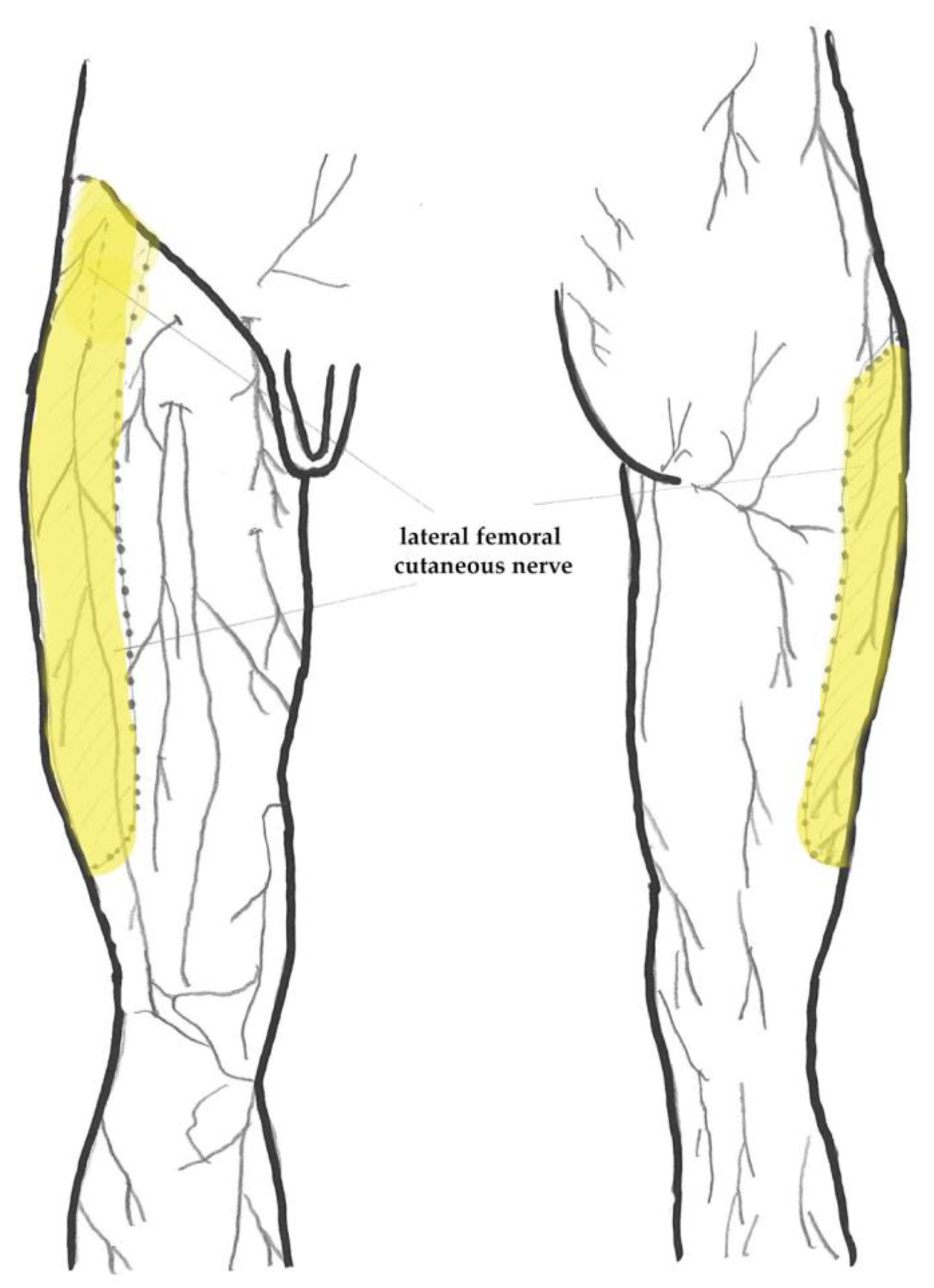
:1. Introduction
2. Materials and Methods
3. Results
4. Discussion
5. Conclusions
Author Contributions
Funding
Institutional Review Board Statement
Informed Consent Statement
Data Availability Statement
Conflicts of Interest
References
- Moreau, P. Minimally Invasive Total Hip Arthroplasty Using Hueter’s Direct Anterior Approach. Eur. J. Orthop. Surg. Traumatol. 2018, 28, 771–779. [Google Scholar] [CrossRef] [PubMed]
- Rachbauer, F.; Kain, M.S.H.; Leunig, M. The History of the Anterior Approach to the Hip. Orthop. Clin. N. Am. 2009, 40, 311–320. [Google Scholar] [CrossRef] [PubMed]
- Wang, Z.; Bao, H.W.; Hou, J.Z. Direct Anterior versus Lateral Approaches for Clinical Outcomes after Total Hip Arthroplasty: A Meta-Analysis. J. Orthop. Surg. Res. 2019, 14, 63. [Google Scholar] [CrossRef]
- Rivera, F.; Comba, L.C.; Bardelli, A. Direct Anterior Approach Hip Arthroplasty: How to Reduce Complications—A 10-Years Single Center Experience and Literature Review. World J. Orthop. 2022, 13, 388–399. [Google Scholar] [CrossRef] [PubMed]
- Nairn, L.; Gyemi, L.; Gouveia, K.; Ekhtiari, S.; Khanna, V. The Learning Curve for the Direct Anterior Total Hip Arthroplasty: A Systematic Review. Int. Orthop. 2021, 45, 1971–1982. [Google Scholar] [CrossRef]
- Shen, K.; Feng, E.; Lin, F.; Weng, Y.; Chen, J. Learning Curve of Total Hip Arthroplasty in Direct Anterior Approach without Requiring Corrective Osteotomy for Hip Dysplasia. Orthop. Surg. 2022, 14, 840–850. [Google Scholar] [CrossRef]
- Peters, R.M.; Ten Have, B.L.E.F.; Rykov, K.; Van Steenbergen, L.; Putter, H.; Rutgers, M.; Vos, S.; Van Steijnen, B.; Poolman, R.W.; Vehmeijer, S.B.W.; et al. The Learning Curve of the Direct Anterior Approach Is 100 Cases: An Analysis Based on 15,875 Total Hip Arthroplasties in the Dutch Arthroplasty Register. Acta Orthop. 2022, 93, 775–782. [Google Scholar] [CrossRef]
- Hasija, R.; Kelly, J.J.; Shah, N.V.; Newman, J.M.; Chan, J.J.; Robinson, J.; Maheshwari, A.V. Nerve Injuries Associated with Total Hip Arthroplasty. J. Clin. Orthop. Trauma 2018, 9, 81–86. [Google Scholar] [CrossRef]
- Vajapey, S.P.; Morris, J.; Lynch, D.; Spitzer, A.; Li, M.; Glassman, A.H. Nerve Injuries with the Direct Anterior Approach to Total Hip Arthroplasty. JBJS Rev. 2020, 8, e0109. [Google Scholar] [CrossRef]
- Goulding, K.; Beaulé, P.E.; Kim, P.R.; Fazekas, A. Incidence of Lateral Femoral Cutaneous Nerve Neuropraxia after Anterior Approach Hip Arthroplasty. Clin. Orthop. Relat. Res. 2010, 468, 2397–2404. [Google Scholar] [CrossRef]
- Bhargava, T.; Goytia, R.N.; Jones, L.C.; Hungerford, M.W. Lateral Femoral Cutaneous Nerve Impairment after Direct Anterior Approach for Total Hip Arthroplasty. Orthopedics 2010, 33, 472. [Google Scholar] [CrossRef]
- Restrepo, C.; Parvizi, J.; Pour, A.E.; Hozack, W.J. Prospective Randomized Study of Two Surgical Approaches for Total Hip Arthroplasty. J. Arthroplast. 2010, 25, 671–679.e1. [Google Scholar] [CrossRef] [PubMed]
- Homma, Y.; Baba, T.; Sano, K.; Ochi, H.; Matsumoto, M.; Kobayashi, H.; Yuasa, T.; Maruyama, Y.; Kaneko, K. Lateral Femoral Cutaneous Nerve Injury with the Direct Anterior Approach for Total Hip Arthroplasty. Int. Orthop. 2016, 40, 1587–1593. [Google Scholar] [CrossRef] [PubMed]
- Grob, K.; Manestar, M.; Ackland, T.; Filgueira, L.; Kuster, M.S. Potential Risk to the Superior Gluteal Nerve during the Anterior Approach to the Hip Joint an Anatomical Study. J. Bone Jt. Surg.—Am. Vol. 2014, 97, 1426–1431. [Google Scholar] [CrossRef] [PubMed]
- Starke, V.; Stofferin, H.; Mannschatz, S.; Hörmann, R.; Dammerer, D.; Thaler, M. The Anatomical Course of the Superior Gluteal Nerve with Regard to the Direct Anterior Approach for Primary and Revision Total Hip Arthroplasty. J. Arthroplast. 2021, 36, 1138–1142. [Google Scholar] [CrossRef] [PubMed]
- Rudin, D.; Manestar, M.; Ullrich, O.; Erhardt, J.; Grob, K. The Anatomical Course of the Lateral Femoral Cutaneous Nerve with Special Attention to the Anterior Approach to the Hip Joint. J. Bone Jt. Surg.—Am. Vol. 2016, 98, 561–567. [Google Scholar] [CrossRef] [PubMed]
- Chang, K.V.; Mezian, K.; Naňka, O.; Wu, W.T.; Lou, Y.M.; Wang, J.C.; Martinoli, C.; Özçakar, L. Ultrasound Imaging for the Cutaneous Nerves of the Extremities and Relevant Entrapment Syndromes: From Anatomy to Clinical Implications. J. Clin. Med. 2018, 7, 457. [Google Scholar] [CrossRef] [PubMed]
- Majkrzak, A.; Johnston, J.; Kacey, D.; Zeller, J. Variability of the Lateral Femoral Cutaneous Nerve: An Anatomic Basis for Planning Safe Surgical Approaches. Clin. Anat. 2010, 23, 304–311. [Google Scholar] [CrossRef]
- Aszmann, O.C.; Dellon, E.S.; Dellon, A.L. Anatomical Course of the Lateral Femoral Cutaneous Nerve and Its Susceptibility to Compression and Injury. Plast. Reconstr. Surg. 1997, 100, 600–604. [Google Scholar] [CrossRef]
- Carai, A.; Fenu, G.; Sechi, E.; Crotti, F.M.; Montella, A. Anatomical Variability of the Lateral Femoral Cutaneous Nerve: Findings from a Surgical Series. Clin. Anat. 2009, 22, 365–370. [Google Scholar] [CrossRef]
- Ropars, M.; Morandi, X.; Huten, D.; Thomazeau, H.; Berton, E.; Darnault, P. Anatomical Study of the Lateral Femoral Cutaneous Nerve with Special Reference to Minimally Invasive Anterior Approach for Total Hip Replacement. Surg. Radiol. Anat. 2009, 31, 199–204. [Google Scholar] [CrossRef] [PubMed]
- Moritz, T.; Prosch, H.; Berzaczy, D.; Happak, W.; Lieba-Samal, D.; Bernathova, M.; Auff, E.; Bodner, G. Common Anatomical Variation in Patients with Idiopathic Meralgia Paresthetica: A High Resolution Ultrasound Case-Control Study. Pain. Physician 2013, 16, E287–E293. [Google Scholar] [CrossRef] [PubMed]
- Tomaszewski, K.A.; Popieluszko, P.; Henry, B.M.; Roy, J.; Sanna, B.; Kijek, M.R.; Walocha, J.A. The Surgical Anatomy of the Lateral Femoral Cutaneous Nerve in the Inguinal Region: A Meta-Analysis. Hernia 2016, 20, 649–657. [Google Scholar] [CrossRef] [PubMed]
- Standring, S. Gray’s Anatomy: The Anatomical Basis of Clinical Practice, 41st ed.; Elsevier: Philadelphia, PA, USA, 2015. [Google Scholar]
- Gray, H.; Lewis, W.; Gray, H. Anatomy of the Human Body, 20th ed.; Lea Febiger: Philadelphia, PA, USA, 1918; Volume 20, ISBN 1-58734-102-6. [Google Scholar]
- Ivins, G.K. Meralgia Paresthetica, the Elusive Diagnosis. Ann. Surg. 2000, 232, 281–286. [Google Scholar] [CrossRef]
- Roth, V.K. Meralgia Paræesthetica. Von Dr. Wladimir K. Roth. Williams and Norgate. London: 1895, pp. 24. J. Ment. Sci. 1896, 42, 20–24. [Google Scholar] [CrossRef]
- de Ruiter, G.C.W.; Oosterhuis, J.W.A.; Vissers, T.F.H.; Kloet, A. Unusual Causes for Meralgia Paresthetica: Systematic Review of the Literature and Single Center Experience. Neurosurg. Rev. 2023, 46, 107. [Google Scholar] [CrossRef]
- Moucharafieh, R.; Wehbe, J.; Maalouf, G. Meralgia Paresthetica: A Result of Tight New Trendy Low Cut Trousers (‘taille Basse’). Int. J. Surg. 2008, 6, 164–168. [Google Scholar] [CrossRef]
- Lee, S.H.; Shin, K.J.; Gil, Y.C.; Ha, T.J.; Koh, K.S.; Song, W.C. Anatomy of the Lateral Femoral Cutaneous Nerve Relevant to Clinical Findings in Meralgia Paresthetica. Muscle Nerve 2017, 55, 646–650. [Google Scholar] [CrossRef]
- Peters, G.; Larner, A.J. Meralgia Paresthetica Following Gynecologic and Obstetric Surgery. Int. J. Gynecol. Obstet. 2006, 95, 42–43. [Google Scholar] [CrossRef]
- Kavanagh, D.; Connolly, S.; Fleming, F.; Hill, A.D.K.; McDermott, E.W.; O’Higgins, N.J. Meralgia Paraesthetica Following Open Appendicectomy. Ir. Med. J. 2005, 98, 183–185. [Google Scholar]
- Parisi, T.J.; Mandrekar, J.; Dyck, P.J.B.; Klein, C.J. Meralgia Paresthetica: Relation to Obesity, Advanced Age, and Diabetes Mellitus. Neurology 2011, 77, 1538–1542. [Google Scholar] [CrossRef] [PubMed]
- Williams, P.H.; Trzil, K.P. Management of Meralgia Paresthetica. J. Neurosurg. 1991, 74, 76–80. [Google Scholar] [CrossRef] [PubMed]
- Khalil, N.; Nicotra, A.; Rakowicz, W. Treatment for Meralgia Paraesthetica. Cochrane Database Syst. Rev. 2012, 2012, CD004159. [Google Scholar] [CrossRef] [PubMed]
- Hurdle, M.F.; Weingarten, T.N.; Crisostomo, R.A.; Psimos, C.; Smith, J. Ultrasound-Guided Blockade of the Lateral Femoral Cutaneous Nerve: Technical Description and Review of 10 Cases. Arch. Phys. Med. Rehabil. 2007, 88, 1362–1364. [Google Scholar] [CrossRef] [PubMed]
- De Ruiter, G.C.W.; Wurzer, J.A.L.; Kloet, A. Decision Making in the Surgical Treatment of Meralgia Paresthetica: Neurolysis versus Neurectomy. Acta Neurochir. 2012, 154, 1765–1772. [Google Scholar] [CrossRef]
- Benezis, I.; Boutaud, B.; Leclerc, J.; Fabre, T.; Durandeau, A. Lateral Femoral Cutaneous Neuropathy and Its Surgical Treatment: A Report of 167 Cases. Muscle Nerve 2007, 36, 659–663. [Google Scholar] [CrossRef]
- Payne, R.; Seaman, S.; Sieg, E.; Langan, S.; Harbaugh, K.; Rizk, E. Evaluating the Evidence: Is Neurolysis or Neurectomy a Better Treatment for Meralgia Paresthetica? Acta Neurochir. 2017, 159, 931–936. [Google Scholar] [CrossRef]
- MacKay, M.D.; Mudreac, A.; Varacallo, M. Anatomy, Abdomen and Pelvis, Camper Fascia. Available online: https://www.ncbi.nlm.nih.gov/books/NBK482246/ (accessed on 10 January 2024).
- Matta, J.M.; Sah, A.P. Anterior Hip Replacement: From Origin to Current Advanced Techniques; Springer: Cham, Switzerland, 2022. [Google Scholar] [CrossRef]
- Nogler, M. The History of the Direct Anterior Approach in Innsburck. In Anterior Hip Replacement: From Origin to Current Advanced Techniques; Springer: Cham, Switzerland, 2022. [Google Scholar] [CrossRef]
- Corten, K.; Holzapfel, B.M. Direct Anterior Approach for Total Hip Arthroplasty Using the “Bikini Incision”. Oper. Orthop. Traumatol. 2021, 33, 318–330. [Google Scholar] [CrossRef]
- Leunig, M.; Hutmacher, J.E.; Rüdiger, H.A.; Naal, F.D.; Ricciardi, B.F.; Impellizzeri, F.M. Skin Crease “bikini” Incision for the Direct Anterior Approach in Total Hip Arthroplasty. Bone Jt. J. 2018, 100B, 853–861. [Google Scholar] [CrossRef]
- Sang, W.; Xue, S.; Xu, Y.; Liu, Y.; Zhu, L.; Ma, J. Bikini Incision Increases the Incidence of Lateral Femoral Cutaneous Nerve Injury in Direct Anterior Approach Hip Arthroplasty: A Prospective Ultrasonic, Electrophysiological, and Clinical Study. J. Arthroplast. 2021, 36, 3463–3470. [Google Scholar] [CrossRef]
- Schopper, C.; Traxler, H.; Schauer, B.; Hipmair, G.; Gotterbarm, T.; Luger, M. Minimally Invasive Approaching in Hip Surgery—An Anatomical Investigation of 20 Specimens. Medicina 2021, 57, 1283. [Google Scholar] [CrossRef]
- Van Slobbe, A.M.; Bohnen, A.M.; Bernsen, R.M.D.; Koes, B.W.; Bierma-Zeinstra, S.M.A. Incidence Rates and Determinants in Meralgia Paresthetica in General Practice. J. Neurol. 2004, 251, 294–297. [Google Scholar] [CrossRef] [PubMed]
- Ukai, T.; Suyama, K.; Hayashi, S.; Omura, H.; Watanabe, M. The Anatomical Features of the Lateral Femoral Cutaneous Nerve with Total Hip Arthroplasty: A Comparative Study of Direct Anterior and Anterolateral Supine Approaches. BMC Musculoskelet. Disord. 2022, 23, 267. [Google Scholar] [CrossRef] [PubMed]
- Herndon, C.L.; Drummond, N.; Sarpong, N.O.; Cooper, H.J.; Shah, R.P.; Geller, J.A. Direct Anterior versus Mini-Anterolateral Approach for Primary Total Hip Arthroplasty: Early Postoperative Outcomes and Complications. Arthroplast. Today 2020, 6, 257–261. [Google Scholar] [CrossRef] [PubMed]
- Beaulieu, M.A.; Laurin, C.A. Gluteal Nerve Damage Following Total Hip Arthroplasty: A Prospective Analysis. J. Arthroplast. 1990, 5, 319–322. [Google Scholar] [CrossRef]
- Chomiak, J.; Huráček, J.; Dvořák, J.; Dungl, P.; Kubeš, R.; Schwarz, O.; Munzinger, U. Lesion of Gluteal Nerves and Muscles in Total Hip Arthroplasty through 3 Surgical Approaches. An Electromyographically Controlled Study. HIP Int. 2015, 25, 176–183. [Google Scholar] [CrossRef]
- Yang, I.-H. Neurovascular Injury in Hip Arthroplasty. Hip Pelvis 2014, 26, 74–78. [Google Scholar] [CrossRef]
- Brown, G.D.; Swanson, E.A.; Nercessian, O.A. Neurologic Injuries after Total Hip Arthroplasty. Am. J. Orthop. 2008, 37, 191–197. [Google Scholar]
- Tanabe, H.; Baba, T.; Ozaki, Y.; Yanagisawa, N.; Banno, S.; Watari, T.; Homma, Y.; Nagao, M.; Kaneko, K.; Ishijima, M. Lateral versus Conventional Fasciotomy for Prevention of Lateral Femoral Cutaneous Nerve Injury in Total Hip Arthroplasty with Direct Anterior Approach: A Study Protocol for a Dual-Center, Double-Blind, Randomized Controlled Trial. Trials 2022, 23, 567. [Google Scholar] [CrossRef]
- Ozaki, Y.; Baba, T.; Homma, Y.; Tanabe, H.; Ochi, H.; Bannno, S.; Watari, T.; Kaneko, K. Preoperative Ultrasound to Identify Distribution of the Lateral Femoral Cutaneous Nerve in Total Hip Arthroplasty Using the Direct Anterior Approach. SICOT J. 2018, 4, 42. [Google Scholar] [CrossRef]
- Ozaki, Y.; Homma, Y.; Sano, K.; Baba, T.; Ochi, H.; Desroches, A.; Matsumoto, M.; Yuasa, T.; Kaneko, K. Small Femoral Offset Is a Risk Factor for Lateral Femoral Cutaneous Nerve Injury during Total Hip Arthroplasty Using a Direct Anterior Approach. Orthop. Traumatol. Surg. Res. 2016, 102, 1043–1047. [Google Scholar] [CrossRef] [PubMed]


| FU | 6 Weeks | 6 Months | 12 Months | 24 Months |
|---|---|---|---|---|
| Symptomatic | 17 (4.1%) | 15 (3.5%) | 7 (1.7%) | 0 |
| 1 | 1 | 0 | 0 |
| 12 | 10 | 4 | 0 |
| 1 | 1 | 1 | 0 |
| 5 | 3 | 2 | 0 |
| 6 | 2 | 0 | 0 |
Disclaimer/Publisher’s Note: The statements, opinions and data contained in all publications are solely those of the individual author(s) and contributor(s) and not of MDPI and/or the editor(s). MDPI and/or the editor(s) disclaim responsibility for any injury to people or property resulting from any ideas, methods, instructions or products referred to in the content. |
© 2024 by the authors. Licensee MDPI, Basel, Switzerland. This article is an open access article distributed under the terms and conditions of the Creative Commons Attribution (CC BY) license (https://creativecommons.org/licenses/by/4.0/).
Share and Cite
Almasi, J.; Ambrus, R.; Steno, B. Meralgia Paresthetica—An Approach Specific Neurological Complication in Patients Undergoing DAA Total Hip Replacement: Anatomical and Clinical Considerations. Life 2024, 14, 151. https://doi.org/10.3390/life14010151
Almasi J, Ambrus R, Steno B. Meralgia Paresthetica—An Approach Specific Neurological Complication in Patients Undergoing DAA Total Hip Replacement: Anatomical and Clinical Considerations. Life. 2024; 14(1):151. https://doi.org/10.3390/life14010151
Chicago/Turabian StyleAlmasi, Jozef, Richard Ambrus, and Boris Steno. 2024. "Meralgia Paresthetica—An Approach Specific Neurological Complication in Patients Undergoing DAA Total Hip Replacement: Anatomical and Clinical Considerations" Life 14, no. 1: 151. https://doi.org/10.3390/life14010151