Article

Association of Gut Lachnospiraceae and Chronic Spontaneous Urticaria

1
Department of Dermatology and Venereology, Medikol Clinic, 10000 Zagreb, Croatia
2
School of Dental Medicine, University of Zagreb, 10000 Zagreb, Croatia
3
Department of Dermatology and Venereology, Sestre milosrdnice University Hospital Centre, 10000 Zagreb, Croatia
4
Laboratory for Hereditary Cancer, Division of Molecular Medicine, Ruđer Bošković Institute, 10000 Zagreb, Croatia
5
Croatian Veterinary Institute, 10000 Zagreb, Croatia
6
Department of Surgery, Sestre milosrdnice University Hospital Centre, 10000 Zagreb, Croatia
7
Department of Clinical Microbiology, University Hospital for Infectious Diseases, 10000 Zagreb, Croatia
*
Author to whom correspondence should be addressed.
Life 2023, 13(6), 1280; https://doi.org/10.3390/life13061280
Submission received: 4 May 2023 / Revised: 25 May 2023 / Accepted: 26 May 2023 / Published: 30 May 2023
(This article belongs to the Special Issue Microbiome, Microorganisms and Skin)

Abstract

:
(1) Background: Chronic spontaneous urticaria (CSU) has been linked to the dysbiosis of the gut microbiota. Furthermore, various studies have highlighted the anti-inflammatory properties of short-chain fatty acids (SCFAs), whose production is primarily regulated by the gut microbiota. However, only a few studies have investigated the role of major SCFA producers, such as Lachnospiraceae, in skin inflammatory diseases. (2) Goal: This study aimed to compare the abundance of Lachnospiraceae between CSU patients and healthy controls (HCs). (3) Material and methods: In this case–control study, 16S rRNA sequencing was performed to compare the composition of the gut microbiome between 22 CSU patients and 23 HCs. (4) Results: Beta-diversity revealed significant clustering (p < 0.05) between the CSU patients and HCs. Alpha diversity in the CSU group was significantly decreased according to the Evenness index (p < 0.05). The linear discriminant analysis effect size (LEfSe) identified the significant depletion of the Lachnospiraceae family in CSU patients. (5) Conclusion: Our study revealed the dysbiosis of the gut microbiota in CSU patients, including decreased levels of Lachnospiraceae members, responsible for SCFA production, suggesting that SCFAs may contribute to immune dysfunction in the pathogenesis of CSU. We speculate that the modulation of SCFAs could serve as a prospective additional option in CSU treatment.

1. Introduction

Chronic spontaneous urticaria (CSU) is defined as the presence of urticaria, angioedema, or both, for a period of at least 6 weeks, without identifiable specific triggers [1]. It is characterized by daily or almost daily signs and symptoms or an intermittent course [1]. CSU affects about 1% of the general population and there has been a noticeable increase in the prevalence of this condition in recent years [2]. It is more frequently observed in young and middle-aged adults, affecting women two times more often than men [3]. However, children can be affected in the same proportion too [4]. The average duration of CSU is commonly around 5 years [5], although, in 10–25% of patients, it persists for more than 5 years [6]. The treatment of CSU is focused on “symptom control”, targeting mast cell mediators and activators such as histamine and autoantibodies. Second-generation non-sedating H1 antihistamines are recommended as the initial treatment (up to four-fold dose), followed by omalizumab as a second-line treatment. Other therapies include cyclosporin, glucocorticoids, and alternative treatments with limited evidence of efficacy [1]. Debilitating symptoms and the prolonged duration of the disease significantly impact individual’s quality of life; moreover, it represents large costs in the healthcare system [1,2].
To date, several theories regarding the pathogenesis of CSU have been proposed. However, none of them have been conclusively established. Research suggests that the autoimmune concept, which involves the response of IgE autoantibodies to aeroallergens or IgG antibodies to the patient’s own IgE or its high-affinity receptor-FcεRI, is the cause of a significant number of CSU cases [7]. This finding indicates that the immune system plays an important role in the development and persistence of this condition. However, other factors, such as the cellular defects theory, various infections, pseudoallergies, stress, coagulation, and vitamin D deficiency, have been proposed [8].
Recently, a growing number of studies have suggested a potential link between the development of CSU and the dysbiosis of the gut microbiota [9,10,11,12,13,14]. The human gut microbiota is a complex community of microorganisms, including bacteria, fungi, and viruses, that reside in the human gastrointestinal tract [14,15,16]. It plays a vital role in health maintenance, such as regulating the host immune system through the control of metabolism, the improvement of gut integrity, the prevention of pathogen propagation, and the modulation of components of both innate and adaptive immunity [15,16,17,18,19]. The overuse of antibiotics, poor hygiene, a diet characterized by a low fiber intake and high levels of fat and sugar, a sedentary lifestyle, pollution, and various toxins can disrupt the natural balance of gut bacteria, leading to dysbiosis [20,21].
Dysbiosis refers to an imbalance in the diversity and composition of microorganisms in the gut, which can lead to chronic inflammation, metabolic and immune dysfunction [20,21]. Alterations in the composition of gut microbiota have been linked to multiple infectious and non-infectious diseases such as inflammatory bowel diseases, diabetes mellitus, obesity, cardiovascular diseases, colorectal cancer, etc. [22,23]. Specifically, alterations in the gut microbiota composition and function have been observed in individuals with CSU compared to healthy individuals, suggesting the potential role of the gut microbiome in the pathogenesis of this condition [9,10,11,12,13,14]. While the exact mechanisms underlying this association are not yet fully understood, it is thought that the gut microbiota may impact the immune system and contribute to the development of chronic inflammation, which can trigger CSU symptoms [9,10,11,12,13,14,15].
One of the ways that the gut microbiome interacts with the host is through the production of metabolic products, such as short-chain fatty acids (SCFAs) [24]. SCFAs such as propionate, butyrate, and acetate are produced by gut bacteria through the degradation of non-digestible carbohydrates, vitamins, and immunomodulatory peptides [25]. Recent studies have reported the role of SCFAs in modulating the immune response in inflammatory skin diseases [24]. It has been proposed that SCFAs alleviate inflammation through the interaction and downregulation of components of both innate and adaptive immune systems [25,26]. The genera of the Lachnospiraceae family, part of the phylum Firmicutes, belongs to the core of the gut microbiota and are among the main producers of SCFAs [16]. The human gut has been found to harbor several dominant genera within the Lachnospiraceae family, including Blautia, Coprococcus, Dorea, Lachnospira, Oribacterium, Roseburia, and L-Ruminococcus [16]. Furthermore, their anti-inflammatory and immunomodulating effects on the human gut have been reported [27]. Therefore, there is growing interest in the research of Lachnospiraceae’s role in maintaining gut homeostasis. At present, there is an increasing number of studies reporting the role of SCFAs in chronic inflammatory diseases [24]. There have been a limited number of studies investigating the potential involvement of SCFA-producing bacteria in CSU [10,11,12]. However, it is important to note that certain studies have examined a combined cohort of both CSU patients and individuals with chronic inducible urticaria [11]. Liu et al. have reported a decrease in the relative abundance of SCFA producers in CSU patients and suggested Subdoligranulum and Ruminococcus bromii as potential markers for the diagnosis of CSU [10]. However, no research has specifically focused on Lachnospiraceae members, which are, along with Ruminococceae, the main producers of SCFAs. A study by Lu et al. observed a depletion of Lachnospiraceae in the gut microbiota of patients with alopecia areata [28]. Similarly, a study on the gut–skin axis in hidradenitis suppurativa showed differences in gut abundances of Lachnospiraceae between patients and healthy controls [29].
This study aimed to compare the composition and diversity of the gut microbiome between CSU and healthy controls (HCs) with an emphasis on identifying differences in the abundance of bacteria from the Lachnospiraceae family between the groups.

2. Materials and Methods

This case–control study was conducted at the Department of Dermatology and Venereology, Sestre milosrdnice University Hospital Centre in Zagreb, Croatia, between October 2020 and October 2021. A total of 45 participants were enrolled in the study, including 22 patients with CSU and 23 healthy individuals. The study was approved by the Research Ethics Committee of Sestre milosrdnice’s University Hospital Centre in Zagreb (Approval No 003-06/20-03/008, 2 April 2020). All participants provided written informed consent before participating in the study. The diagnosis of CSU was established according to the EEACI guidelines [1]. Participants with certain conditions that could affect gut microbiota composition were excluded from further investigation. The exclusion criteria were as follows: patients who had taken systemic antibiotics and commercial probiotics in the last 3 months, patients with inflammatory bowel diseases, diabetes, obesity, psychiatric diseases or malignancy, and patients who were pregnant. All 45 participants were instructed to provide gut samples at home and transfer them to the Department of Dermatology and Venereology within 7 days of entering the study. The participants followed the manufacturer’s instructions using the OM-200 OMNIgene GUT sample collection kit (DNA Genotek, Ottawa, ON, Canada). To collect the fecal samples, participants were instructed to use a spatula from the collection kit and transfer the samples into the provided tubes containing stabilizing liquid. After closing the tubes, participants were instructed to vigorously shake them for a minimum of 30 s to ensure the proper mixing of the feces with the stabilizing liquid. The stabilizing liquid rapidly homogenizes and stabilizes samples at the point of collection, ensuring that the microbiota profiles accurately represent the in vivo state. The manufacturer guarantees stabilized DNA at an ambient temperature for 60 days. All received samples were further stored at −80 °C until DNA extraction.

2.1. DNA Extraction and 16S rRNA Gene Sequencing

Microbial DNA from stool was isolated using a Zymo BIOMICS DNA Miniprep Kit #4300 (Zymo Research, Irvine, CA, USA), according to the manufacturer’s instructions [30]. Briefly, 250 mL of stool and 750 µL of Zymo BIOMICS Lysis Solution were put in ZR BashingBead Lysis Tubes and homogenized using a FastPrep FP120 Cell Disrupter (Thermo Electron Corporation, Milford, MA, USA) twice for 20 s, with a 30 s pause in between, at a speed of 4 [31]. All the steps followed were the same as those mentioned in the instruction manual, and at the end, the microbial DNA was eluted once in 50 µL of ZymoBIOMICS DNase/RNase-free water. The DNA concentration was measured on a Qubit 4 Fluorometer (Thermo Fisher Scientific, Waltham, MA, USA) using a Qubit 1× dsDNA High Sensitivity (HS) Assay Kit Q33230 (Thermo Fisher Scientific). DNA aliquots with a 5 ng/µL concentration were used for sequencing the library preparation. All extracted DNA samples were stored at −20 °C until further analysis.
The NGS libraries were prepared and sequenced following Illumina’s 16S metagenomic sequencing library preparation protocol (Part # 15,044,223 Rev. B; Illumina, San Diego, CA, USA) [32]. First, the 16S RNA gene amplicons were prepared using the primer pair targeting the V3–V4 hypervariable region of 16S rRNA genes [33]. Then, the PCR products were cleaned using MagSi-NGS PREP Plus (Magtivo, Nuth, The Netherlands), and Illumina dual-index barcodes were added to the amplicons using a Nextera XT Index Kit v2 set A (Illumina, (FC-131-2001)). The final pooled normalized library (4 nM), including controls, was diluted, denatured to 2 pM, and spiked with 15% PhiX (PhiX Control v3). A paired-end 300 bp sequencing run (600 cycles) was performed using the MiSeq platform (Illumina), using MiSeq Reagent Kit v3 chemicals (Illumina, (MS-102-3003)). All DNA quantity measurements were performed on a Qubit 4.0 Fluorometer (Thermo Fisher Scientific, Waltham, MA, USA) using the Qubit dsDNA HS Assay Kit (Thermo Fisher Scientifc).

2.2. Bioinformatics and Statistical Analysis

To analyze the 16S rRNA gene sequencing data, we utilized the QIIME2 bioinformatics platform [34]. The α-diversity of the bacterial populations, including the species richness and evenness, was calculated using several indices, including the Chao 1, Evenness, Faith’s phylogenetic diversity (PD), Observed OTUs, Shannon, and Simpson indices. Furthermore, we used Jaccard distance to determine the β-diversity between the two groups. Linear discriminant analysis (LDA) effect size (LEfSe) was performed to identify statistically significant differences in the relative abundance of gut microbial families and genera between the CSU patients and healthy individuals [35]. Only those LDA values greater than 2.5 and a p-value less than 0.05 were considered significantly enriched.
The normality of continuous data was tested using the D’Agostino–Pearson test. Data with a non-normal distribution were presented with a median (range). Categorical data were presented as a number of samples (percentage). Mann–Whitney and Chi-squared tests were used to test the differences between two groups of continuous or categorical data, respectively. Statistical analyses were performed using Med Calc v20.218 (Med Calc Software, Ostend, Belgium) and R software v3.4.4 (R Foundation for Statistical Computing, Vienna, Austria). p-values < 0.05 were considered statistically significant.

3. Results

3.1. Characterization of Participants

The study included 45 participants, 22 patients with CSU, and 23 HCs. The observed groups did not exhibit significant differences in terms of gender and age. The basic information regarding the demographic and clinical data of the included participants is summarized in Table 1, Table 2 and Table 3. In terms of the appearance of urticaria, more than half of the CSU patients reported hives every day. The majority of patients (91%) reported taking second-generation non-sedating antihistamines daily.
About one-third of the CSU patients had associated angioedema or atopic disorders. Half of the patients had elevated TPO-Ab, and a significant proportion had elevated anti-Tg, which are both markers of autoimmune thyroid disease. Vitamin D deficiency was highly prevalent in CSU patients, with over two-thirds having hypovitaminosis.

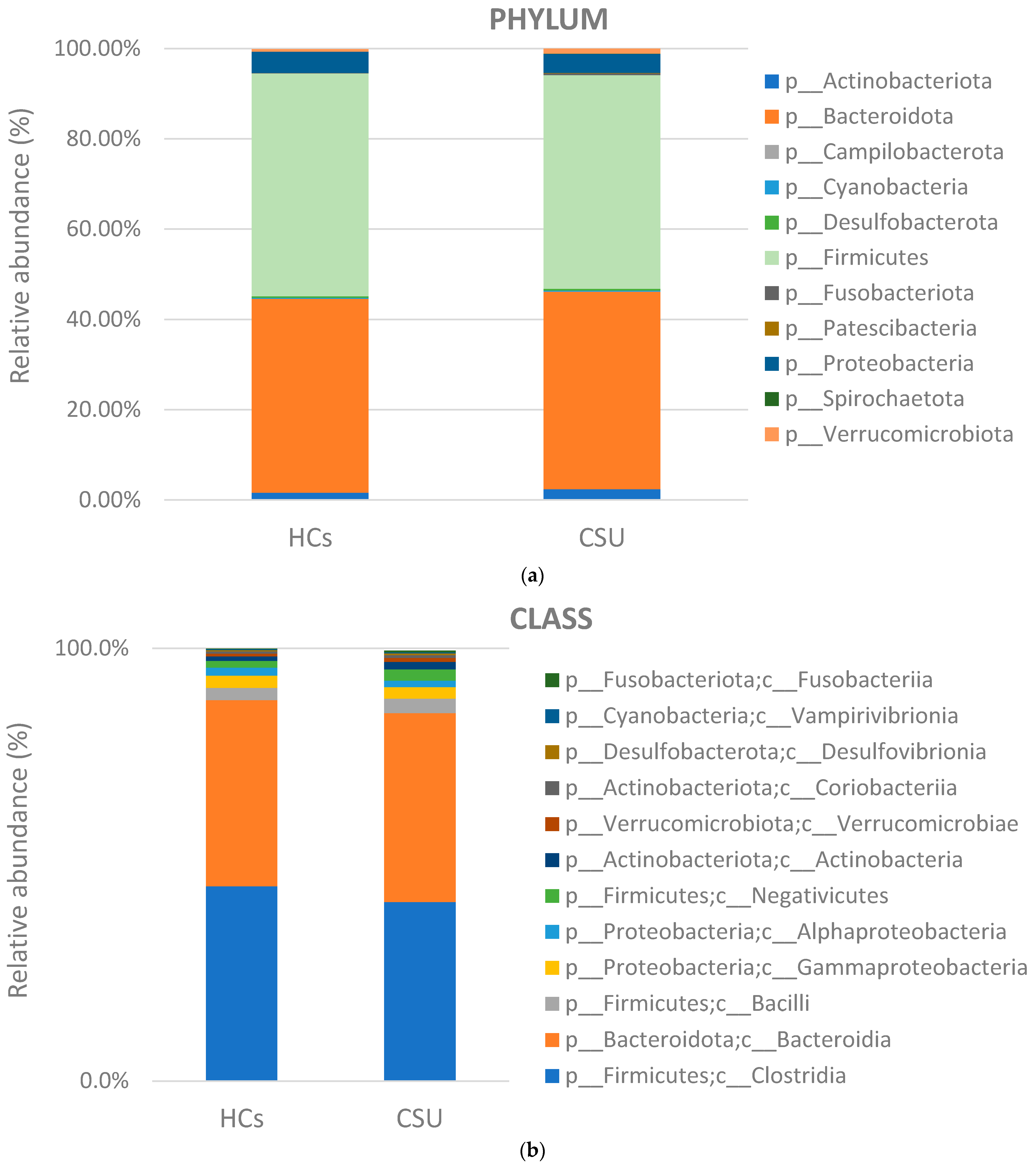
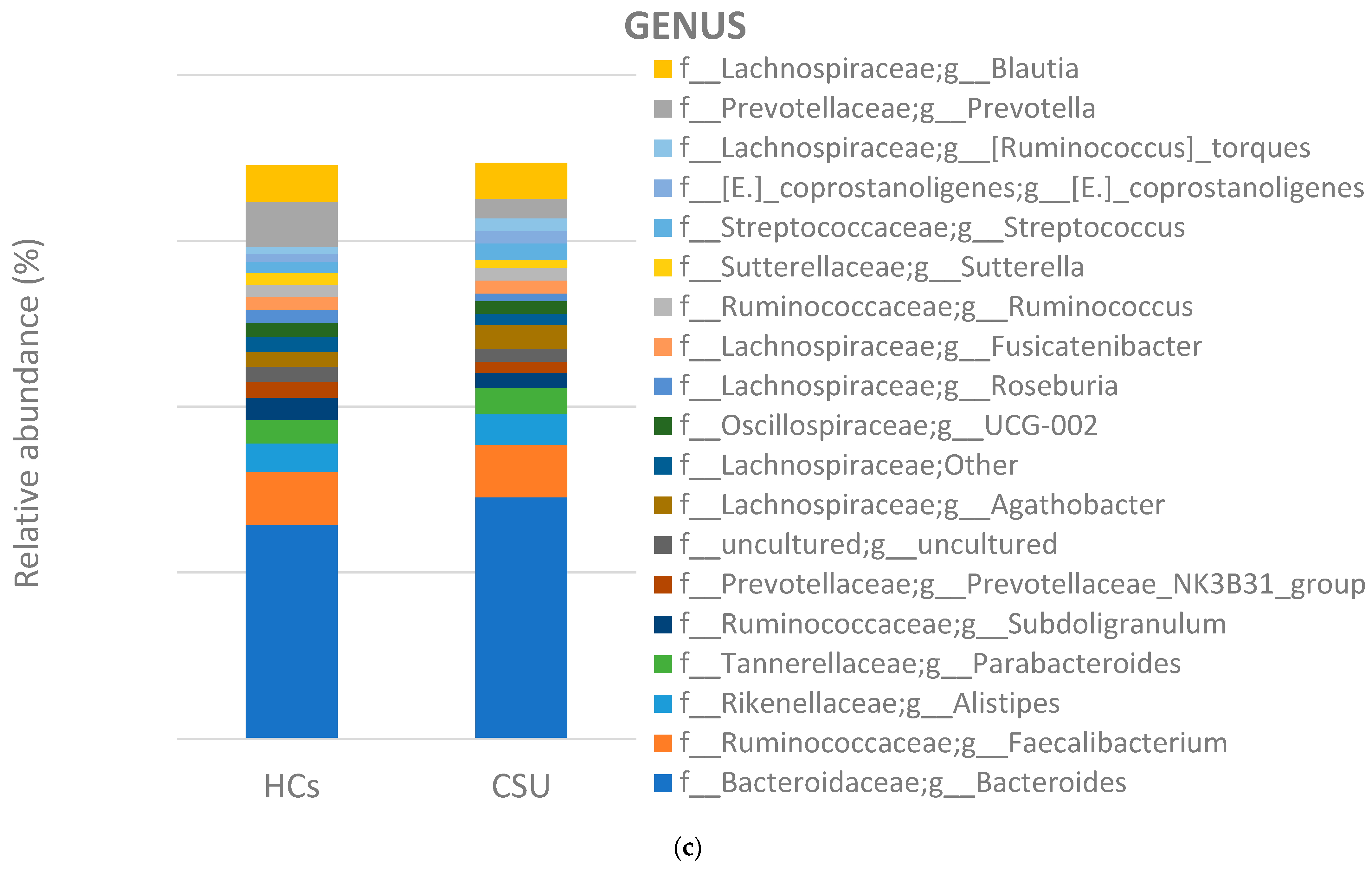
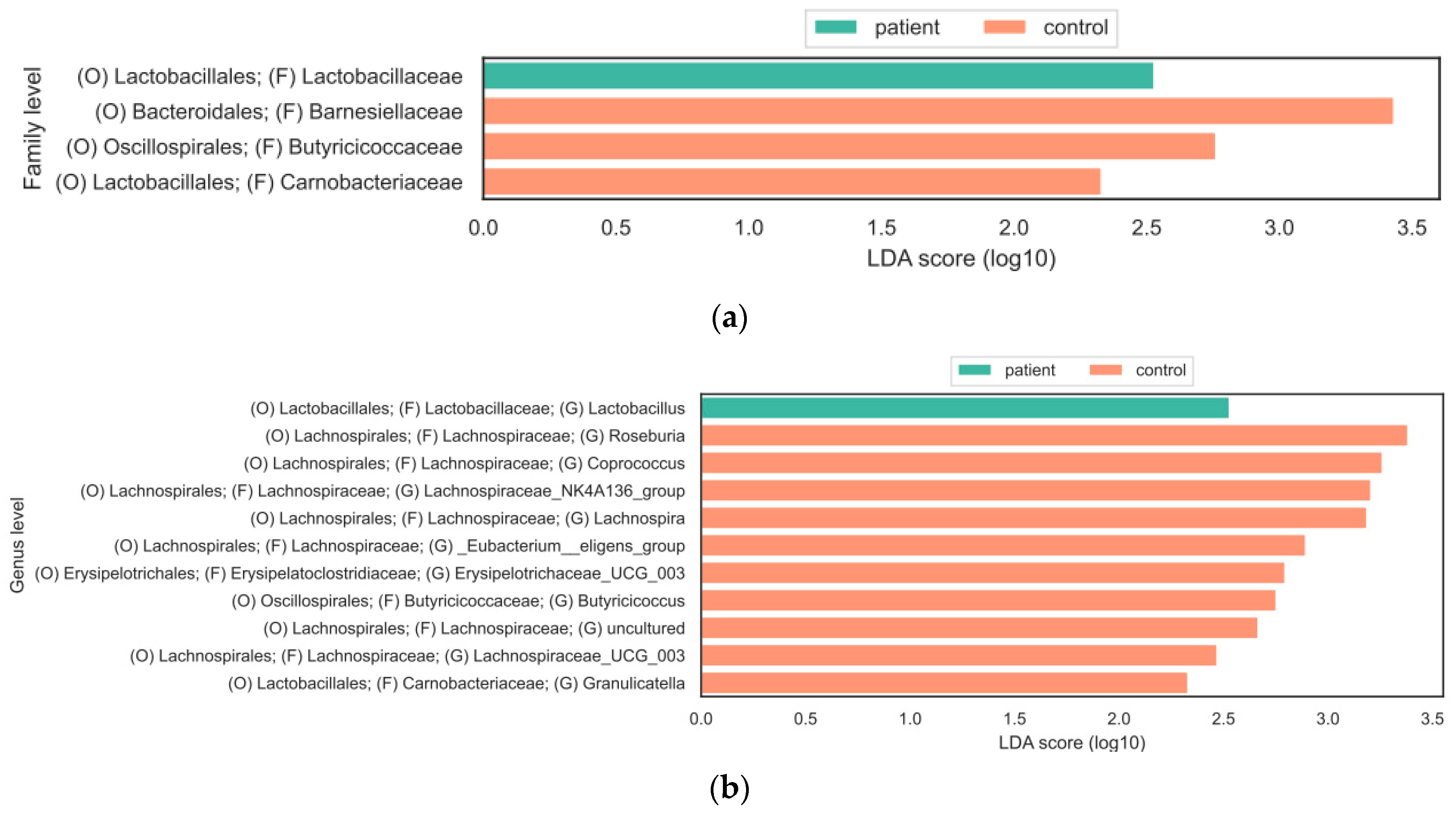
3.2. Composition of Gut Microbiota

After quality control, a total of 3,633,362 sequences were obtained from the fecal samples of 45 subjects, resulting in an average of 80,741 sequences per sample. We examined the bacterial communities and relative abundance in the two groups at different taxonomic levels. At the phylum level, Firmicutes, Bacteroidetes, Proteobacteria, and Actinobacteria were found to be the dominant phyla in both CSU patients and healthy individuals. The most abundant taxes at different levels are shown in Figure 1a–c. The relative abundance of Firmicutes was increased in the HC group, while Bacteroidetes and Actinobacteria were more abundant in the CSU group. At the class level, the relative abundance of Clostridia was decreased in the CSU group. At the genus level, the relative abundances of Bacteroides, Streptococcus, Agathobacter, and Bifidobacterium were increased in the CSU group, while Roseburia, Faecalibacterium, Ruminococcus, Lachnospira, Prevotella, Blautia, Coprococcus and Subdoligranulum were decreased in the CSU group, compared to HCs. To evaluate the alterations in the gut microbial diversities between the CSU patients and HCs, we examined the α-diversity using the Chao 1, Evenness, Faith’s phylogenetic diversity (PD), Observed OTUs, Shannon and Simpson indices. Only the Evenness index showed a statistically significant difference (p < 0.05) between the two groups (Figure 2a), while the other metrics did not show a difference. The beta diversity, measured by the Jaccard distance between the CSU patients and the HCs, revealed the significant clustering (p < 0.05) of the CSU and HC groups. This finding supports the conclusion that there were notable differences in the gut microbiota composition between the two groups, as illustrated in Figure 2b.
To explore the statistically significant differences in the relative abundance of gut bacteria between the two groups, LEfSe was performed. A bar plot was utilized to present the log10 LDA scores. At the family level, the results of the LEfSe analysis revealed that Lactobacillaceae were significantly more abundant in the gut microbiota of CSU patients compared to HCs. On the other hand, Barnesiellaceae, Butyricicoccaceae, and Carnobacteriaceae were more abundant in the control group compared to the patient group (Figure 3a). At the genus level, LEfSe identified a statistically significant increased abundance of bacteria from the Lachnospiraceae family in the HC group, including Lachnospira, Roseburia, Ruminococcus, Coporcoccus, and the Eubacterium eligens group (Figure 3b). On the other hand, the genus Lactobacillus was significantly increased in the CSU group.

4. Discussion

The gut microbiome has been the subject of intense research in recent years, and there is growing evidence that its composition and diversity are associated with various health and pathologic conditions, including CSU. In this study, we aimed to investigate the composition and diversity of the gut microbiome in CSU patients in comparison with HCs, using Illumina-based 16S rRNA gene sequencing. Our findings revealed significant alterations in the composition and diversity of the gut microbiome in CSU patients, compared to HCs. It is noteworthy that half of the included patients had elevated anti-TPO antibodies and one-third had anti-Tg antibodies, which is in line with the concept of CSU as an immune-mediated disease.
To assess the differences in the diversity of the gut microbiome, we used different indices for alpha and beta diversity. Alpha diversity refers to the diversity of bacterial species within a single individual. Beta diversity, on the other hand, refers to the diversity of bacterial species between individuals. Analysis of beta diversity showed that the microbiota composition differed significantly between the groups, as was shown in the principal co-ordinates analysis (PCoA). This clustering is in line with previously published papers on gut microbiota in CSU [10,12,13]. We observed a significant decrease in the alpha diversity of the gut microbiota in the CSU group, according to the Evenness index, while other indices did not show statistically significant differences. We speculate that this bias may be due to a large range of ages within the group, differences in dietary habits, and demographic data. The CSU and HCs groups were mainly composed of Firmicutes, Bacteroidetes, Actinobacteria, and Proteobacteria, consistent with similar studies. At the phylum level, the relative abundance of Bacteroidetes and Actinobacteriota was slightly increased in the CSU group, while phylum Firmicutes was decreased, which has also been reported in studies by Wang et al. [11] and Liu et al. [10]. At the class level, Clostridia was relatively more abundant in the HC group, while the relative abundance of Bacteroides was slightly more increased in the CSU group. Genera of the Lachnospiraceae family, as well as Subdoligranulum, from the Ruminococcaceae family, were relatively decreased in the CSU group. Of note, we found decreased Prevotella spp. in the CSU group compared to healthy individuals, which is in concordance with a study by Lu et al. [9]. Emerging studies have linked the increased abundance of Prevotella with metabolic changes in the gut microbiota, resulting in chronic inflammation [36,37]. In contrast, Hilty et al. reported a reduced abundance of Prevotella in the lung microbiota of asthma patients and patients with chronic obstructive pulmonary disease [38]. Furthermore, a study by Chen et al. investigated the relationship between the composition of the gut microbiota and its ability to utilize dietary fiber. The results showed that the Prevotella-dominated group had a higher fiber-utilizing capacity compared to the Bacteroides-dominated group. The authors observed that a high-fiber diet increased the abundance of Prevotella and the expression of fiber-utilizing genes [39]. Therefore, the role of Prevotella in disease development requires further investigation.
Furthermore, we used LefSe analysis to identify specific bacterial taxa that were significantly more abundant in either the CSU group or the healthy controls. We found significant changes in the bacterial composition at the family and genus levels. Interestingly we found statistically significant increased levels of Lactobacillaceae at the family level and Lactobacillus at the genus level in the CSU group. On the other hand, Coprococcus, Lachnospira, Roseburia, Lachnospiraceae NK4A136, Lachnospiraceae_UCG_003, Eubacterium eligens, Erysipelotrichaceae_UCG_003, and Granulicatella were significantly more abundant in the healthy controls. An increased abundance of Lactobacillaales in CSU patients has also been reported in a study by Lu et al. [9], while Wang et al. reported an increased genus of Lactobacillus in the CSU group [12]. The potential role of Lactobacillus in the prevention and treatment of allergic diseases has been observed [40]. However, the results of clinical trials and observational studies examining the efficacy of Lactobacillus administration in reducing the risk of allergic diseases have been inconsistent. Some studies have reported no significant effect of Lactobacillus supplementation on the incidence of eczema, asthma, or cow’s milk allergy [41,42]. It is important to note that the inconsistencies in the findings of these studies may be due to differences in the study design. Despite these mixed findings, the potential beneficial role of Lactobacillus in preventing or treating allergic diseases should not be overlooked. Further research is needed to better understand the mechanisms by which Lactobacillus may exert its beneficial effects on the host.
Coprococcus, Lachnospira, Roseburia, Lachnospiraceae NK4A136, Lachnospiraceae_UCG_003, and Eubacterium eligens are members of the Lachnospiraceae family, which belongs to the clostridial cluster XIVa of the phylum Firmicutes [16,43]. The Lachnospiraceae family comprises strictly anaerobic bacteria, which belong to the core of gut microbiota and are one of the most abundant bacteria in the human gut. About 10% of all microbes present in the human gut are members of the Lachnospiraceae family [44]. Lachnospiraceae start to inhabit the intestinal lumen from birth, and their abundance increases during the host’s life [16], so they have been detected in individuals of all age groups, from infants to elderly people [27,45,46].
Due to its high abundance and lifelong association with the host, studies have emphasized the role of the Lachnospiraceae family in maintaining gut homeostasis and the overall health of the host [27]. However, a reduction in Lachnospiraceae within the gut microbiota has been associated with a range of conditions, including allergies, inflammatory bowel disease, and metabolic disorders [16,47].
Along with Lactobacillaceae and Ruminococcaceae, Lachnospiraceae are the main bacteria in charge of producing SCFAs [48,49,50].
SCFAs are a group of fatty acids with fewer than six carbon atoms that are produced by the bacterial fermentation of non-digestible carbohydrates in the gut, such as resistant starch, polysaccharide plant cell walls, soluble oligosaccharides, etc. [21,51,52]. The main SCFAs produced by gut bacteria are acetate, propionate, and butyrate, with butyrate being the most abundant. The main butyrate-producing bacteria in the human gut belong to the phylum Firmicutes, including Faecalibacterium prausnitzii, Subdoligranulum, and Clostridium leptum, which belong to the families Ruminococcaceae, Eubacterium rectale and Roseburia spp. from the family Lachnospireacae [53,54]. Our study reported decreased genus levels of all the above-mentioned bacteria in CSU patients compared to healthy individuals.
Following fermentation, the intestinal lumen contains millimolar concentrations of short-chain fatty acids (SCFAs), which are absorbed by the epithelium through both active and passive transport mechanisms. The SCFAs are subsequently transported to distant organs and tissues via peripheral circulation [55,56]. SCFAs play various roles in host metabolism and the immune system. They have an impact on the metabolism of lipids and glucose. Furthermore, they are important for the integrity and protection of the intestinal barrier, with butyrate being the dominant SCFA in charge of this function. Similar beneficial effects in improving the intestinal barrier were found after the administration of the probiotic Butyrococcus pullicaecorum to patients with Crohn’s disease [57]. Our study also showed a lower abundance of Butyrococcus at the genus level in CSU patients.
SCFAs can mediate their effects via multiple mechanisms, including binding to G-protein coupled receptors (GPCRs). GPCRs are the largest family of receptors with seven-transmembrane domains. These receptors play a crucial role in regulating various important cellular functions, including cell proliferation and survival, metabolism, and neuronal signal transmission [58].
SCFAs bind to GPCRs such as GPR41, GPR41, and GPR109A. These receptors are expressed in various cells, including neutrophils, leukocytes, skin cells, etc. [59,60]. SCFA-mediated GPCRs initiate signaling pathways that play a vital role in regulating various cellular responses, immune functions, and inflammatory processes; they, therefore, have an important role in regulating the inflammatory response of the host [61]. Furthermore, the anti-inflammatory effects of SCFAs, especially butyrate, have been attributed to their ability to inhibit histone deacetylase (HDAC) activity. This leads to a decrease in the production of pro-inflammatory mediators such as TNF-α, IL-6, and IL-12 while increasing the production of anti-inflammatory mediators such as IL-10. Additionally, butyrate has been found to promote FOXP3 expression in naïve CD4+ T-cells and facilitate their differentiation into Tregs by inhibiting HDACs. Similarly, acetate, propionate, and butyrate have been shown to modulate the immune response of dendritic cells, macrophages, and Treg cells by inhibiting HDACs [62,63]. Treg cells can suppress pro-inflammatory cytokines mediated by various T-cells, including Th2 cells, and can induce the secretion of anti-inflammatory mediators, thereby reducing inflammation [14]. Reduced Treg cells have been observed in patients with CSU [64,65,66]. GPCRs are also expressed in mast cells, the key cells in the development of urticaria. SCFAs can bind to receptors on mast cells, resulting in the inhibition of mast cell activation and inflammatory response [67,68]. Moreover, studies have shown that not only receptors such as GPR41, GPR43, or peroxisome proliferator-activated receptors (PPARs) are responsible for mast cell inhibition, but that HDAC also independently inhibits their maturation and degranulation [69]. Liu et al. suggest that decreased levels of Subdoligranulum and Ruminococcus bromii can promote mast cell activation and degranulation, leading to the development of hives and itching in CSU patients [10]. Recent studies have shown that CSU patients have lower levels of fecal isobutyrate and serum butyrate compared to healthy individuals [11,12]. The Roseburia/Eubacterium group and Lachnospiraceae NK4A136 group produce a high amount of butyrate, which is involved in controlling gut inflammatory processes and immune system maturation [70]. Our study reported a reduction in all the above-mentioned bacterial groups in CSU patients, which is consistent with emerging studies suggesting that a reduction in the number of SCFA-producing bacteria may contribute to the development of CSU. In addition to SCFAs, several other mechanisms underlying the impact of Lachnospiraceae bacteria on the gut microbiota have been proposed. Certain Lachnospiraceae species have been found to utilize mucin, a glycoprotein from the protective mucus layer of the gut. By breaking down mucin, Lachnospiraceae can promote the turnover of the mucus layer, which is important for protecting the gut from pathogens [16]. Lachnospiraceae can engage in cross-feeding interactions with other members of the gut microbiota by consuming the byproducts of other bacteria and producing metabolites that benefit gut stability and functionality. They also can promote Treg cells and downregulate pro-inflammatory cytokines and Toll-like receptor 4 (TLR4) [16,45]. It is important to note that the specific effects of Lachnospiraceae bacteria on the gut microbiota may vary depending on the species and strains within this family. Additionally, individual variations in the gut microbial composition and host factors can influence the impact of Lachnospiraceae on gut health. The gut microbiota is influenced by various factors such as infections, antibiotics, age, lifestyle, diet, and environmental exposures [20]. Moreover, changes in the composition of the gut microbiota have been associated with many diseases and cancers [71]. Our findings suggest that alterations in the gut microbiota may contribute to the pathogenesis of CSU. Furthermore, we found a lower abundance of Lachnospiraceae, a core family of the gut microbiota known for producing SCFAs, in CSU subjects. This study has several limitations: a small sample size which may not be large enough for strong conclusions; the study was conducted in a single center, which may limit the generalizability of the findings; and while the study excluded participants with certain medical conditions and medication use, it did not report information regarding the dietary habits of the participants, which can have a significant impact on the gut microbiome. Considering the limitations encountered in our study, we plan to expand our sample size and classify participants into groups based on age and dietary habits. By undertaking these measures, our objective is to obtain a more comprehensive representation of the diverse microbial profiles, thereby augmenting the validity of our findings.
The gut microbiome and its interaction with the host are critical for maintaining the health and homeostasis of the organism. SCFAs have been shown to play an important role in the regulation of metabolism and the integrity of the intestinal barrier, as well as in the modulation of the immune response. Moreover, SCFAs have shown promising results in reducing inflammation in inflammatory skin diseases. Thus, the reduced abundance of Lachnospiraceae, SCFA-producing bacteria, may contribute to the development of CSU. Herein, we speculate that the modulation of SCFAs and SCFA-producing organisms could be a potential additional option in the treatment of CSU.
These findings provide important insights into the potential role of gut microbiota in the pathogenesis of CSU and highlight the need for continued research in this area.

Author Contributions

Conceptualization was performed by D.Ć. and L.L.M. Methodology was developed by P.O., I.L. and A.T.A. Validation was carried out by D.Ć., I.L. and P.O. Formal analysis and an investigation were conducted by P.O. and I.L. Resources and data curation were provided by D.Ć. and L.L.M.; D.Ć. and P.O. were responsible for writing the original draft. L.L.M., M.B., M.Š., D.V., M.Z. and A.M. contributed to reviewing and editing the manuscript. Visualization was performed by N.G. and A.T.A.; A.T.A. supervised the entire process. All authors have read and agreed to the published version of the manuscript.

Funding

This study did not receive any external funding.

Institutional Review Board Statement

This study was conducted following the guidelines of the Declaration of Helsinki and approved by the Research Ethics Committee of Sestre milosrdnice University Hospital Centre (Approval No 003-06/20-03/008, 2 April 2020).

Informed Consent Statement

Informed consent was obtained from all study participants.

Data Availability Statement

The corresponding author can be contacted for access to the data presented in this study as they are not publicly available due to the inclusion of the personal information of the study participants.

Acknowledgments

The authors appreciate the support of the Department of Dermatology and Venereology, Sestre milosrdnice University Hospital Centre, Zagreb, the Division of Molecular Medicine, Ruđer Bošković Institute, Zagreb, and the Croatian Veterinary Institute for providing access and performing the experiments. Special thanks go to Slave Trajanoski from the Medical University of Graz for performing the data analysis.

Conflicts of Interest

All the authors declare no conflict of interest.

References

  1. Zuberbier, T.; Abdul Latiff, A.H.; Abuzakouk, M.; Aquilina, S.; Asero, R.; Baker, D.; Ballmer-Weber, B.; Bangert, C.; Ben-Shoshan, M.; Bernstein, J.A.; et al. The international EAACI/GA2LEN/EuroGuiDerm/APAAACI guideline for the definition, classification, diagnosis, and management of urticaria. Allergy 2022, 77, 734–766. [Google Scholar] [CrossRef] [PubMed]
  2. Gonçalo, M.; Gimenéz-Arnau, A.; Al-Ahmad, M.; Ben-Shoshan, M.; Bernstein, J.; Ensina, L.; Fomina, D.; Galvàn, C.A.; Godse, K.; Grattan, C.; et al. The global burden of chronic urticaria for the patient and society. Br. J. Dermatol. 2021, 184, 226–236. [Google Scholar] [CrossRef] [PubMed]
  3. Maurer, M.; Weller, K.; Bindslev-Jensen, C.; Giménez-Arnau, A.; Bousquet, P.J.; Bousquet, J.; Canonica, G.W.; Church, M.K.; Godse, K.V.; Grattan, C.E.H.; et al. Unmet clinical needs in chronic spontaneous urticaria. A GA2LEN task force report1. Allergy 2010, 66, 317–330. [Google Scholar] [CrossRef]
  4. Poddighe, D. The prevalence of chronic spontaneous urticaria (CSU) in the pediatric population. J. Am. Acad. Dermatol. 2019, 81, e149. [Google Scholar] [CrossRef] [PubMed]
  5. Maurer, M.; Abuzakouk, M.; Bérard, F.; Canonica, W.; Oude Elberink, H.; Giménez-Arnau, A.; Grattan, C.; Hollis, K.; Knulst, A.; Lacour, J.-P.; et al. The burden of chronic spontaneous urticaria is substantial: Real-world evidence from ASSURE-CSU. Allergy 2017, 72, 2005–2016. [Google Scholar] [CrossRef]
  6. Rabelo-Filardi, R.; de Oliveira, R.D.; Campos, R.A. Parameters Associated with Chronic Spontaneous Urticaria Duration and Severity: A Systematic Review. Int. Arch. Allergy Immunol. 2013, 161, 197–204. [Google Scholar] [CrossRef]
  7. Bracken, S.J.; Abraham, S.; MacLeod, A.S. Autoimmune Theories of Chronic Spontaneous Urticaria. Front. Immunol. 2019, 10, 627. [Google Scholar] [CrossRef]
  8. Chu, C.-Y.; Zuberbier, T. Urticaria and the gut. Curr. Opin. Allergy Clin. Immunol. 2020, 20, 381–385. [Google Scholar]
  9. Lu, T.; Chen, Y.; Guo, Y.; Sun, J.; Shen, W.; Yuan, M.; Zhang, S.; He, P.; Jiao, X. Altered gut microbiota diversity and composition in chronic urticaria. Dis. Markers 2019, 2019, 6417471. [Google Scholar] [CrossRef]
  10. Liu, R.; Peng, C.; Jing, D.; Xiao, Y.; Zhu, W.; Zhao, S.; Zhang, J.; Chen, X.; Li, J. Biomarkers of Gut Microbiota in Chronic Spontaneous Urticaria and Symptomatic Dermographism. Front. Cell. Infect. Microbiol. 2021, 11, 703126. [Google Scholar] [CrossRef]
  11. Wang, D.; Guo, S.; He, H.; Gong, L.; Cui, H. Gut microbiome and serum metabolome analyses identify unsaturated fatty acids and butanoate metabolism induced by gut microbiota in patients with chronic spontaneous urticaria. Front. Cell. Infect. Microbiol. 2020, 10, 24. [Google Scholar] [CrossRef] [PubMed]
  12. Wang, X.; Yi, W.; He, L.; Luo, S.; Wang, J.; Jiang, L.; Long, H.; Zhao, M.; Lu, Q. Abnormalities in Gut Microbiota and Metabolism in Patients with Chronic Spontaneous Urticaria. Front. Immunol. 2021, 12, 691304. [Google Scholar] [CrossRef] [PubMed]
  13. Zhang, X.; Zhang, J.; Chu, Z.; Shi, L.; Geng, S.; Guo, K. Gut Microbiome Alterations and Functional Prediction in Chronic Spontaneous Urticaria Patients. J. Microbiol. Biotechnol. 2021, 31, 747–755. [Google Scholar] [CrossRef] [PubMed]
  14. Krišto, M.; Lugović-Mihić, L.; Muñoz, M.; Rupnik, M.; Mahnic, A.; Ozretić, P.; Jaganjac, M.; Ćesić, D.; Kuna, M. Gut Microbiome Composition in Patients with Chronic Urticaria: A Review of Current Evidence and Data. Life 2023, 13, 152. [Google Scholar] [CrossRef]
  15. Al Bander, Z.; Nitert, M.D.; Mousa, A.; Naderpoor, N. The Gut Microbiota and Inflammation: An Overview. Int. J. Environ. Res. Public Health 2020, 17, 7618. [Google Scholar] [CrossRef] [PubMed]
  16. Vacca, M.; Celano, G.; Calabrese, F.M.; Portincasa, P.; Gobbetti, M.; De Angelis, M. The Controversial Role of Human Gut Lachnospiraceae. Microorganisms 2020, 8, 573. [Google Scholar] [CrossRef] [PubMed]
  17. Gensollen, T.; Iyer, S.S.; Kasper, D.L.; Blumberg, R.S. How Colonization by Microbiota in Early Life Shapes the Immune System. Science 2016, 352, 539–544. [Google Scholar] [CrossRef]
  18. Power, S.E.; O’Toole, P.W.; Stanton, C.; Ross, R.P.; Fitzgerald, G.F. Intestinal Microbiota, Diet and Health. Br. J. Nutr. 2014, 111, 387–402. [Google Scholar] [CrossRef]
  19. Valdes, A.M.; Walter, J.; Segal, E.; Spector, T.D. Role of the Gut Microbiota in Nutrition and Health. BMJ 2018, 361, k2179. [Google Scholar] [CrossRef]
  20. Sommer, F.; Bäckhed, F. The Gut Microbiota—Masters of Host Development and Physiology. Nat. Rev. Microbiol. 2013, 11, 227–238. [Google Scholar] [CrossRef]
  21. Thorburn, A.N.; Macia, L.; Mackay, C.R. Diet, Metabolites, and “Western-Lifestyle” Inflammatory Diseases. Immunity 2014, 40, 833–842. [Google Scholar] [CrossRef] [PubMed]
  22. Vrancken, G.; Gregory, A.C.; Huys, G.R.B.; Faust, K.; Raes, J. Synthetic Ecology of the Human Gut Microbiota. Nat. Rev. Microbiol. 2019, 15, 754–763. [Google Scholar] [CrossRef] [PubMed]
  23. Eisenstein, M. A Slow-Motion Epidemic. Nature 2016, 540, S98–S99. [Google Scholar] [CrossRef]
  24. Xiao, X.; Hu, X.; Yao, J.; Cao, W.; Zou, Z.; Wang, L. The Role of Short-Chain Fatty Acids in Inflammatory Skin Diseases. Front. Microbiol. 2023, 13, 1083432. [Google Scholar] [CrossRef] [PubMed]
  25. Yao, Y.; Cai, X.; Fei, W.; Ye, Y.; Zhao, M.; Zheng, C. The Role of Short-Chain Fatty Acids in Immunity, Inflammation, and Metabolism. Crit. Rev. Food Sci. Nutr. 2022, 62, 1–12. [Google Scholar] [CrossRef]
  26. Davila, A.-M.; Blachier, F.; Gotteland, M.; Andriamihaja, M.; Benetti, P.-H.; Sanz, Y.; Tomé, D. Intestinal Luminal Nitrogen Metabolism: Role of the Gut Microbiota and Consequences for the Host. Pharmacol. Res. 2013, 69, 114–126. [Google Scholar] [CrossRef]
  27. Abdugheni, R.; Wang, W.-Z.; Wang, Y.-J.; Du, M.-X.; Liu, F.-L.; Zhou, N.; Jiang, C.-Y.; Wang, C.-Y.; Wu, L.; Ma, J.; et al. Metabolite profiling of human-originated Lachnospiraceae at the strain level. iMeta 2022, 1, 100019. [Google Scholar] [CrossRef]
  28. Lu, J.; Zhang, P.; Hu, R.; Qi, S.; Zhao, Y.; Miao, Y.; Han, Y.; Zhou, L.; Yang, Q. Gut Microbiota Characterization in Chinese Patients with Alopecia Areata. J. Dermatol. Sci. 2021, 102, 109–115. [Google Scholar] [CrossRef]
  29. Lam, S.Y.; Radjabzadeh, D.; Eppinga, H.; Nossent, Y.R.A.; van der Zee, H.H.; Kraaij, R.; Konstantinov, S.R.; Fuhler, G.M.; Prens, E.P.; Thio, H.B.; et al. A Microbiome Study to Explore the Gut-Skin Axis in Hidradenitis Suppurativa. J. Dermatol. Sci. 2021, 101, 218–220. [Google Scholar] [CrossRef]
  30. Kazantseva, J.; Malv, E.; Kaleda, A.; Kallastu, A.; Meikas, A. Optimisation of Sample Storage and DNA Extraction for Human Gut Microbiota Studies. BMC Microbiol. 2021, 21, 158. [Google Scholar] [CrossRef]
  31. Milaković, M.; Križanović, S.; Petrić, I.; Šimatović, A.; González-Plaza, J.J.; Gužvinec, M.; Andrašević, A.T.; Pole, L.; Fuka, M.M.; Udiković-Kolić, N. Characterization of Macrolide Resistance in Bacteria Isolated from Macrolide-Polluted and Unpolluted River Sediments and Clinical Sources in Croatia. Sci. Total Environ. 2020, 749, 142357. [Google Scholar] [CrossRef] [PubMed]
  32. Panek, M.; Čipčić Paljetak, H.; Barešić, A.; Perić, M.; Matijašić, M.; Lojkić, I.; Vranešić Bender, D.; Krznarić, Ž.; Verbanac, D. Methodology Challenges in Studying Human Gut Microbiota—Effects of Collection, Storage, DNA Extraction and Next Generation Sequencing Technologies. Sci. Rep. 2018, 8, 5143. [Google Scholar] [CrossRef] [PubMed]
  33. Klindworth, A.; Pruesse, E.; Schweer, T.; Peplies, J.; Quast, C.; Horn, M.; Glöckner, F.O. Evaluation of General 16S Ribosomal RNA Gene PCR Primers for Classical and Next-Generation Sequencing-Based Diversity Studies. Nucleic Acids Res. 2013, 41, e1. [Google Scholar] [CrossRef]
  34. Caporaso, J.G.; Kuczynski, J.; Stombaugh, J.; Bittinger, K.; Bushman, F.D.; Costello, E.K.; Fierer, N.; Peña, A.G.; Goodrich, J.K.; Gordon, J.I.; et al. QIIME Allows Analysis of High-Throughput Community Sequencing Data. Nat. Methods 2010, 7, 335–336. [Google Scholar] [CrossRef]
  35. Thomas, F.; Hehemann, J.H.; Rebuffet, E.; Czjzek, M.; Michel, G. Environmental and Gut Bacteroidetes: The Food Connection. Front. Microbiol. 2011, 2, 93. [Google Scholar] [CrossRef] [PubMed]
  36. Iljazovic, A.; Roy, U.; Gálvez, E.J.C.; Lesker, T.R.; Zhao, B.; Gronow, A.; Amend, L.; Will, S.E.; Hofmann, J.D.; Pils, M.C.; et al. Perturbation of the gut microbiome by Prevotella spp. enhances host susceptibility to mucosal inflammation. Mucosal Immunol. 2021, 14, 113–124. [Google Scholar] [CrossRef] [PubMed]
  37. Larsen, J.M. The immune response to Prevotella bacteria in chronic inflammatory disease. Immunology 2017, 151, 363–374. [Google Scholar] [CrossRef]
  38. Hilty, M.; Burke, C.; Pedro, H.; Cardenas, P.; Bush, A.; Bossley, C.; Davies, J.; Ervine, A.; Poulter, L.; Pachter, L.; et al. Disordered microbial communities in asthmatic airways. PLoS ONE 2010, 5, e8578. [Google Scholar] [CrossRef]
  39. Chen, T.; Long, W.; Zhang, C.; Liu, S.; Zhao, L.; Hamaker, B.R. Fiber-Utilizing Capacity Varies in Prevotella- versus Bacteroides-Dominated Gut Microbiota. Sci. Rep. 2017, 7, 2594. [Google Scholar] [CrossRef]
  40. Wickens, K.; Barthow, C.; Mitchell, E.A.; Kang, J.; Van Zyl, N.; Purdie, G.; Stanley, T.; Fitzharris, P.; Murphy, R.; Crane, J. Effects of Lactobacillus Rhamnosus HN001 in Early Life on the Cumulative Prevalence of Allergic Disease to 11 Years. Pediatr. Allergy Immunol. 2018, 29, 808–814. [Google Scholar]
  41. Cabana, M.D.; McKean, M.; Caughey, A.B.; Fong, L.; Lynch, S.; Wong, A.; Leong, R.; Boushey, H.A.; Hilton, J.F. Early Probiotic Supplementation for Eczema and Asthma Prevention: A Randomized Controlled Trial. Pediatrics 2017, 140, e20163000. [Google Scholar] [CrossRef]
  42. Hol, J.; Van Leer, E.H.; Elink Schuurman, B.E.; De Ruiter, L.F.; Samsom, J.N.; Hop, W.; Neijens, H.J.; de Jongste, J.C.; Nieuwenhuis, E.E.S.; Cow’s Milk Allergy Modified by Elimination and Lactobacilli Study Group. The Acquisition of Tolerance Toward Cow’s Milk Through Probiotic Supplementation: A Randomized, Controlled Trial. J. Allergy Clin. Immunol. 2008, 121, 1448–1454. [Google Scholar] [CrossRef] [PubMed]
  43. Rainey, F.A.; Family, V. Lachnospiraceae fam. nov. In Bergey’s Manual of Systematic Bacteriology, 3rd ed.; De Vos, P., Garrity, G.M., Jones, D., Krieg, N.R., Ludwig, W., Rainey, F.A., Schleifer, K.H., Whitman, W.B., Eds.; Springer: Berlin/Heidelberg, Germany, 2009; pp. 921–968. [Google Scholar]
  44. Almeida, A.; Mitchell, A.L.; Boland, M.; Forster, S.C.; Gloor, G.B.; Tarkowska, A.; Lawley, T.D.; Finn, R.D. A New Genomic Blueprint of the Human Gut Microbiota. Nature 2019, 568, 499–504. [Google Scholar] [CrossRef] [PubMed]
  45. Oliphant, K.; Ali, M.; D’Souza, M.; Hughes, P.D.; Sulakhe, D.; Wang, A.Z.; Xie, B.; Yeasin, R.; Msall, M.E.; Andrews, B.; et al. Bacteroidota and Lachnospiraceae Integration into the Gut Microbiome at Key Time Points in Early Life Are Linked to Infant Neurodevelopment. Gut Microbes 2021, 13, 1997560. [Google Scholar] [CrossRef] [PubMed]
  46. Cox, N.J.; Bowyer, R.C.E.; Ni Lochlainn, M.; Wells, P.M.; Roberts, H.C.; Steves, C.J. The Composition of the Gut Microbiome Differs Among Community Dwelling Older People with Good and Poor Appetite. J. Cachexia Sarcopenia Muscle 2021, 12, 368–377. [Google Scholar] [CrossRef]
  47. Suchodolski, J.S. Gastrointestinal Microbiota. In Canine and Feline Gastroenterology; Steiner, J.M., Ed.; Elsevier: St. Louis, MO, USA, 2013; pp. 32–41. [Google Scholar] [CrossRef]
  48. Biddle, A.; Stewart, L.; Blanchard, J.; Leschine, S. Untangling the genetic basis of fibrolytic specialization by Lachnospiraceae and Ruminococcaceae in diverse gut communities. Diversity 2013, 5, 627–640. [Google Scholar] [CrossRef]
  49. Devillard, E.; McIntosh, F.M.; Duncan, S.H.; Wallace, R.J. Metabolism of linoleic acid by human gut bacteria: Different routes for biosynthesis of conjugated linoleic acid. J. Bacteriol. 2007, 189, 2566–2570. [Google Scholar] [CrossRef]
  50. Wong, J.; Piceno, Y.M.; DeSantis, T.Z.; Pahl, M.; Andersen, G.L.; Vaziri, N.D. Expansion of urease-and uricase-containing, indole-and p-cresol-forming and contraction of short-chain fatty acid-producing intestinal microbiota in ESRD. Am. J. Nephrol. 2014, 39, 230–237. [Google Scholar] [CrossRef]
  51. Tan, J.; McKenzie, C.; Potamitis, M.; Thorburn, A.N.; Mackay, C.R.; Macia, L. The role of short-chain fatty acids in health and disease. Adv. Immunol. 2014, 121, 91–119. [Google Scholar]
  52. Rauf, A.; Khalil, A.A.; Rahman, U.; Khalid, A.; Naz, S.; Shariati, M.A.; Rebezov, M.; Urtecho, E.Z.; Ricardo Diego Galhardo de Albuquerque, R.D.D.; Anwar, S.; et al. Recent advances in the therapeutic application of short-chain fatty acids (SCFAs): An updated review. Crit. Rev. Food Sci. Nutr. 2022, 62, 6034–6054. [Google Scholar] [CrossRef]
  53. Louis, P.; Flint, H.J. Diversity, Metabolism and Microbial Ecology of Butyrate-Producing Bacteria from the Human Large Intestine. FEMS Microbiol. Lett. 2009, 294, 1–8. [Google Scholar] [CrossRef] [PubMed]
  54. Louis, P.; Flint, H.J. Formation of Propionate and Butyrate by the Human Colonic Microbiota. Environ. Microbiol. 2017, 19, 29–41. [Google Scholar] [CrossRef] [PubMed]
  55. Canfora, E.E.; Jocken, J.W.; Blaak, E.E. Short-chain fatty acids in control of body weight and insulin sensitivity. Nat. Rev. Endocrinol. 2015, 11, 577–591. [Google Scholar] [CrossRef] [PubMed]
  56. Hee, B.V.D.; Wells, J.M. Microbial regulation of host physiology by short-chain fatty acids. Trends Microbiol. 2021, 29, 700–712. [Google Scholar] [PubMed]
  57. Eeckhaut, V.; Machiels, K.; Perrier, C.; Romero, C.; Maes, S.; Flahou, B.; Steppe, M.; Haesebrouck, F.; Sas, B.; Ducatelle, R.; et al. Butyricicoccus pullicaecorum in Inflammatory Bowel Disease. Gut 2013, 62, 1745–1752. [Google Scholar] [CrossRef]
  58. Wootten, D.; Christopoulos, A.; Marti-Solano, M.; Babu, M.M.; Sexton, P.M. Mechanisms of Signalling and Biased Agonism in G Protein-Coupled Receptors. Nat. Rev. Mol. Cell Biol. 2018, 19, 638–653. [Google Scholar] [CrossRef] [PubMed]
  59. Krejner, A.; Bruhs, A.; Mrowietz, U.; Wehkamp, U.; Schwarz, T.; Schwarz, A. Decreased Expression of G-protein-coupled Receptors GPR43 and GPR109a in Psoriatic Skin Can be Restored by Topical Application of Sodium Butyrate. Arch. Dermatol. Res. 2018, 310, 751–758. [Google Scholar] [CrossRef]
  60. Schlatterer, K.; Peschel, A.; Kretschmer, D. Short-Chain Fatty Acid and FFAR2 Activation—A New Option for Treating Infections? Front. Cell. Infect. Microbiol. 2021, 11, 785833. [Google Scholar] [CrossRef]
  61. Marinissen, M.J.; Gutkind, J.S. G-Protein-Coupled Receptors and Signaling Networks: Emerging Paradigms. Trends Pharmacol. Sci. 2001, 22, 368–376. [Google Scholar] [CrossRef]
  62. Martin-Gallausiaux, C.; Béguet-Crespel, F.; Marinelli, L.; Jamet, A.; Ledue, F.; Blottière, H.M.; Lapaque, N. Butyrate produced by gut commensal bacteria activates TGF-beta1 expression through the transcription factor SP1 in human intestinal epithelial cells. Sci. Rep. 2018, 8, 9742. [Google Scholar] [CrossRef]
  63. Thiruvengadam, M.; Subramanian, U.; Venkidasamy, B.; Thirupathi, P.; Samynathan, R.; Shariati, M.A.; Rebezov, M.; Chung, I.M.; Rengasamy, K.R. Emerging Role of Nutritional Short-Chain Fatty Acids (SCFAs) against Cancer via Modulation of Hematopoiesis. Crit. Rev. Food Sci. Nutr. 2021, 63, 827–844. [Google Scholar] [CrossRef] [PubMed]
  64. Ćesić, D.; Lugović-Mihić, L.; Ferček, I.; Grginić, A.G.; Jelić, M.; Bešlić, I.; Tambić Andrašević, A. Salivary Microbiota Is Significantly Less Diverse in Patients with Chronic Spontaneous Urticaria Compared to Healthy Controls: Preliminary Results. Life 2021, 11, 1329. [Google Scholar] [CrossRef] [PubMed]
  65. Arshi, S.; Babaie, D.; Nabavi, M.; Tebianian, M.; Ghalehbaghi, B.; Jalali, F.; Ahmadvand, A.; Gholami, R. Circulating Level of CD4+ CD25+ FOXP3+ T Cells in Patients with Chronic Urticaria. Int. J. Dermatol. 2014, 53, 561–566. [Google Scholar] [CrossRef] [PubMed]
  66. Chen, W.-C.; Chiang, B.-L.; Liu, H.E.; Leu, S.-J.; Lee, Y.-L. Defective functions of circulating CD4+CD25+ and CD4+CD25− T cells in patients with chronic ordinary urticaria. J. Dermatol. Sci. 2008, 51, 121–130. [Google Scholar] [CrossRef] [PubMed]
  67. Sugiyama, H.; Nonaka, T.; Kishimoto, T.; Komoriya, K.; Tsuji, K.; Nakahata, T. Peroxisome Proliferator-Activated Receptors Are Expressed in Mouse Bone Marrow-Derived Mast Cells. Jpn. J. Pharmacol. 2000, 82, 259–262. [Google Scholar] [CrossRef] [PubMed]
  68. Tachibana, M.; Wada, K.; Katayama, K.; Kamisaki, Y.; Maeyama, K.; Kadowaki, T.; Blumberg, R.S.; Nakajima, A. Activation of Peroxisome Proliferator-Activated Receptor Gamma Suppresses Mast Cell Maturation Involved in Allergic Diseases. Allergy 2008, 63, 1136–1147. [Google Scholar] [CrossRef] [PubMed]
  69. Folkerts, J.; Redegeld, F.; Folkerts, G.; Blokhuis, B.; van den Berg, M.P.M.; de Bruijn, M.J.W.; van IJcken, W.F.; Junt, T.; Tam, S.Y.; Galli, S.J.; et al. Butyrate Inhibits Human Mast Cell Activation via Epigenetic Regulation of FcεRI-Mediated Signaling. Allergy 2020, 75, 1966–1978. [Google Scholar] [CrossRef]
  70. Kasahara, K.; Krautkramer, K.A.; Org, E.; Romano, K.A.; Kerby, R.L.; Vivas, E.I.; Mehrabian, M.; Denu, J.M.; Bäckhed, F.; Lusis, A.J.; et al. Interactions between Roseburia intestinalis and Diet Modulate Atherogenesis in a Murine Model. Nat. Microbiol. 2018, 3, 1461. [Google Scholar] [CrossRef]
  71. Fan, Y.; Pedersen, O. Gut microbiota in human metabolic health and disease. Nat. Rev. Microbiol. 2021, 19, 55–71. [Google Scholar] [CrossRef]
Figure 1. (a) The composition of the gut microbiota in CSU patients and HCs. Relative abundance at the phylum level. (b) Relative abundance at the class level. The most abundant taxa at the class level in both groups were Clostridia and Bacteroidia. Clostridia taxa were decreased in the CSU group. (c) Relative abundance at the genus level. The relative abundance of Bacteroides, Blautia, and Bifidobacterium was increased in the CSU group, while Roseburia, Ruminococcus, Faecalibarium, Lachnospira, Prevotella, Coprococcus and Subdoligranulum were decreased in the CSU group compared to the HCs.
Life 13 01280 g001aLife 13 01280 g001b
Figure 2. (a) Alpha diversity, measured by Evenness index. (b) Beta diversity, measured by Jaccard distance.
Life 13 01280 g002
Figure 3. Linear discriminant analysis effect size (LEfSe) identified bacterial taxa that were significantly more abundant in the CSU group and HC group at the (a) family level and (b) genus level.
Life 13 01280 g003
Table 1. Characteristics of the CSU patients and healthy controls.
VariableCSU Patients (N = 22)Healthy Controls (N = 23)p-Value
Median age, years (range)42 (20–73)40 (19–74)0.928
Sex, n (%)
Male6 (27.3%)7 (30.4%)0.817
Female16 (72.7%)16 (69.6%)
Table 2. Clinical parameters of CSU patients.
VariableCSU Patients (N = 22)
Duration of symptoms
6 weeks–5 months, n (%)15 (68%)
6 months–12 months, n (%)7 (32%)
Appearance of urticaria
everyday, n (%)13 (59%)
2–4 times a week, n (%)8 (36%)
once a week, n (%)1 (5%)
Taking non-sedating antihistamines n (%)20 (91%)
1 tablet daily, n (%)5 (23%)
2 tablets daily, n (%)8 (36%)
3 tablets daily, n (%)3 (14%)
4 tablets daily, n (%)4 (18%)
Table 3. Laboratory parameters of CSU patients.
VariableCSU Patients (N = 22)
Associated angioedema, n (%)7 (32%)
Associated atopic disorders, n (%)7 (32%)
Elevated Anti-Tg, n (%)6 (27%)
Elevated TPO-Ab, n (%)11 (50%)
Vitamin D deficiency, n (%)15(68%)
Elevated IgE, n (%)9 (41%)
Abbreviation: Anti-Tg—anti-thyroglobulin; TgTPO-Ab—thyroid peroxidase antibodies.
Disclaimer/Publisher’s Note: The statements, opinions and data contained in all publications are solely those of the individual author(s) and contributor(s) and not of MDPI and/or the editor(s). MDPI and/or the editor(s) disclaim responsibility for any injury to people or property resulting from any ideas, methods, instructions or products referred to in the content.

