Article

Natural Perylenequinone Compounds as Potent Inhibitors of Schistosoma mansoni Glutathione S-Transferase

by
Benson Otarigho
1,* and
Mofolusho O. Falade
2,*
1
Department of Molecular Microbiology and Immunology, Oregon Health and Science University, Portland, OR 97239, USA
2
Department of Biology, Transylvania University, Lexington, KY 40508, USA
*
Authors to whom correspondence should be addressed.
Life 2023, 13(10), 1957; https://doi.org/10.3390/life13101957
Submission received: 25 August 2023 / Revised: 19 September 2023 / Accepted: 21 September 2023 / Published: 25 September 2023

Abstract

:
The existing treatment strategy for Schistosomiasis centers on praziquantel, a single drug, but its effectiveness is limited due to resistance and lack of preventive benefits. Thus, there is an urgent need for novel antischistosomal agents. Schistosoma glutathione S-transferase (GST) is an essential parasite enzyme, with a high potential for targeted drug discovery. In this study, we conducted a screening of compounds possessing antihelminth properties, focusing on their interaction with the Schistosoma mansoni glutathione S-transferase (SmGST) protein. We demonstrated the unique nature of SmGST in comparison to human GST. Evolutionary analysis indicated its close relationship with other parasitic worms, setting it apart from free-living worms such as C. elegans. Through an assessment of binding pockets and subsequent protein–ligand docking, we identified Scutiaquinone A and Scutiaquinone B, both naturally derived Perylenequinones, as robust binders to SmGST. These compounds have exhibited effectiveness against similar parasites and offer promising potential as antischistosomal agents.

1. Introduction

Schistosomiasis is a debilitating tropical disease caused by Schistosoma parasites, posing a significant burden on global health, particularly in regions with poor access to sanitation and clean water [1,2,3,4]. Schistosomiasis affects millions of people in developing countries, particularly in sub-Saharan Africa, Asia, and South America [1,3,4,5]. Schistosoma mansoni is one of the species responsible for causing schistosomiasis in humans. Despite efforts to control and eliminate the disease, schistosomiasis remains a significant global health concern [1,2,3,4,5]. Current treatment options primarily rely on a limited number of drugs, such as praziquantel, which has been effective in reducing morbidity and controlling the spread of the disease. However, the continuous reliance on praziquantel has led to concerns about the potential emergence of drug-resistant strains of Schistosoma, posing a serious threat to the efficacy of this drug and the overall management of the disease [6,7,8,9,10,11]. On the other hand, developing a vaccine against Schistosoma parasites has been a challenging endeavor due to the complex life cycle of these parasites and the way they interact with the human immune system. Therefore, there is currently no vaccine available to prevent schistosomiasis. Considering the scarcity of verified drug targets for schistosomes, compounds with distinct mechanisms of action from praziquantel represent a valuable asset in the battle against these parasites and offer new therapeutic strategies to combat schistosomiasis effectively [12,13,14]. One promising avenue of research involves targeting a key enzyme present in the parasite, Schistosoma mansoni Glutathione S-Transferase (SmGST) [15]. SmGST is crucial for the parasite’s survival as it helps the worm neutralize and eliminate harmful molecules, including reactive oxygen species and toxic xenobiotics, which can be detrimental to its cellular functions. By targeting this enzyme with specific inhibitors, it is possible to disrupt the detoxification process, leading to an accumulation of toxic molecules within the parasite, impairing its redox homeostasis, and ultimately causing its death [16,17].
In further response to the challenges associated with the utilization of praziquantel, scientists have redirected their focus towards other synthetic agents and, notably, natural compounds sourced from diverse origins like plants, fungi, and marine organisms [18,19,20,21]. These natural compounds have long been recognized for their diverse biological activities and have been extensively studied for their potential medicinal applications [19,22]. However, the development of natural compounds as antischistosomal agents is not without challenges [23]. One significant obstacle is the limited availability of some of these compounds, as they might be found in only specific plant species or organisms [19,22]. Additionally, the low bioavailability of certain natural compounds can hinder their effective delivery to the target sites in the parasite. Besides formulation strategies, structural optimization and molecular docking of these compounds against targeted proteins are necessary to enhance the pharmacological properties and bioavailability of the compounds [24,25]. Molecular docking of natural compounds offers a compelling and innovative approach in the treatment of diseases such as Schistosomiasis [26,27]. Hence, docking that results in strong affinity binding could target Schistosoma mansoni-specific proteins, thereby disrupting detoxification processes, impairing redox homeostasis, and weakening the parasite’s survival mechanisms [28]. Moreover, the multifaceted nature of these compounds, encompassing antioxidant, anti-inflammatory, and immunomodulatory properties, presents a comprehensive therapeutic strategy against schistosomiasis [29].
A compelling approach in drug development involves targeting enzymes specific to parasites, distinguishing them from their human homologs. In the current study, we also assessed the similarity between SmGST and its human homologs. The evolutionary relationship of SmGST with other parasitic worms and free-living worms like Caenorhabditis elegans was also explored. Protein–ligand docking studies demonstrated that Scutiaquinone A and Scutiaquinone B, natural compounds categorized as Perylenequinones, exhibited strong binding affinities to SmGST. These compounds have the potential to serve as potent antischistosomal agents, targeting the parasite’s GST enzyme. This specificity offers promising advantages for targeted treatments against schistosomiasis, addressing current therapeutic challenges, and advancing the fight against this disease.

2. Methods

2.1. Exploring and Extracting Antihelminth Compounds and SmGST Protein Sequences from Databases

One of the objectives of this study was to explore and identify compounds that exhibit anthelmintic activities. To achieve this goal, we conducted an extensive search across multiple databases renowned for their comprehensive collection of chemical and biological information. The databases used for this purpose included SANCDB [30,31], which specializes in natural compounds; PubChem [32,33], a vast repository of chemical structures and biological activities; and DrugBank [34], a comprehensive database of drugs and their associated properties. We obtained the relevant data from these databases, and the compounds of interest were extracted in the Protein Data Bank (PDB) format, which allowed for us to analyze and visualize the atomic arrangements of the compounds, which can provide valuable insights into their interactions with biological targets. To facilitate further analysis and make the findings accessible, we meticulously documented common names and accessions or unique identifiers of the results in a tabular form (Table 1). To conduct molecular docking studies involving the extracted compounds and SmGST, the protein’s sequence and structure (SmGST) were acquired from the Protein Database, accessible at https://www.rcsb.org/, (accessed on 10 June 2023) using the accession number 1U3I.

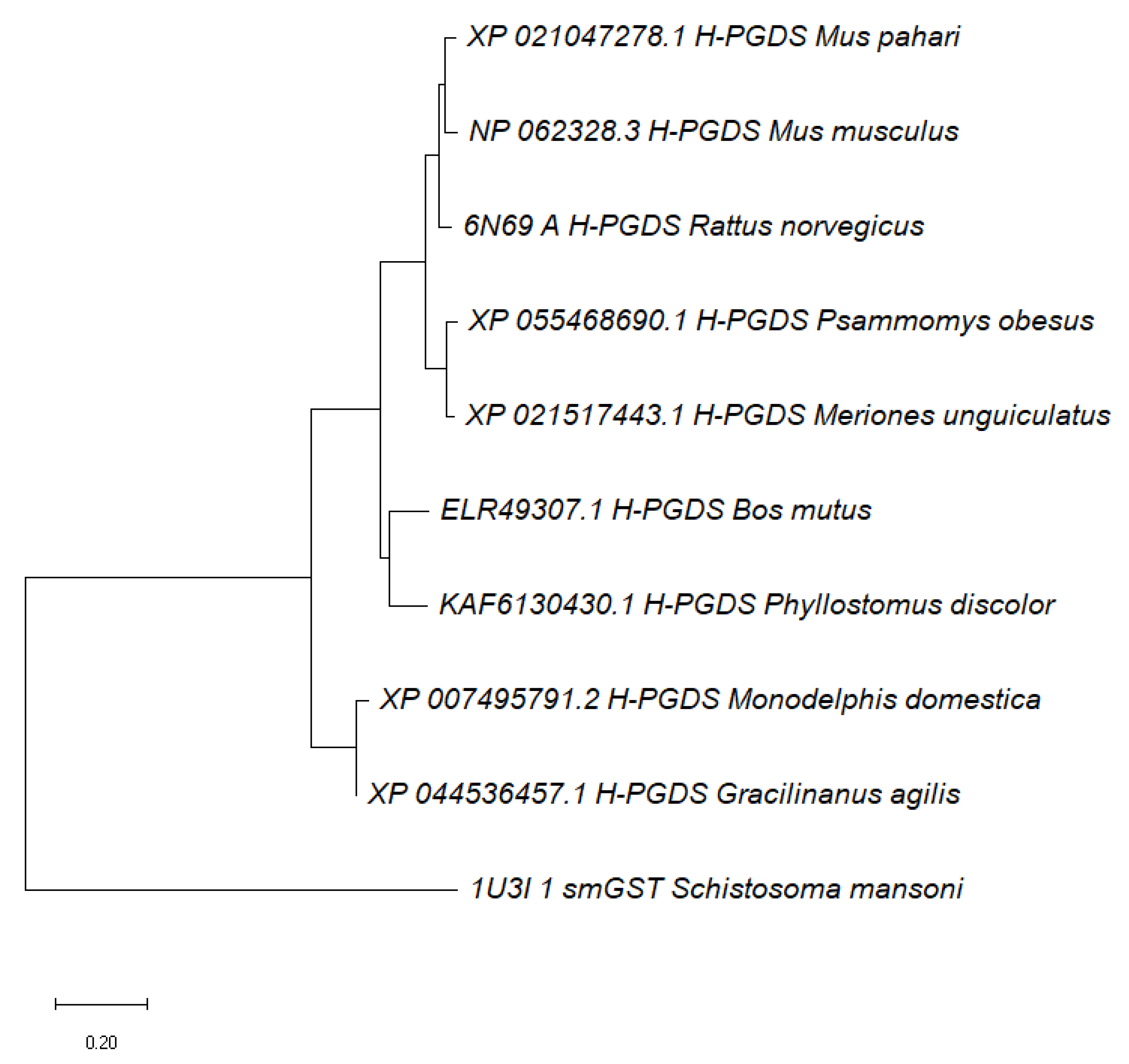
2.2. Alignment of Schistosoma mansoni GST Protein Sequences to Mammalian Homologs

Given that Schistosoma parasites are known to infect a wide range of hosts, we aimed to investigate the similarity of the SmGST protein sequences with GST sequences from mammals and other vertebrates. To achieve this, we used the BLASTP program, which is available on the NCBI BLAST (Basic Local Alignment Search Tool) platform [35]. We used the SmGST protein sequences as the query and searched against a dataset of GST protein sequences from mammals and other vertebrates. The BLASTP analysis allowed for us to identify similar sequences in the mammalian and vertebrate datasets that shared significant homology with the SmGST sequences. These similar sequences might indicate potential conserved functions or regions among different organisms. After obtaining similar protein sequences from the BLASTP search, the next step was to perform a multiple sequence alignment, which was subjected to a phylogenetic tree analysis using MEGA [36,37].

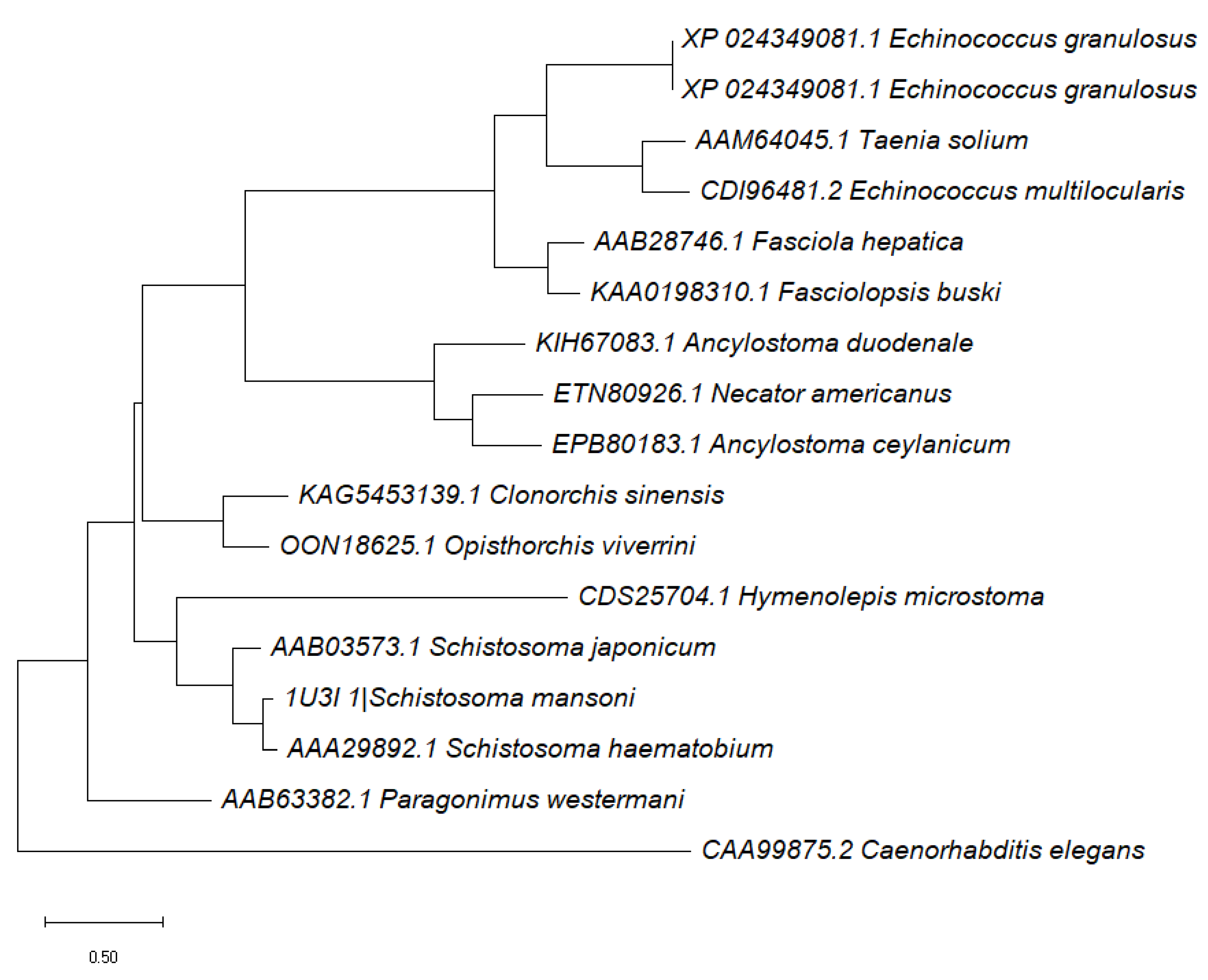
2.3. Schistosoma mansoni GST Relationship to Other Parasitic Worms as Well as Caenorhabditis elegans

We also aimed to analyze the similarity between SmGST and its homologs in other helminths. To accomplish this, we conducted a BLAST search of the SmGST protein against a range of worm parasites, including liver flukes (Clonorchis sinensis, Fasciola hepatica, and Opisthorchis viverrine), which cause liver diseases; lung flukes (Paragonimus westermani), which cause lung infections; intestinal flukes (Fasciolopsis buski), which cause intestinal disorders; tapeworms (Taenia solium, Echinococcus granulosus, and Echinococcus multilocularis), which cause cystic diseases; hookworms (Necator americanus, Ancylostoma duodenale, and Ancylostoma ceylanicum), which cause anemia and malnutrition; and a free-living worm (Caenorhabditis elegans), which is a model organism for studying biology. We obtained the GST protein sequences of these worms from the NCBI database, which is a public repository of biological data. We then used ClustalW version 2 (https://www.ebi.ac.uk/Tools/msa/clustalw2/) (accessed on 13 June 2023), which is a software tool that aligns multiple sequences based on their similarities and differences, to perform a multiple alignment of the GST sequences. This allowed us to identify regions of conservation and variation among the GST sequences, as well as possible functional or structural domains. We then used the MEGA software version 11.0.10, which is a software tool that infers evolutionary relationships among sequences based on various methods and models, to construct a phylogenetic tree of the GST sequences. This allowed for us to delineate possible common ancestries and divergences among the worms.

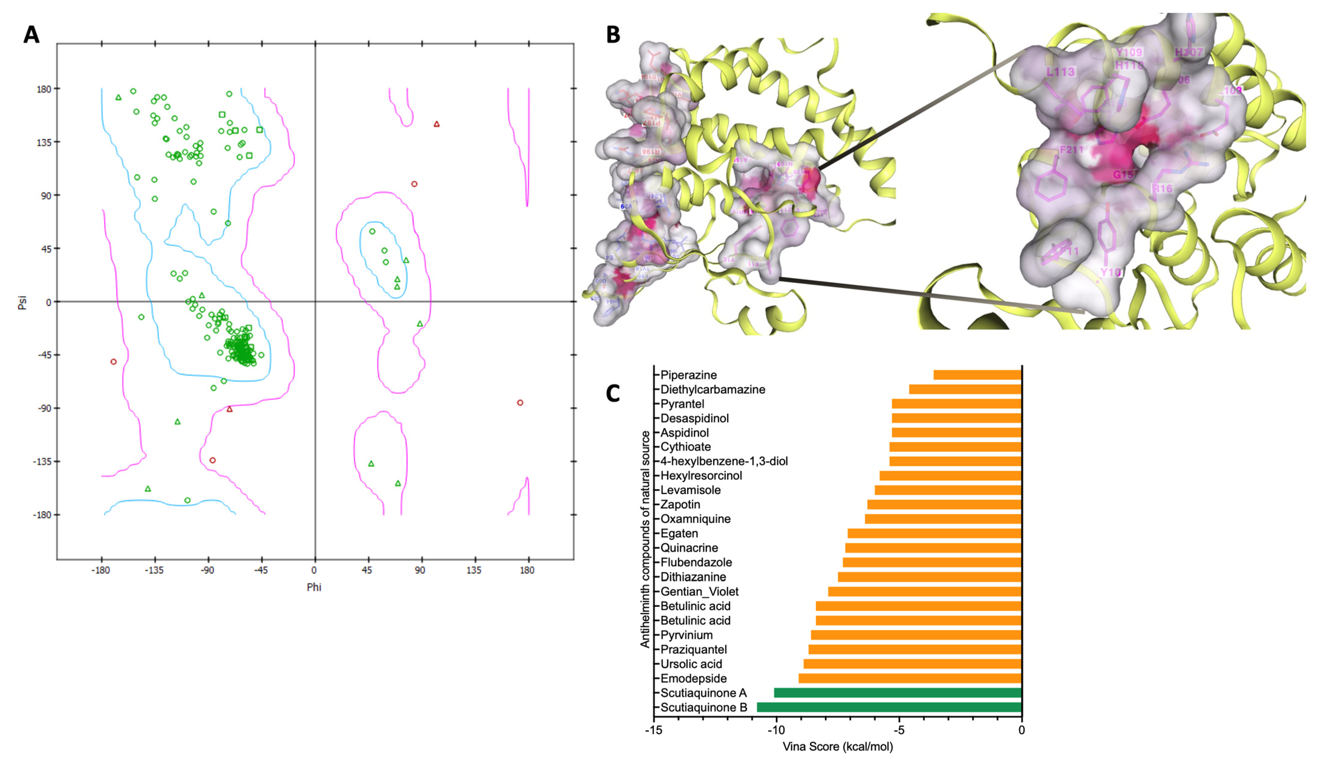
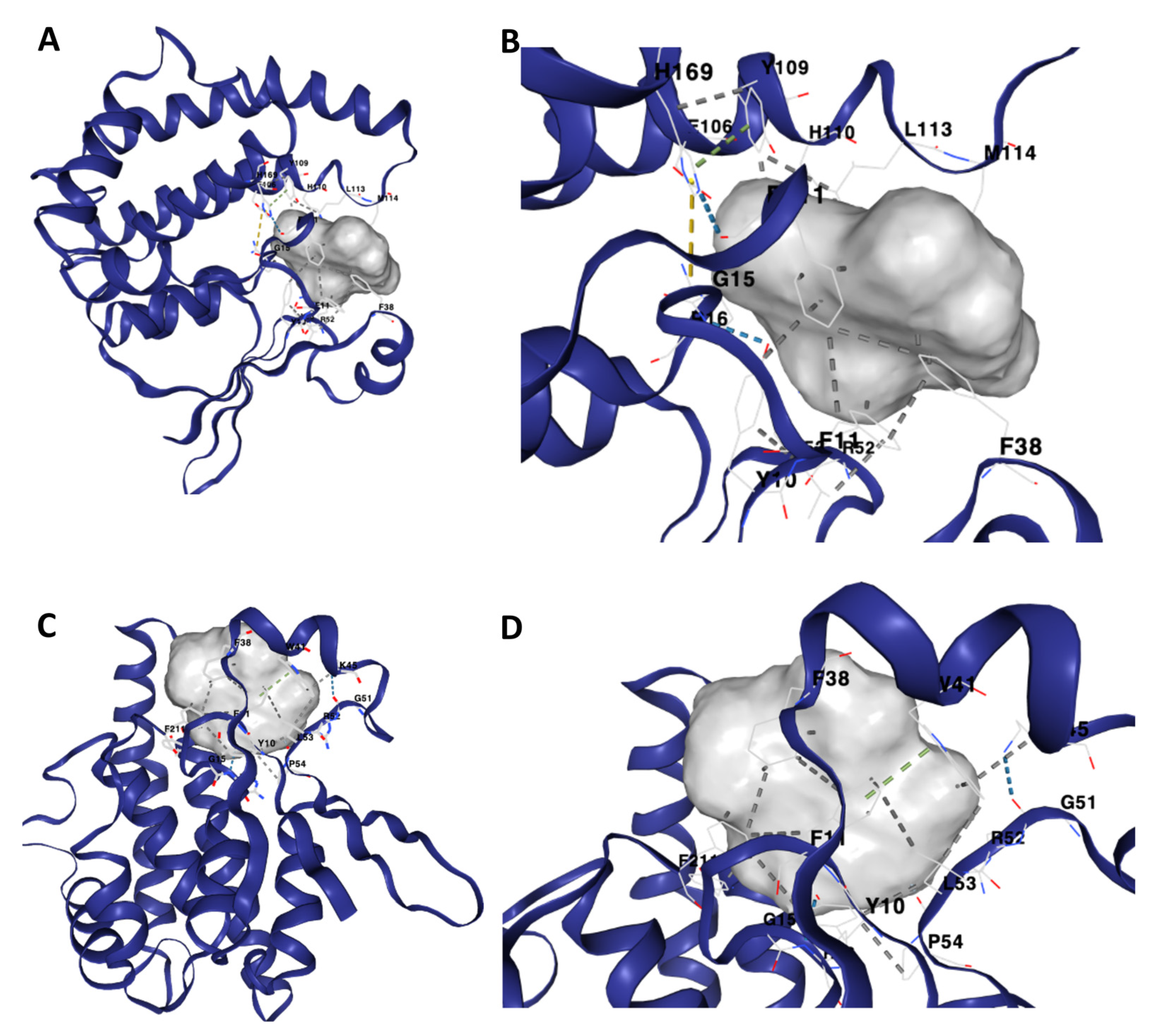
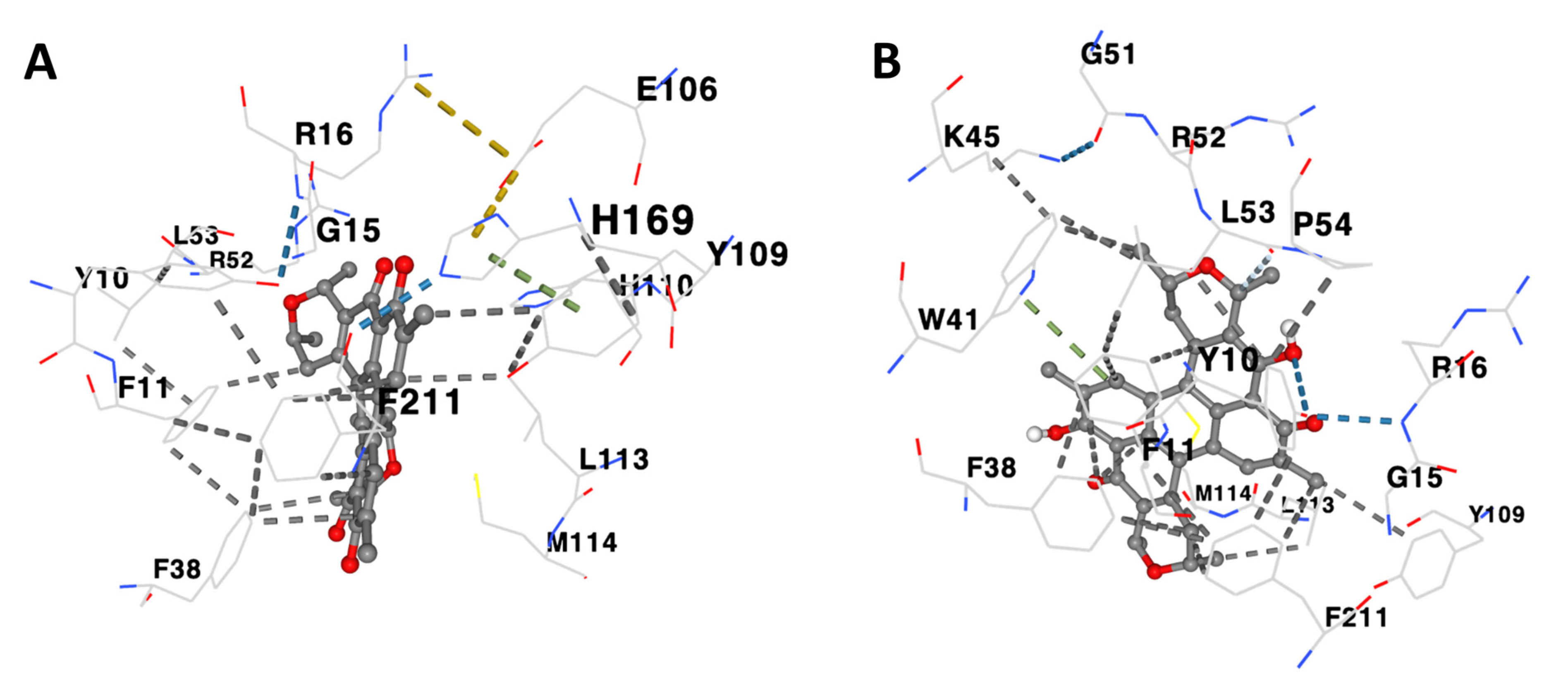
2.4. Protein–Ligand Docking of Compounds against Schistosoma mansoni GST

The compounds obtained from the previous step were subjected to molecular docking against the SmGST proteins using the CB-Dock2 server, accessible at https://cadd.labshare.cn/cb-dock2/php/blinddock.php (accessed on 20 June 2023) [38]. CB-Dock2 is an advanced protein–ligand blind docking server that incorporates improvements in binding site identification and binding pose prediction. The CB-Dock2 server utilizes a combination of techniques to enhance the accuracy of the docking process. It integrates cavity detection, docking algorithms, and homologous template fitting to improve the reliability of the docking predictions. By employing these techniques, CB-Dock2 aims to provide more accurate predictions of protein–ligand interactions. The blind docking approach implemented by CB-Dock2 allows for the exploration of potential binding sites on the target proteins without prior knowledge of their exact locations. This enables the identification of potential binding pockets that may not be readily apparent based solely on the protein’s structure.
We submitted the PDB file of the SmGST protein and the MOL2 file of the compounds to CB-Dock2. During the docking process, the compounds were systematically evaluated for their binding affinity with the target proteins. The potential binding pockets were subsequently validated using the DrugRep tool (http://cao.labshare.cn/drugrep/) (accessed on 29 June 2023). This tool facilitates the virtual screening of drug libraries, encompassing FDA-approved, experimental, and traditional Chinese medicine compounds, against specific selected binding pockets of the target protein. Its capabilities include an automatic molecular 3D structure construction, binding pocket prediction, docking, similarity comparison, and binding affinity screening against hundreds to thousands of compounds in a shorter time, ensuring a comprehensive and automated analysis. Only compounds that exhibited a strong affinity, with Vina scores of −10 kcal/mol and below, were selected for further analysis and were further validated using Swissdock (http://www.swissdock.ch/) and PatchDock (http://bioinfo3d.cs.tau.ac.il/PatchDock/php.php) (accessed on 29 June 2023). Based on the initial data showing a strong binding affinity between binding pocket 3 and the established anthelminthic compounds, we proceeded to utilize the DrugReg tool for the screening of both FDA-approved drugs and naturally sourced compounds from the Traditional Chinese Medicine Library [39]. The Vina scoring function is a widely used empirical scoring function that estimates the binding affinity between a protein and ligand-based on their interaction energy and spatial complementarity. Selecting compounds with high binding affinity is crucial as it indicates a stronger potential for specific interactions with the target proteins. These compounds are more likely to exhibit favorable binding and potentially possess greater therapeutic efficacy against the target proteins.

2.5. Analyses of the Binding Affinity of the SmGST Protein to the Compounds

In this study, we utilized computational tools to analyze and visualize the binding affinities of 27 compounds against the SmGST proteins. The Vina scores of these compounds were computed and arranged in an Excel spreadsheet for further analysis. To create visually appealing data visualizations, we employed the Datawrapper tool (https://www.datawrapper.de/) (accessed on 9 August 2023), which is an online platform known for its user-friendly interface and interactive visualization capabilities. By inputting the Vina scores into Datawrapper, we generated a chart to represent the binding affinities of the 27 compounds against the target proteins. This visualization provided an overview of the compound–protein interactions and allowed for easy comparison and interpretation of the data. To further explore and illustrate this finding, we focused on visualizing the Vina scores of these 27 compounds specifically against the SmGST protein. Using Datawrapper, we generated another chart that specifically highlighted the binding affinities of these compounds to SmGST.

2.6. Evaluation of Pharmacokinetic and Solubility Profiles for Scutiaquinone A and B

To assess the pharmacokinetic and solubility characteristics of Scutiaquinone A and B, we employed the SwissADME platform (http://www.swissadme.ch/) (accessed on 10 September 2023) [40]. SwissADME is an online resource specialized for applications in drug discovery and development, providing valuable data and prediction regarding the pharmacokinetic and physicochemical attributes of chemical compounds. This versatile tool scrutinizes essential parameters encompassing absorption, distribution, metabolism, and excretion, all of which significantly impact how compounds behave within the human body. We input the SMILES notation for Scutiaquinone A and B into SwissADME to compute their pharmacokinetics and solubility. The results, including lipophilicity, water solubility, drug-likeness, and other pharmacokinetic properties, were extracted and presented in a tabular form.

3. Results

A search across various databases was carried out to mine compounds with anthelmintic properties. Upon analyzing the data, we observed that anthelmintic compounds can be grouped into three main categories: natural compounds constituted 29.63% of the total, synthetic compounds made up 62.96%, and the remaining 7.41% comprised semi-synthetic compounds (Figure 1A). Among the natural compounds, a significant portion was derived from specific natural sources, with notable contributions stemming from Scutia myrtina, Leucosidea sericea, and Termitomyces microcarpus (Figure 1B). These sources likely harbor bioactive compounds known for their anthelmintic properties, rendering them appealing candidates for further exploration and potential drug developments. Additionally, the identified compounds were classified based on their chemical structures. This classification encompassed Quinones, Sesquiterpenes, and Triterpenes (Figure 1C). These categories represent distinct classes of organic compounds, each characterized by unique molecular structures and potentially novel mechanisms of action against helminth parasites. Conversely, synthetic compounds constituted the largest proportion of the identified anthelmintic compounds. The majority of these synthetic compounds belonged to the Sesquiterpenes category, followed by Phenol and Tetrahydropyrimidine, among others. Overall, this study’s findings shed light on the diversity of anthelmintic compounds originating from different sources, encompassing natural products derived from specific plants and chemically synthesized compounds.
Upon conducting a protein BLAST analysis focused on mammalian proteins associated with SmGST, we identified the protein hematopoietic prostaglandin D synthase isoform from Bos mutus (wild yak) as the closest match. This protein exhibited a sequence similarity of 36.13% to SmGST. This indicates a distinction between SmGST and its mammalian homologs, potentially implying that SmGST could serve as a promising target for drug developments against Schistosoma mansoni. Furthermore, upon scrutinizing the constructed phylogenetic tree, it became evident that SmGST exhibited a marked divergence from its analogous counterparts in humans and other mammalian species (Figure 2). Previous studies indicate that the search for novel potential drug targets has been focused on biochemical and metabolic pathways that show differences between pathogens and their hosts.
The constructed phylogenetic tree illustrates a connection between SmGST and various parasitic worms, including Hymenolepis microstoma and diverse Schistosoma species, emphasizing the shared ancestral lineage between SmGST and other parasites that rely on animals as hosts. In contrast, the evolutionary origin of C. elegans, an organism that lives independently in the soil, deviates from these parasitic worms (Figure 3). The divergence in the phylogenetic placement of these two species reinforces the parasitic lifestyle of Schistosoma mansoni in contrast to the free-living nature of C. elegans.
Table 1 illustrates the binding characteristics of distinct anthelmintic compounds against the GST proteins of Schistosoma mansoni. Prior to commencing the molecular docking experiment, we conducted an initial validation process for the three-dimensional structure of the SmGST protein. To achieve this, we utilized the Ramachandran plot (Figure 4A). We further utilized DrugRep tools to conduct a comprehensive analysis of the binding pocket depicted in Figure 4B. Throughout the docking process, we methodically evaluated the binding affinity of compounds with the target proteins. Subsequently, for a more in-depth analysis emphasizing their binding strengths with SmGST (Figure 4C), we exclusively selected compounds exhibiting robust affinity, characterized by Vina scores below −10 kcal/mol. Interestingly, our investigation revealed that none of the FDA-approved drugs or traditional Chinese medicine compounds displayed a Vina score lower than −10, suggesting a comparatively weaker or moderate binding affinity to the SmGST protein. Consequently, we concentrated our scrutiny on anthelmintic compounds boasting Vina scores less than −10 kcal/mol with the SmGST protein. Notably, the compounds exhibiting the most elevated binding affinities, as depicted in Figure 5, hold a higher potential for precise interactions with the target proteins, thereby potentially leading to enhanced therapeutic effects (Figure 6 and Table 1). Moreover, our findings underscore the robustness of the binding between the SmGST protein and the natural compounds, Scutiaquinone A and B, as evidenced by the depiction of their respective interacting residues in Figure 7. Table 2 displays the pharmacokinetic and solubility profiles of Scutiaquinone A and B. Their drug-like properties were evaluated using Lipinski’s Rule of Five and Veber’s Rules. Both compounds meet these criteria, suggesting they have characteristics commonly found in pharmaceuticals and could be potential drug candidates. The pharmacokinetic data include factors like GI absorption, BBB permeability, P-glycoprotein substrate status, and interactions with various CYP enzymes. Notably, Scutiaquinone B exhibits enhanced GI absorption and is not a P-gp substrate, which could impact its transport in the body. However, both compounds exhibit poor water solubility, confirming their limited solubility and stability in methanol.

4. Discussion

In the current study, we explored various databases to identify anthelmintic compounds, which are drugs that inhibit the growth of parasitic worms. We searched for compounds that have been reported to have anthelmintic activity against Schistosoma or other helminths, or that share structural similarity with known anthelmintic drugs. Additionally, we conducted a molecular docking investigation involving these compounds and the SmGST protein. GSTs are enzymes that catalyze the conjugation of glutathione to xenobiotics, which are foreign substances that enter the body. This process helps to detoxify the xenobiotics and make them more soluble for excretion, making GSTs potential drug targets [41,42]. GSTs are widely distributed in living organisms, but they have different evolutionary origins and functions [43]. In Schistosoma parasites, GSTs play a crucial role in protecting them from oxidative stress and host immune responses [41,42].
Our search for antihelminth compounds and docking studies identified Scutiaquinone A and B as compounds that most likely possess antischistosomal activity. Scutiaquinone A and B are isolated from the roots of the Scutia myrtina plant, which is native to South Africa [44]. These compounds belong to a class of chemicals known as perylenequinones, a group of organic compounds that contain a perylene nucleus, which is a polycyclic aromatic system [45,46]. Interestingly, these compounds have previously demonstrated anthelmintic activities against Haemonchus contortus [44], a common and harmful parasite that infects the stomachs of ruminant animals like sheep and goats. It is a major cause of gastrointestinal parasitic infections in these animals, leading to significant economic losses in livestock industries worldwide [47]. Further, it has been shown that Scutiaquinone A and B possess complex chemical structures and diverse pharmacological properties, making them valuable starting points for drug discovery and development [44,46]. Based on our protein–ligand docking studies, Scutiaquinone A and Scutiaquinone B exhibited strong binding affinities to Schistosoma mansoni GST. Our results seem to corroborate the pharmacological effectiveness of Scutiaquinone A and B as lead compounds for drug discovery and development.
The pharmacokinetics and solubility profiles of Scutiaquinone A and B are presented, with an assessment of their drug-likeness based on Lipinski’s Rule of Five and Veber’s Rules, revealing that both compounds conform to these criteria [48]. This implies that Scutiaquinone A and B possess characteristics commonly observed in pharmaceuticals, suggesting their potential as viable drug candidates. The provided pharmacokinetic data encompass various parameters, including gastrointestinal (GI) absorption, blood–brain barrier (BBB) permeability, P-glycoprotein (P-gp) substrate status, and interactions with various cytochrome P450 enzymes (CYP) [49,50]. Notably, Scutiaquinone B demonstrates enhanced GI absorption, indicative of its propensity for absorption in the gastrointestinal tract.
Numerous studies have concentrated on finding therapeutic targets for enzymes with active sites that are distinct from those of their mammalian hosts and peculiar to helminths [51,52,53]. These studies suggest that parasite-specific metabolic pathways that demonstrate variations between helminths and their hosts can be the focus of the search for novel potential drug targets [51,53]. As a consequence, in our evolutionary analysis, we aimed to evaluate the distinctions between SmGST and its homologs in other parasitic helminths as well as a representative free-living worm. The results of our work demonstrate that SmGST had a common ancestor with other animal-parasitic worms, including Hymenolepis microstoma and several Schistosoma species. The free-living C. elegans, on the other hand, followed a different evolutionary path, showing a divergence from these parasitic worms. This may justify SmGST as a more parasite-specific drug target than a host target.
Previously, several studies have employed computational methods to identify natural compounds and metabolites as potential inhibitors of various drug targets within the Schistosoma mansoni parasite [54,55,56,57,58,59]. In a recent study, a computational approach was employed to conduct a screening analysis involving several natural compounds sourced from medicinal plants indigenous to Saudi Arabia [54]. This screening utilized the docking technique, which serves to predict the optimal orientation and interactions between a ligand in this context, natural compounds, and a Schistosoma mansoni histone deacetylase 8 (SmHDAC8) protein. Notably, the screening process unveiled nine previously undiscovered compounds demonstrating a robust binding affinity for SmHDAC8. In another study, the researchers focused on SmHDAC1, an enzyme belonging to the class I histone deacetylase (HDAC) category [55], which exhibits expression throughout all phases of the parasite’s life cycle and plays a vital role in the regulation of genes. In this screening process, they utilized a set of established class I HDAC inhibitors as molecular ligands and compared their affinities for binding to both SmHDAC1 and HsHDAC1 (human HDAC1), along with their respective concentrations required for inhibition. Their investigation revealed that a particular compound, namely, N,8-dihydroxy-8-(naphthalen-2-yl) octanamide (ZINC13474421), exhibited the highest level of selectivity and specificity for SmHDAC1 in comparison to HsHDAC1. This observation suggests that ZINC13474421 holds promise as a potential candidate for further development as an anti-parasitic agent.
In our current study, we have employed a structure-based docking strategy to discern the anthelmintic potential of a selection of natural and synthetic compounds against various helminth parasites. This investigation entailed the computational screening of a compound library, primarily sourced from South Africa. Our findings have revealed that two distinct perylenequinone compounds, namely, Scutiaquinone A and B, exhibit robust binding affinities to a common binding site on SmGST proteins. The binding interactions between Scutiaquinone A and SmGST primarily encompassed amino acid residues GLU106, HIS110, and HIS169, while Scutiaquinone B’s interactions predominantly involved residues TRP41, LYS45, GLY51, and PRO54. It is noteworthy that the majority of the interacting residues were shared by both compounds. The outcomes of our ongoing investigations strongly suggest the potential suitability of these two Perylenequinone compounds as druggable candidates for combating schistosomiasis.

5. Conclusions

In our study on natural Perylenequinone compounds as potential inhibitors of SmGST, we address the important need for novel antischistosomal agents due to the limitations of the current treatment strategy relying on praziquantel. Our work focused on SmGST, an essential parasite enzyme, and compared it to its human homolog, highlighting its distinctness. Our evolutionary analysis revealed that SmGST shares a closer relationship with other parasitic worms than with free-living worms like C. elegans, emphasizing its potential as a drug target specific to parasites. Utilizing protein–ligand docking and binding pocket assessments, we identified Scutiaquinone A and Scutiaquinone B as robust binders to SmGST. These naturally derived compounds, known for their effectiveness against similar parasites, demonstrate promising potential as antischistosomal agents. The unique characteristics of SmGST make it a compelling target for drug discovery, and compounds like Scutiaquinone A and Scutiaquinone B offer hope for the development of alternative treatments for Schistosomiasis, addressing the urgent need for improved therapeutic options.

Author Contributions

Both B.O. and M.O.F. contributed significantly to the conceptualization, methodology, formal analysis, investigation, data curation, writing of the original draft, and review and editing of the manuscript. All authors have read and agreed to the published version of the manuscript.

Funding

This work was supported by the Bingham Start-up Program of Transylvania University to MOF.

Institutional Review Board Statement

Not applicable.

Informed Consent Statement

Not applicable.

Data Availability Statement

All data underlying the results are included as part of the published article.

Conflicts of Interest

The authors declare no conflict of interest. The funders had no role in the design of the study; in the collection, analyses, or interpretation of data; in the writing of the manuscript; or in the decision to publish the results.

References

  1. Colley, D.G.; Bustinduy, A.L.; Secor, W.E.; King, C.H. Human schistosomiasis. Lancet 2014, 383, 2253–2264. [Google Scholar] [CrossRef] [PubMed]
  2. Lo, N.C.; Addiss, D.G.; Hotez, P.J.; King, C.H.; Stothard, J.R.; Evans, D.S.; Colley, D.G.; Lin, W.; Coulibaly, J.T.; Bustinduy, A.L. A call to strengthen the global strategy against schistosomiasis and soil-transmitted helminthiasis: The time is now. Lancet Infect. Dis. 2017, 17, e64–e69. [Google Scholar] [CrossRef] [PubMed]
  3. Lo, N.C.; Bezerra, F.S.M.; Colley, D.G.; Fleming, F.M.; Homeida, M.; Kabatereine, N.; Kabole, F.M.; King, C.H.; Mafe, M.A.; Midzi, N. Review of 2022 WHO guidelines on the control and elimination of schistosomiasis. Lancet Infect. Dis. 2022, 22, e327–e335. [Google Scholar] [CrossRef] [PubMed]
  4. Wiegand, R.E.; Fleming, F.M.; de Vlas, S.J.; Odiere, M.R.; Kinung’hi, S.; King, C.H.; Evans, D.; French, M.D.; Montgomery, S.P.; Straily, A. Defining elimination as a public health problem for schistosomiasis control programmes: Beyond prevalence of heavy-intensity infections. Lancet Glob. Health 2022, 10, e1355–e1359. [Google Scholar] [CrossRef]
  5. Colley, D.G.; Andros, T.S.; Campbell, C.H. Schistosomiasis is more prevalent than previously thought: What does it mean for public health goals, policies, strategies, guidelines and intervention programs? Infect. Dis. Poverty 2017, 6, 63. [Google Scholar] [CrossRef] [PubMed]
  6. Cioli, D.; Pica-Mattoccia, L.; Basso, A.; Guidi, A. Schistosomiasis control: Praziquantel forever? Mol. Biochem. Parasitol. 2014, 195, 23–29. [Google Scholar] [CrossRef]
  7. Fenwick, A.; Webster, J.P.; Bosque-Oliva, E.; Blair, L.; Fleming, F.; Zhang, Y.; Garba, A.; Stothard, J.; Gabrielli, A.F.; Clements, A. The Schistosomiasis Control Initiative (SCI): Rationale, development and implementation from 2002–2008. Parasitology 2009, 136, 1719–1730. [Google Scholar] [CrossRef]
  8. Stothard, J.R.; Sousa-Figueiredo, J.C.; Betson, M.; Green, H.K.; Seto, E.Y.; Garba, A.; Sacko, M.; Mutapi, F.; Nery, S.V.; Amin, M.A. Closing the praziquantel treatment gap: New steps in epidemiological monitoring and control of schistosomiasis in African infants and preschool-aged children. Parasitology 2011, 138, 1593–1606. [Google Scholar] [CrossRef]
  9. Caffrey, C.R. Chemotherapy of schistosomiasis: Present and future. Curr. Opin. Chem. Biol. 2007, 11, 433–439. [Google Scholar] [CrossRef]
  10. Summers, S.; Bhattacharyya, T.; Allan, F.; Stothard, J.R.; Edielu, A.; Webster, B.L.; Miles, M.A.; Bustinduy, A.L. A review of the genetic determinants of praziquantel resistance in Schistosoma mansoni: Is praziquantel and intestinal schistosomiasis a perfect match? Front. Trop. Dis. 2022, 3, 933097. [Google Scholar] [CrossRef]
  11. Botros, S.; Sayed, H.; Amer, N.; El-Ghannam, M.; Bennett, J.L.; Day, T.A. Current status of sensitivity to praziquantel in a focus of potential drug resistance in Egypt. Int. J. Parasitol. 2005, 35, 787–791. [Google Scholar] [CrossRef] [PubMed]
  12. Zhang, S.-M.; Coultas, K.A. Identification of plumbagin and sanguinarine as effective chemotherapeutic agents for treatment of schistosomiasis. Int. J. Parasitol. Drugs Drug Resist. 2013, 3, 28–34. [Google Scholar] [CrossRef] [PubMed]
  13. Engels, D.; Chitsulo, L.; Montresor, A.; Savioli, L. The global epidemiological situation of schistosomiasis and new approaches to control and research. Acta Trop. 2002, 82, 139–146. [Google Scholar] [CrossRef]
  14. Faust, C.L.; Osakunor, D.N.; Downs, J.A.; Kayuni, S.; Stothard, J.R.; Lamberton, P.H.; Reinhard-Rupp, J.; Rollinson, D. Schistosomiasis control: Leave no age group behind. Trends Parasitol. 2020, 36, 582–591. [Google Scholar] [CrossRef]
  15. Nogueira, R.A.; Lira, M.G.S.; Licá, I.C.L.; Frazão, G.C.C.G.; Dos Santos, V.A.F.; Mendes Filho, A.C.C.; Rodrigues, J.G.M.; Miranda, G.S.; Carvalho, R.C.; Nascimento, F.R.F. Praziquantel: An update on the mechanism of its action against schistosomiasis and new therapeutic perspectives. Mol. Biochem. Parasitol. 2022, 252, 111531. [Google Scholar] [CrossRef] [PubMed]
  16. Kremer, L.; Riveau, G.; Baulard, A.; Capron, A.; Locht, C. Neutralizing antibody responses elicited in mice immunized with recombinant bacillus Calmette-Guérin producing the Schistosoma mansoni glutathione S-transferase. J. Immunol. 1996, 156, 4309–4317. [Google Scholar] [CrossRef] [PubMed]
  17. Chen, W.Q.; Liu, S.S.; Cheng, C.; Cui, J.; Wang, Z.Q.; Zhang, X. Molecular characteristics of glutathione transferase gene family in a neglect medical Spirometra tapeworm. Front. Vet. Sci. 2022, 9, 1035767. [Google Scholar] [CrossRef]
  18. Adegboye, O.; Field, M.A.; Kupz, A.; Pai, S.; Sharma, D.; Smout, M.J.; Wangchuk, P.; Wong, Y.; Loiseau, C. Natural-product-based solutions for tropical infectious diseases. Clin. Microbiol. Rev. 2021, 34, e00348-20. [Google Scholar] [CrossRef]
  19. Wangchuk, P.; Loukas, A. Techniques and technologies for the biodiscovery of novel small molecule drug lead compounds from natural products. In Natural Products and Drug Discovery; Elsevier: Amsterdam, The Netherlands, 2018; pp. 435–465. [Google Scholar]
  20. Neves, B.J.; Andrade, C.H.; Cravo, P.V. Natural products as leads in schistosome drug discovery. Molecules 2015, 20, 1872–1903. [Google Scholar] [CrossRef]
  21. Mtemeli, F.; Ndlovu, J.; Mugumbate, G.; Makwikwi, T.; Shoko, R. Advances in schistosomiasis drug discovery based on natural products. All Life 2022, 15, 608–623. [Google Scholar] [CrossRef]
  22. Wangchuk, P. Therapeutic applications of natural products in herbal medicines, biodiscovery programs, and biomedicine. J. Biol. Act. Prod. Nat. 2018, 8, 1–20. [Google Scholar] [CrossRef]
  23. Cheuka, P.M.; Mayoka, G.; Mutai, P.; Chibale, K. The role of natural products in drug discovery and development against neglected tropical diseases. Molecules 2016, 22, 58. [Google Scholar] [CrossRef] [PubMed]
  24. Racz, L.Z.; Racz, C.P.; Pop, L.-C.; Tomoaia, G.; Mocanu, A.; Barbu, I.; Sárközi, M.; Roman, I.; Avram, A.; Tomoaia-Cotisel, M. Strategies for improving bioavailability, bioactivity, and physical-chemical behavior of curcumin. Molecules 2022, 27, 6854. [Google Scholar] [CrossRef] [PubMed]
  25. Shoaib, A.; Javed, S.; Wahab, S.; Azmi, L.; Tabish, M.; Sultan, M.H.; Abdelsalam, K.; Alqahtani, S.S.; Ahmad, M.F. Cellular, Molecular, Pharmacological, and Nano-Formulation Aspects of Thymoquinone—A Potent Natural Antiviral Agent. Molecules 2023, 28, 5435. [Google Scholar] [CrossRef]
  26. Adekiya, T.A.; Aruleba, R.T.; Klein, A.; Fadaka, A.O. In silico inhibition of SGTP4 as a therapeutic target for the treatment of schistosomiasis. J. Biomol. Struct. Dyn. 2022, 40, 3697–3705. [Google Scholar] [CrossRef] [PubMed]
  27. Ferreira, L.G.; Oliva, G.; Andricopulo, A.D. Target-based molecular modeling strategies for schistosomiasis drug discovery. Future Med. Chem. 2015, 7, 753–764. [Google Scholar] [CrossRef]
  28. Huang, H.-H.; Rigouin, C.; L Williams, D. The redox biology of schistosome parasites and applications for drug development. Curr. Pharm. Des. 2012, 18, 3595–3611. [Google Scholar] [CrossRef]
  29. Anibogwu, R.; Jesus, K.D.; Pradhan, S.; Pashikanti, S.; Mateen, S.; Sharma, K. Extraction, isolation and characterization of bioactive compounds from Artemisia and their biological significance: A review. Molecules 2021, 26, 6995. [Google Scholar] [CrossRef]
  30. Diallo, B.; Glenister, M.; Musyoka, T.M.; Lobb, K.; Tastan Bishop, Ö. SANCDB: An update on South African natural compounds and their readily available analogs. J. Cheminform. 2021, 13, 37. [Google Scholar] [CrossRef]
  31. Hatherley, R.; Brown, D.K.; Musyoka, T.M.; Penkler, D.L.; Faya, N.; Lobb, K.A.; Tastan Bishop, Ö. SANCDB: A South African natural compound database. J. Cheminform. 2015, 7, 29. [Google Scholar] [CrossRef]
  32. Sayers, E.W.; Beck, J.; Bolton, E.E.; Bourexis, D.; Brister, J.R.; Canese, K.; Comeau, D.C.; Funk, K.; Kim, S.; Klimke, W. Database resources of the national center for biotechnology information. Nucleic Acids Res. 2021, 49, D10. [Google Scholar] [CrossRef] [PubMed]
  33. Sayers, E.W.; Bolton, E.E.; Brister, J.R.; Canese, K.; Chan, J.; Comeau, D.C.; Connor, R.; Funk, K.; Kelly, C.; Kim, S. Database resources of the national center for biotechnology information. Nucleic Acids Res. 2022, 50, D20. [Google Scholar] [CrossRef] [PubMed]
  34. Wishart, D.S.; Feunang, Y.D.; Guo, A.C.; Lo, E.J.; Marcu, A.; Grant, J.R.; Sajed, T.; Johnson, D.; Li, C.; Sayeeda, Z. DrugBank 5.0: A major update to the DrugBank database for 2018. Nucleic Acids Res. 2018, 46, D1074–D1082. [Google Scholar] [CrossRef] [PubMed]
  35. Johnson, M.; Zaretskaya, I.; Raytselis, Y.; Merezhuk, Y.; McGinnis, S.; Madden, T.L. NCBI BLAST: A better web interface. Nucleic Acids Res. 2008, 36, W5–W9. [Google Scholar] [CrossRef] [PubMed]
  36. Kumar, S.; Tamura, K.; Nei, M. MEGA: Molecular evolutionary genetics analysis software for microcomputers. Bioinformatics 1994, 10, 189–191. [Google Scholar] [CrossRef]
  37. Tamura, K.; Stecher, G.; Kumar, S. MEGA11: Molecular evolutionary genetics analysis version 11. Mol. Biol. Evol. 2021, 38, 3022–3027. [Google Scholar] [CrossRef]
  38. Liu, Y.; Yang, X.; Gan, J.; Chen, S.; Xiao, Z.-X.; Cao, Y. CB-Dock2: Improved protein–ligand blind docking by integrating cavity detection, docking and homologous template fitting. Nucleic Acids Res. 2022, 50, W159–W164. [Google Scholar] [CrossRef]
  39. Gan, J.-H.; Liu, J.-X.; Liu, Y.; Chen, S.-W.; Dai, W.-T.; Xiao, Z.-X.; Cao, Y. DrugRep: An automatic virtual screening server for drug repurposing. Acta Pharmacol. Sin. 2023, 44, 888–896. [Google Scholar] [CrossRef]
  40. Daina, A.; Michielin, O.; Zoete, V. SwissADME: A free web tool to evaluate pharmacokinetics, drug-likeness and medicinal chemistry friendliness of small molecules. Sci. Rep. 2017, 7, 42717. [Google Scholar] [CrossRef]
  41. Riveau, G.; Poulain-Godefroy, O.; Dupré, L.; Remoué, F.; Mielcarek, N.; Locht, C.; Capron, A. Glutathione S-transferases of 28kDa as major vaccine candidates against schistosomiasis. Mem. Inst. Oswaldo Cruz 1998, 93, 87–94. [Google Scholar] [CrossRef]
  42. Angelucci, F.; Baiocco, P.; Brunori, M.; Gourlay, L.; Morea, V.; Bellelli, A. Insights into the catalytic mechanism of glutathione S-transferase: The lesson from Schistosoma haematobium. Structure 2005, 13, 1241–1246. [Google Scholar] [CrossRef] [PubMed]
  43. Sheehan, D.; Meade, G.; Foley, V.M.; Dowd, C.A. Structure, function and evolution of glutathione transferases: Implications for classification of non-mammalian members of an ancient enzyme superfamily. Biochem. J. 2001, 360, 1–16. [Google Scholar] [CrossRef] [PubMed]
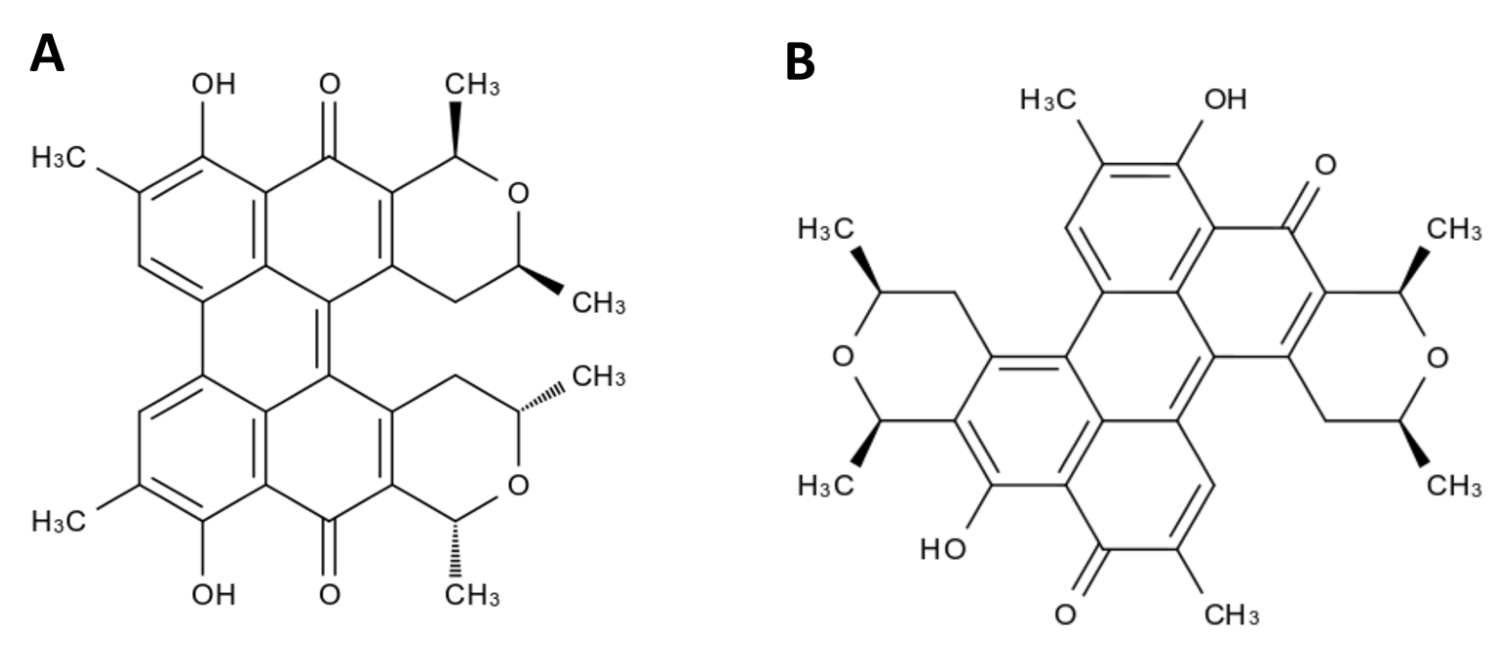
  44. Ayers, S.; Zink, D.L.; Mohn, K.; Powell, J.S.; Brown, C.M.; Murphy, T.; Brand, R.; Pretorius, S.; Stevenson, D.; Thompson, D. Scutiaquinones A and B, Perylenequinones from the Roots of Scutia myrtina with Anthelmintic Activity. J. Nat. Prod. 2007, 70, 425–427. [Google Scholar] [CrossRef]
  45. Mulrooney, C.A.; O’Brien, E.M.; Morgan, B.J.; Kozlowski, M.C. Perylenequinones: Isolation, synthesis, and biological activity. Eur. J. Org. Chem. 2012, 2012, 3887–3904. [Google Scholar] [CrossRef] [PubMed]
  46. Mulrooney, C.A.; Morgan, B.J.; Li, X.; Kozlowski, M.C. Perylenequinone natural products: Enantioselective synthesis of the oxidized pentacyclic core. J. Org. Chem. 2010, 75, 16–29. [Google Scholar] [CrossRef]
  47. Emery, D.L.; Hunt, P.W.; Le Jambre, L.F. Haemonchus contortus: The then and now, and where to from here? Int. J. Parasitol. 2016, 46, 755–769. [Google Scholar] [CrossRef]
  48. Bickerton, G.R.; Paolini, G.V.; Besnard, J.; Muresan, S.; Hopkins, A.L. Quantifying the chemical beauty of drugs. Nat. Chem. 2012, 4, 90–98. [Google Scholar] [CrossRef]
  49. Daina, A.; Zoete, V. A boiled-egg to predict gastrointestinal absorption and brain penetration of small molecules. ChemMedChem 2016, 11, 1117–1121. [Google Scholar] [CrossRef]
  50. Daina, A.; Michielin, O.; Zoete, V. iLOGP: A simple, robust, and efficient description of n-octanol/water partition coefficient for drug design using the GB/SA approach. J. Chem. Inf. Model. 2014, 54, 3284–3301. [Google Scholar] [CrossRef]
  51. Lautens, M.J.; Tan, J.H.; Serrat, X.; Del Borrello, S.; Schertzberg, M.R.; Fraser, A.G. Identification of enzymes that have helminth-specific active sites and are required for Rhodoquinone-dependent metabolism as targets for new anthelmintics. PLoS Neglected Trop. Dis. 2021, 15, e0009991. [Google Scholar] [CrossRef]
  52. Rana, A.K.; Misra-Bhattacharya, S. Current drug targets for helminthic diseases. Parasitol. Res. 2013, 112, 1819–1831. [Google Scholar] [CrossRef] [PubMed]
  53. J Timson, D. Metabolic enzymes of helminth parasites: Potential as drug targets. Curr. Protein Pept. Sci. 2016, 17, 280–295. [Google Scholar] [CrossRef] [PubMed]
  54. Alzain, A.A.; Mukhtar, R.M.; Abdelmoniem, N.; Elbadwi, F.A.; Hussien, A.; Garelnabi, E.A.; Osman, W.; Sherif, A.E.; Khedr, A.I.; Ghazawi, K.F. Computational Insights into Natural Antischistosomal Metabolites as SmHDAC8 Inhibitors: Molecular Docking, ADMET Profiling, and Molecular Dynamics Simulation. Metabolites 2023, 13, 658. [Google Scholar] [CrossRef] [PubMed]
  55. Singh, R.; Pandey, P.N. Molecular docking and molecular dynamics study on SmHDAC1 to identify potential lead compounds against Schistosomiasis. Mol. Biol. Rep. 2015, 42, 689–698. [Google Scholar] [CrossRef]
  56. Akachukwu, I.; O Olubiyi, O.; Kosisochukwu, A.; John, M.C.; Justina, N.N. Structure-Based study of Natural products with anti-Schistosoma activity. Curr. Comput.-Aided Drug Des. 2017, 13, 91–100. [Google Scholar] [CrossRef]
  57. Mafud, A.C.; Ferreira, L.G.; Mascarenhas, Y.P.; Andricopulo, A.D.; de Moraes, J. Discovery of novel antischistosomal agents by molecular modeling approaches. Trends Parasitol. 2016, 32, 874–886. [Google Scholar] [CrossRef]
  58. Menezes, R.P.B.d.; Viana, J.d.O.; Muratov, E.; Scotti, L.; Scotti, M.T. Computer-assisted discovery of alkaloids with schistosomicidal activity. Curr. Issues Mol. Biol. 2022, 44, 383–408. [Google Scholar] [CrossRef]
  59. Rocha, J.A.; Rego, N.C.; Carvalho, B.T.; Silva, F.I.; Sousa, J.A.; Ramos, R.M.; Passos, I.N.; de Moraes, J.; Leite, J.R.; Lima, F.C. Computational quantum chemistry, molecular docking, and ADMET predictions of imidazole alkaloids of Pilocarpus microphyllus with schistosomicidal properties. PLoS ONE 2018, 13, e0198476. [Google Scholar] [CrossRef]
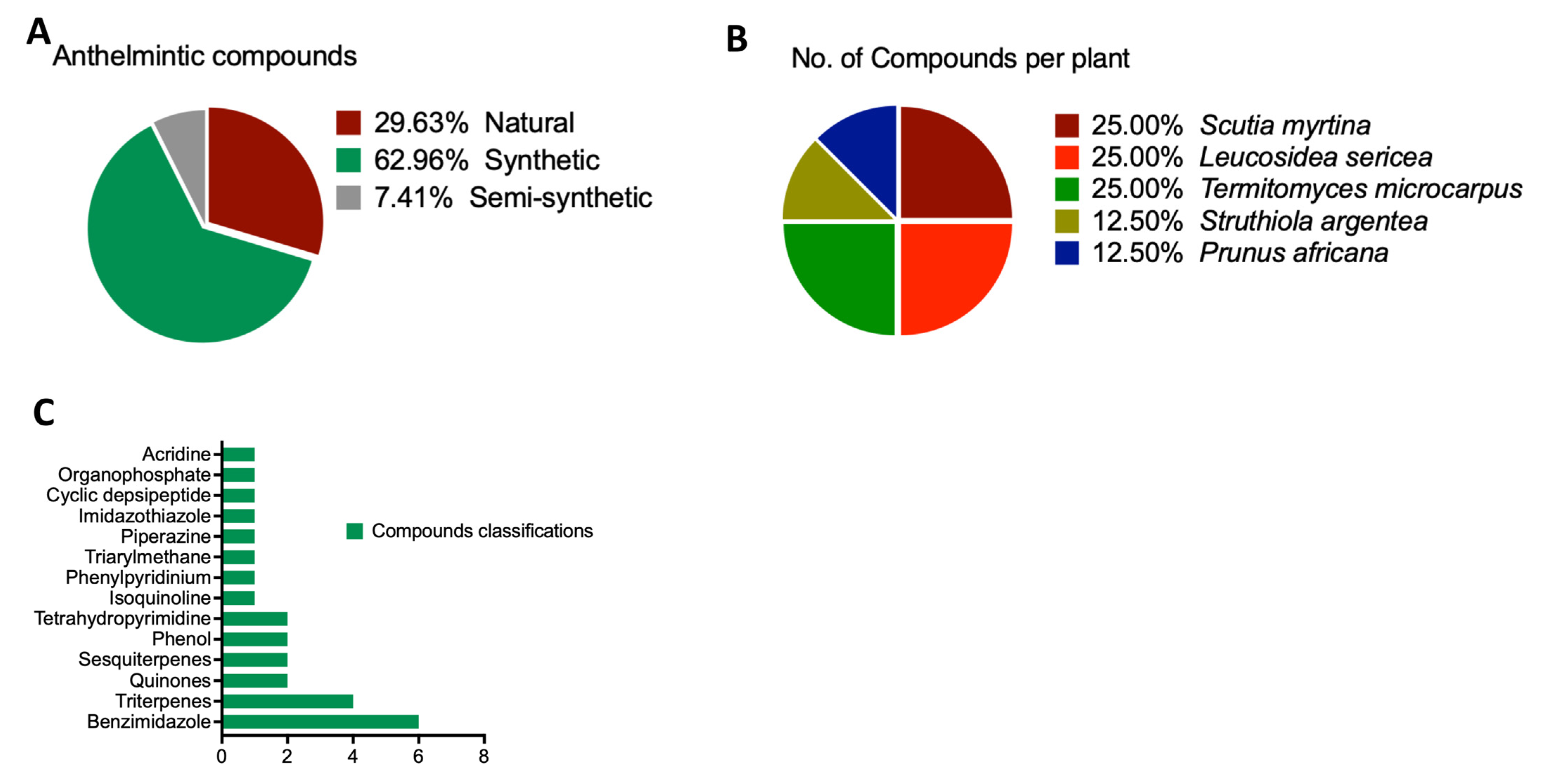
Figure 1. Scientific representation of compound characteristics. (A) The origin of anthelmintic compounds. (B) The botanical origins of the compounds. This aspect of the figure presents details about the particular plants from which the organic compounds are sourced. It emphasizes the wide range of botanical origins. (C) Categorization of the compounds.
Life 13 01957 g001
Figure 2. A phylogenetic tree depicting the distinct evolutionary divergence of the SmGST from its mammalian homolog.
Life 13 01957 g002
Figure 3. A phylogenetic tree depicting the close evolutionary relationship of the SmGST protein to other parasitic worms in comparison to free-living worms.
Life 13 01957 g003
Figure 4. Binding sites and Vina score of the SmGST protein and comparative anthelmintic compound affinities. (A). Ramachandran plot of the SmGST protein showing the angles for each residue of the smGST. (B). Potential binding pockets of the SmGST protein, with a focus on zooming out from pocket 3. (C). Bar chart illustrating the Vina score of the different anthelmintic compounds against the SmGST protein. The bars highlighted in green were chosen for further analysis and discussion due to their Vina scores of less than −10 kcal/mol. The orange bars were not included as their Vina scores exceeded −10 kcal/mol.
Life 13 01957 g004
Figure 5. Scientific depiction of natural compounds (A) Scutiaquinone A and (B) Scutiaquinone B, derived from the South African Natural Compounds Database.
Life 13 01957 g005
Figure 6. The three-dimensional binding structure of SmGST protein shows its strong binding affinity to the two natural compounds. (A,B) Scutiaquinone A strongly binds to SmGST and (C,D) Scutiaquinone B strongly binds to SmGST. The compounds are shown in a gray-colored surface model, while the protein, SmGST, is shown in a purple-colored cartoon model.
Life 13 01957 g006
Figure 7. Residues of the SmGST protein actively interacting with (A) Scutiaquinone A and (B) Scutiaquinone B. The interacting amino acid of SmGST to Scutiaquinone A: TYR10 PHE11 GLY15 ARG16 PHE38 ARG52 LEU53 GLU106 TYR109 HIS110 LEU113 MET114 HIS169 PHE211. The interacting amino acid of SmGST to Scutiaquinone B: TYR10 PHE11 GLY15 ARG16 PHE38 TRP41 LYS45 GLY51 ARG52 LEU53 PRO54 TYR109 LEU113 MET114 PHE211.
Life 13 01957 g007
Table 1. The binding affinity of the SmGST protein and 27 anthelmintic compounds.
DatabaseActivitiesS/NCompound NameAccession NoSourcesClassVina Score Cavity Volume (Å3) Center (x, y, z)Docking Size (x, y, z)
SANCDBAnthelmintic Compound1Scutiaquinone ASANC00584Scutia myrtinaQuinones−10.129716, 51, 3422, 22, 22
2Scutiaquinone BSANC00585Scutia myrtinaQuinones−10.829716, 51, 3423, 23, 23
3AspidinolSANC00741Leucosidea sericeaSesquiterpenes−5.311714, 56, 1319, 19, 19
4DesaspidinolSANC00742Leucosidea sericeaSesquiterpenes−5.311714, 56, 1319, 19, 19
5Betulinic acidSANC00743Termitomyces microcarpusTriterpenes−8.411714, 56, 1322, 22, 22
6ZapotinSANC01031Struthiola argenteaTriterpenes−6.311714, 56, 1321, 21, 21
7Betulinic acidSANC00743Termitomyces microcarpusTriterpenes−8.411714, 56, 1322, 22, 22
8Ursolic acidSANC00744Prunus africanaTriterpenes−8.911714, 56, 1323, 23, 23
pubchem 94-hexylbenzene-1,3-diolCompound CID: 3610 Anacardium occidentale, Sargassum muticumPhenol−5.411714, 56, 1320, 20, 20
drugbank 10AlbendazoleDB00518Synthetic SourceBenzimidazole
11PyrantelDB11156Synthetic SourceTetrahydropyrimidine−5.311714, 56, 1320, 20, 20
12PiperazineDB00592Synthetic SourcePiperazine−3.629716, 51, 3414, 14, 14
13MebendazoleDB00643Synthetic SourceBenzimidazole
14PraziquantelDB01058Synthetic SourceIsoquinoline−8.729716, 51, 3422, 22, 22
15OxamniquineDB01096Semi-syntheticTetrahydroquinoline−6.429716, 51, 3421, 21, 21
16EgatenDB12245Synthetic SourceBenzimidazole−7.129716, 51, 3422, 22, 22
17PyrviniumDB06816Synthetic SourcePhenylpyridinium−8.629716, 51, 3424, 24, 24
18Gentian_VioletDB00406Synthetic SourceTriarylmethane−7.929716, 51, 3423, 23, 23
19DiethylcarbamazineDB00711Synthetic SourcePiperazine−4.611714, 56, 1318, 18, 18
20LevamisoleDB00848Synthetic SourceImidazothiazole−611714, 56, 1319, 19, 19
21HexylresorcinolDB11254Synthetic SourcePhenol−5.829716, 51, 3420, 20, 20
22EmodepsideDB11403Semi-syntheticCyclic depsipeptide−9.111714, 56, 1329, 29, 29
23FlubendazoleDB08974Synthetic SourceBenzimidazole−7.329716, 51, 3423, 23, 23
24CythioateDB11392Synthetic SourceOrganophosphate−5.429716, 51, 3420, 20, 20
25QuinacrineDB01103Synthetic SourceAcridine−7.229716, 51, 3422, 22, 22
26FenbendazoleDB11410Synthetic SourceBenzimidazole
27DithiazanineDB11516Synthetic SourceBenzothiazole−7.529716, 51, 3425, 25, 25
Table 2. The Pharmacokinetic and Solubility Profiles of Scutiaquinone A and B.
Scutiaquinone AScutiaquinone B
LipophilicityLog Po/w (iLOGP) 4.384.39
Log Po/w (XLOGP3) 5.726.17
Log Po/w (WLOGP) 6.326.05
Log Po/w (MLOGP) 2.622.62
Log Po/w (SILICOS-IT) 6.686.71
Consensus Log Po/w 5.155.19
Water SolubilityLog S (ESOL) −6.88−7.17
Solubility6.7 × 10−5 mg/mL; 1.31 × 10⁻⁷ mol/L3.49 × 10−5 mg/mL; 6.83 × 10⁻⁸ mol/L
Class Poorly solublePoorly soluble
Log S (Ali) −7.44−7.91
Solubility1.85 × 10−5 mg/mL; 3.62 × 10⁻⁸ mol/L6.31 × 10−6 mg/mL; 1.24 × 10⁻⁸ mol/L
Class Poorly solublePoorly soluble
Log S (SILICOS-IT) −8.33−8.11
Solubility2.39 × 10−6 mg/mL; 4.69 × 10⁻⁹ mol/L3.96 × 10−6 mg/mL; 7.76 × 10⁻⁹ mol/L
Class Poorly solublePoorly soluble
DruglikenessLipinskiYesYes
VeberYesYes
Bioavailability Score 0.550.55
PharmacokineticsGI absorption LowHigh
BBB permeant NoNo
P-gp substrate NoNo
CYP1A2 inhibitor NoNo
CYP2C19 inhibitor YesNo
CYP2C9 inhibitor YesYes
CYP2D6 inhibitor NoNo
CYP3A4 inhibitor YesNo
Log Kp (skin permeation) −5.35 cm/s−5.03 cm/s
Disclaimer/Publisher’s Note: The statements, opinions and data contained in all publications are solely those of the individual author(s) and contributor(s) and not of MDPI and/or the editor(s). MDPI and/or the editor(s) disclaim responsibility for any injury to people or property resulting from any ideas, methods, instructions or products referred to in the content.

