Article

Analysis of Types of Skin Lesions and Diseases in Everyday Infectious Disease Practice—How Experienced Are We?

1
University Hospital for Infectious Diseases “Dr. Fran Mihaljević”, 10000 Zagreb, Croatia
2
School of Dental Medicine, University of Zagreb, 10000 Zagreb, Croatia
3
Department of Pharmacology, School of Medicine, University of Zagreb, 10000 Zagreb, Croatia
4
School of Medicine, University of Zagreb, 10000 Zagreb, Croatia
*
Author to whom correspondence should be addressed.
Life 2022, 12(7), 978; https://doi.org/10.3390/life12070978
Submission received: 31 May 2022 / Revised: 24 June 2022 / Accepted: 27 June 2022 / Published: 29 June 2022
(This article belongs to the Special Issue Microbiome, Microorganisms and Skin)

Abstract

:
Rashes and skin lesions are a common reason for patient visits to emergency departments and physicians’ offices. The differential diagnosis includes a variety of infectious and non-infectious diseases, some of which can be life-threatening. The aim of this retrospective study was to evaluate the quantity and type of skin lesions among outpatients and inpatients at a tertiary care university-affiliated teaching hospital for infectious diseases over a three-year period to assess disease burden and physicians’ experience in diagnosing skin lesions. Diagnoses (by ICD-10 codes) were classified into three groups: infectious diseases that include skin lesions, non-infectious skin lesions and undiagnosed skin lesions. During the observed period, out of the total of 142,416 outpatients, 14.8% presented with some form of skin lesion. Among them, 68% had skin lesions inherent to infectious disease, 10.8% suffered from non-infectious skin lesions and 21.2% remained with undiagnosed skin lesions. The most common infectious diagnoses were chickenpox, herpes zoster and unspecified viral infections characterized by skin and mucous membrane lesions. The most common non-infectious diagnoses were urticaria and atopic dermatitis. Overall, the most common individual diagnosis (ICD-10 code) was “nonspecific skin eruption” (n = 4448, 21.1%), which was followed by chickenpox and herpes zoster. Among the 17,401 patients hospitalized over the observed period, 13.1% had skin lesion as the main reason for hospitalization, almost all (97.5%) of which were infectious in etiology. The most common diagnoses were cellulitis, erysipelas and herpes zoster. The presented data suggest that the burden of diseases presenting with skin lesions is significant in everyday infectious disease practice, but the overwhelming number of undiagnosed patients implies the need for further education in this area.

1. Introduction

Rashes and skin lesions are common reasons for seeking medical services. Generally, skin diseases are among the most frequently occurring illnesses in humans. Skin and subcutaneous disorders were the fourth leading cause of nonfatal disease burden worldwide in the last decade [1,2,3] with increasing prevalence [4]. In 2013, skin conditions contributed 1.79% to the total international burden of disease measured in disability-adjusted life expectancy across 306 different diseases and injuries. An important part of those conditions refers to certain skin conditions caused by infectious agents, such as cellulitis, pyoderma, scabies, and various other fungal and viral skin diseases [2].
The most predominant types of skin lesions in everyday infectious disease practice are skin and soft tissue infections caused by bacteria, viruses, fungi and parasites, and different types of exanthems presenting with fever [5,6,7,8]. Skin and soft tissue infections are among the most widespread types of infections and include cellulitis, erysipelas, impetigo, ecthyma, folliculitis, furuncles, carbuncles, abscesses, trauma-related infections, decubital ulcers, necrotizing fasciitis, Fournier gangrene and infections from human or animal bites. Their presentation, etiology, and severity are variable. The incidence of these infections, which are a common reason for hospitalizations, has increased in the previous decades by up to 65% [9,10,11,12].
In the pediatric population, a combination of characteristic rash and other symptoms is sufficient for the diagnosis of classical exanthems such as measles, scarlet fever, rubella, erythema infectiosum, exanthema subitum and chickenpox [13,14,15]. There are also “atypical exanthems” that may be associated with fever or other constitutional symptoms and may be caused by infectious or toxic agents through hypersensitivity reactions [15,16,17]. Some of them are a diagnostic challenge, but the ones present in medical emergencies and those that pose an urgent diagnostic and therapeutic dilemma are especially important [18].
Differential diagnosis of all the aforementioned conditions is extensive and includes a variety of infectious and non-infectious causes. In some cases, skin lesions are a prominent symptom of systemic or localized disease and are ample for diagnosing certain infections or non-infectious conditions [19,20,21], but a variety of infectious and non-infectious diseases can produce similar skin lesions, and a single condition can present with different types of skin lesions due to a limited number of skin manifestations [6,19,20,21].
The pathogenesis of skin lesions associated with infectious diseases can be variable: (a) direct inoculation and multiplication of microorganisms within the skin, (b) dissemination of microorganisms from a different site, (c) production of toxins that can directly affect skin structure, (d) vascular effects of toxins, (e) inflammatory/immune response to microorganisms or toxins, (f) disseminated intravascular coagulation and coagulopathy, (g) direct vascular invasion and occlusion by microorganisms, and (h) reaction to medications used to treat infectious diseases [10,17,22,23]. The pathogenesis in other, non-infectious skin lesions is even more complex. However, knowing possible mechanisms in the development of skin lesions is not a direct clue in establishing an etiological diagnosis.
Even though a detailed physical examination is crucial, a thorough medical history is also essential where a number of factors must be taken into account in establishing the diagnosis of skin lesions. These include the appearance of skin lesions together with other symptoms, distribution of skin changes, accompanying pruritus, potential exposure to infectious agents, occurrence in family members and colleagues, drug intake, immunization, seasonal occurrence, history of allergy, immune status of the host, contact with animals and exposure to sexually transmitted diseases [6,20,21].
Some skin lesions, such as herpes zoster, psoriasis and atopic dermatitis can be correctly recognized even without significant experience, but many others are more demanding in this respect [24,25]. Training and clinical exposure to dermatological conditions leads to a greater ability to establish the correct diagnosis not only by dermatologists but also by other physicians. Infectious skin diseases are among the most common dermatologic conditions reported by non-dermatologists [25,26]. In certain diseases, skin lesions can be an early sign of life-threatening infections (meningococcal infection, toxic shock syndrome, necrotizing fasciitis, staphylococcal scalded skin syndrome) or non-infectious diseases (toxic epidermal necrolysis, Steven–Johnson syndrome, angio-edema). Rapid diagnosis is essential in order to start timely treatment. Some skin lesions are present during diseases that must be treated to prevent serious consequences (syphilis, cellulitis, scarlet fever, varicella in adults, exanthema chronicum migraines, etc.), and some skin lesions are part of self-limited diseases (exanthema subitum, enteroviral infections, erythema infectiosum). It is crucial for clinicians to have a basic knowledge of infectious diseases while dealing with skin lesions, but a comprehensive knowledge of other non-infectious diseases is also required. Experience and appropriate education in these areas are critically important and can be lifesaving.
We aimed to assess disease burden and physicians’ experience in diagnosing skin lesions in outpatients and inpatients at a tertiary care university-affiliated teaching hospital for infectious diseases.

2. Patients and Methods
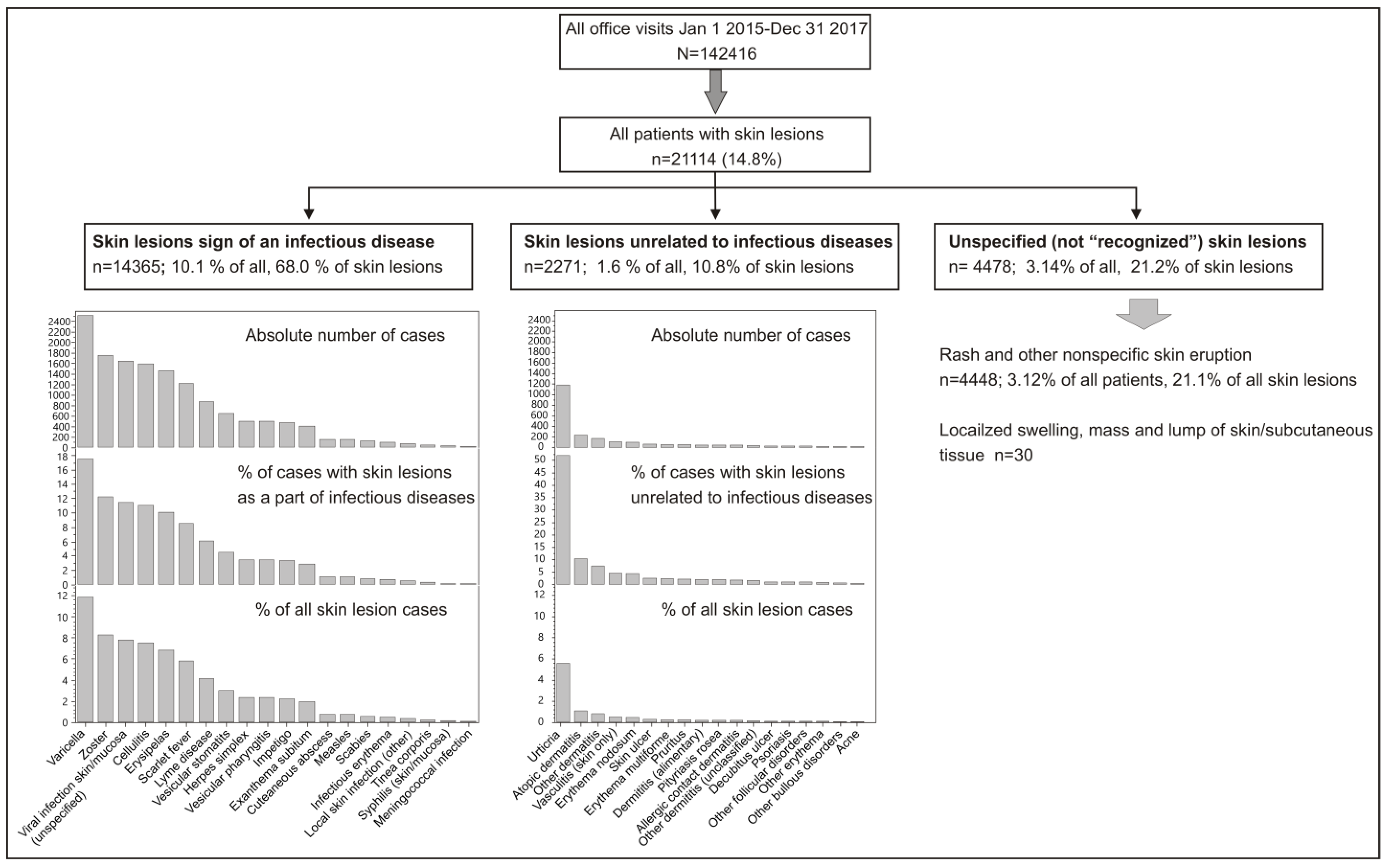
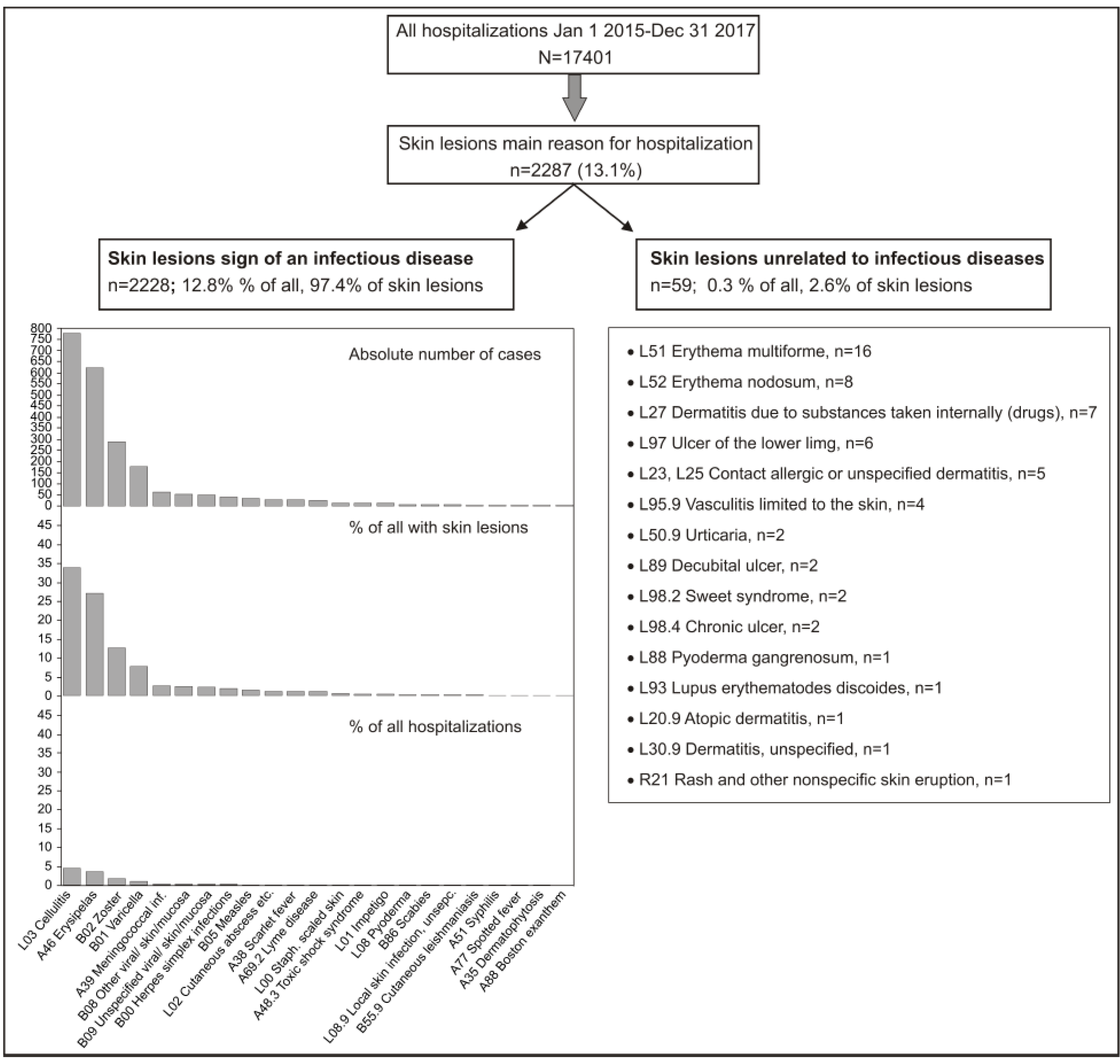
We retrospectively searched institutional administrative data collected between 1 January 2015 and 31 December 2017 on all outpatient visits and hospitalizations at a tertiary care university-affiliated teaching hospital in order to identify the International Classification of Disease version 10 (ICD-10, 2015) codes assigned to: (a) infectious diseases in which rash/skin lesions are a major element of clinical presentation; (b) infectious diseases in which rash/skin lesions are a common element of clinical presentation; and (c) rash/skin lesions unrelated to infectious diseases classified in the chapter “Diseases of the skin and subcutaneous tissues” (L00-L99) of the ICD-10—if they were the main reason for the patient’s visit/hospitalization. The searched ICD-10 codes are listed in Table 1 and Table 2. We disregarded: (a) infectious diseases in which rash/skin lesions may occasionally be present; and (b) diseases classified as ICD-10 codes L00-L99, in which skin is typically not affected, even if they were the main reason (leading diagnosis) for the patient’s visit. All included patients were first examined in our emergency department specifically organized for patients with confirmed or suspected infectious diseases. The patients who were diagnosed and treated there were considered outpatients, while those who had to be hospitalized were considered inpatients. We used routinely collected anonymized data; hence, the need for ethical approval was waived by the Institutional Ethics Committee.

3. Results

3.1. Outpatients

Of the 142,416 outpatient visits recorded during the observed period, 21,114 (14.8%) presented with some form of a skin lesion (Figure 1). Among them, 14,365 patients (68.0%) presented with skin lesions inherent to an infectious disease, 2271 (10.8%) were deemed to have suffered from non-infectious skin lesions and 4478 patients (21.2% of managed patients with skin lesions) remained with undiagnosed skin lesions (Figure 1). The overall most common individual ICD-10 code (diagnosis) was “rash and other nonspecific skin eruption” or, in other words, not recognized skin lesions—in 4448 patients (21.1% of those who presented with skin lesions and 3.1% of all outpatient visits) (Figure 1). The other most common diagnoses pertaining to infectious diseases included: chickenpox (n = 2513, 11.9% of all skin lesions), herpes zoster (n = 1754, 8.3%), unspecified viral infections characterized by skin and mucous membrane lesions (n = 1646, 7.8%), cellulitis (n = 1590, 7.5%), erysipelas (n = 1451, 6.9%), scarlet fever (n = 1223, 5.8%) and Lyme disease (n = 876, 4.1%) (Figure 1). The most common non-infectious diagnoses were urticaria (n = 1180, 5.6%), atopic dermatitis (n = 232, 1.1%) and “other” dermatitis (n = 168, 0.8%) (Figure 1).

3.2. Inpatients

Of the 17,401 patients hospitalized over the observed period, 2287 (13.1%) had skin lesions as the main reason for hospitalization (Figure 2): almost all (n = 2228; 97.4% of skin lesions, 12.8% of all hospitalizations) were infectious in etiology; in only 59 patients (2.6% or 0.3% of all hospitalizations), skin lesions were not related to an infectious disease. The most common discharge diagnoses were (Figure 2) cellulitis (various locations, typically lower limb) (n = 776, 33.9% of skin lesions), erysipelas (n = 620, 27.1%), herpes zoster (with or without complications) (n = 287, 12.5%), chickenpox (mostly with, sporadically without complications) (n = 177, 7.7%) and meningococcal infections (n = 61, 2.7%). There were 50 patients (2.2% of all skin lesions) in whom only a rather “vague” diagnosis of “unspecified viral infection characterized by skin and mucous membrane lesions” (ICD-10 code B09) was the leading discharge diagnosis (Figure 2). Individual non-infectious skin lesions such as erythema multiforme or nodosum, dermatitis due to substances taken internally (mostly drugs), contact allergic or non-allergic dermatitis and ulcer of the lower limb were sporadic and the only conditions observed in more than just a few patients (Figure 2).
The age of hospitalized patients ranged from <1 to 101 years (Table 3). Patients with infectious lesions appeared somewhat older than those with non-infections lesions, but age varied in respect to individual diagnoses, since some of the conditions are characteristic for children and younger adults (e.g., chickenpox, meningococcal infections), while other typically affect older people (e.g., cellulitis, erysipelas, herpes zoster) (Table 3). The median length of hospital stay was consistently 8–10 days, which was a bit shorter in patients hospitalized for (complicated) chickenpox (Table 3).

4. Discussion

There are a few studies documenting the profile of patients with skin lesions in emergency departments (ED), all of which report an impressive percentage (2.5–8%) of all visits to the ED as being due to skin complaints [27,28,29,30,31,32,33,34,35,36,37,38].
In our infectious diseases ED, the profile of patients is different than in "classical" EDs that are mostly focused on internal medicine, surgical or neurological diseases. Patients with rash, with or without fever, as well as those with skin and soft tissue infections are quite common in our practice and according to our data, they make up almost 15% of all outpatients, which is higher than in other previously mentioned studies. A possible reason could be the fact that skin lesions are routinely ascribed an infectious etiology, especially when associated with constitutional symptoms.
The proportion of patients hospitalized due to skin lesions is high in our setting (13.1% of all hospitalizations). Among those, 13.1% and 61% are due to skin and soft tissue infections such as erysipelas and cellulitis, respectively. This is consistent with the overall increase in the incidence of skin and soft tissue infections, especially in populations over 65 years of age [9,39,40]. The median age of hospitalized patients with erysipelas and cellulitis in our study was 64 and 66 years, respectively.
The highest median age was recorded in patients hospitalized due to herpes zoster (73 years), the incidence of which has also been increasing worldwide [41,42]. Age is the most important risk factor for the development of herpes zoster with a dramatic increase in the incidence rate after 50 years of age [43].
The incidence of life-threatening infectious diseases presenting with skin lesions (meningococcal infection, toxic shock syndrome, necrotizing fasciitis and staphylococcal scalded skin syndrome) was low in both outpatients (n = 37, 0.03%) and inpatients (n = 82, 0.5%). The number of patients diagnosed with meningococcal diseases, toxic shock syndrome and staphylococcal scalded skin syndrome was lower in ambulatory settings (n = 61, n = 10, n = 11, respectively) than in hospital settings (n = 23, n = 6, n = 8, respectively), which indicates that not all were recognized in the ED, but the diagnoses were established during hospitalization.
To date, the information we aim to present (the proportion of infectious, non-infectious and undiagnosed skin lesions) was also found in four published studies [16,44,45,46] but with a few differences, as shown in Table 4:
(1)
Study population
In their study, Goodyear et al. [44] included only children, Tabak et al. [46] included only adults and Drago et al. [16,45] included both adults and children. Our study included adults and children as well.
(2)
Inclusion criteria
Goodyear et al. [44] included children with acute erythematous rash with fever, but children with recognizable rashes such as classical measles and chickenpox were excluded. Drago et al. [16,45] included adults and children with clinical features which differ from classical exanthems. Finally, Tabak et al. [46] studied adult patients with fever and rash. We included all patients with infectious diseases that included skin lesions and those with common non-infectious skin lesions.
(3)
Aim
Goodyear et al. [44] aimed to establish the etiology of non-classical erythematous rashes with fever in the pediatric population. Drago et al. [16,45] aimed to investigate whether morphology, other clinical features, laboratory results and other characteristics can help to determine the etiology of atypical exanthems. Tabak et al. [46] aimed to determine the etiology of fever and rash in adult patients. Our aim was to evaluate the quantity and type of skin lesions among outpatients and inpatients in order to assess disease burden and physicians’ experience in diagnosing skin lesions.
(4)
Study design
Both Goodyear et al. [44] and Drago et al. [16,45] provided laboratory testing together with routine procedures (patient history and physical examination) in order to establish the etiology of exanthems. Tabak et al. [46] performed routine procedures and necessary tests according to suspected diagnoses. In our study, ICD codes were used to detect infectious disease patients with skin lesions, non-infectious skin lesions and undiagnosed skin lesions.
Although there is a significant difference between these four studies and ours, they were the only ones that used the term “undiagnosed” skin lesions/rashes in their analysis. Even though the results are not comparable with our own, due to the differences in study design and patient characteristics, the proportion of undiagnosed skin lesions in our study was smaller than Goodyear et al. [44] and Drago et al. [45] reported, as it only included patients with atypical exanthems. This difference is possibly due to the exclusion of patients with typical, recognizable rashes and the presence of more challenging patients. The proportion of undiagnosed skin lesions was higher than reported by Tabak et al. [46] in a study that included all adult patients with fever and rash. Finally, the proportion of undiagnosed skin lesions was similar to Drago et al. [16], even though it only included patients with atypical exanthems.
As dermatological disorders are quite commonly seen by almost all clinicians in their clinical practice, it seems logical to try to evaluate their ability in diagnosing skin conditions. Our clinicians are not the only ones commonly faced with diagnostic failures. Onsoi et al. [47] reported a large percentage (48.2%) of misdiagnoses of common cutaneous diseases in pediatric population.
Antic et al. [25] reported that their internists recognized 51.1% of the cutaneous manifestations during examinations and 49% when presented with slides showing common dermatoses.
Because of the large number of illnesses that can manifest with different types of skin lesions, it is unreasonable to expect physicians to recognize all that comes into consideration while establishing a differential diagnosis. Working in pairs with more experienced colleagues during the learning period and practicing clinical image-based teaching as a supplement to the patient-based clinical teaching is the approach we suggest. Additionally, residents in infectious diseases should be exposed to dermatology training during residency, as it is currently not a required component of their training program. In addition, consultations with dermatologists are highly recommended in certain cases, especially if the physicians handling them are not routinely exposed to patients presenting with skin changes. This was already proven to be efficient in the study by Zhang et al. [48]. In the future, we might hope for a computational neural network to be developed in order to help with diagnosing skin lesions consistent with infectious diseases, as such a tool already exists for skin cancer [49,50].
The limitations of our study, listed below, arise from the use of ICD codes and bias introduced by potential misclassification and coding errors:
(1)
It is possible that the number of patients with skin lesions is underreported due to the exclusion of diagnoses that include skin lesions as a non-compulsory part of the clinical presentation. These diagnoses include leptospirosis, bartonellosis, rotavirus infections, dengue fever, infections caused by mycoplasma pneumoniae, human immunodeficiency virus infection, Epstein–Barr virus infections, toxoplasmosis, malaria, etc.
(2)
It is possible that there was a greater number of hospitalized patients with skin lesions that were not included in this study, as the skin lesions they may have presented with were not the cause of hospitalization or part of the primary diagnosis.
(3)
The proportion of skin lesions classified as unspecified (R21 and R22) might be higher than reported due to individual classification preferences of clinicians. An example of such a preference is the fact that an unspecified cause can be also reported under L30 (Other dermatitis).
(4)
Not all diagnoses could be classified unanimously as infectious or unrelated to infections. One such diagnosis is erythema nodosum (L52), which can occur secondary to a wide variety of conditions; however, an infection (primarily streptococcal) is the most commonly identified etiology [51].
(5)
The reasons for using codes for unspecified skin lesions (R21, R22) can be multiple: (a) a skin lesion not being recognized; (b) uncertainty in the diagnosis—the coding system cannot determine the physician’s confidence in certain diagnoses; (c) a lack of code to accurately describe the condition. It is possible that not all skin lesions classified as “undiagnosed” were unrecognized.
Few studies have addressed the diagnosis of exanthems in adult and pediatric populations together with the proportion of infectious, non-infectious and undiagnosed exanthems as well as proportion of skin lesions caused by infectious agents among patients in emergency department. To date, no other study included all types of skin lesions that are infectious in etiology (mostly rashes with or without fever and local skin lesions such as skin and soft tissue infections) among patients visiting infectious disease settings including inpatients and outpatients. That is why, to our knowledge, our study is unique.

5. Conclusions

The presented data suggest that the burden of diseases presenting with skin lesions in everyday infectious disease practice is large, especially in a specific setting that includes an emergency department and a hospital department for infectious diseases, and that the overwhelming number of undiagnosed outpatients implies the need for further education in this area.

Author Contributions

Conceptualization, T.S., V.T., M.R., A.D. and B.D.; Data curation, T.S., V.T. and M.R.; Formal analysis, V.T.; Methodology, T.S. and A.D.; Supervision, B.D.; Writing—original draft, T.S. and M.R.; Writing—review & editing, T.S., V.T. and B.D. All authors have read and agreed to the published version of the manuscript.

Funding

This research received no external funding.

Institutional Review Board Statement

This study did not require ethical approval.

Informed Consent Statement

Patient consent was waived due to data anonymity.

Data Availability Statement

We used institutional data that is not available for sharing.

Conflicts of Interest

The authors declare no conflict of interest.

References

  1. Hay, R.J.; Johns, N.E.; Williams, H.C.; Bolliger, I.W.; Dellavalle, R.P.; Margolis, D.J.; Marks, R.; Naldi, L.; Weinstock, M.A.; Wulf, S.K.; et al. The global burden of skin disease in 2010: An analysis of the prevalence and impact of skin conditions. J. Investig. Dermatol. 2014, 134, 1527–1534. [Google Scholar] [CrossRef] [PubMed] [Green Version]
  2. Karimkhani, C.; Dellavalle, R.P.; Coffeng, L.E.; Flohr, C.; Hay, R.J.; Langan, S.M.; Nsoesie, E.O.; Ferrari, A.J.; Erskine, H.E.; Silverberg, J.I.; et al. Global Skin Disease Morbidity and Mortality: An Update from the Global Burden of Disease Study 2013. JAMA Dermatol. 2017, 153, 406–412. [Google Scholar] [CrossRef]
  3. Seth, D.; Cheldize, K.; Brown, D.; Freeman, E.F. Global Burden of Skin Disease: Inequities and Innovations. Curr. Dermatol. Rep. 2017, 6, 204–210. [Google Scholar] [CrossRef] [PubMed]
  4. Verhoeven, E.W.; Kraaimaat, F.W.; van Weel, C.; van de Kerkhof, P.C.; Duller, P.; van der Valk, P.G.; van den Hoogen, H.J.; Bor, J.H.; Schers, H.J.; Evers, A.W. Skin diseases in family medicine: Prevalence and health care use. Ann. Fam. Med. 2008, 6, 349–354. [Google Scholar] [CrossRef] [Green Version]
  5. Yeroushalmi, S.; Shirazi, J.Y.; Friedman, A. New Developments in Bacterial; Viral; and Fungal Cutaneous Infections. Curr. Dermatol. Rep. 2020, 9, 152–165. [Google Scholar] [CrossRef]
  6. Sanders, C.V.; Lopez, F.A. Cutaneous manifestations of infectious diseases: Approach to the patient with fever and rash. Trans. Am. Clin. Climatol. Assoc. 2001, 112, 235–251, discussion 251–252. [Google Scholar]
  7. Miró, E.M.; Sánchez, N.P. Cutaneous Manifestations of Infectious Diseases. In Atlas of Dermatology in Internal Medicine; Sánchez, N., Ed.; Springer: New York, NY, USA, 2012; pp. 77–119. [Google Scholar]
  8. Mims, C.A. Pathogenesis of rashes in virus diseases. Bacteriol. Rev. 1966, 30, 739–760. [Google Scholar] [CrossRef]
  9. Esposito, S.; Noviello, S.; Leone, S. Epidemiology and microbiology of skin and soft tissue infections. Curr. Opin. Infect. Dis. 2016, 29, 109–115. [Google Scholar] [CrossRef]
  10. Ki, V.; Rotstein, C. Bacterial skin and soft tissue infections in adults: A review of their epidemiology; pathogenesis; diagnosis; treatment and site of care. Can. J. Infect. Dis. Med. Microbiol. 2008, 19, 173–184. [Google Scholar] [CrossRef] [Green Version]
  11. Silverberg, B. A Structured Approach to Skin and Soft Tissue Infections (SSTIs) in an Ambulatory Setting. Clin. Pract. 2021, 11, 65–74. [Google Scholar] [CrossRef]
  12. Ramakrishnan, K.; Salinas, R.C.; Agudelo Higuita, N.I. Skin and Soft Tissue Infections. Am. Fam. Physician. 2015, 92, 474–483. [Google Scholar] [PubMed]
  13. Cherry, J.D. Contemporary infectious exanthems. Clin. Infect. Dis. 1993, 16, 199–205. [Google Scholar] [CrossRef] [PubMed]
  14. Soman, L. Fever with Rashes. Indian. J. Pediatr. 2018, 85, 528–534. [Google Scholar] [CrossRef] [PubMed]
  15. Muzumdar, S.; Rothe, M.J.; Grant-Kels, J.M. The rash with maculopapules and fever in children. Clin. Dermatol. 2019, 37, 119–128. [Google Scholar] [CrossRef] [PubMed]
  16. Drago, F.; Paolino, S.; Rebora, A.; Broccolo, F.; Drago, F.; Cardo, P.; Parodi, A. The challenge of diagnosing atypical exanthems: A clinico-laboratory study. J. Am. Acad. Dermatol. 2012, 67, 1282–1288. [Google Scholar] [CrossRef]
  17. Drago, F.; Ciccarese, G.; Gasparini, G.; Cogorno, L.; Javor, S.; Toniolo, A.; Broccolo, F. Contemporary infectious exanthems: An update. Future Microbiol. 2017, 12, 171–193. [Google Scholar] [CrossRef] [Green Version]
  18. Patel, T.; Quow, K.; Cardones, A.R. Management of Infectious Emergencies for the Inpatient Dermatologist. Curr. Dermatol. Rep. 2021, 10, 232–242. [Google Scholar] [CrossRef]
  19. Ely, J.W.; Seabury Stone, M. The generalized rash: Part I. Differential diagnosis. Am. Fam. Physician 2010, 81, 726–734. [Google Scholar]
  20. Ely, J.W.; Seabury Stone, M. The generalized rash: Part II. Diagnostic approach. Am. Fam. Physician 2010, 81, 735–739. [Google Scholar]
  21. Kang, J.H. Febrile Illness with Skin Rashes. Infect. Chemother. 2015, 47, 155–166. [Google Scholar] [CrossRef] [Green Version]
  22. Kingston, M.E.; Mackey, D. Skin clues in the diagnosis of life-threatening infections. Rev. Infect. Dis. 1986, 8, 1–11. [Google Scholar] [CrossRef] [PubMed]
  23. Bigby, M. Rates of cutaneous reactions to drugs. Arch. Dermatol. 2001, 137, 765–770. [Google Scholar] [PubMed]
  24. Fenton, A.; Elliott, E.; Shahbandi, A.; Ezenwa, E.; Morris, C.; McLawhorn, J.; Jackson, J.G.; Allen, P.; Murina, A. Medical students’ ability to diagnose common dermatologic conditions in skin of color. J. Am. Acad. Dermatol. 2020, 83, 957–958. [Google Scholar] [CrossRef]
  25. Antic, M.; Conen, D.; Itin, P.H. Teaching effects of dermatological consultations on nondermatologists in the field of internal medicine. A study of 1290 inpatients. Dermatology 2004, 208, 32–37. [Google Scholar] [CrossRef]
  26. Wilmer, E.N.; Gustafson, C.J.; Ahn, C.S.; Davis, S.A.; Feldman, S.R.; Huang, W.W. Most common dermatologic conditions encountered by dermatologists and nondermatologists. Cutis 2014, 94, 285–292. [Google Scholar] [PubMed]
  27. Kilic, D.; Yigit, O.; Kilic, T.; Buyurgan, C.S.; Dicle, O. Epidemiologic Characteristics of Patients Admitted to Emergency Department with Dermatological Complaints; a Retrospective Cross sectional Study. Arch. Acad. Emerg Med. 2019, 7, e47. [Google Scholar] [PubMed]
  28. Isnard, C.; Ingen-Housz-Oro, S.; Fardet, L.; Matteodo, E.; Duval, S.; Hemery, F.; Khellaf, M.; Duong, T.-A.; Chosidow, O.; Wolkenstein, P. Dermatological emergencies: Evolution from 2008 to 2014 and perspectives. J. Eur. Acad. Dermatol. Venereol. 2017, 31, 274–279. [Google Scholar] [CrossRef]
  29. Martínez-Martínez, M.L.; Escario-Travesedo, E.; Rodríguez-Vázquez, M.; Azaña-Defez, J.M.; Martín de Hijas-Santos, M.C.; Juan-Pérez-García, L. Dermatology consultations in an emergency department prior to establishment of emergency dermatology cover. Actas Dermosifiliogr. 2011, 102, 39–47. [Google Scholar] [CrossRef]
  30. Rubegni, P.; Cevenini, G.; Lamberti, A.; Bruni, F.; Tiezzi, R.; Verzuri, A.; Barbini, P.; Manzi, P.; Fimiani, M. Dermatological conditions presenting at the Emergency Department in Siena University Hospital from 2006 to 2011. J. Eur. Acad. Dermatol. Venereol. 2015, 29, 164–168. [Google Scholar] [CrossRef]
  31. Grillo, E.; Vañó-Galván, S.; Jiménez-Gómez, N.; Ballester, A.; Muñoz-Zato, E.; Jaén, P. Dermatologic emergencies: Descriptive analysis of 861 patients in a tertiary care teaching hospital. Actas. Dermosifiliogr. 2013, 104, 316–324. [Google Scholar] [CrossRef]
  32. Kim, J.Y.; Cho, H.H.; Hong, J.S.; Jin, S.P.; Park, H.S.; Lee, J.H.; Cho, S. Skin conditions presenting in emergency room in Korea: An eight-year retrospective analysis. J. Eur. Acad. Dermatol. Venereol. 2013, 27, 479–485. [Google Scholar] [CrossRef] [PubMed]
  33. Bancalari-Díaz, D.; Gimeno-Mateos, L.I.; Cañueto, J.; Andrés-Ramos, I.; Fernández-López, E.; Román-Curto, C. Dermatologic emergencies in a tertiary hospital: A descriptive study. Actas Dermosifiliogr. 2016, 107, 666–673. [Google Scholar] [CrossRef] [PubMed]
  34. Ozkur, E.; Altunay, I.; Sekerlisoy, G.; Erdem, Y. Evaluation of Dermatology Consultations in a Tertiary Care Centre Emergency Service. Med. Bull. Sisli Etfal Hosp. 2020, 54, 197–200. [Google Scholar]
  35. Drago, F.; Gasparini, G.; Signori, A.; Campisi, C.; Cozzani, E.; Parodi, A. Dermatological consultations in an observation unit of an emergency department in Italy. J. Eur. Acad. Dermatol. Venereol. 2015, 29, 973–980. [Google Scholar] [CrossRef] [PubMed]
  36. Jack, A.R.; Spence, A.A.; Nichols, B.J.; Chong, S.; Williams, D.T.; Swadron, S.P.; Peng, D.H. Cutaneous conditions leading to dermatology consultations in the emergency department. West. J. Emerg. Med. 2011, 12, 551–555. [Google Scholar] [CrossRef] [Green Version]
  37. Gupta, S.; Sandhu, K.; Kumar, B. Evaluation of emergency dermatological consultations in a tertiary care centre in North India. J. Eur. Acad. Dermatol. Venereol. 2003, 17, 303–305. [Google Scholar] [CrossRef]
  38. Murr, D.; Bocquet, H.; Bachot, N.; Bagot, M.; Revuz, J.; Roujeau, J.C. Medical activity in an emergency outpatient department dermatology. Ann. Dermatol. Venereol. 2003, 130, 167–170. [Google Scholar]
  39. Tun, K.; Shurko, J.F.; Ryan, L.; Lee, G.C. Age-based health and economic burden of skin and soft tissue infections in the United States, 2000 and 2012. PLoS ONE 2018, 13, e0206893. [Google Scholar] [CrossRef]
  40. Kaye, K.S.; Petty, L.A.; Shorr, A.F.; Zilberberg, M.D. Current Epidemiology, Etiology, and Burden of Acute Skin Infections in the United States. Clin. Infect. Dis. 2019, 68, S193–S199. [Google Scholar] [CrossRef] [Green Version]
  41. Kawai, K.; Gebremeskel, B.G.; Acosta, C.J. Systematic review of incidence and complications of herpes zoster: Towards a global perspective. BMJ Open 2014, 4, e004833. [Google Scholar] [CrossRef] [Green Version]
  42. Kawai, K.; Yawn, B.P.; Wollan, P.; Harpaz, R. Increasing Incidence of Herpes Zoster Over a 60-year Period from a Population-based Study. Clin. Infect. Dis. 2016, 63, 221–226. [Google Scholar] [CrossRef] [PubMed] [Green Version]
  43. Schmader, K. Herpes zoster in older adults. Clin. Infect. Dis. 2001, 32, 1481–1486. [Google Scholar] [PubMed]
  44. Goodyear, H.M.; Laidler, P.W.; Price, E.H.; Kenny, P.A.; Harper, J.I. Acute infectious erythemas in children: A clinico-microbiological study. Br. J. Dermatol. 1991, 124, 433–438. [Google Scholar] [PubMed]
  45. Drago, F.; Rampini, P.; Rampini, E.; Rebora, A. Atypical exanthems: Morphology and laboratory investigations may lead to an aetiological diagnosis in about 70% of cases. Br. J. Dermatol. 2002, 147, 255–260. [Google Scholar] [CrossRef]
  46. Tabak, F.; Murtezaoglu, A.; Tabak, O.; Ozaras, R.; Mete, B.; Kutlubay, Z.; Mert, A.; Ozturk, R. Clinical Features and Etiology of Adult Patients with Fever and Rash. Ann. Dermatol. 2012, 24, 420–425. [Google Scholar] [CrossRef] [Green Version]
  47. Onsoi, W.; Chaiyarit, J.; Techasatian, L. Common misdiagnoses and prevalence of dermatological disorders at a pediatric tertiary care center. J. Int. Med. Res. 2020, 48, 300060519873490. [Google Scholar] [CrossRef] [Green Version]
  48. Zhang, H.; Tang, K.; Fang, R.; Jin, H.; Sun, Q. Inpatient Dermatology Consultations in a General Surgery Ward in a Tertiary Hospital in China: A Retrospective Study of 251 Patients. Dermatol Ther. 2021, 11, 961–970. [Google Scholar] [CrossRef]
  49. Maron, R.C.; Utikal, J.S.; Hekler, A.; Hauschild, A.; Sattler, E.; Sondermann, W.; Haferkamp, S.; Schilling, B.; Heppt, M.V.; Jansen, P.; et al. Artificial Intelligence and Its Effect on Dermatologists’ Accuracy in Dermoscopic Melanoma Image Classification: Web-Based Survey Study. J. Med. Internet Res. 2020, 22, e18091. [Google Scholar] [CrossRef]
  50. Dildar, M.; Akram, S.; Irfan, M.; Khan, H.U.; Ramzan, M.; Mahmood, A.R.; Alsaiari, S.A.; Saeed, A.H.M.; Alraddadi, M.O.; Mahnashi, M.H. Skin Cancer Detection: A Review Using Deep Learning Techniques. Int. J. Environ. Res. Public Health. 2021, 18, 5479. [Google Scholar] [CrossRef]
  51. Requena, L.; Requena, C. Erythema nodosum. Dermatol. Online J. 2002, 8, 4. [Google Scholar] [CrossRef]
Figure 1. Breakdown of outpatient visits included in the present analysis.
Life 12 00978 g001
Figure 2. Breakdown of hospitalized patients included in the present analysis.
Life 12 00978 g002
Table 1. List of infectious diseases/ICD-10 codes searched.
Bacterial diseases
A25 Rat-bite fevers
B35.0 Tinea barbae and tinea capitis
B35.1 Tinea unguium
A26 ErysipeloidB35.3 Tinea pedis
A31.1 Cutaneous mycobacterial infectionB35.4 Tinea corporis
A32.0 Cutaneous listeriosisB35.5 Tinea imbricata
A36.3 Cutaneous diphtheriaB35.6 Tinea inguinalis (Tinea cruris)
A39 Meningococcal infectionB35.8 Other dermatophytoses
A43.1 Cutaneous nocardiosisB35.9 Dermatophytosis, unspecified
A44.1 Cutaneous and mucocutaneous bartonellosisB36 Other superficial mycoses
A46 ErysipelasB36.0 Pityriasis versicolor
A48.3 Toxic shock syndromeB36.1 Tinea nigra
A51.3 Secondary syphilis of skin and mucous membranesB36.2 White piedra
A69.2 Lyme diseaseB36.3 Black piedra
A75 Typhus feverB36.8 Other specified superficial mycoses
A77.1 Spotted fever due to Rickettsia conoriiB36.9 Superficial mycosis, unspecified
A79.1 Rickettsial pox due to Rickettsia akariB37.2 Candidiasis of skin and nail
Viral diseasesB38.3 Cutaneous coccidioidomycosis
A88.0 Enteroviral exanthematous fever (Boston exanthem)B40.3 Cutaneous blastomycosis
A91 Dengue hemorrhagic fever B43.0 Cutaneous chromomycosis
A92.0 Chikungunya virus diseaseB45.2 Cutaneous cryptococcosis
B00 Herpes viral (herpes simplex) infectionsB46.3 Cutaneous mucormycosis
B01 Varicella (chickenpox)Parasitic diseases
B02 Zoster (herpes zoster)B55.1 Cutaneous leishmaniasis
B03 SmallpoxB55.2 Mucocutaneous leishmaniasis
B04 MonkeypoxB65.3 Cercarial dermatitis
B05 MeaslesB72 Dracunculiasis
B06 RubellaB74 Filariasis
B07 Viral wartsB78.1 Cutaneous strongyloidiasis
B08 Other viral infections characterized by skin and mucous membrane lesions, not elsewhere classifiedB85 Pediculosis and phthiriasis
B08.0 Other orthopoxvirus infectionsB86 Scabies
B08.1 Molluscum contagiosumB88.0 Other acariasis
B08.2 Exanthema subitum (sixth disease)Diseases caused by infectious agent
B08.3 Erythema infectiosum (fifth disease)L00 Staphylococcal scalded skin syndrome
B08.4 Enteroviral vesicular stomatitis with exanthemL01 Impetigo
B08.5 Enteroviral vesicular pharyngitisL02 Cutaneous abscess, furuncle and carbuncle
B08.8 Other specified viral infections characterized by skin and mucous membrane lesionsL03 Cellulitis
B09 Unspecified viral infection characterized by skin and mucous membrane lesionsL08 Other local infections of skin and subcutaneous tissue
Fungal diseasesL30.3 Infective dermatitis
B35 DermatophytosisL44.4 Infantile papular acrodermatitis (Giannotti–Crosti)
Table 2. List of non-infectious or non-specific skin lesions/ICD-10 codes searched.
L10 Pemphigus
L12 Pemphigoid
L13 Other bullous disorders
L20 Atopic dermatitis
L21 Seborrhoeic dermatitis
L22 Diaper [napkin] dermatitis
L23 Allergic contact dermatitis
L24 Irritant contact dermatitis
L25 Unspecified contact dermatitis
L26 Exfoliative dermatitis
L27 Dermatitis due to substances taken internally
L29 Pruritus
L30 Other dermatitis (except L30.3)
L40 Psoriasis
L41 Parapsoriasis
L42 Pityriasis rosea
L43 Lichen planus
L50 Urticaria
L51 Erythema multiforme
L52 Erythema nodosum
L53 Other erythematous conditions
L54 Erythema in diseases classified elsewhere
L55 Sunburn
L56 Other acute skin changes due to ultraviolet radiation
L70 Acne
L71 Rosacea
L73 Other follicular disorders
L84 Corns and callosities
L85 Other epidermal thickening
L88 Pyoderma gangrenosum
L89 Decubitus ulcer and pressure area
L90 Atrophic disorders of skin
L91 Hypertrophic disorders of skin
L92 Granulomatous disorders of skin and subcutaneous tissue
L93 Lupus erythematosus
L94 Other localized connective tissue disorders
L95 Vasculitis limited to skin, not elsewhere classified
L97 Ulcer of lower limb, not elsewhere classified
L98 Other disorders of skin and subcutaneous tissue, not elsewhere classified
R21 Rash and other nonspecific skin eruption
R22 Localized swelling, mass and lump of skin and subcutaneous tissue
Table 3. Age, gender and length of hospital stay overall and across most common diagnoses. Data are median (quartiles, range) or count (percent).
DiagnosisnAge (Years)MenHospital Stay (Days)
All 228759 (34–74; <1–101)1119 (48.9)8 (6–11; 1–119)
All infectious222859 (34–74; <1–101)1097 (49.2)8 (6–11; 1–119)
All non-infectious5944 (15–60; <1–90)22 (37.3)8 (5–14; 1–40)
Cellulitis77666 (51–76; <1–101)355 (45.8)9 (7–12; 1–33)
Erysipelas62064 (54–75; 2–94)316 (51.0)8 (6–10; 1–44)
Herpes zoster28773 (58–79; 8–97)135 (47.0)8 (6–11; 2–60)
Chickenpox 1776 (2–28; <1–69)100 (56.5)5 (3–7; 1–23)
Meningococcal infection612 (<1–15; <1–69)31 (50.8)10 (8–13; 1–49)
Erythema multiforme/nodosum2444 (29–55; 8–72)9 (37.5)8 (5–12; 3–22)
Table 4. Comparison of similar studies that reported undiagnosed skin rashes.
AuthorsYear of PublicationNo. of PatientsCharacteristics of PatientsInclusion CriteriaUndiagnosed
Goodyear HM, Laidler PW,
Price EH,
Kenny PA, Harper JI
1991100ChildrenAcute erythematous rash and a febrile illness of short duration
Patients with recognizable rashes were excluded
n= 35, 35%
Drago F,
Rampini P, Rampini E, Rebora A
2002112Children and adultsRash
other than classical exanthems
n = 36, 32%
Tabak F, Murtezaoglu A,
Tabak O,
Ozaras R,
Mete B,
Kutlubay Z,
Mert A,
Ozturk R
2012100AdultsRash and fevern = 10, 10%
Drago F,
Paolino S, Rebora A, Broccolo F, Drago F,
Cardo P,
Parodi A
2012260Children and adultsAtypical exanthems
other than classical exanthems
n = 59, 23%
Skuhala T, Trkulja V,
Rimac M,
Dragobratović A,
Desnica B
21,114Children and adultsAll types of skin lesionsn= 4478, 21.1%
Publisher’s Note: MDPI stays neutral with regard to jurisdictional claims in published maps and institutional affiliations.

