
Caucasian Blueberry: Comparative Study of Phenolic Compounds and Neuroprotective and Antioxidant Potential of Vaccinium myrtillus and Vaccinium arctostaphylos Leaves

 ,
,
Abstract
:1. Introduction
2. Materials and Methods
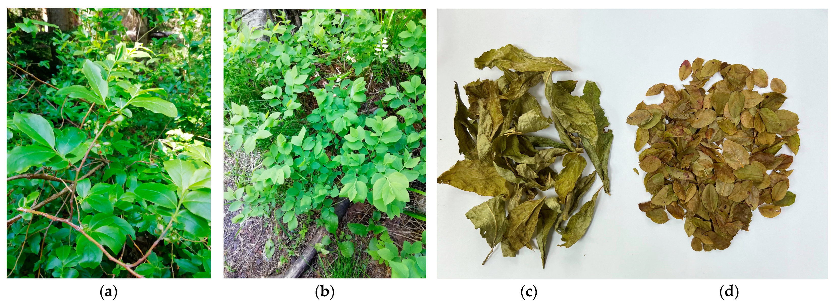
2.1. Plant Material and Chemicals
2.2. Plant Extract Preparation
2.3. High-Performance Liquid Chromatography with Photodiode Array Detection and Electrospray Ionization Triple Quadrupole Mass Spectrometric Detection (HPLC–PDA–ESI–QQQ–MS)
2.4. Neuroprotective Activity
2.4.1. Animals
2.4.2. Brain Ischemia Model
2.4.3. Study Design
2.4.4. Cerebral Blood Flow Evaluation
2.4.5. Biomaterial Sampling and Preparation
2.4.6. Evaluation of Necrosis Zone
2.5. Antioxidant Potential
2.5.1. Thiobarbituric Acid Reactive Substances (TBARS) Evaluation
2.5.2. Superoxide Dismutase (SOD) Activity
2.5.3. Succinate Dehydrogenase (SDH) Activity
2.5.4. Cytochrome-C-Oxidase (COX) Activity
2.6. Statistical Analysis
3. Results
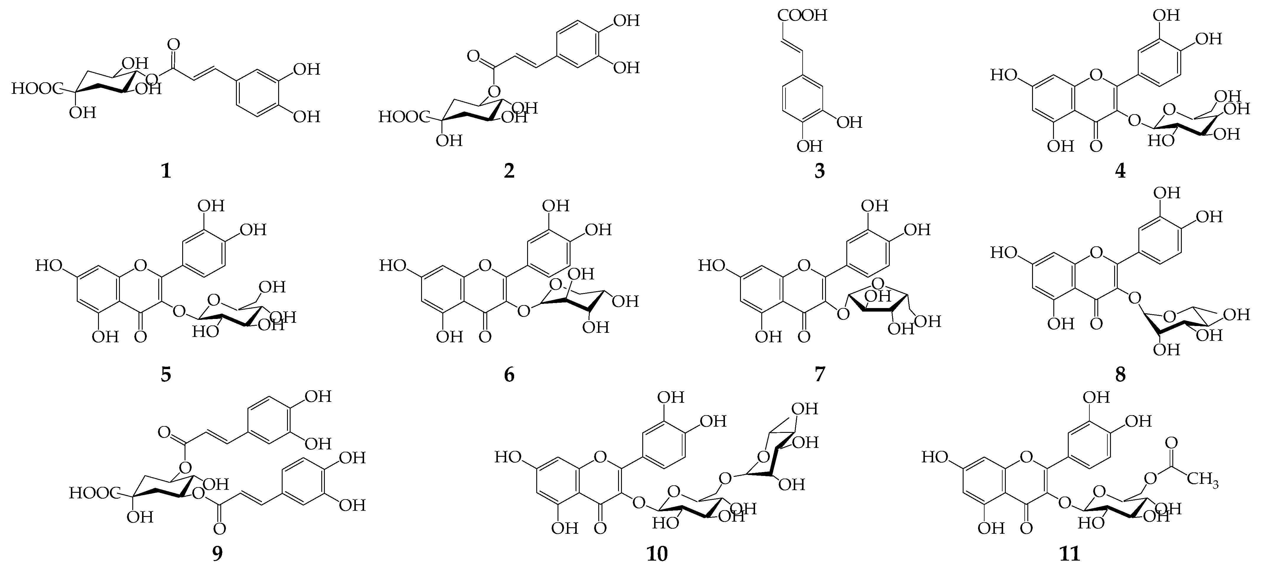
3.1. Phenolic Compounds of V. myrtillus and V. arctostaphylos Leaves
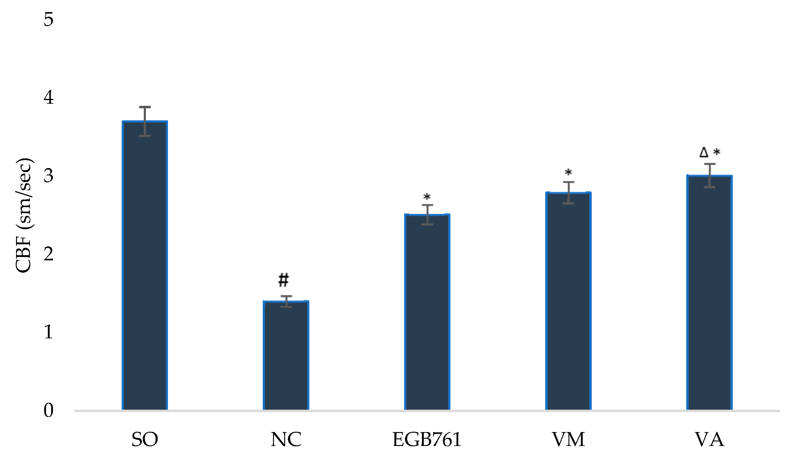
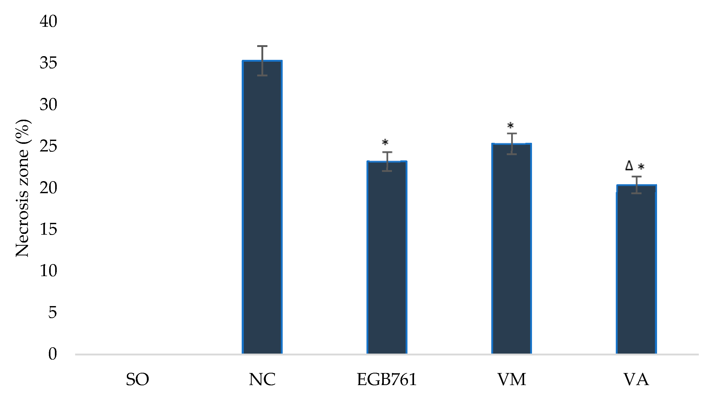
3.2. Neuroprotective Activity of V. myrtillus and V. arctostaphylos Leaf Extracts
3.3. Antioixdant Potential of V. myrtillus and V. arctostaphylos Leaf Extracts
4. Discussion
Author Contributions
Funding
Institutional Review Board Statement
Informed Consent Statement
Data Availability Statement
Acknowledgments
Conflicts of Interest
References
- WHO: The World Flora Online. Available online: http://www.worldfloraonline.org/ (accessed on 12 November 2022).
- Takhtadjan, A. Flowering Plants, 2nd ed.; Springer Science & Business Media: Berlin/Heidelberg, Germany, 2009; pp. 188–193. ISBN 978-1-4020-9608-2. [Google Scholar]
- Czerepanov, S.K. Vascular Plants of Russia and Neighboring Countries; Word and Family-95: Saint-Petersburg, Russia, 1995; p. 424. [Google Scholar]
- Kasotea, D.M.; Duncanb, G.J.; Neacsua, M.; Russella, W.R. Rapid method for quantification of anthocyanidins and anthocyanins in human biological samples. Food Chem. 2019, 290, 56–63. [Google Scholar] [CrossRef] [Green Version]
- Chan, S.W.; Tomlinson, B. Effects of bilberry supplementation on metabolic and cardiovascular disease risk. Molecules 2020, 25, 1653. [Google Scholar] [CrossRef] [Green Version]
- Satoh, Y.; Ishihara, K. Investigation of the antimicrobial activity of bilberry (Vaccinium myrtillus L.) extract against periodontopathic bacteria. J. Oral Biosci. 2020, 62, 169–174. [Google Scholar] [CrossRef]
- Sezer, E.D.; Oktay, L.M.; Karadadaş, E.; Memmedov, H.; Gunel, N.S.; Sözmen, E. Assessing anticancer potential of blueberry flavonoids, quercetin, kaempferol, and gentisic acid, through oxidative stress and apoptosis parameters on HCT-116 cells. J. Med. Food. 2019, 22, 1118–1126. [Google Scholar] [CrossRef]
- Vaneková, Z.; Vanek, M.; Škvarenina, J.; Nagy, M. The influence of local habitat and microclimate on the levels of secondary metabolites in slovak bilberry (Vaccinium myrtillus L.) Fruits. Plants 2020, 9, 436. [Google Scholar] [CrossRef] [Green Version]
- Zheng, W.; Wang, S.Y. Oxygen radical absorbing capacity of phenolics in blueberries, cranberries, chokeberries, and lingonberries. J. Agric. Food Chem. 2003, 51, 502–509. [Google Scholar] [CrossRef]
- Viljanen, K.; Kylli, P.; Kivikari, R.; Heinonen, M. Inhibition of protein and lipid oxidation in liposomes by berry phenolics. J. Agric. Food Chem. 2004, 52, 7419–7424. [Google Scholar] [CrossRef]
- Puupponen-Pimiä, R.; Nohynek, L.; Hartmann-Schmidlin, S.; Kähkönen, M.; Heinonen, M.; Määttä-Riihinen, K.; Oksman-Caldentey, K.M. Berry phenolics selectively inhibit the growth of intestinal pathogens. J. Appl. Microbiol. 2005, 98, 991–1000. [Google Scholar] [CrossRef]
- Ayaz, F.A.; Ayaz, S.H.; Gruz, J.; Novak, O.; Strnad, M. Separation, characterization, and quantitation of phenolic acids in a little-known blueberry (Vaccinium arctostaphylos L.) fruit by HPLC-MS. J. Agric. Food Chem. 2005, 53, 8116–8122. [Google Scholar] [CrossRef]
- Lätti, A.K.; Kainulainen, P.S.; Hayirlioglu-Ayaz, S.; Ayaz, F.A.; Riihinen, K.R. Characterization of anthocyanins in Caucasian blueberries (Vaccinium arctostaphylos L.) native to Turkey. J. Agric. Food Chem. 2009, 57, 5244–5249. [Google Scholar] [CrossRef]
- Chkhikvishvili, I.D.; Kharebava, G.I. Chicoric and chlorogenic acids in plant species from Georgia. Appl. Biochem. Microbiol. 2001, 37, 188–191. [Google Scholar] [CrossRef]
- Mohtashami, R.; Huseini, H.F.; Nabati, F.; Hajiaghaee, R.; Kianbakht, S. Effects of standardized hydro-alcoholic extract of Vaccinium arctostaphylos leaf on hypertension and biochemical parameters in hypertensive hyperlipidemic type 2 diabetic patients: A randomized, double-blind and placebo-controlled clinical trial. Avicenna J. Phytomed. 2019, 9, 44–53. [Google Scholar]
- Nickavar, B.; Amin, G. Enzyme assay guided isolation of an α-amylase inhibitor flavonoid from Vaccinium arctostaphylos leaves. Iran J. Pharm. Res. 2011, 10, 849–853. [Google Scholar]
- Yang, L.; Wang, Z.M.; Wang, Y.; Li, R.S.; Wang, F.; Wang, K. Phenolic constituents with neuroprotective activities from Hypericum wightianum. Phytochemistry 2019, 165, 112049. [Google Scholar] [CrossRef]
- Xie, Y.; Yang, W.; Tang, F.; Chen, X.; Ren, L. Antibacterial activities of flavonoids: Structure-activity relationship and mechanism. Curr. Med. Chem. 2015, 22, 132–149. [Google Scholar] [CrossRef]
- Aryal, S.; Skinner, T.; Bridges, B.; Weber, J.T. The pathology of Parkinson’s disease and potential benefit of dietary polyphenols. Molecules 2020, 25, 4382. [Google Scholar] [CrossRef]
- Kempuraj, D.; Thangavel, R.; Kempuraj, D.D.; Ahmed, M.E.; Selvakumar, G.P.; Raikwar, S.P.; Zaheer, S.A.; Iyer, S.S.; Govindarajan, R.; Chandrasekaran, P.N.; et al. Neuroprotective effects of flavone luteolin in neuroinflammation and neurotrauma. Biofactors 2021, 47, 190–197. [Google Scholar] [CrossRef]
- Zhou, Y.; Zhang, S.; Fan, X. Role of polyphenols as antioxidant supplementation in ischemic stroke. Oxid. Med. Cell Longev. 2021, 2021, 5471347. [Google Scholar] [CrossRef]
- Xie, Y.; Wang, H.; He, Z. Recent advances in polyphenols improving vascular endothelial dysfunction induced by endogenous toxicity. J. Appl. Toxicol. 2021, 41, 701–712. [Google Scholar] [CrossRef]
- Gao, Q.; Dong, J.Y.; Cui, R.; Muraki, I.; Yamagishi, K.; Sawada, N.; Iso, H.; Tsugane, S.; Japan Public Health Center-based Prospective study group. consumption of flavonoid-rich fruits, flavonoids from fruits and stroke risk: A prospective cohort study. Br. J. Nutr. 2021, 126, 1717–1724. [Google Scholar] [CrossRef]
- Kalt, W.; Cassidy, A.; Howard, L.R.; Krikorian, R.; Stull, A.J.; Tremblay, F.; Zamora-Ros, R. Recent research on the health benefits of blueberries and their anthocyanins. Adv. Nutr. 2020, 11, 224–236. [Google Scholar] [CrossRef]
- Rabinstein, A.A. Update on treatment of acute ischemic stroke. Continuum 2020, 26, 268–286. [Google Scholar] [CrossRef]
- Ting, H.C.; Chang, C.Y.; Lu, K.Y.; Chuang, H.M.; Tsai, S.F.; Huang, M.H.; Liu, C.A.; Lin, S.Z.; Harn, H.J. Targeting cellular stress mechanisms and metabolic homeostasis by chinese herbal drugs for neuroprotection. Molecules 2018, 23, 259. [Google Scholar] [CrossRef] [Green Version]
- Riihinen, K.; Jaakola, L.; Kärenlampi, S.; Hohtola, A. Organ-specific distribution of phenolic compounds in bilberry (Vaccinium myrtillus) and ‘northblue’ blueberry (Vaccinium corymbosum × V. angustifolium). Food Chem. 2008, 110, 156–160. [Google Scholar] [CrossRef]
- Rahman, M.M.; Ichiyanagi, T.; Komiyama, T.; Sato, S.; Konishi, T. Effects of anthocyanins on psychological stress-induced oxidative stress and neurotransmitter status. J. Agric. Food Chem. 2008, 56, 7545–7550. [Google Scholar] [CrossRef]
- Olennikov, D.N.; Shamilov, A.A. New compounds from Vaccinium vitis-idaea. Chem. Nat. Compd. 2022, 58, 240–244. [Google Scholar] [CrossRef]
- Olennikov, D.N.; Shamilov, A.A. Catechin-O-rhamnosides from Vaccinium vitis-idaea stems. Chem. Nat. Compd. 2022, 58, 269–273. [Google Scholar] [CrossRef]
- Shamilov, A.A.; Olennikov, D.N.; Pozdnyakov, D.I.; Bubenchikova, V.N.; Garsiya, E.R. Investigation of phenolic compounds at the leaves and shoots Arctostaphylos spp. and their antioxidant and antityrosinase activities. Nat. Prod. Res. 2022, 36, 6312–6317. [Google Scholar] [CrossRef]
- Kashchenko, N.I.; Jafarova, G.S.; Isaev, J.I.; Olennikov, D.N.; Chirikova, N.K. Caucasian dragonheads: Phenolic compounds, polysaccharides, and bioactivity of Dracocephalum austriacum and Dracocephalum botryoides. Plants 2022, 11, 2126. [Google Scholar] [CrossRef]
- Pozdnyakov, D.I. 4-Hydroxy-3,5-di-tret-butyl cinnamic acid restores the activity of the hippocampal mitochondria in rats under permanent focal cerebral ischemia. Iran. J. Basic. Med. Sci. 2021, 24, 1590–1601. [Google Scholar] [CrossRef]
- Wang, N.; Chen, X.; Geng, D.; Huang, H.; Zhou, H. Ginkgo biloba leaf extract improves the cognitive abilities of rats with D-galactose induced dementia. J. Biomed. Res. 2013, 27, 29–36. [Google Scholar] [CrossRef] [PubMed] [Green Version]
- Voronkov, A.V.; Dyakova, I.N.; Pozdnyakov, D.I. The influence of natural compounds of polyphenolic structure on the vasodilating function of the vascular endothelium of the rat brain in conditions of its focal ischemia. Eksp. Klin. Farmakol. 2016, 79, 7–9. [Google Scholar] [PubMed]
- Janero, D.R. Malondialdehyde and thiobarbituric acid-reactivity as diagnostic indices of lipid peroxidation and peroxidative tissue injury. Free Rad. Biol. Med. 1990, 9, 515–540. [Google Scholar] [CrossRef] [PubMed]
- Woolliams, J.A.; Wiener, G.; Anderson, P.H.; McMurray, C.H. Variation in the activities of glutathione peroxidase and superoxide dismutase and in the concentration of copper in the blood in various breed crosses of sheep. Res. Vet. Sci. 1983, 34, 253–256. [Google Scholar] [CrossRef] [PubMed]
- Wang, H.; Huwaimel, B.; Verma, K.; Miller, J.; Germain, T.M.; Kinarivala, N.; Pappas, D.; Brookes, P.S.; Trippier, P.C. Synthesis and antineoplastic evaluation of mitochondrial complex II (succinate dehydrogenase) inhibitors derived from atpenin A5. ChemMedChem 2017, 12, 1033–1044. [Google Scholar] [CrossRef]
- Li, Y.; D’Aurelio, M.; Deng, J.H.; Park, J.S.; Manfredi, G.; Hu, P.; Lu, J.; Bai, Y. An assembled complex IV maintains the stability and activity of complex I in mammalian mitochondria. J. Biol. Chem. 2007, 282, 17557–17562. [Google Scholar] [CrossRef] [Green Version]
- Ștefănescu, B.E.; Szabo, K.; Mocan, A.; Crişan, G. Phenolic compounds from five Ericaceae species leaves and their related bioavailability and health benefits. Molecules 2019, 24, 2046. [Google Scholar] [CrossRef] [Green Version]
- Mzhavanadze, V.V.; Targamadze, I.L.; Dranik, L.I. Polyphenols of the leaves of Vaccinium arctostaphylos. Chem. Nat. Compd. 1971, 7, 536–537. [Google Scholar] [CrossRef]
- Liu, P.; Lindstedt, A.; Markkinen, N.; Sinkkonen, J.; Suomela, J.-P.; Yang, B. Characterization of metabolite profiles of leaves of bilberry (Vaccinium myrtillus L.) and lingonberry (Vaccinium vitis-idaea L.). J. Agric. Food Chem. 2014, 62, 12015–12026. [Google Scholar] [CrossRef]
- Mzhavanadze, V.V.; Targamadze, I.L.; Dranik, L.I. Phenolic compounds of the leaves of Vaccinium arctostaphylos. Chem. Nat. Compd. 1972, 8, 125–126. [Google Scholar] [CrossRef]
- Tadić, V.M.; Nešić, I.; Martinović, M.; Rój, E.; Brašanac-Vukanović, S.; Maksimović, S.; Žugić, A. Old plant, new possibilities: Wild bilberry (Vaccinium myrtillus L., Ericaceae) in topical skin preparation. Antioxidants 2021, 10, 465. [Google Scholar] [CrossRef] [PubMed]
- Ieri, F.; Martini, S.; Innocwnti, M.; Mulinacci, N. Phenolic distribution in liquid preparations of Vaccinium myrtillus L. and Vaccinium vitis idaea L. Phytochem. Anal. 2013, 24, 467–475. [Google Scholar] [CrossRef] [PubMed]
- Hasaloo, T.; Sepehrifar, R.; Hajimehdipoor, H. Levels of phenolic compounds and their effects on antioxidant capacity of wild Vaccinium arctostaphylos L. (Qare-Qat) collected from different regions of Iran. Turk. J. Biol. 2011, 35, 13. [Google Scholar] [CrossRef]
- Oszmianski, J.; Wojdyło, A.; Gorzelany, J.; Kapusta, I. Identification and characterization of low molecular weight polyphenols in berry leaf extracts by HPLC-DAD and LC-ESI/MS). J. Agric. Food Chem. 2011, 59, 12830–12835. [Google Scholar] [CrossRef]
- Taram, F.; Winter, A.N.; Linseman, D.A. Neuroprotection comparison of chlorogenic acid and its metabolites against mechanisti-cally distinct cell death-inducing agents in cultured cerebellar granule neurons. Brain Res. 2016, 1648, 69–80. [Google Scholar] [CrossRef]
- Zheng, Y.; Li, L.; Chen, B.; Fang, Y.; Lin, W.; Zhang, T.; Feng, X.; Tao, X.; Wu, Y.; Fu, X.; et al. Chlorogenic acid exerts neuroprotective effect against hypoxia-ischemia brain injury in neonatal rats by activating Sirt1 to regulate the Nrf2-NF-κB signaling pathway. Cell Commun. Signal. 2022, 20, 84. [Google Scholar] [CrossRef]
- Singh, S.S.; Rai, S.N.; Birla, H.; Zahra, W.; Rathore, A.S.; Dilnashin, H.; Singh, R.; Singh, S.P. Neuroprotective Effect of chlorogenic acid on mitochondrial dysfunction-mediated apoptotic death of DA neurons in a Parkinsonian mouse model. Oxid. Med. Cell Longev. 2020, 2020, 6571484. [Google Scholar] [CrossRef]
- Khan, H.; Ullah, H.; Aschner, M.; Cheang, W.S.; Akkol, E.K. Neuroprotective effects of quercetin in Alzheimer’s disease. Biomolecules 2019, 10, 59. [Google Scholar] [CrossRef] [Green Version]
- Alvarez-Arellano, L.; Salazar-García, M.; Corona, J.C. Neuroprotective effects of quercetin in pediatric neurological diseases. Molecules 2020, 25, 5597. [Google Scholar] [CrossRef]
- Pu, F.; Mishima, K.; Irie, K.; Motohashi, K.; Tanaka, Y.; Orito, K.; Egawa, T.; Kitamura, Y.; Egashira, N.; Iwasaki, K.; et al. Neuroprotective effects of quercetin and rutin on spatial memory impairment in an 8-arm radial maze task and neuronal death induced by repeated cerebral ischemia in rats. J. Pharmacol. Sci. 2007, 104, 329–334. [Google Scholar] [CrossRef] [Green Version]
- Gaire, B.P. Herbal medicine in ischemic stroke: Challenges and prospective. Chin. J. Integr. Med. 2018, 24, 243–246. [Google Scholar] [CrossRef] [PubMed]
- Mishra, A.; Mishra, P.S.; Bandopadhyay, R.; Khurana, N.; Angelopoulou, E.; Paudel, Y.N.; Piperi, C. Neuroprotective potential of chrysin: Mechanistic insights and therapeutic potential for neurological disorders. Molecules 2021, 26, 6456. [Google Scholar] [CrossRef] [PubMed]
- Wang, J.; Zhang, J.; Li, S.; Huang, C.; Xie, Y.; Cao, Y. Anthocyanins decrease the internalization of TiO2 nanoparticles into 3D Caco-2 spheroids. Food Chem. 2020, 331, 127360. [Google Scholar] [CrossRef] [PubMed]
- Wu, S.; Wu, B.; Liu, M.; Chen, Z.; Wang, W.; Anderson, C.S.; Sandercock, P.; Wang, Y.; Huang, Y.; Cui, L.; et al. China stroke study collaboration. Stroke in China: Advances and challenges in epidemiology, prevention, and management. Lancet Neurol. 2019, 18, 394–405. [Google Scholar] [CrossRef]
- Silva, S.; Costa, E.M.; Veiga, M.; Morais, R.M.; Calhau, C.; Pintado, M. Health promoting properties of blueberries: A review. Crit. Rev. Food Sci. Nutr. 2020, 60, 181–200. [Google Scholar] [CrossRef]
- He, Z.; Ning, N.; Zhou, Q.; Khoshnam, S.E.; Farzaneh, M. Mitochondria as a therapeutic target for ischemic stroke. Free Radic. Biol. Med. 2020, 146, 45–58. [Google Scholar] [CrossRef]


| Compound | Regression Equation a | r2 | SYX | LOD/LOQ (µg/mL) | Linear Range (µg/mL) | |
|---|---|---|---|---|---|---|
| a | b × 106 | |||||
| 4-O-Caffeoylquinic acid | 0.162 | −0.011 | 0.9973 | 1.83 × 10⁻² | 0.37/1.12 | 2–100 |
| 5-O-Caffeoylquinic acid | 0.150 | −0.010 | 0.9982 | 1.67 × 10⁻² | 0.36/1.11 | 2–100 |
| Caffeic acid | 0.189 | −0.017 | 0.9988 | 1.26 × 10⁻² | 0.22/0.67 | 1–100 |
| Quercetin 3-O-rutinoside (rutin) | 0.085 | −0.062 | 0.9873 | 3.89 × 10⁻² | 1.51/4.57 | 5–100 |
| Quercetin 3-O-galactoside (hyperoside) | 0.090 | −0.052 | 0.9891 | 3.52 × 10⁻² | 1.29/3.91 | 4–100 |
| Quercetin 3-O-glucoside (isoquercitrin) | 0.098 | −0.053 | 0.9889 | 3.37 × 10⁻² | 1.14/3.43 | 4–100 |
| Quercetin 3-O-arabinopyranoside (guaiaverin) | 0.121 | −0.031 | 0.9953 | 2.59 × 10⁻² | 0.70/2.14 | 3–100 |
| Quercetin 3-O-arabinofuranoside (avicularin) | 0.114 | −0.026 | 0.9927 | 2.80 × 10⁻² | 0.81/2.46 | 3–100 |
| Quercetin 3-O-rhamnoside (quercitrin) | 0.109 | −0.043 | 0.9912 | 3.16 × 10⁻² | 0.96/2.90 | 3–100 |
| Quercetin 3-O-(6″-acetyl)-glucoside | 0.095 | −0.050 | 0.9880 | 3.28 × 10⁻² | 1.14/3.45 | 4–100 |
| 4,5-Di-O-caffeoylquinic acid | 0.163 | −0.016 | 0.9975 | 1.93 × 10⁻² | 0.39/1.18 | 2–100 |
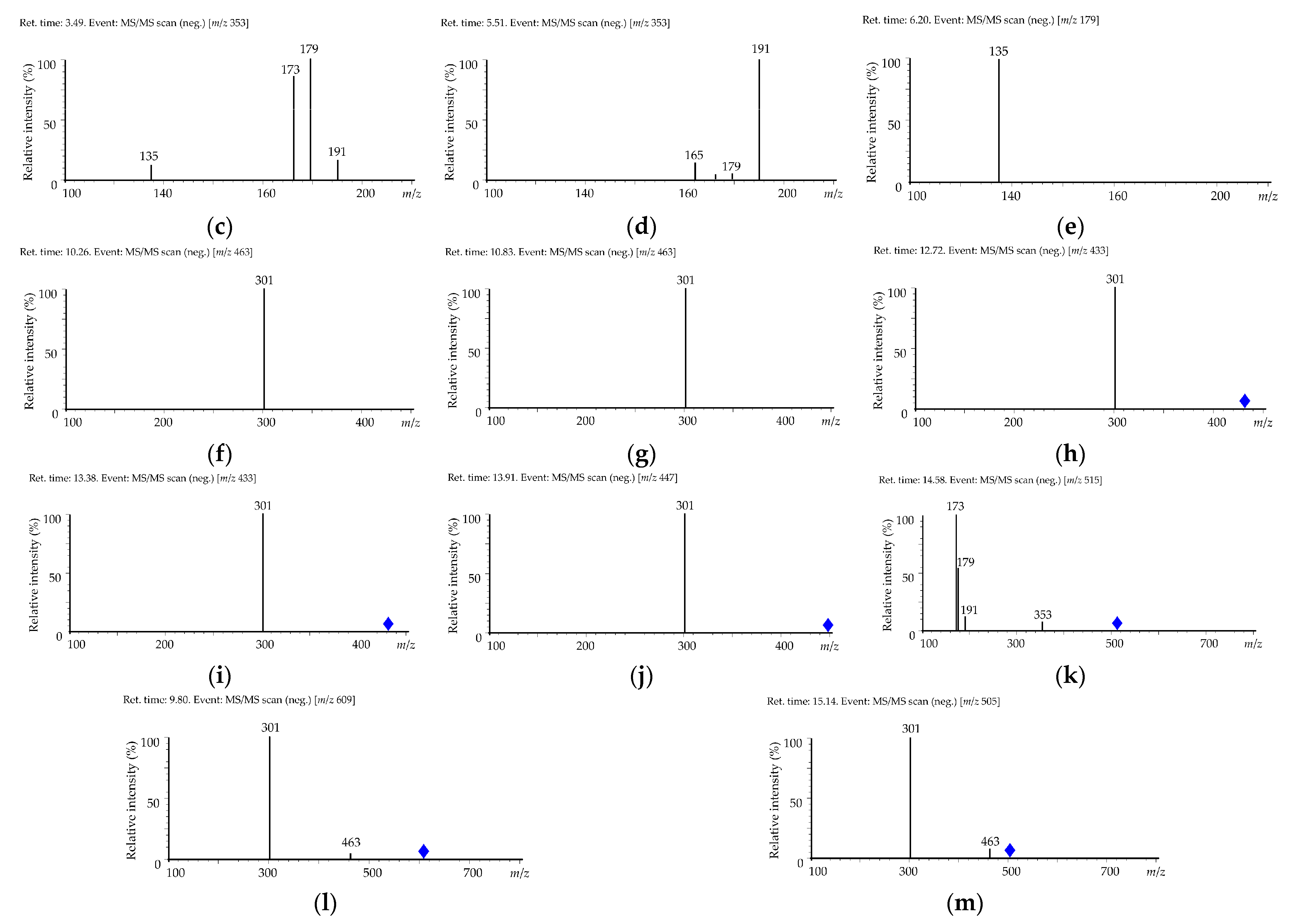
| No | t, min | Compound * | UV, λmax, nm | ESI-MS, [M − H]−, m/z | MS/MS, m/z |
|---|---|---|---|---|---|
| 1 | 3.49 | 4-O-Caffeoylquinic acid | 322 | 353 | [353]: 191, 179, 173, 135 |
| 2 | 5.51 | 5-O-Caffeoylquinic acid | 322 | 353 | [353]: 191, 179, 165 |
| 3 | 6.20 | Caffeic acid | 320 | 179 | [179]: 135 |
| 4 | 10.26 | Quercetin 3-O-galactoside (hyperoside) | 255, 267, 355 | 463 | [463]: 301 |
| 5 | 10.83 | Quercetin 3-O-glucoside (isoquercitrin) | 255, 267, 356 | 463 | [463]: 301 |
| 6 | 12.72 | Quercetin 3-O-arabinopyranoside (guaiaverin) | 255, 267, 354 | 433 | [433]: 301 |
| 7 | 13.38 | Quercetin 3-O-arabinofuranoside (avicularin) | 255, 267, 354 | 433 | [433]: 301 |
| 8 | 13.91 | Quercetin 3-O-rhamnoside (quercitrin) | 255, 267, 352 | 447 | [447]: 301 |
| 9 | 14.58 | 3,5-Di-O-caffeoylquinic acid | 322 | 515 | [515]: 353, 191, 179, 173 |
| 10 | 9.80 | Quercetin 3-O-rutinoside (rutin) | 255, 267, 355 | 609 | [609]: 463, 301 |
| 11 | 15.14 | Quercetin 3-O-(6″-acetyl)-glucoside | 256, 268, 351 | 505 | [505]: 463, 301 |
| Compound | V. myrtillus | V. arctostaphylos |
|---|---|---|
| 4-O-Caffeoylquinic acid | <0.01 | 8.01 ± 0.14 |
| 5-O-Caffeoylquinic acid | 226.85 ± 5.21 | 105.32 ± 2.41 |
| Caffeic acid | <0.01 | 4.29 ± 0.08 |
| Quercetin 3-O-rutinoside (rutin) | <0.01 | 1.46 ± 0.03 |
| Quercetin 3-O-galactoside (hyperoside) | 4.69 ± 0.09 | 3.99 ± 0.06 |
| Quercetin 3-O-glucoside (isoquercitrin) | 12.02 ± 0.24 | 2.38 ± 0.05 |
| Quercetin 3-O-arabinopyranoside (guaiaverin) | 1.34 ± 0.02 | 1.29 ± 0.02 |
| Quercetin 3-O-arabinofuranoside (avicularin) | <0.01 | <0.01 |
| Quercetin 3-O-rhamnoside (quercitrin) | 2.77 ± 0.05 | 1.23 ± 0.02 |
| Quercetin 3-O-(6″-acetyl)-glucoside | <0.01 | 0.70 ± 0.01 |
| 4,5-Di-O-caffeoylquinic acid | <0.01 | <0.01 |
| Experimental Group | SOD, U/Protein mg | COX, U/Protein mg | SDH, U/Protein mg | TBARS, µM/Protein mg |
|---|---|---|---|---|
| Sham-operated animals | 300.5 ± 7.1(29) | 4.3 ± 0.0(47) | 2.7 ± 0.062 | 2.4 ± 0.124 |
| Negative control group | 125.6 ± 9.5(41) # | 2.1 ± 0.0(14) # | 1.1 ± 0.097 # | 9.8 ± 0.663 # |
| EGB761 reference group | 190.5 ± 11.2(84) * | 2.5 ± 0.1(17) * | 1.6 ± 0.018 * | 5.2 ± 0.541 * |
| V. myrtillus group | 181.0 ± 10.9(37) * | 2.6 ± 0.0(37) * | 1.5 ± 0.09 * | 4.4 ± 0.364 * |
| V. arctostaphylos group | 221.5 ± 10.2(32) * | 3.0 ± 0.0(58) * Δα | 2.1 ± 0.071 * Δα | 3.5 ± 0.193 * |
Publisher’s Note: MDPI stays neutral with regard to jurisdictional claims in published maps and institutional affiliations. |
© 2022 by the authors. Licensee MDPI, Basel, Switzerland. This article is an open access article distributed under the terms and conditions of the Creative Commons Attribution (CC BY) license (https://creativecommons.org/licenses/by/4.0/).
Share and Cite
Shamilov, A.A.; Olennikov, D.N.; Pozdnyakov, D.I.; Bubenchikova, V.N.; Garsiya, E.R.; Larskii, M.V. Caucasian Blueberry: Comparative Study of Phenolic Compounds and Neuroprotective and Antioxidant Potential of Vaccinium myrtillus and Vaccinium arctostaphylos Leaves. Life 2022, 12, 2079. https://doi.org/10.3390/life12122079
Shamilov AA, Olennikov DN, Pozdnyakov DI, Bubenchikova VN, Garsiya ER, Larskii MV. Caucasian Blueberry: Comparative Study of Phenolic Compounds and Neuroprotective and Antioxidant Potential of Vaccinium myrtillus and Vaccinium arctostaphylos Leaves. Life. 2022; 12(12):2079. https://doi.org/10.3390/life12122079
Chicago/Turabian StyleShamilov, Arnold A., Daniil N. Olennikov, Dmitryi I. Pozdnyakov, Valentina N. Bubenchikova, Ekaterina R. Garsiya, and Mikhail V. Larskii. 2022. "Caucasian Blueberry: Comparative Study of Phenolic Compounds and Neuroprotective and Antioxidant Potential of Vaccinium myrtillus and Vaccinium arctostaphylos Leaves" Life 12, no. 12: 2079. https://doi.org/10.3390/life12122079