

Wound Healing and Anti-Inflammatory Effects of a Newly Developed Ointment Containing Jujube Leaves Extract

 , , ,
, , ,
Abstract
:1. Introduction
2. Materials and Methods
2.1. Materials
2.2. Methods
2.2.1. Ointment Production and Pharmacotechnical Assessment
Formulation
Production
Quality Control
pH
Ultra-High-Performance Liquid Chromatography Electrospray Ionization Tandem Mass Spectrometry
Spreadability
2.2.2. In Vivo Evaluation
Local Tolerability
Determination of Acute Dermal Irritation/Corrosion (OECD 404)
Determination of Dermal Irritation after Repeated Administration (OECD 410)
- Group 1F: ZIZ-L-F: consisting of 4 females, who received the lipophilic ointment ZIZ-L;
- Group 1M: ZIZ-L-M: consisting of 4 males, who received the lipophilic ointment ZIZ-L;
- Group 2F: B-L-F: consisting of 4 females, who received the lipophilic base B-L;
- Group 2M: B-L-M: consisting of 4 males, who received the lipophilic base B-L.
Wound Healing Activity
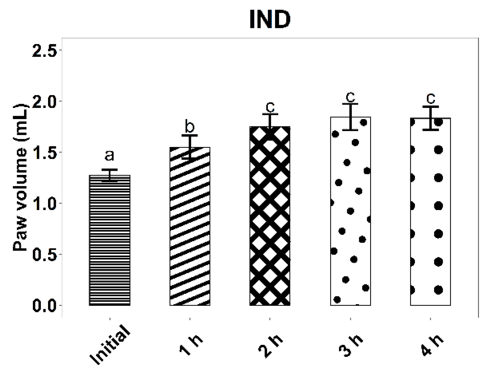
Anti-Inflammatory Activity
- Group ZIZ-L: ZIZ-L: lipophilic ointment;
- Group B-L: B-L lipophilic base;
- Group IND: Indomethacin HYPERION, ointment, 40 mg/g.
Statistical Analysis
3. Results and Discussion
3.1. Ointment Quality Control
3.1.1. Organoleptic Properties and Homogeneity
3.1.2. pH
3.1.3. Ultra-High-Performance Liquid Chromatography Electrospray Ionization Tandem Mass Spectrometry
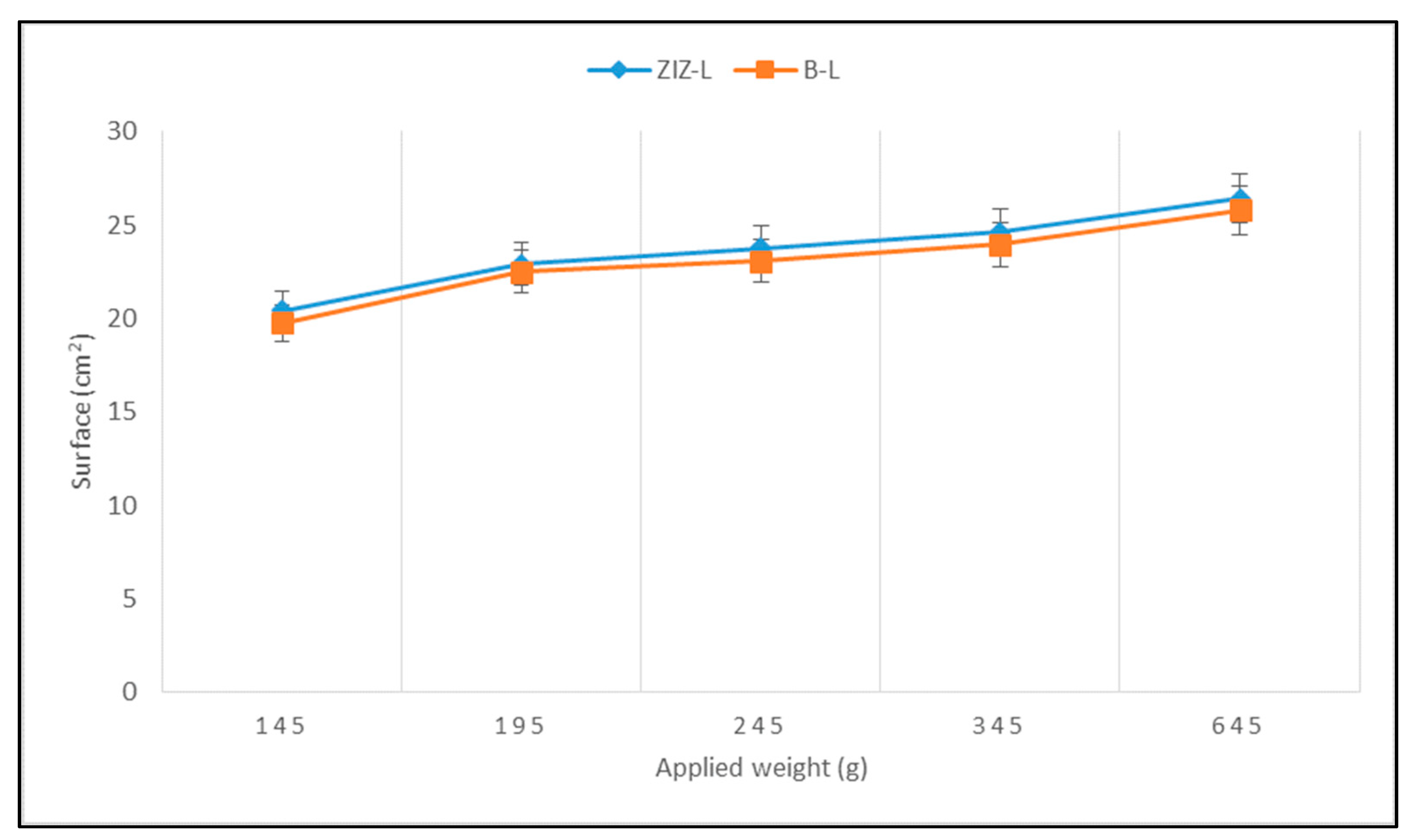
3.1.4. Spreadability
3.2. Local Tolerability
3.2.1. Determination of Acute Dermal Irritation/Corrosion (OECD 404)
3.2.2. Determination of Dermal Irritation after Repeated Administration (OECD 410)
3.3. Wound Healing Activity
3.4. Anti-Inflammatory Activity
4. Conclusions
Author Contributions
Funding
Institutional Review Board Statement
Informed Consent Statement
Data Availability Statement
Conflicts of Interest
References
- Shedoeva, A.; Leavesley, D.; Upton, Z.; Fan, C. Wound Healing and the Use of Medicinal Plants. Evid.-Based Complementary Altern. Med. 2019, 2019, 2684108. [Google Scholar] [CrossRef] [PubMed] [Green Version]
- Dorai, A.A. Wound Care with Traditional, Complementary and Alternative Medicine. Indian J. Plast. Surg. 2012, 45, 418–424. [Google Scholar] [CrossRef] [PubMed]
- Jarić, S.; Kostić, O.; Mataruga, Z.; Pavlović, D.; Pavlović, M.; Mitrović, M.; Pavlović, P. Traditional Wound-Healing Plants Used in the Balkan Region (Southeast Europe). J. Ethnopharmacol. 2018, 211, 311–328. [Google Scholar] [CrossRef] [PubMed]
- Kumar, B.; Vijayakumar, M.; Govindarajan, R.; Pushpangadan, P. Ethnopharmacological Approaches to Wound Healing—Exploring Medicinal Plants of India. J. Ethnopharmacol. 2007, 114, 103–113. [Google Scholar] [CrossRef]
- Rhoads, D.D.; Cox, S.B.; Rees, E.J.; Sun, Y.; Wolcott, R.D. Clinical Identification of Bacteria in Human Chronic Wound Infections: Culturing vs. 16S Ribosomal DNA Sequencing. BMC Infect. Dis. 2012, 12, 321. [Google Scholar] [CrossRef] [PubMed] [Green Version]
- Maxson, S.; Lopez, E.A.; Yoo, D.; Danilkovitch-Miagkova, A.; LeRoux, M.A. Concise Review: Role of Mesenchymal Stem Cells in Wound Repair. Stem Cells Transl. Med. 2012, 1, 142–149. [Google Scholar] [CrossRef]
- Reinke, J.M.; Sorg, H. Wound Repair and Regeneration. Eur. Surg. Res. 2012, 49, 35–43. [Google Scholar] [CrossRef]
- Edeoga, H.O.; Okwu, D.E.; Mbaebie, B.O. Phytochemical Constituents of Some Nigerian Medicinal Plants. Afr. J. Biotechnol. 2005, 4, 685–688. [Google Scholar] [CrossRef] [Green Version]
- Chen, W.-C.; Liou, S.-S.; Tzeng, T.-F.; Lee, S.-L.; Liu, I.-M. Effect of Topical Application of Chlorogenic Acid on Excision Wound Healing in Rats. Planta Med. 2013, 79, 616–621. [Google Scholar] [CrossRef]
- Maver, T.; Maver, U.; Stana Kleinschek, K.; Smrke, D.M.; Kreft, S. A Review of Herbal Medicines in Wound Healing. Int. J. Dermatol. 2015, 54, 740–751. [Google Scholar] [CrossRef]
- Sharma, A.; Khanna, S.; Kaur, G.; Singh, I. Medicinal Plants and Their Components for Wound Healing Applications. Future J. Pharm. Sci. 2021, 7, 53. [Google Scholar] [CrossRef]
- Appendino, G.; Ottino, M.; Marquez, N.; Bianchi, F.; Giana, A.; Ballero, M.; Sterner, O.; Fiebich, B.L.; Munoz, E. Arzanol, an Anti-Inflammatory and Anti-HIV-1 Phloroglucinol Alpha-Pyrone from Helichrysum Italicum Ssp. Microphyllum. J. Nat. Prod. 2007, 70, 608–612. [Google Scholar] [CrossRef]
- Wu, F.; Bian, D.; Xia, Y.; Gong, Z.; Tan, Q.; Chen, J.; Dai, Y. Identification of Major Active Ingredients Responsible for Burn Wound Healing of Centella Asiatica Herbs. Evid.-Based Complementary Altern. Med. 2012, 2012, 848093. [Google Scholar] [CrossRef] [Green Version]
- Süntar, I.; Akkol, E.K.; Nahar, L.; Sarker, S.D. Wound Healing and Antioxidant Properties: Do They Coexist in Plants? Free. Radic. Antioxid. 2012, 2, 1–7. [Google Scholar] [CrossRef] [Green Version]
- Sen, C.K.; Roy, S. Redox Signals in Wound Healing. Biochim. Biophys. Acta 2008, 1780, 1348–1361. [Google Scholar] [CrossRef] [Green Version]
- Thakur, R.; Jain, N.; Pathak, R.; Sandhu, S.S. Practices in Wound Healing Studies of Plants. Evid.-Based Complementary Altern. Med. 2011, 2011, 438056. [Google Scholar] [CrossRef] [Green Version]
- Manjunatha, B.; Vidya, S.; Rashmi, K.; Mankani, K.; Shilpa, H.; Singh, S.J. Evaluation of Wound-Healing Potency of Vernonia Arborea Hk. Indian J. Pharmacol. 2005, 37, 223. [Google Scholar] [CrossRef]
- Shikov, A.N.; Pozharitskaya, O.N.; Makarov, V.G.; Wagner, H.; Verpoorte, R.; Heinrich, M. Medicinal Plants of the Russian Pharmacopoeia; Their History and Applications. J. Ethnopharmacol. 2014, 154, 481–536. [Google Scholar] [CrossRef] [Green Version]
- Dinda, M.; Mazumdar, S.; Das, S.; Ganguly, D.; Dasgupta, U.B.; Dutta, A.; Jana, K.; Karmakar, P. The Water Fraction of Calendula officinalis Hydroethanol Extract Stimulates In Vitro and In Vivo Proliferation of Dermal Fibroblasts in Wound Healing: In Vitro and In Vivo Wound Healing Activity of Calendula officinalis. Phytother. Res. 2016, 30, 1696–1707. [Google Scholar] [CrossRef]
- Givol, O.; Kornhaber, R.; Visentin, D.; Cleary, M.; Haik, J.; Harats, M. A Systematic Review of Calendula officinalis Extract for Wound Healing. Wound Repair Regen. 2019, 27, 548–561. [Google Scholar] [CrossRef]
- Quave, C.L. Wound Healing with Botanicals: A Review and Future Perspectives. Curr. Dermatol. Rep. 2018, 7, 287–295. [Google Scholar] [CrossRef]
- Wölfle, U.; Seelinger, G.; Schempp, C. Topical Application of St. John’s Wort (Hypericum perforatum). Planta Med. 2013, 80, 109–120. [Google Scholar] [CrossRef] [Green Version]
- Idolo, M.; Motti, R.; Mazzoleni, S. Ethnobotanical and Phytomedicinal Knowledge in a Long-History Protected Area, the Abruzzo, Lazio and Molise National Park (Italian Apennines). J. Ethnopharmacol. 2010, 127, 379–395. [Google Scholar] [CrossRef]
- Nayak, B.S.; Raju, S.S.; Rao, A.V.C. Wound Healing Activity of Matricaria recutita L. Extract. J. Wound Care 2007, 16, 298–302. [Google Scholar] [CrossRef]
- Niknam, S.; Tofighi, Z.; Faramarzi, M.A.; Abdollahifar, M.A.; Sajadi, E.; Dinarvand, R.; Toliyat, T. Polyherbal Combination for Wound Healing: Matricaria chamomilla L. and Punica granatum L. DARU J. Pharm. Sci. 2021, 29, 133–145. [Google Scholar] [CrossRef]
- Shetty, B.S.; Udupa, S.L.; Udupa, A.L.; Somayaji, S.N. Effect of Centella asiatica L (Umbelliferae) on Normal and Dexamethasone-Suppressed Wound Healing in Wistar Albino Rats. Int. J. Low. Extrem. Wounds 2006, 5, 137–143. [Google Scholar] [CrossRef]
- Araújo, L.U.; Reis, P.G.; Barbosa, L.C.O.; Saúde-Guimarães, D.A.; Grabe-Guimarães, A.; Mosqueira, V.C.F.; Carneiro, C.M.; Silva-Barcellos, N.M. In Vivo Wound Healing Effects of Symphytum officinale L. Leaves Extract in Different Topical Formulations. Pharmazie 2012, 67, 355–360. [Google Scholar]
- Antunes Viegas, D.; Palmeira-de-Oliveira, A.; Salgueiro, L.; Martinez-de-Oliveira, J.; Palmeira-de-Oliveira, R. Helichrysum Italicum: From Traditional Use to Scientific Data. J. Ethnopharmacol. 2014, 151, 54–65. [Google Scholar] [CrossRef]
- Sala, A.; Recio, M.; Giner, R.M.; Máñez, S.; Tournier, H.; Schinella, G.; Ríos, J.-L. Anti-Inflammatory and Antioxidant Properties of Helichrysum Italicum. J. Pharm. Pharmacol. 2002, 54, 365–371. [Google Scholar] [CrossRef]
- Hamedi, S.; Shams-Ardakani, M.R.; Sadeghpour, O.; Amin, G.; Hajighasemali, D.; Orafai, H. Designing Mucoadhesive Discs Containing Stem Bark Extract of Ziziphus Jujuba Based on Iranian Traditional Documents. Iran. J. Basic Med. Sci. 2016, 19, 330. [Google Scholar] [CrossRef]
- Guo, S.; Duan, J.; Qian, D.; Tang, Y.; Wu, D.; Su, S.; Wang, H.; Zhao, Y. Content Variations of Triterpenic Acid, Nucleoside, Nucleobase, and Sugar in Jujube (Ziziphus Jujuba) Fruit during Ripening. Food Chem. 2015, 167, 468–474. [Google Scholar] [CrossRef] [PubMed]
- Preeti; Tripathi, S. Ziziphus Jujuba: A Phytopharmacological Review. IJRDPL 2014, 3, 959–966. [Google Scholar]
- Hossain, M.A. A Phytopharmacological Review on the Omani Medicinal Plant: Ziziphus Jujube. J. King Saud Univ. Sci. 2019, 31, 1352–1357. [Google Scholar] [CrossRef]
- Hamedi, S.; Sadeghpour, O.; Shamsardekani, M.R.; Amin, G.; Hajighasemali, D.; Feyzabadi, Z. The Most Common Herbs to Cure the Most Common Oral Disease: Stomatitis Recurrent Aphthous Ulcer (RAU). Iran. Red Crescent Med. J. 2016, 18, e21694. [Google Scholar] [CrossRef] [PubMed] [Green Version]
- Masullo, M.; Cerulli, A.; Montoro, P.; Pizza, C.; Piacente, S. In Depth LC-ESIMSn-Guided Phytochemical Analysis of Ziziphus Jujuba Mill. Leaves. Phytochemistry 2019, 159, 148–158. [Google Scholar] [CrossRef]
- Damiano, S.; Forino, M.; De, A.; Vitali, L.A.; Lupidi, G.; Taglialatela-Scafati, O. Antioxidant and Antibiofilm Activities of Secondary Metabolites from Ziziphus Jujuba Leaves Used for Infusion Preparation. Food Chem. 2017, 230, 24–29. [Google Scholar] [CrossRef] [PubMed]
- Hovaneţ, M.-V.; Oprea, E.; Ancuceanu, R.V.; Duţu, L.E.; Budura, E.A.; Şeremet, O.; Ancu, I.; Moroşan, E. Wound Healing Properties of Ziziphus Jujuba Mill. Leaves. Rom. Biotechnol. Lett. 2016, 21, 11842–11849. [Google Scholar]
- Hovaneţ, M.-V.; Ancuceanu, R.V.; Dinu, M.; Oprea, E.; Budura, E.A.; Negreş, S.; Velescu, B.; Duţu, L.; Anghel, I.A.; Ancu, I.; et al. Toxicity and Anti-Inflammatory Activity of Ziziphus Jujuba Mill. Leaves. Farmacia 2016, 64, 802–808. [Google Scholar]
- Andjić, M.; Božin, B.; Draginić, N.; Kočović, A.; Jeremić, J.N.; Tomović, M.; Milojević Šamanović, A.; Kladar, N.; Čapo, I.; Jakovljević, V.; et al. Formulation and Evaluation of Helichrysum Italicum Essential Oil-Based Topical Formulations for Wound Healing in Diabetic Rats. Pharmaceuticals 2021, 14, 813. [Google Scholar] [CrossRef]
- Han, X.; Beaumont, C.; Stevens, N. Chemical Composition Analysis and in vitro Biological Activities of Ten Essential Oils in Human Skin Cells. Biochim. Open 2017, 5, 1–7. [Google Scholar] [CrossRef]
- Atzmony, L.; Lim, Y.H.; Hamilton, C.; Leventhal, J.S.; Wagner, A.; Paller, A.S.; Choate, K.A. Topical cholesterol/lovastatin for the treatment of porokeratosis: A pathogenesis-directed therapy. J. Am. Acad. Dermatol. 2020, 82, 123–131. [Google Scholar] [CrossRef] [PubMed]
- Murota, H.; Itoi, S.; Terao, M.; Matsui, S.; Kawai, H.; Satou, Y.; Suda, K.; Katayama, I. Topical cholesterol treatment ameliorates hapten-evoked cutaneous hypersensitivity by sustaining expression of 11β-HSD1 in epidermis. Exp. Dermatol. 2014, 23, 68–70. [Google Scholar] [CrossRef] [PubMed] [Green Version]
- Elder, R.L. Final Report on the Safety Assessment of Cetearyl Alcohol, Cetyl Alcohol, lsostearyl Alcohol, Myristyl Alcohol, and Behenyl Alcohol. J. Am. Coll. Toxicol. 1988, 7, 359–413. [Google Scholar] [CrossRef]
- American Academy of Dermatology. Proper Wound Care. 2017. Available online: https://www.aad.org/public/everyday-care/injured-skin/burns/wound-care-minimize-scars (accessed on 10 November 2022).
- Yeap, S.K.; Beh, B.K.; Ali, N.M.; Yusof, H.M.; Ho, W.Y.; Koh, S.P.; Alitheen, N.B.; Long, K. Antistress and Antioxidant Effects of Virgin Coconut Oil in Vivo. Exp. Ther. Med. 2015, 9, 39–42. [Google Scholar] [CrossRef] [PubMed]
- Ghani, N.A.A.; Channip, A.-A.; Chok Hwee Hwa, P.; Ja’afar, F.; Yasin, H.M.; Usman, A. Physicochemical Properties, Antioxidant Capacities, and Metal Contents of Virgin Coconut Oil Produced by Wet and Dry Processes. Food Sci. Nutr. 2018, 6, 1298–1306. [Google Scholar] [CrossRef] [PubMed]
- Burnett, C.L.; Bergfeld, W.F.; Belsito, D.V.; Klaassen, C.D.; Marks, J.G.; Shank, R.C.; Slaga, T.J.; Snyder, P.W.; Andersen, F.A. Final Report on the Safety Assessment of Cocos Nucifera (Coconut) Oil and Related Ingredients. Int. J. Toxicol. 2011, 30, 5S–16S. [Google Scholar] [CrossRef]
- Gavriloaia, M.-R.; Budura, E.-A.; Toma, C.C.; Mitu, M.A.; Karampelas, O.; Arama, C.; Lupuleasa, D. In Vitro Evaluation of Diffusion and Rheological Profiles for Dexamethasone Inclusion Complexes with β-Cyclodextrin or Hydroxypropyl β-Cyclodextrin. Farmacia 2012, 60, 895–904. [Google Scholar]
- Balaci, T.D.; Ozon, E.A.; Baconi, D.L.; Nițulescu, G.; Velescu, B.; Bălălău, C.; Păunică, I.; Fița, C.A. Study on the Formulation and Characterization of a Photoprotective Cream Containing a New Synthetized Compound. J. Mind Med. Sci. 2020, 7, 193–200. [Google Scholar]
- Aiyalu, R.; Govindarjan, A.; Ramasamy, A. Formulation and Evaluation of Topical Herbal Gel for the Treatment of Arthritis in Animal Model. Braz. J. Pharm. Sci. 2016, 52, 493–507. [Google Scholar] [CrossRef]
- Akhtar, N.; Khan, B.; Khan, M.; Mahmood, T.; Khan, H.; Iqbal, M.; Bashir, S. Formulation Development and Moiturising Effects of a Topical Cream of Aloe Vera Extract. World Acad. Eng. Technol. 2011, 51, 172–179. [Google Scholar]
- Comisia Farmacopeei Române. Farmacopeea Română (Romanian Pharmacopoeia), 10th ed.; Editura Medicală: Bucharest, Romania, 1993. [Google Scholar]
- European Directorate for the Quality of Medicine & Health Care of the Council of Europe (EDQM). European Pharmacopoeia, 10th ed.; EDQM: Strasbourg, France, 2019. [Google Scholar]
- Mănescu, O.; Lupuleasa, D.; Miron, D.; Budura, E.; Rădulescu, F. In Vitro Drug Release from Topical Antifungal Pharmaceutical Formulations. Farmacia 2011, 59, 15–23. [Google Scholar]
- Kulawik-Pióro, A.; Drabczyk, A.K.; Kruk, J.; Wróblewska, M.; Winnicka, K.; Tchórzewska, J. Thiolated Silicone Oils as New Components of Protective Creams in the Prevention of Skin Diseases. Materials 2021, 14, 4723. [Google Scholar] [CrossRef] [PubMed]
- Gore, E.; Picard, C.; Savary, G. Spreading Behavior of Cosmetic Emulsions: Impact of the Oil Phase. Biotribology 2018, 16, 17–24. [Google Scholar] [CrossRef]
- Cimino, M.C. New OECD Genetic Toxicology Guidelines and Interpretation of Results. In Genetic Toxicology and Cancer Risk Assessment; CRC Press: Boca Raton, FL, USA, 2001; pp. 237–262. [Google Scholar]
- Dinu, M.; Anghel, A.-I.; Olaru, O.-T.; Şeremet, O.C.; Calalb, T.; Cojocaru-Toma, M.; Negreş, S.; Hovaneţ, V.; Zbarcea, C.E.; Ancuceanu, R. Toxicity Investigation of an Extract of Amaranthus retroflexus L.(Amaranthaceae) Leaves. Farmacia 2017, 65, 289–294. [Google Scholar]
- OECD. Test No. 404: Acute Dermal Irritation/Corrosion; OECD Guidelines for the Testing of Chemicals, Section 4; OECD: Paris, France, 2015; ISBN 978-92-64-24267-8. [Google Scholar]
- OECD. Test No. 410: Repeated Dose Dermal Toxicity: 21/28-Day Study; OECD Guidelines for the Testing of Chemicals; OECD: Paris, France, 1981. [Google Scholar]
- Masson-Meyers, D.S.; Andrade, T.A.M.; Caetano, G.F.; Guimaraes, F.R.; Leite, M.N.; Leite, S.N.; Frade, M.A.C. Experimental Models and Methods for Cutaneous Wound Healing Assessment. Int. J. Exp. Pathol. 2020, 101, 21–37. [Google Scholar] [CrossRef] [PubMed]
- Abdullahi, A.; Amini-Nik, S.; Jeschke, M.G. Animal Models in Burn Research. Cell. Mol. Life Sci. 2014, 71, 3241–3255. [Google Scholar] [CrossRef] [Green Version]
- Jassim, R.A.; Mihele, D.; Dogaru, E. Study Regarding the Influence of Vitis Vinifera Fruit (Muscat of Hamburg Species) on Some Biochemical Parameters. Cancer 2010, 1, 6–7. [Google Scholar]
- Mihai, D.P.; Seremet, O.C.; Nitulescu, G.; Ivopol, M.; Sevastre, A.-S.; Negres, S.; Ivopol, G.; Nitulescu, G.M.; Olaru, O.T. Evaluation of Natural Extracts in Animal Models of Pain and Inflammation for a Potential Therapy of Hemorrhoidal Disease. Sci. Pharm. 2019, 87, 14. [Google Scholar] [CrossRef] [Green Version]
- Negres, S.; Dinu, M.; Ancuceanu, R.; Olaru, T.O.; Ghica, M.V.; Seremet, O.C.; Zbarcea, C.E.; Velescu, B.S.; Stefanescu, E.; Chirita, C. Correlations in Silico/in Vitro/in Vivo Regarding Determinating Acute Toxicity in Non-Clinical Experimental Trial, According to Bioethic Regulations Inforced by the European Union. Farmacia 2015, 63, 877–885. [Google Scholar]
- Xue, X.; Zhao, A.; Wang, Y.; Ren, H.; Du, J.; Li, D.; Li, Y. Composition and Content of Phenolic Acids and Flavonoids among the Different Varieties, Development Stages, and Tissues of Chinese Jujube (Ziziphus jujuba Mill.). PLoS ONE 2021, 16, e0254058. [Google Scholar] [CrossRef]
- Saeedi-Boroujeni, A.; Mahmoudian-Sani, M.-R. Anti-Inflammatory Potential of Quercetin in COVID-19 Treatment. J. Inflamm. 2021, 18, 3. [Google Scholar] [CrossRef]
- Lin, C.-F.; Leu, Y.-L.; Al-Suwayeh, S.A.; Ku, M.-C.; Hwang, T.-L.; Fang, J.-Y. Anti-Inflammatory Activity and Percutaneous Absorption of Quercetin and Its Polymethoxylated Compound and Glycosides: The Relationships to Chemical Structures. Eur. J. Pharm. Sci. 2012, 47, 857–864. [Google Scholar] [CrossRef]
- Choi, J.K.; Kim, S.-H. Rutin Suppresses Atopic Dermatitis and Allergic Contact Dermatitis. Exp. Biol. Med. 2013, 238, 410–417. [Google Scholar] [CrossRef]
- Girsang, E.; Ginting, C.N.; Lister, I.N.E.; Gunawan, K.Y.; Widowati, W. Anti-Inflammatory and Antiaging Properties of Chlorogenic Acid on UV-Induced Fibroblast Cell. PeerJ 2021, 9, e11419. [Google Scholar] [CrossRef]
- Bae, J.; Kim, N.; Shin, Y.; Kim, S.-Y.; Kim, Y.-J. Activity of Catechins and Their Applications. Biomed. Dermatol. 2020, 4, 8. [Google Scholar] [CrossRef] [Green Version]
- Patel, N.K.; Jaiswal, G.; Bhutani, K.K. A Review on Biological Sources, Chemistry and Pharmacological Activities of Pinostrobin. Nat. Prod. Res. 2016, 30, 2017–2027. [Google Scholar] [CrossRef] [PubMed]
- Cavalcanti, G.R.; Duarte, F.I.C.; Converti, A.; de Lima, Á.A.N. Ferulic Acid Activity in Topical Formulations: Technological and Scientific Prospecting. Curr. Pharm. Des. 2021, 27, 2289–2298. [Google Scholar] [CrossRef] [PubMed]
- Majnooni, M.B.; Fakhri, S.; Shokoohinia, Y.; Mojarrab, M.; Kazemi-Afrakoti, S.; Farzaei, M.H. Isofraxidin: Synthesis, Biosynthesis, Isolation, Pharmacokinetic and Pharmacological Properties. Molecules 2020, 25, 2040. [Google Scholar] [CrossRef] [PubMed]
- Petrônio, M.; Zeraik, M.; Fonseca, L.; Ximenes, V. Apocynin: Chemical and Biophysical Properties of a NADPH Oxidase Inhibitor. Molecules 2013, 18, 2821–2839. [Google Scholar] [CrossRef] [Green Version]
- Jamuna, S.; Karthika, K.; Paulsamy, S.; Thenmozhi, K.; Kathiravan, S.; Venkatesh, R. Confertin and Scopoletin from Leaf and Root Extracts of Hypochaeris Radicata Have Anti-Inflammatory and Antioxidant Activities. Ind. Crops Prod. 2015, 70, 221–230. [Google Scholar] [CrossRef]
- Hovanet, M.-V.; Dociu, N.; Dinu, M.; Ancuceanu, R.; Morosan, E.; Oprea, E. A Comparative Physico-Chemical Analysis of Acer Platanoides and Acer Pseudoplatanus Seed Oils. Rev. Chim. (Buchar.) 2015, 66, 987–991. [Google Scholar]
- Shelke, U.; Mahajan, A. Review on: An Ointment. Int. J. Pharm. Pharm. Sci. 2015, 4, 170–192. [Google Scholar]
- Abeje, B.A.; Bekele, T.; Getahun, K.A.; Asrie, A.B. Evaluation of Wound Healing Activity of 80% Hydromethanolic Crude Extract and Solvent Fractions of the Leaves of Urtica simensis in Mice. J. Exp. Pharmacol. 2022, 7, 221–241. [Google Scholar] [CrossRef]
- Imran, H.; Sohail, T.; Shaukat, S.; Khokar, A. Wound Healing Potential/Activity of Polyherbal Ointment Containing Salvadora persica, Azadirachta indica and Calendula officinalis Extracts: An Experimental Study: Wound Healing Potential: An Experimental Study. Biol. Sci.-PJSIR 2022, 65, 55–61. [Google Scholar] [CrossRef]
- Demilew, W.; Adinew, G.M.; Asrade, S. Evaluation of the Wound Healing Activity of the Crude Extract of Leaves of Acanthus polystachyus Delile (Acanthaceae). Evid.-Based Complementary Altern. Med. 2018, 11, 2047896. [Google Scholar] [CrossRef] [Green Version]
- Ghosh, S.; Samanta, A.; Mandal, N.B.; Bannerjee, S.; Chattopadhyay, D. Evaluation of the wound healing activity of methanol extract of Pedilanthus tithymaloides (L.) Poit leaf and its isolated active constituents in topical formulation. J. Ethnopharmacol. 2012, 142, 714–722. [Google Scholar] [CrossRef]
- Wang, J.; Li, Z.; Sun, F.; Tang, S.; Zhang, S.; Lv, P.; Li, J.; Cao, X. Evaluation of Dermal Irritation and Skin Sensitization Due to Vitacoxib. Toxicol. Rep. 2017, 4, 287–290. [Google Scholar] [CrossRef]
- Aafi, E.; Reza, M.; Mirabzadeh, M. Jujube (Ziziphus jujuba Mill. (Rhamnaceae)): A review on its pharmacological properties and phytochemistry. Tradit. Med. Res. 2022, 7, 38. [Google Scholar] [CrossRef]
- Soni, H.; Malik, J.K. Phyto-pharmacological potential of Zizyphus jujube: A review. Sch. Int. J. Biochem. 2021, 4, 1–5. [Google Scholar] [CrossRef]
- Chopda, M.Z.; Nemade, N.V.; Mahajan, R.T. Wound healing activity of root of Ziziphus jujuba mill in rat model. World J. Pharm. Pharm. Sci. (WJPPS) 2014, 3, 830–836. [Google Scholar]
- Moghadam, S.E.; Ebrahimi, S.N.; Salehi, P.; Moridi Farimani, M.; Hamburger, M.; Jabbarzadeh, E. Wound Healing Potential of Chlorogenic Acid and Myricetin-3-O-β-Rhamnoside Isolated from Parrotia persica. Molecules 2017, 22, 1501. [Google Scholar] [CrossRef] [PubMed] [Green Version]
- Lim, H.; Kim, H.P. Inhibition of mammalian collagenase, matrix metalloproteinase-1, by naturally-occurring flavonoids. Planta Med. 2007, 73, 1267–1274. [Google Scholar] [CrossRef] [PubMed]
- Doersch, K.M.; Newell-Rogers, M.K. The impact of quercetin on wound healing relates to changes in αV and β1 integrin expression. Exp. Biol. Med. 2017, 42, 1424–1431. [Google Scholar] [CrossRef] [PubMed] [Green Version]
- Osama, M.A.; Tarek, M.; Hala, M.; Hany, H.; Rasha, R.A.; Ebtsam, A. Quercetin and low level laser therapy promote wound healing process in diabetic rats via structural reorganization and modulatory effects on inflammation and oxidative stress. Biomed. Pharmacother. 2018, 101, 58–73. [Google Scholar] [CrossRef]
- Chen, L.Y.; Huang, C.N.; Liao, C.K.; Chang, H.M.; Kuan, Y.H.; Tseng, T.J.; Yen, K.J.; Yang, K.L.; Lin, H.C. Effects of Rutin on Wound Healing in Hyperglycemic Rats. Antioxidants 2020, 9, 1122. [Google Scholar] [CrossRef]
- Moreira de Castro, M.E.; Pereira, R.G.F.A.; Danielle Ferreira Dias Gontijo, V.S.; Vilela, F.C.; Isac de Moraes, G.; Giusti-Paiva, A.; Henrique dos Santos, M. Anti-inflammatory effect of aqueous extracts of roasted and green Coffea arabica L. J. Funct. Foods 2013, 5, 466–474. [Google Scholar] [CrossRef]
- Bagdas, D.; Gul, Z.; Meade, J.A.; Cam, B.; Cinkilic, N.; Gurun, M.S. Pharmacologic Overview of Chlorogenic Acid and its Metabolites in Chronic Pain and Inflammation. Curr. Neuropharmacol. 2020, 18, 216–228. [Google Scholar] [CrossRef]
- Kim, H.P.; Mani, I.; Iversen, L.; Ziboh, V.A. Effects of naturally-occurring flavonoids and biflavonoids on epidermal cyclooxygenase and lipoxygenase from guinea-pigs. Prostaglandins Leukot. Essent. Fat. Acids 1998, 58, 17–24. [Google Scholar] [CrossRef]
- Lee, K.M.; Hwang, M.K.; Lee, D.E.; Lee, K.W.; Lee, H.J. Protective effect of quercetin against arsenite-induced COX-2 expression by targeting PI3K in rat liver epithelial cells. J. Agric. Food Chem. 2010, 58, 5815–5820. [Google Scholar] [CrossRef]
- Manjeet, K.R.; Ghosh, B. Quercetin inhibits LPS-induced nitric oxide and tumor necrosis factor-alpha production in murine macrophages. Int. J. Immunopharmacol. 1999, 21, 435–443. [Google Scholar] [CrossRef]
- Geraets, L.; Moonen, H.J.; Brauers, K.; Wouters, E.F.; Bast, A.; Hageman, G.J. Dietary flavones and flavonoles are inhibitors of poly(ADP-ribose)polymerase-1 in pulmonary epithelial cells. J. Nutr. 2007, 137, 2190–2195. [Google Scholar] [CrossRef] [Green Version]
- Kempuraj, D.; Madhappan, B.; Christodoulou, S.; Boucher, W.; Cao, J.; Papadopoulou, N.; Cetrulo, C.L.; Theoharides, T.C. Flavonols inhibit proinflammatory mediator release, intracellular calcium ion levels and protein kinase C theta phosphorylation in human mast cells. Br. J. Pharmacol. 2005, 145, 934–944. [Google Scholar] [CrossRef] [Green Version]
- Kim, J.H.; Park, S.H.; Beak, E.J.; Han, C.H.; Kang, N.J. Anti-oxidant and Anti-inflammatory Effects of Rutin and Its Metabolites. Curr. Res. Agric. Life Sci. 2013, 31, 165–169. [Google Scholar]
- Yoo, H.; Ku, S.K.; Baek, Y.D.; Bae, J.S. Anti-inflammatory effects of rutin on HMGB1-induced inflammatory responses in vitro and in vivo. Inflamm. Res. 2014, 63, 197–206. [Google Scholar] [CrossRef]
- Adler, M.; Mayo, A.; Zhou, X.; Franklin, R.A.; Meizlish, M.L.; Medzhitov, R.; Kallenberger, S.M.; Alon, U. Principles of Cell Circuits for Tissue Repair and Fibrosis. iScience 2020, 23, 100841. [Google Scholar] [CrossRef]
- Wynn, T.A. Cellular and molecular mechanisms of fibrosis. J. Pathol. 2008, 214, 199–210. [Google Scholar] [CrossRef] [Green Version]
- Wynn, T.A.; Vannella, K.M. Macrophages in Tissue Repair, Regeneration, and Fibrosis. Immunity 2016, 44, 450–462. [Google Scholar] [CrossRef]


| Ingredients | Quantity (g) |
|---|---|
| Ziziphus jujuba dried ethanolic leaves extract | 10.00 |
| Cetyl alcohol | 2.00 |
| Cholesterol | 2.00 |
| Petrolatum | 80.70 |
| Coconut oil | 5.00 |
| Butylhydroxyanisole | 0.30 |
| TOTAL | 100.00 |
| Dermal Reactions to the ZIZ-L and B-L Application | |
|---|---|
| Erythema or ulceration | Score |
| No erythema | 0 |
| Mild erythema (difficult to perceive) | 1 |
| Well defined erythema | 2 |
| Medium to severe erythema | 3 |
| Severe erythema until ulceration is formed | 4 |
| Edema occurs | |
| No edema | 0 |
| Very mild edema (difficult to perceive) | 1 |
| Mild edema | 2 |
| Medium edema (height approx. 1 mm) | 3 |
| Severe edema (greater than 1 mm in height and extending beyond the exposed surface) | 4 |
| A histopathological examination should be performed to clarify an equivocal response | |
| Compound | Retention Time [min] | Exact Mass | Accurate Mass [M-H]− | Experimental Adduct Ion (m/z) | Concentration (µg/g) |
|---|---|---|---|---|---|
| Phenolic acids | |||||
| Gallic acid | 1.65 | 170.0215 | 169.0142 | 169.0130 | 11.72 |
| Syringic acid | 3.61 | 198.0528 | 197.0455 | 197.0444 | 80.42 |
| 3,4-dihydroxybenzoic acid | 3.92 | 154.0266 | 153.0193 | 153.0189 | 81.03 |
| 4-hydroxy benzoic acid | 6.72 | 138.0316 | 137.0243 | 137.0230 | 69.51 |
| p-coumaric acid | 8.16 | 164.0473 | 163.0400 | 163.0387 | 31.39 |
| Ferulic acid | 8.84 | 194.0579 | 193.0506 | 193.0495 | 29.31 |
| Caffeic acid | 9.40 | 180.0422 | 179.0349 | 179.0337 | 24.19 |
| Chlorogenic acid | 9.749 | 354.0950 | 353.0877 | 353.0871 | 350.96 |
| Cinnamic acid | 10.18 | 148.0524 | 147.0451 | 147.0440 | 20.61 |
| Flavonoids | |||||
| Catechin | 8.78 | 290.0790 | 289.0717 | 289.0712 | 96.45 |
| Epicatechin | 10.93 | 20.73 | |||
| Pinocembrin | 19.69 | 256.0735 | 255.0662 | 255.0656 | 1.29 |
| Pinostrobin | 15.39 | 270.0892 | 269.0819 | 269.0821 | 87.64 |
| Chrysin | 20.66 | 254.0579 | 253.0506 | 253.0498 | 7.66 |
| Apigenin | 18.89 | 270.0528 | 269.0455 | 269.0450 | 1.10 |
| Quercetin | 17.29 | 302.2357 | 301.0354 | 301.0347 | 15180.65 |
| Isorhamnetin | 18.01 | 316.0582 | 315.0509 | 315.0500 | 3.48 |
| Kaemferol | 18.57 | 286.0477 | 285.0404 | 285.0399 | 16.05 |
| Galangin | 20.93 | 270.0528 | 269.0455 | 269.0450 | 2.63 |
| Rutin | 15.18 | 610.1533 | 609.1460 | 609.1447 | 29,836.97 |
| Naringin | 16.86 | 580.1791 | 579.1718 | 579.1703 | 9.79 |
| Quercetin-3-glucoside | 15.06 | 464.0954 | 463.0881 | 463.0873 | - |
| Kaempferol-3-glucoside (astragalin) | 11.60 | 448.10056 | 447.0932 | 447.0955 | - |
| Kaempferol-7-O-glucoside | 16.11 | 448.10056 | 447.0932 | 447.0923 | - |
| Quercetin 3,4′-diglucoside | 14.04 | 626.1483 | 625.1410 | 625.1401 | - |
| Procyanidin C | 8.51 | 866.2058 | 865.1985 | 865.1974 | - |
| Isorhamnetin-3-rutinoside | 16.41 | 624.1690 | 623.1617 | 623.1609 | - |
| Quercetin-3-(6-O-acetyl-beta-glucoside) | 15.53 | 506.1060 | 505.0987 | 505.0978 | - |
| Quercetin-3-D-xyloside | 15.61 | 434.0849 | 433.0776 | 433.0769 | - |
| Kaempferol-3-O-arabinoside | 16.36 | 418.0899 | 417.0827 | 417.0822 | - |
| Kaempferol-O-rhamnoside | 17.17 | 432.1056 | 431.0983 | 431.0974 | - |
| Fatty acids | |||||
| Trihydroxy octadecadienoic acid | 19.69 | 328.2249 | 327.2177 | 327.2170 | - |
| Trihydroxy octadecenoic acid | 20.33 | 330.2406 | 329.2333 | 329.2327 | - |
| Hydroxy octadecadienoic acid | 24.10 | 296.2351 | 295.2278 | 295.2271 | - |
| Linolenic acid | 25.89 | 278.2245 | 277.2173 | 277.2166 | - |
| Organic acids | |||||
| Aconitic acid | 0.90 | 174.0164 | 173.0091 | 173.0077 | - |
| Itaconic acid | 1.26 | 130.0266 | 129.0193 | 129.0177 | - |
| Uric acid | 0.86 | 168.0283 | 167.0210 | 167.0197 | - |
| Quinic acid | 0.69 | 192.0633 | 191.0561 | 191.0549 | - |
| Malic acid | 0.74 | 134.0215 | 133.0142 | 133.0127 | - |
| Gluconic acid | 0.69 | 196.0583 | 195.0510 | 195.0498 | - |
| Other compounds | |||||
| Apocynin | 10.18 | 166.0629 | 165.0557 | 165.0542 | - |
| Scopoletin | 12.13 | 192.0422 | 191.0349 | 191.0338 | - |
| Isofraxidin | 8.44 | 222.0528 | 221.0455 | 221.0445 | - |
| Azelaic acid | 15.20 | 188.1048 | 187.0975 | 87.0963 | - |
| 3-p-Coumaroylquinic acid | 12.23 | 338.1001 | 337.0929 | 337.0924 | - |
| Sample | Erythema/Edema Score | ||||
|---|---|---|---|---|---|
| Immediately * | 1 h * | 24 h * | 48 h * | 72 h * | |
| ZIZ-L | 0 | 0 | 0 | 0 | 0 |
| B-L | 0 | 0 | 0 | 0 | 0 |
| Group | Initially | Day 7 | Day 14 | Day 21 | |
|---|---|---|---|---|---|
| Group 1 | M ± SD | 231.4 ± 23.22 | 254.6 ± 16.72 | 272.5 ± 21.37 | 294.4 ± 16.47 |
| Δ% vs. initial | - | 10.03 | 17.76 | 27.23 | |
| t Student test (p) | - | *** 0.0006 | *** <0.0001 | *** <0.0001 | |
| Group 2 | M ± SD | 236.9 ± 28.52 | 253.9 ± 24.66 | 273.0 ± 20.82 | 287.8 ± 21.06 |
| Δ% vs. initial | - | 7.18 | 15.24 | 21.49 | |
| t Student test (p) | - | *** 0.0005 | *** <0.0001 | *** <0.0001 | |
| Sample | Wound Surface (mm2) ± SD | |||||||
|---|---|---|---|---|---|---|---|---|
| Group 1—control | Day 1 | Day 2 | Day 4 | Day 6 | Day 8 | Day 10 | Day 12 | |
| 99 ± 1.41 | 82.4 ± 4.33 | 73.6 ± 4.92 | 62.8 ± 4.20 | 49.2 ± 3.27 | 33.8 ± 5.44 | 23 ± 7.81 | ||
| E% | - | 16.76 | 25.65 | 37.37 | 50.30 | 66.85 | 76.76 | |
| Group 2—ZIZ-L | 95.6 ± 5.17 | 75 ± 4.15 | 66.6 ± 4.21 * | 53.6 ± 2.96 * | 43.8 ± 2.04 * | 34.4 ± 3.20 | 20 ± 3.24 | |
| E% | - | 21.54 | 30.33 | 43.93 | 54.18 | 64.01 | 79.07 | |
| Group 3—L-B | 96 ± 5.47 | 79.6 ± 4.92 * | 70.8 ± 7.59 * | 56.6 ± 3.50 * | 46.6 ± 1.812 | 40 ± 5.14 * | 28.2 ± 2.94 * | |
| E% | 17.08 | 26.25 | 41.04 | 51.45 | 58.33 | 70.62 | ||
| Group 4—Cicatrizin | 92.6 ± 7.92 | 77 ± 7.81 * | 61.4 ± 7.30 * | 47 ± 4.94 * | 37 ± 4.11 * | 20.2 ± 4.38 ** | 13.8 ± 5.67 ** | |
| E% | - | 16.84 | 33.69 | 49.24 | 60.04 | 78.18 | 85.09 | |
| Group | Moment of determination | ||||
|---|---|---|---|---|---|
| Initial | 1 h | 2 h | 3 h | 4 h | |
| ZIZ-L (Mean ± SD) | 1.25 ± 0.09 | 1.61 ± 0.12 | 1.73 ± 0.12 | 1.76 ± 0.09 | 1.78 ± 0.10 |
| Paw volume increase (%) # | 28.8 *** | 38.56 *** | 40.56 *** | 42.24 *** | |
| B-L (Mean ± SD) | 1.21 ± 0.07 | 1.6 ± 0.06 | 1.75 ± 0.1 | 1.87 ± 0.12 | 1.91 ± 0.14 |
| Paw volume increase (%) # | 32.13 *** | 44.24 *** | 54.37 *** | 58.02 *** | |
| IND (Mean ± SD) | 1.27 ± 0.05 | 1.55 ± 0.11 | 1.75 ± 0.12 | 1.85 ± 0.13 | 1.83 ± 0.11 |
| Paw volume increase (%) # | 22.05 *** | 37.64 *** | 45.33 *** | 44.33 *** | |
| ZIZ-L vs. B-L (%) a# | 3.33 | 5.68 | 13.81 * | 15.78 * | |
| IND vs. B-L (%) b# | 10.08 | 6.60 | 9.04 | 13.69 | |
Publisher’s Note: MDPI stays neutral with regard to jurisdictional claims in published maps and institutional affiliations. |
© 2022 by the authors. Licensee MDPI, Basel, Switzerland. This article is an open access article distributed under the terms and conditions of the Creative Commons Attribution (CC BY) license (https://creativecommons.org/licenses/by/4.0/).
Share and Cite
Hovaneț, M.-V.; Ozon, E.A.; Moroșan, E.; Șeremet, O.C.; Oprea, E.; Geană, E.-I.; Anghel, A.I.; Bădiceanu, C.; Duțu, L.E.; Stoicescu, C.S.; et al. Wound Healing and Anti-Inflammatory Effects of a Newly Developed Ointment Containing Jujube Leaves Extract. Life 2022, 12, 1947. https://doi.org/10.3390/life12121947
Hovaneț M-V, Ozon EA, Moroșan E, Șeremet OC, Oprea E, Geană E-I, Anghel AI, Bădiceanu C, Duțu LE, Stoicescu CS, et al. Wound Healing and Anti-Inflammatory Effects of a Newly Developed Ointment Containing Jujube Leaves Extract. Life. 2022; 12(12):1947. https://doi.org/10.3390/life12121947
Chicago/Turabian StyleHovaneț, Marilena-Viorica, Emma Adriana Ozon, Elena Moroșan, Oana Cristina Șeremet, Eliza Oprea, Elisabeta-Irina Geană, Adriana Iuliana Anghel, Carmellina Bădiceanu, Ligia Elena Duțu, Cristina Silvia Stoicescu, and et al. 2022. "Wound Healing and Anti-Inflammatory Effects of a Newly Developed Ointment Containing Jujube Leaves Extract" Life 12, no. 12: 1947. https://doi.org/10.3390/life12121947