

Fungal Biodeterioration of a Historical Manuscript Dating Back to the 14th Century: An Insight into Various Fungal Strains and Their Enzymatic Activities

 , , , and
Abstract
:1. Introduction
2. Materials and Methods
2.1. Materials
2.1.1. New Whatman Paper (Control)
2.1.2. New Vegetable-Tanned Leather Samples (Control)
2.1.3. Historical Manuscript Studied
2.2. Methods
2.2.1. Photographic Documentation
2.2.2. ATR-FTIR Analysis
2.2.3. SEM Analysis
2.2.4. XRD Analysis
2.2.5. Measurement of Color Change
2.2.6. Measurement of pH Value
Measurement of pH Value of the Leather Samples
Measurement of the pH of the Paper Samples
2.2.7. Microbiological Analysis
Fungal Isolation
Fungal Identification
2.2.8. Extracellular Enzyme Activities
2.2.9. Statistical Analysis
3. Results and Discussion
3.1. Assessment of the Deterioration Aspects of the Selected Historical Manuscript
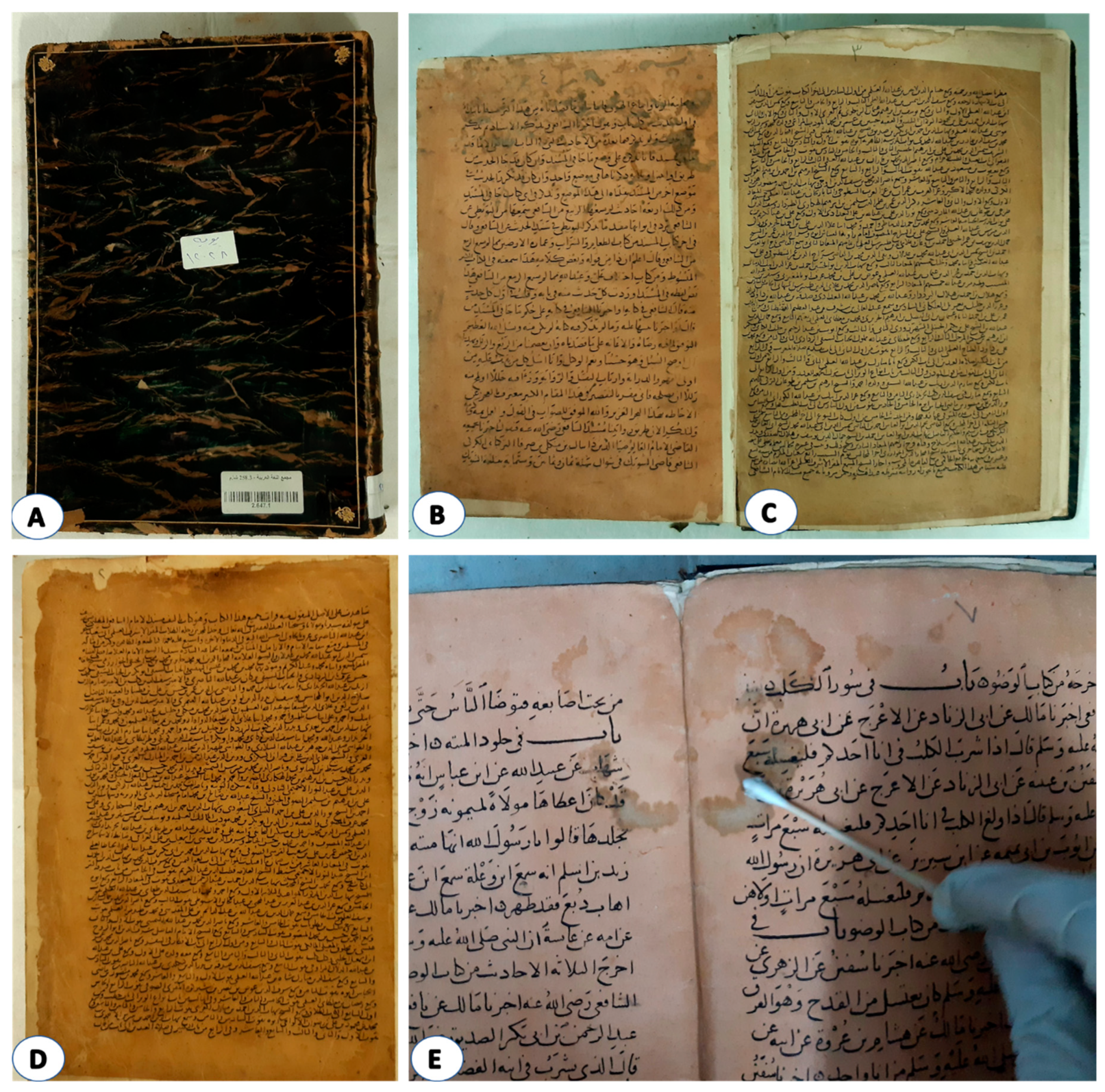
3.1.1. Photographic Documentation
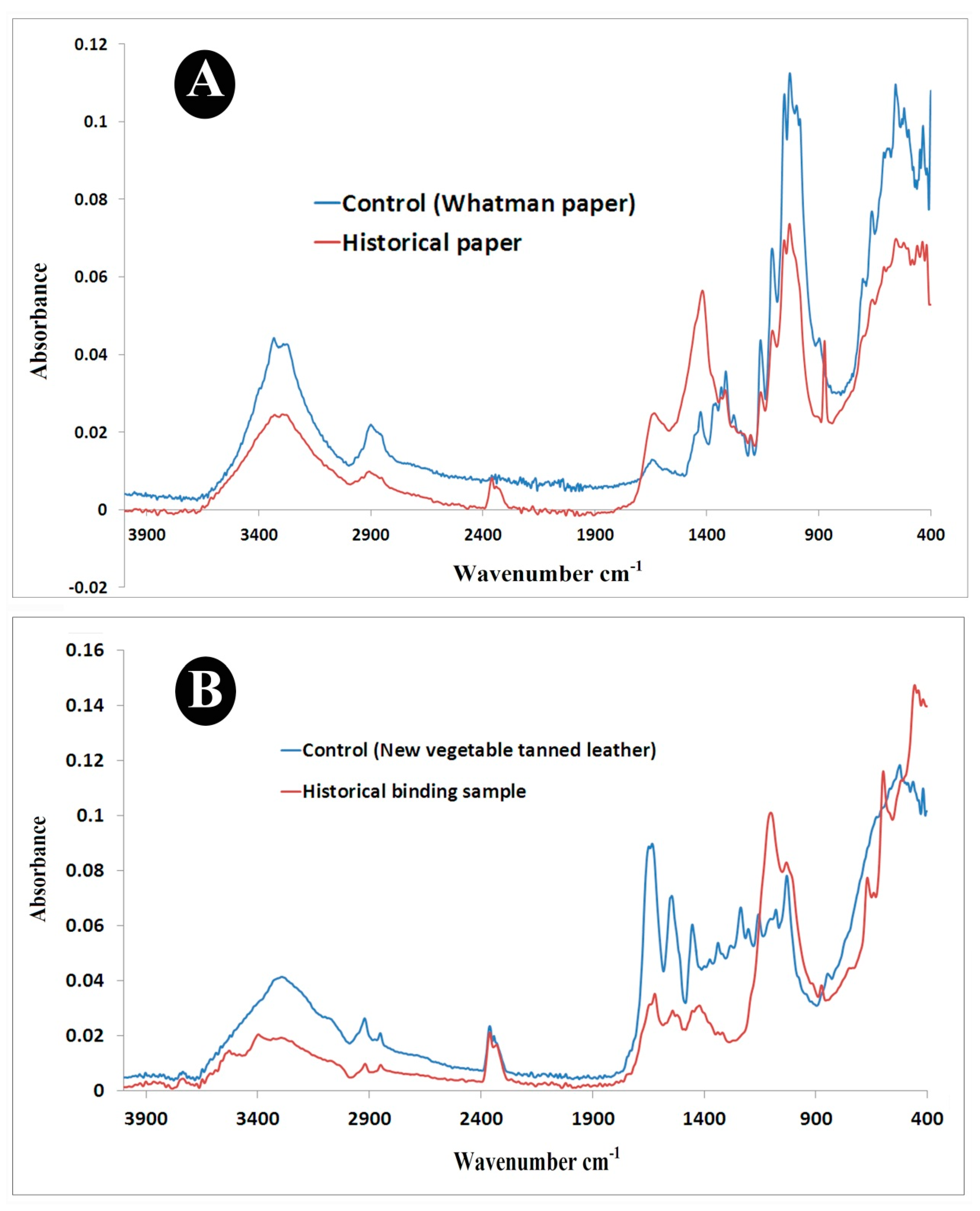
3.1.2. ATR-FTIR Analysis
- A.
- ATR/FTIR Analysis of Paper Samples
- B.
- ATR/FTIR Analysis Leather Samples
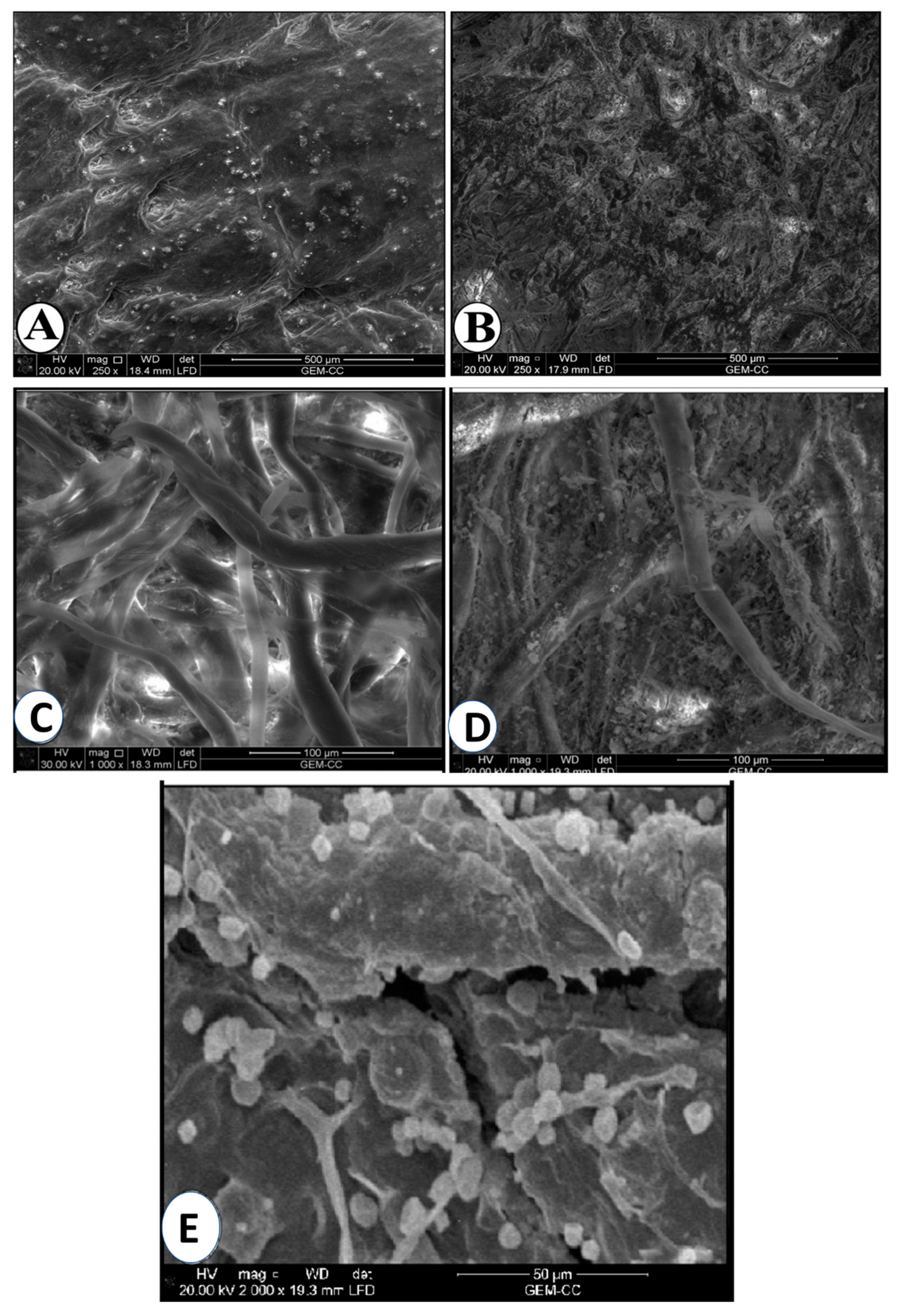
3.1.3. SEM Analysis
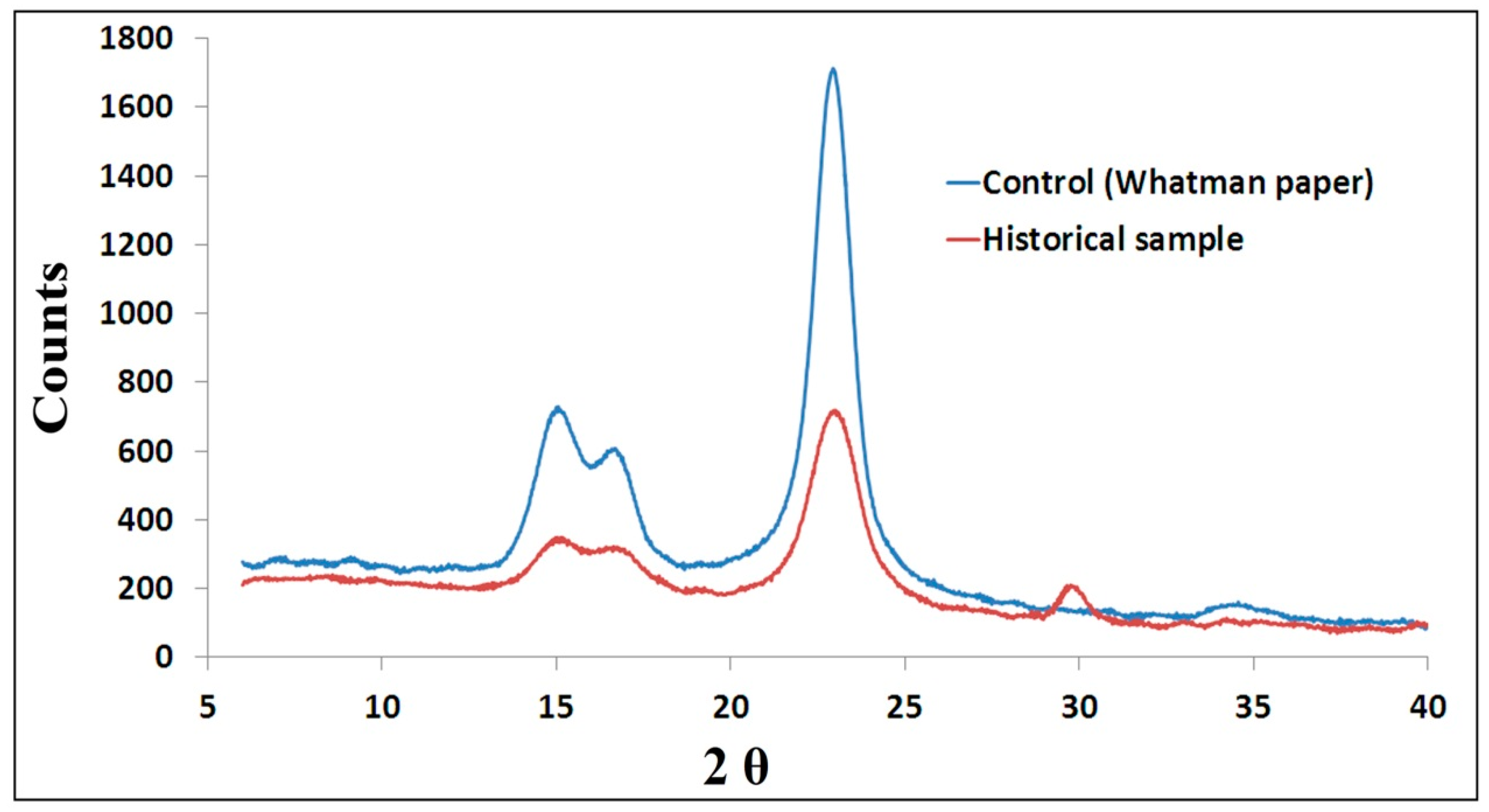
3.1.4. XRD Analysis
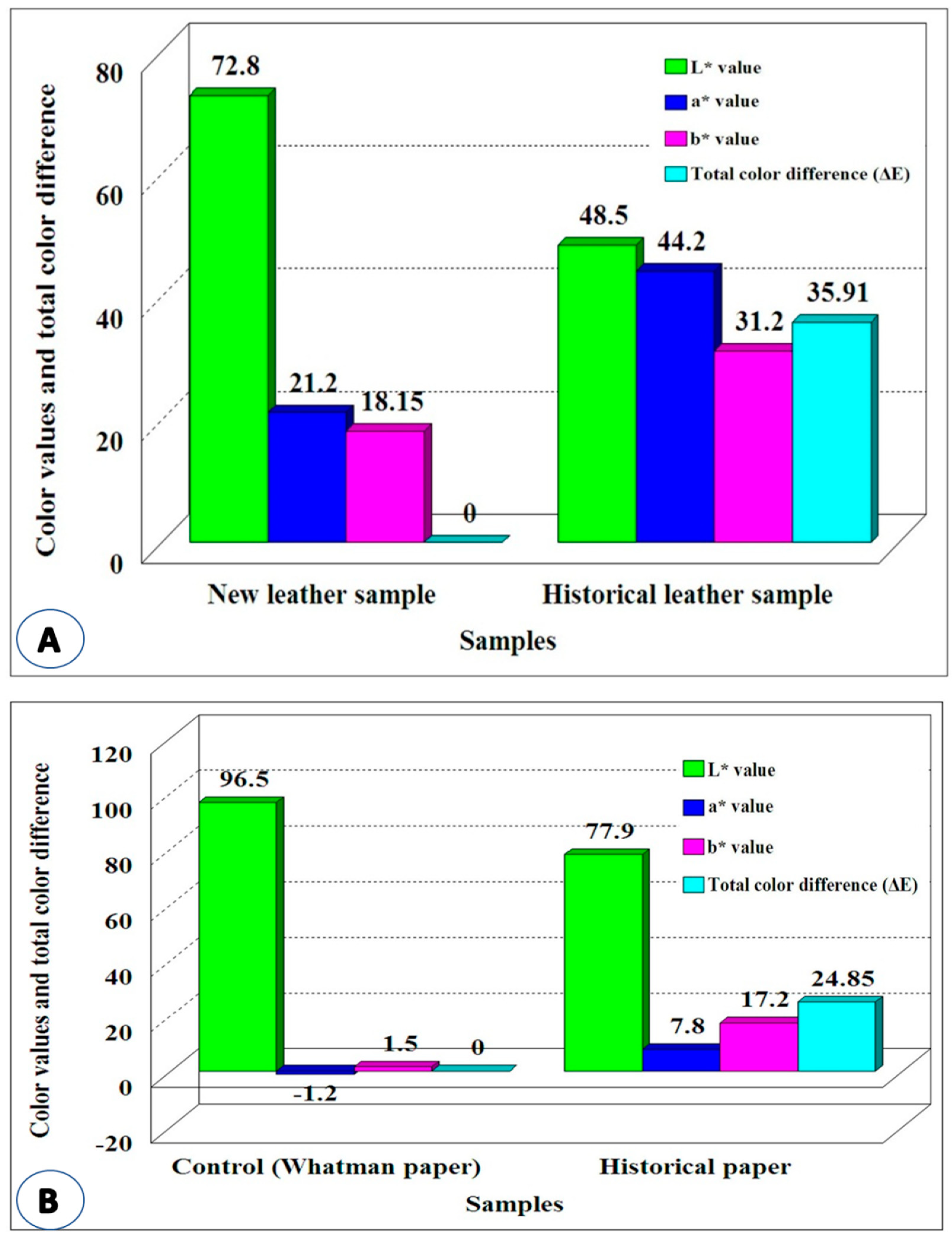
3.1.5. Measurement of Color Change
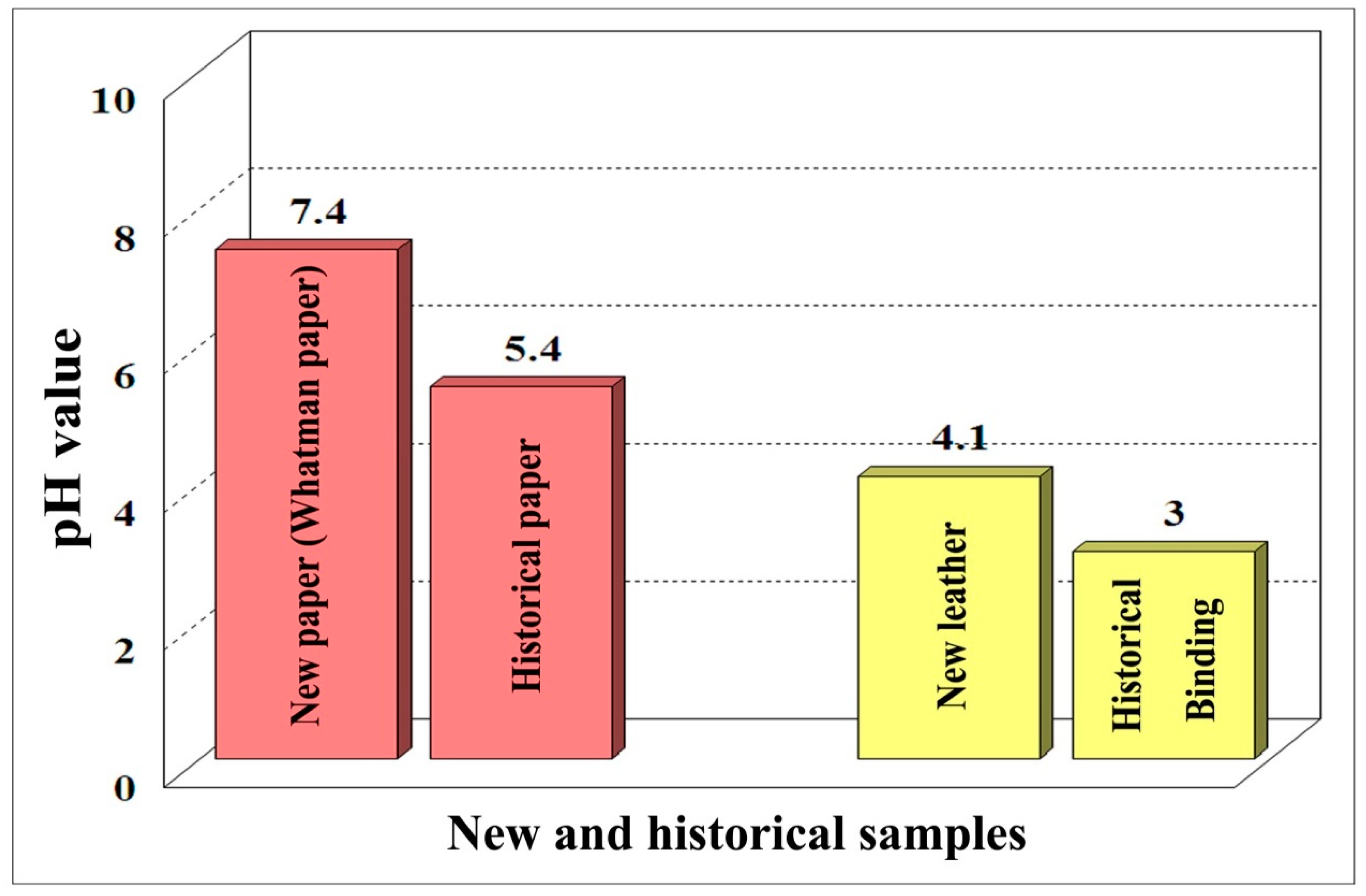
3.1.6. Measurement of pH Values
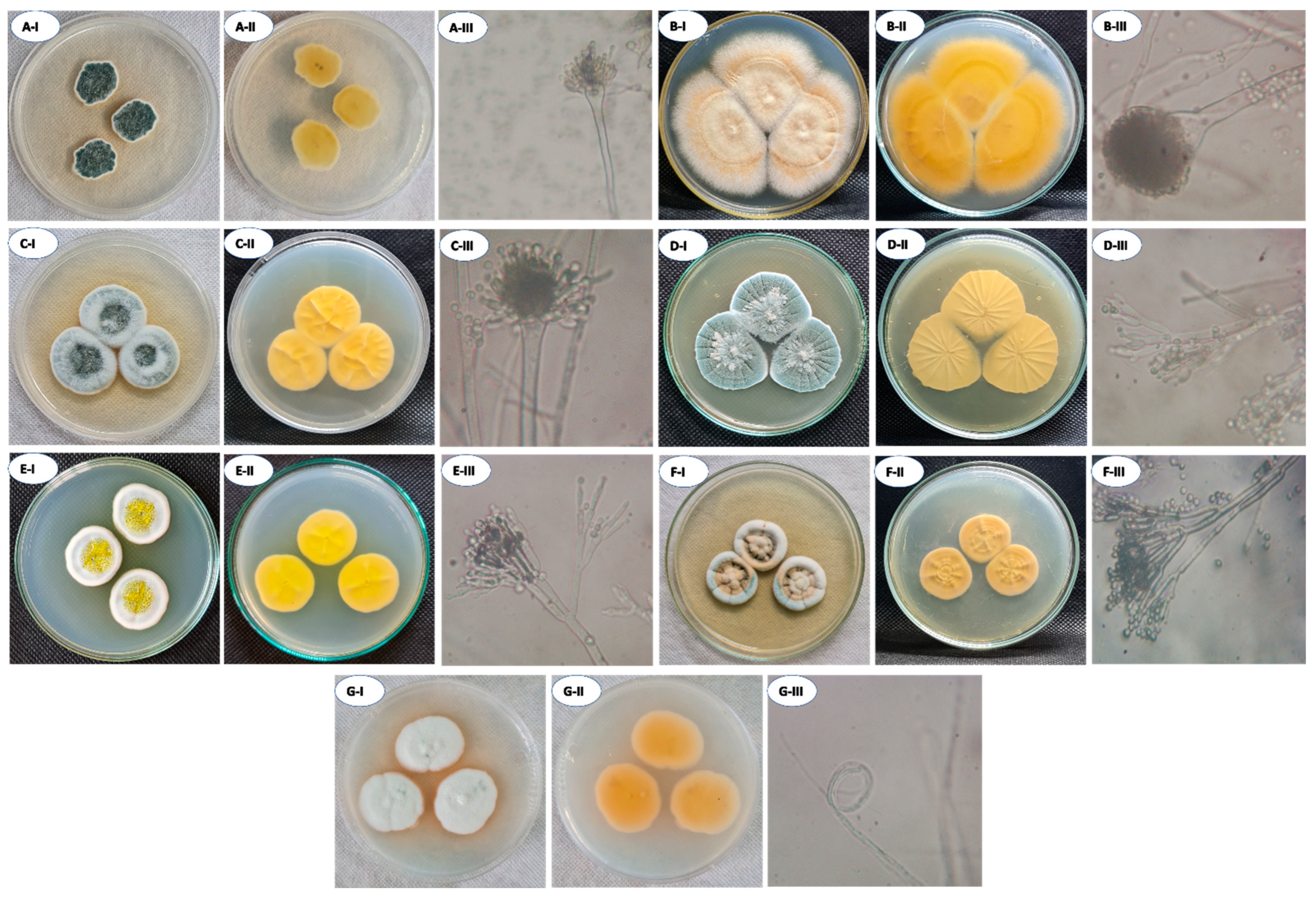
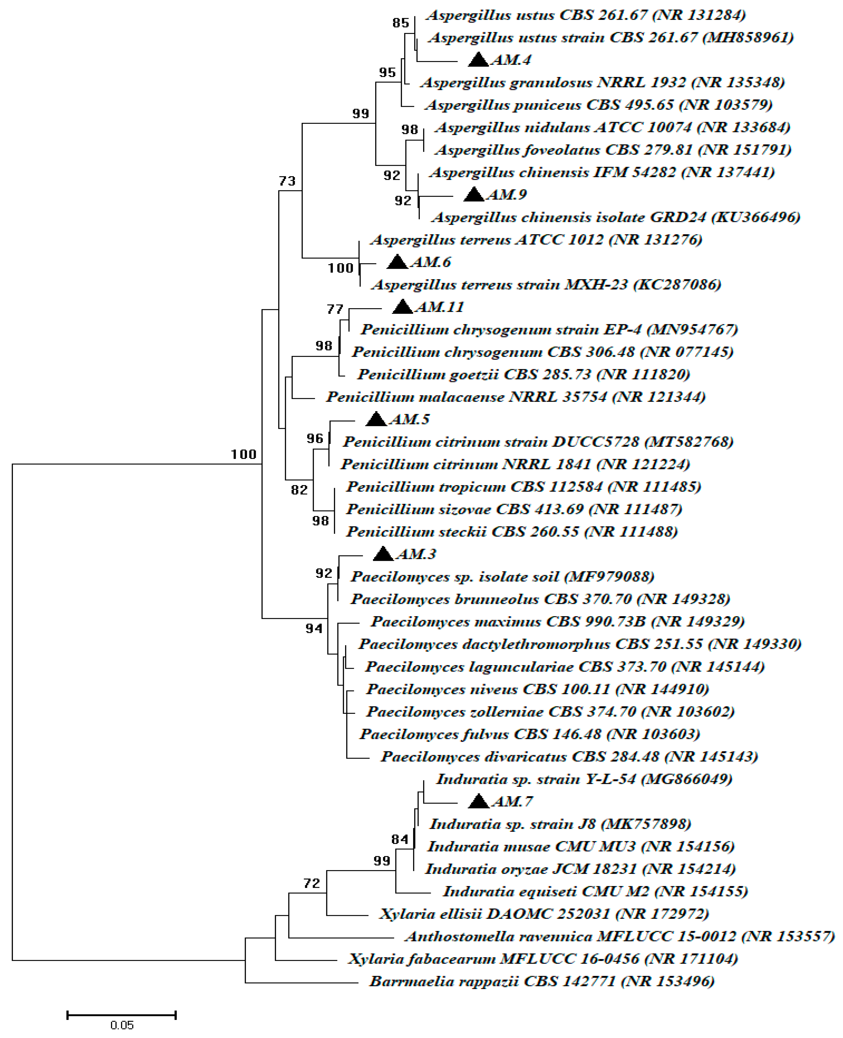
3.2. Fungal Isolations and Identifications
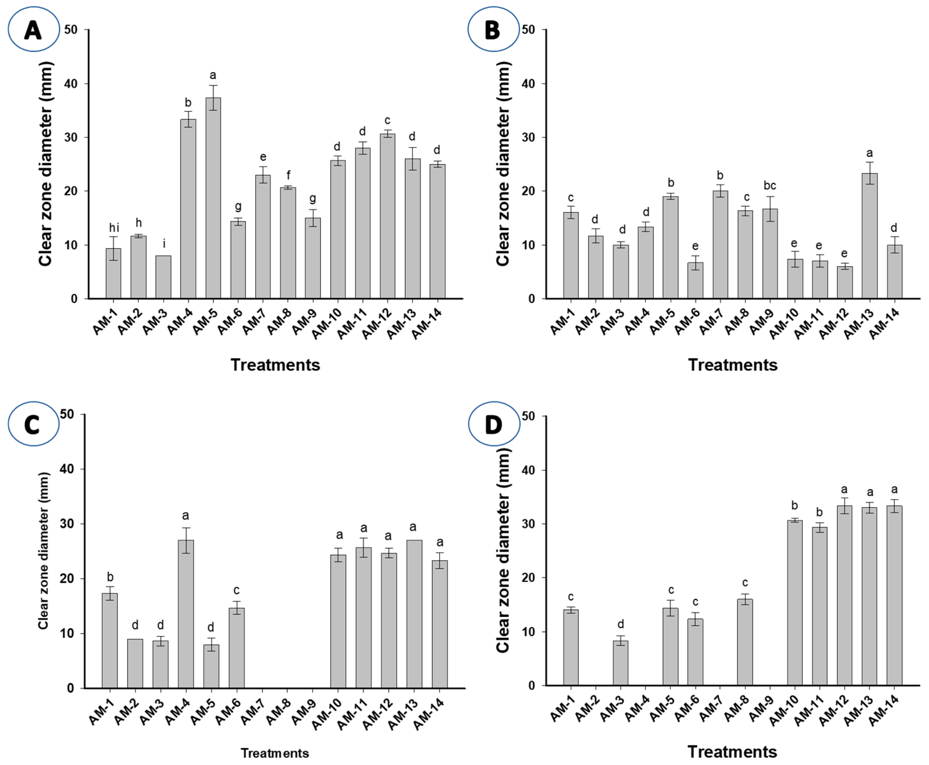
3.3. Hydrolytic Enzyme Activities
4. Conclusions
Author Contributions
Funding
Institutional Review Board Statement
Informed Consent Statement
Data Availability Statement
Acknowledgments
Conflicts of Interest
References
- Sequeira, S.; Cabrita, E.J.; Macedo, M.F. Antifungals on paper conservation: An overview. Int. Biodeterior. Biodegrad. 2012, 74, 67–86. [Google Scholar] [CrossRef]
- Stanaszek-Tomal, E. Environmental Factors Causing the Development of Microorganisms on the Surfaces of National Cultural Monuments Made of Mineral Building Materials—Review. Coatings 2020, 10, 1203. [Google Scholar] [CrossRef]
- Fouda, A.; Abdel-Maksoud, G.; Abdel-Rahman, M.A.; Eid, A.M.; Barghoth, M.G.; El-Sadany, M.A.-H. Monitoring the effect of biosynthesized nanoparticles against biodeterioration of cellulose-based materials by Aspergillus niger. Cellulose 2019, 26, 6583–6597. [Google Scholar] [CrossRef]
- Abdel-Maksoud, G. Evaluation of wax or oil/fungicide formulations for preservation of vegetable-tanned leather artifacts. J. Soc. Leather Technol. Chem. 2006, 90, 58–67. [Google Scholar]
- Wilson, W.K. Environmental Guidelines for the Storage of Paper Records; NISO Press: Bethesda, MD, USA, 1995. [Google Scholar]
- Saada, N.S.; Abdel-Maksoud, G.; Abd El-Aziz, M.S.; Youssef, A.M. Evaluation and utilization of lemongrass oil nanoemulsion for disinfection of documentary heritage based on parchment. Biocatal. Agric. Biotechnol. 2020, 29, 101839. [Google Scholar] [CrossRef]
- Fouda, A.; Abdel-Maksoud, G.; Saad, H.A.; Gobouri, A.A.; Mohammedsaleh, Z.M.; Abdel-Haleem El-Sadany, M. The Efficacy of Silver Nitrate (AgNO3) as a Coating Agent to Protect Paper against High Deteriorating Microbes. Catalysts 2021, 11, 310. [Google Scholar] [CrossRef]
- Sterflinger, K. Fungi: Their role in deterioration of cultural heritage. Fungal Biol. Rev. 2010, 24, 47–55. [Google Scholar] [CrossRef]
- Abdel-Maksoud, G.; Tharwat, N.; Gad, H. The Role of Fungi Isolated from Historical Vegetable-Tanned Leather on the Degradation of Peptides and Amino Acids. J. Soc. Leather Technol. Chem. 2014, 98, 1–9. [Google Scholar]
- Solanki, P.; Putatunda, C.; Kumar, A.; Bhatia, R.; Walia, A. Microbial proteases: Ubiquitous enzymes with innumerable uses. 3 Biotech 2021, 11, 428. [Google Scholar] [CrossRef]
- Koul, B.; Upadhyay, H. Fungi-Mediated Biodeterioration of Household Materials, Libraries, Cultural Heritage and Its Control. In Fungi and their Role in Sustainable Development: Current Perspectives; Gehlot, P., Singh, J., Eds.; Springer: Singapore, 2018; pp. 597–615. [Google Scholar]
- Pinheiro, A.; Sequeira, S. Mycological Studies in Cultural Heritage. In Reference Module in Life Sciences; Elsevier: Amsterdam, The Netherlands, 2020. [Google Scholar]
- Kirtzel, J.; Ueberschaar, N.; Deckert-Gaudig, T.; Krause, K.; Deckert, V.; Gadd, G.M.; Kothe, E. Organic acids, siderophores, enzymes and mechanical pressure for black slate bioweathering with the basidiomycete Schizophyllum commune. Environ. Microbiol. 2020, 22, 1535–1546. [Google Scholar] [CrossRef] [Green Version]
- Abdel-Maksoud, G.; Sobh, R.A.; Tarek, A. Evaluation of MMI/acrylate nanocomposite with hydroxyapatite as a novel paste for gap filling of archaeological bones. J. Cult. Herit. 2022, 57, 194–204. [Google Scholar] [CrossRef]
- Borrego, S.; Lavin, P.; Perdomo, I.; Gómez de Saravia, S.; Guiamet, P. Determination of indoor air quality in archives and biodeterioration of the documentary heritage. ISRN Microbiol. 2012, 2012, 680598. [Google Scholar] [CrossRef] [PubMed]
- Palermo, A.M.; Gentile, A.; Pellegrino, G. Documentary heritage: Fungal deterioration in Compact Discs. Herit. Sci. 2021, 9, 133. [Google Scholar] [CrossRef]
- Mohi, M.A.; Ismail, S.; Hassan, A.; Tawfik, A.M.; Mohamed, W. Assessment of the Applicability of Cellulolytic Enzyme in Disassembling of Caked Papers. Egypt. J. Chem. 2022, 65, 581–591. [Google Scholar] [CrossRef]
- Abdel-Maksoud, G. Study of cleaning materials and methods for stains on parchment. J. Soc. Leather Technol. Chem. 2006, 90, 146–154. [Google Scholar]
- Beyene, D.; Chae, M.; Dai, J.; Danumah, C.; Tosto, F.; Demesa, A.G.; Bressler, D.C. Characterization of Cellulase-Treated Fibers and Resulting Cellulose Nanocrystals Generated through Acid Hydrolysis. Matererials 2018, 11, 1272. [Google Scholar] [CrossRef] [Green Version]
- Abdel-Maksoud, G.; Marcinkowska, E. Changes in Some Properties of Aged and Historical Parchment. Restaurator 2000, 21, 138–157. [Google Scholar] [CrossRef]
- Reis-Menezes, A.A.; Gambale, W.; Giudice, M.C.; Shirakawa, M.A. Accelerated testing of mold growth on traditional and recycled book paper. Int. Biodeterior. Biodegrad. 2011, 65, 423–428. [Google Scholar] [CrossRef]
- Abdel-Maksoud, G.; Hussien, N.M.; Al-Arif, N.; Gouda, A.; Mohamed, W.S.; Ibrahim, M. Evaluation of a Mixture of Castor Oil and Polyvinylpyrrolidone for the Lubrication of Dry Vegetable Tanned Leather Artifacts. J. Soc. Leather Technol. Chem. 2022, 106, 20–31. [Google Scholar]
- Wouters, J.; Claeys, J.; Lamens, K.; Van Bos, M. Evaluation of methods for the micro-analysis of materials added to parchment. In Handbook in the Microanalysis of Parchments; Larsen, R., Ed.; Archetype Pub. Ltd.: London, UK, 2002; pp. 112–116. [Google Scholar]
- Abdel-Maksoud, G. Analytical techniques used for the evaluation of a 19th century quranic manuscript conditions. Measurement 2011, 44, 1606–1617. [Google Scholar] [CrossRef]
- Rojas, J.A.; Cruz, C.; Mikán, J.F.; Villalba, L.S.; Cepero de García, M.C.; Restrepo, S. Isoenzyme characterization of proteases and amylases and partial purification of proteases from filamentous fungi causing biodeterioration of industrial paper. Int. Biodeterior. Biodegrad. 2009, 63, 169–175. [Google Scholar] [CrossRef]
- Ismail, M.A.; Amin, M.A.; Eid, A.M.; Hassan, S.E.; Mahgoub, H.A.M.; Lashin, I.; Abdelwahab, A.T.; Azab, E.; Gobouri, A.A.; Elkelish, A.; et al. Comparative Study between Exogenously Applied Plant Growth Hormones versus Metabolites of Microbial Endophytes as Plant Growth-Promoting for Phaseolus vulgaris L. Cells 2021, 10, 1059. [Google Scholar] [CrossRef] [PubMed]
- Diba, K.; Kordbacheh, P.; Mirhendi, S.; Rezaie, S.; Mahmoudi, M. Identification of Aspergillus species using morphological characteristics. Pak. J. Med. Sci. 2007, 23, 867. [Google Scholar]
- Frisvad, J.C.; Samson, R.A. Polyphasic taxonomy of Penicillium subgenus Penicillium. A guide to identification of food and air-borne terverticillate Penicillia and their mycotoxins. Stud. Mycol. 2004, 49, 1–174. [Google Scholar]
- Samson, R.A.; Houbraken, J.; Varga, J.; Frisvad, J.C. Polyphasic taxonomy of the heat resistant ascomycete genus Byssochlamys and its Paecilomyces anamorphs. Persoonia 2009, 22, 14–27. [Google Scholar] [CrossRef] [PubMed] [Green Version]
- Wendt, L.; Sir, E.B.; Kuhnert, E.; Heitkämper, S.; Lambert, C.; Hladki, A.I.; Romero, A.I.; Luangsa-ard, J.J.; Srikitikulchai, P.; Peršoh, D.; et al. Resurrection and emendation of the Hypoxylaceae, recognised from a multigene phylogeny of the Xylariales. Mycol. Prog. 2018, 17, 115–154. [Google Scholar] [CrossRef]
- Khalil, A.M.A.; Hassan, S.E.; Alsharif, S.M.; Eid, A.M.; Ewais, E.E.; Azab, E.; Gobouri, A.A.; Elkelish, A.; Fouda, A. Isolation and Characterization of Fungal Endophytes Isolated from Medicinal Plant Ephedra pachyclada as Plant Growth-Promoting. Biomolecules 2021, 11, 140. [Google Scholar] [CrossRef]
- Mahgoub, H.A.M.; Fouda, A.; Eid, A.M.; Ewais, E.E.-D.; Hassan, S.E.-D. Biotechnological application of plant growth-promoting endophytic bacteria isolated from halophytic plants to ameliorate salinity tolerance of Vicia faba L. Plant Biotechnol. Rep. 2021, 15, 819–843. [Google Scholar] [CrossRef]
- Abdel-Maksoud, G.; Abed al-Sameh Al-Shazly, E.E.; El-Amin, A.-R. Damage caused by insects during the mummification process: An experimental study. Archaeol. Anthropol. Sci. 2011, 3, 291–308. [Google Scholar] [CrossRef]
- Abdel-Maksoud, G.; Abdel-Hamied, M.; Abdelhafez, A.A. Condition Assessment of a Mamluk HistoricalIlluminated Leather Binding at the Libraryof Mashiakht El-Azhar, Egypt. J. Soc. Leather Technol. Chem. 2021, 105, 248–256. [Google Scholar]
- Vichi, A.; Eliazyan, G.; Kazarian, S.G. Study of the Degradation and Conservation of Historical Leather Book Covers with Macro Attenuated Total Reflection–Fourier Transform Infrared Spectroscopic Imaging. ACS Omega 2018, 3, 7150–7157. [Google Scholar] [CrossRef] [PubMed]
- Sebestyén, Z.; Badea, E.; Carsote, C.; Czégény, Z.; Szabó, T.; Babinszki, B.; Bozi, J.; Jakab, E. Characterization of historical leather bookbindings by various thermal methods (TG/MS, Py-GC/MS, and micro-DSC) and FTIR-ATR spectroscopy. J. Anal. Appl. Pyrolysis 2022, 162, 105428. [Google Scholar] [CrossRef]
- El-Naggar, M.E.; Gaballah, S.; Abdel-Maksoud, G.; El-Sayed, H.S.; Youssef, A.M. Preparation of bactericidal zinc oxide nanoparticles loaded carboxymethyl cellulose/polyethylene glycol cryogel for gap filling of archaeological bones. J. Mater. Res. Technol. 2022, 20, 114–127. [Google Scholar] [CrossRef]
- Saada, N.S.; Abdel-Maksoud, G.; Abd El-Aziz, M.S.; Youssef, A.M. Green synthesis of silver nanoparticles, characterization, and use for sustainable preservation of historical parchment against microbial biodegradation. Biocatal. Agric. Biotechnol. 2021, 32, 101948. [Google Scholar] [CrossRef]
- Saada, N.; Gomaa, A.-M.G.; Youssef, A.M.; AZIZ, M.S.A. The Hydrolytic Activities of Two Fungal Species Isolatedf rom Historical Quranic Parchment Manuscript. J. Soc. Leather Technol. Chem. 2018, 102, 141–148. [Google Scholar]
- Helmi, F.M.; Wahba, W.N.; Brania, A.; Abdelnasser, M.; Elkobasy, M. Characterization of a Historical Leather Cover of Manuscript ‘Ensaan Elaauon fe sert Elameen Elmaamon’, Al-Azhar Library, Eygpt. Adv. Res. Conserv. Sci. 2021, 2, 40–52. [Google Scholar] [CrossRef]
- Garside, P.; Wyeth, P. Identification of Cellulosic Fibres by FTIR Spectroscopy—Thread and Single Fibre Analysis by Attenuated Total Reflectance. Stud. Conserv. 2003, 48, 269–275. [Google Scholar] [CrossRef] [Green Version]
- Cao, Q.; Zhu, S.; Zhao, H.; Tu, H. Application of Fourier transform attenuated total reflection infrared spectroscopy to identifying archaeological fibres. Res. J. Text. Appar. 2010, 14, 38–41. [Google Scholar] [CrossRef]
- Cocca, M.; D’Arienzo, L.; D’Orazio, L. Effects of different artificial agings on structure and properties of Whatman paper samples. Int. Sch. Res. Not. 2011, 2011, 863083. [Google Scholar] [CrossRef] [Green Version]
- Librando, V.; Minniti, Z.; Lorusso, S. Ancient and modern paper characterization by FTIR and Micro-Raman spectroscopy. Conserv. Sci. Cult. Herit. 2011, 11, 249–268. [Google Scholar]
- Abdel-Maksoud, G. Investigation techniques and conservation methods for a historical parchment document. J. Soc. Leather Technol. Chem. 2011, 95, 23–34. [Google Scholar]
- Carşote, C.; Budrugeac, P.; Decheva, R.; Haralampiev, N.S.; Miu, L.; Badea, E. Characterization of a byzantine manuscript by infrared spectroscopy and thermal analysis. Rev. Roum. Chim. 2014, 59, 429–436. [Google Scholar]
- Liu, Y.; Li, Y.; Chang, R.; Zheng, H.; Zhou, Y.; Li, M.; Hu, Z.; Wang, B. Species identification of ancient leather objects by the use of the enzyme-linked immunosorbent assay. Anal. Methods 2016, 8, 7689–7695. [Google Scholar] [CrossRef]
- Vyskočilová, G.; Ebersbach, M.; Kopecká, R.; Prokeš, L.; Příhoda, J. Model study of the leather degradation by oxidation and hydrolysis. Herit. Sci. 2019, 7, 26. [Google Scholar] [CrossRef] [Green Version]
- Jawahar, M.; Vani, K.; Babu, N.C. Leather species identification based on surface morphological characteristics using image analysis technique. J. Am. Leather Chem. Assoc. 2016, 111, 308–314. [Google Scholar]
- Orlita, A. Microbial biodeterioration of leather and its control: A review. Int. Biodeterior. Biodegrad. 2004, 53, 157–163. [Google Scholar] [CrossRef]
- Wang, H.; Farooq, A.; Memon, H. Influence of cotton fiber properties on the microstructural characteristics of mercerized fibers by regression analysis. Wood Fiber Sci 2020, 52, 13–27. [Google Scholar] [CrossRef]
- Borrego, S.; Guiamet, P.; Vivar, I.; Battistoni, P. Fungi involved in biodeterioration of documents in paper and effect on substrate. Acta Microsc. 2017, 27, 37–44. Available online: http://bdigital2.ula.ve:8080/xmlui/654321/1670 (accessed on 5 November 2022).
- Fouda, A.; Abdel-Maksoud, G.; Abdel-Rahman, M.A.; Salem, S.S.; Hassan, S.E.-D.; El-Sadany, M.A.-H. Eco-friendly approach utilizing green synthesized nanoparticles for paper conservation against microbes involved in biodeterioration of archaeological manuscript. Int. Biodeterior. Biodegrad. 2019, 142, 160–169. [Google Scholar] [CrossRef]
- Agarwal, U.P.; Ralph, S.A.; Reiner, R.S.; Baez, C. New cellulose crystallinity estimation method that differentiates between organized and crystalline phases. Carbohydr. Polym. 2018, 190, 262–270. [Google Scholar] [CrossRef]
- Abdel-Nasser, M.; Abdel-Maksoud, G.; Abdel-Aziz, M.S.; Darwish, S.S.; Hamed, A.A.; Youssef, A.M. Evaluation of the efficiency of nanoparticles for increasing α-amylase enzyme activity for removing starch stain from paper artifacts. J. Cult. Herit. 2022, 53, 14–23. [Google Scholar] [CrossRef]
- Sandy, M.; Manning, A.; Bollet, F. Changes in the Crystallinity of Cellulose in Response to Changes in Relative Humidity and Acid Treatment. Restaurator 2010, 31, 1–18. [Google Scholar] [CrossRef]
- Whitmore, P.M.; Bogaard, J. Determination of the Cellulose Scission Route in the Hydrolytic and Oxidative Degradation of Paper. Restaurator 1994, 15, 26–45. [Google Scholar] [CrossRef]
- Lama, A.; Fletcher, Y.; Guthrie-Strachan, J.; Covington, A.D.; Antunes, A.P.M. A New Formulation for the Treatment of Acid-Deterioration (Red Rot) in Historic Leathers. In XXXIII International Union of Leather Technologist and Chemists Societies (IULTCS) Congress; IULTCS: Novo Hamburgo, Brazil, 2015. [Google Scholar]
- Lama, A.; Antunes, A.P.M.; Fletcher, Y.; Guthrie-Strachan, J.; Vidler, K. Investigation of Acid-Deterioration in Leather Leading towards Finding a Suitable Product for Treatment. In Proceedings of the 114th Society of Leather Technologists and Chemists (SLTC) Conference 2011, Northampton, UK, 7 May 2011. [Google Scholar]
- Mochizuki, Y.; Itsumura, H.; Enomae, T. Mechanism of Acidification that Progresses in Library Collections of Books Made of Alkaline Paper. Restaurator 2020, 41, 153–172. [Google Scholar] [CrossRef]
- Smith, R.D. Paper impermanence as a consequence of pH and storage conditions. Libr. Q. 1969, 39, 153–195. [Google Scholar] [CrossRef]
- Hagaggi, N.; Salah, T.A. Microbial deterioration of a 13 AH-century manuscript housed in Al-Azhar library in Egypt: A case study. J. Basic Environ. Sci. 2016, 3, 65–73. [Google Scholar]
- Marín, E.; Sistach, M.C.; Jiménez, J.; Clemente, M.; Garcia, G.; García, J.F. Distribution of Acidity and Alkalinity on Degraded Manuscripts Containing Iron Gall Ink. Restaurator 2015, 36, 229–247. [Google Scholar] [CrossRef]
- Oetari, A.; Natalius, A.; Komalasari, D.; Susetyo-Salim, T.; Sjamsuridzal, W. Fungal Deterioration of Old Manuscripts of European Paper Origin. In Proceedings of the AIP Conference Proceedings 2023, Stanford, CA, USA, 19−20 May 1989; AIP Publishing LLC: Melville, NY, USA, 2018; p. 020156. [Google Scholar]
- Hassan, R.R. Analytical study of a manuscript,’Tafsir Al khazin’-the seventeenth century AD. Curr. Sci. Int. 2015, 4, 196–207. [Google Scholar]
- Mansour, M.; Hassan, R.; Salem, M. Characterization of historical bookbinding leather by FTIR, SEM-EDX and investigation of fungal species isolated from the leather. Egypt. J. Archaeol. Restor. Stud. 2017, 7, 1. [Google Scholar]
- Shamsian, A.; Fata, A.; Mohajeri, M.; Ghazvini, K. Fungal contaminations in historical manuscripts at Astan Quds museum library, Mashhad, Iran. Int. J. Agric. Biol. 2006, 8, 420–422. [Google Scholar]
- Ferrándiz-Pulido, C.; Martin-Gomez, M.T.; Repiso, T.; Juárez-Dobjanschi, C.; Ferrer, B.; López-Lerma, I.; Aparicio, G.; González-Cruz, C.; Moreso, F.; Roman, A.; et al. Cutaneous infections by dematiaceous opportunistic fungi: Diagnosis and management in 11 solid organ transplant recipients. Mycoses 2019, 62, 121–127. [Google Scholar] [CrossRef] [PubMed]
- Mesquita, N.; Portugal, A.; Videira, S.; Rodríguez-Echeverría, S.; Bandeira, A.M.L.; Santos, M.J.A.; Freitas, H. Fungal diversity in ancient documents. A case study on the Archive of the University of Coimbra. Int. Biodeterior. Biodegrad. 2009, 63, 626–629. [Google Scholar] [CrossRef]
- Swapna, P.; Lalch, P. Fungal biodiversity of a library and cellulolytic activity of some fungi. Indian J. Pharm. Sci. 2017, 78, 849–854. [Google Scholar] [CrossRef] [Green Version]
- Chadiesh, N.; Jishnu, B.T.; Tupaki-Sreepurna, A.; Ramanan, R.; Thanneru, V.; Kindo, A.J. Aspergillus quadrilineatus infection in an elderly debilitated patient. J. Acad. Clin. Microbiol. 2017, 19, 62. [Google Scholar]
- Hubbe, M.A.; Bowden, C. Handmade paper: A review of its history, craft, and science. BioResources 2009, 4, 1736–1792. [Google Scholar] [CrossRef]
- Sabatini, L.; Sisti, M.; Campana, R. Evaluation of fungal community involved in the bioderioration process of wooden artworks and canvases in Montefeltro area (Marche, Italy). Microbiol. Res. 2018, 207, 203–210. [Google Scholar] [CrossRef]
- El Bergadi, F.; Laachari, F.; Elabed, S.; Mohammed, I.H.; Ibnsouda, S.K. Cellulolytic potential and filter paper activity of fungi isolated from ancients manuscripts from the Medina of Fez. Ann. Microbiol. 2014, 64, 815–822. [Google Scholar] [CrossRef]
- Pinheiro, A.C.; Sequeira, S. Mycological Studies in Cultural Heritage. J. Encycl. Mycol. 2021, 27–39. [Google Scholar]
- Anaya, M.; Borrego, S.F.; Gámez, E.; Castro, M.; Molina, A.; Valdés, O. Viable fungi in the air of indoor environments of the National Archive of the Republic of Cuba. Aerobiologia 2016, 32, 513–527. [Google Scholar] [CrossRef]
- Savković, Ž.; Stupar, M.; Unković, N.; Ivanović, Ž.; Blagojević, J.; Vukojević, J.; Ljaljević Grbić, M. In vitro biodegradation potential of airborne Aspergilli and Penicillia. Sci. Nat. 2019, 106, 8. [Google Scholar] [CrossRef]
- Caneva, G.; Maggi, O.; Nugari, M.P.; Pietrini, A.M.; Piervittori, R.; Ricci, S.; Roccardi, A. The Biological Aerosol as a Factor of Biodeterioration. In Cultural Heritage and Aerobiology: Methods and Measurement Techniques for Biodeterioration Monitoring; Mandrioli, P., Caneva, G., Sabbioni, C., Eds.; Springer Netherlands: Dordrecht, The Netherlands, 2003; pp. 3–29. [Google Scholar]
- Trovão, J.; Mesquita, N.; Paiva, D.S.; Paiva de Carvalho, H.; Avelar, L.; Portugal, A. Can arthropods act as vectors of fungal dispersion in heritage collections? A case study on the archive of the University of Coimbra, Portugal. Int. Biodeterior. Biodegrad. 2013, 79, 49–55. [Google Scholar] [CrossRef]
- Ruga, L.O.F.F.M. Preventive Conservation of Cultural Heritage Biodeteriogens Control by Aerobiological Monitoring. Sensors 2019, 19, 3467. [Google Scholar] [CrossRef] [PubMed]


| Fungal Code | GenBank Accession Number | Homolog Sequences (Sequence Identity%) | Closest Accession Number |
|---|---|---|---|
| AM.3 | ON527926 | Paecilomyces sp. (98.96%) | MF979088 |
| AM.4 | ON527927 | Aspergillus ustus (98.82%) | NR131284 |
| AM.5 | ON527930 | Penicillium citrinum (98.53%) | NR121224 |
| AM.6 | ON527928 | Aspergillus terreus (98.21%) | NR131276 |
| AM.7 | ON527932 | Induratia sp. (98.76%) | NR154156 |
| AM.9 | ON527929 | Aspergillus chinensis (99.01%) | NR137441 |
| AM.11 | ON527931 | Penicillium chrysogenum (98.24%) | NR077145 |
Publisher’s Note: MDPI stays neutral with regard to jurisdictional claims in published maps and institutional affiliations. |
© 2022 by the authors. Licensee MDPI, Basel, Switzerland. This article is an open access article distributed under the terms and conditions of the Creative Commons Attribution (CC BY) license (https://creativecommons.org/licenses/by/4.0/).
Share and Cite
Abdel-Maksoud, G.; Abdel-Nasser, M.; Sultan, M.H.; Eid, A.M.; Alotaibi, S.H.; Hassan, S.E.-D.; Fouda, A. Fungal Biodeterioration of a Historical Manuscript Dating Back to the 14th Century: An Insight into Various Fungal Strains and Their Enzymatic Activities. Life 2022, 12, 1821. https://doi.org/10.3390/life12111821
Abdel-Maksoud G, Abdel-Nasser M, Sultan MH, Eid AM, Alotaibi SH, Hassan SE-D, Fouda A. Fungal Biodeterioration of a Historical Manuscript Dating Back to the 14th Century: An Insight into Various Fungal Strains and Their Enzymatic Activities. Life. 2022; 12(11):1821. https://doi.org/10.3390/life12111821
Chicago/Turabian StyleAbdel-Maksoud, Gomaa, Mahmoud Abdel-Nasser, Mahmoud H. Sultan, Ahmed M. Eid, Saad H. Alotaibi, Saad El-Din Hassan, and Amr Fouda. 2022. "Fungal Biodeterioration of a Historical Manuscript Dating Back to the 14th Century: An Insight into Various Fungal Strains and Their Enzymatic Activities" Life 12, no. 11: 1821. https://doi.org/10.3390/life12111821