Article

The Genus Chlorosplenium (Helotiales, Leotiomycetes) from China with Notes on C. chlora Complex

State Key Laboratory of Mycology, Institute of Microbiology, Chinese Academy of Sciences, Beijing 100101, China
*
Author to whom correspondence should be addressed.
Life 2021, 11(11), 1167; https://doi.org/10.3390/life11111167
Submission received: 31 August 2021 / Revised: 22 October 2021 / Accepted: 28 October 2021 / Published: 1 November 2021

Abstract

:
The small fruitbodies of Chlorosplenium are greenish yellow and mainly grow on woody substrates. The species diversity of the genus in China was investigated based on specimens formerly deposited in the Herbarium Mycologicum Academiae Sinicae as well as new collections gained in recent years. Our phylogenetic results revealed the species diversity of the genus is underestimated and the commonly known Chlorosplenium chlora is a species complex. Based on morphology studies and sequence analyses of three regions (ITS, LSU and RPB1), the Chinese collections represent two new species which are described and illustrated here as C. sinicum and C. sinochlora. Chlorosplenium fusisporum is quite possibly a species of the genus Chlorociboria, and C. hyperici-maculati should be excluded from the genus.

Graphical Abstract

1. Introduction

Chlorosplenium Fr. is an inoperculate discomycete genus in Helotiales (Leotiomycetes, Ascomycota, Fungi). It was established in 1849, typified by C. chlora (Schwein.) M.A. Curtis, and originally included three species (Peziza chlora Schwein., P. chlorascens Schwein. and P. torta Schwein.) [1]. Although only three species were firstly involved, their taxonomic and nomenclatural changes were complicated which were detailed by Dixon in his monographic treatments of the genera Chlorsplenium, Chlorociboria Seaver and Chlorencoelia Dixon [1,2]. In this study, Dixon’s taxonomic concept of Chlorosplenium is generally followed.
Many saprotrophic inoperculate cup-fungi have been placed in Chlorosplenium on the basis of the phenotypic character grayish- to greenish-yellow apothecia, and 57 names were published under the genus (http://www.indexfungorum.org/Names/Names.asp (accessed on 15 August 2021)). However, many of them are obviously not congeneric with the type species C. chlora, and they were even scattered in 11 different families according to the recent taxonomic treatments [3]. Chlorosplenium was once treated as a member of Helotiaceae or Dermateaceae [1,4]. According to the results of multi-locus phylogenetic analysis, the genus appeared as a clade distinct from any other fungal groups, thus a new family Chlorospleniaceae was proposed [5].
Five species are currently known in Chlorosplenium [4], i.e., C. chlora, C. hypochlorum (Berk. & M.A. Curtis ex W. Phillips) J.R. Dixon [1,2], C. cenangium (De Not.) Korf [6], C. fusisporum Liou & Z.C. Chen [7], and C. hyperici-maculati Svrček [8], among which two were previously recorded from China [9]. Species of the genus are somewhat easily recognized in the field, and are characterized by discoid to shallowly cupulate, grayish to greenish yellow apothecia with enrolled margin, usually sessile, reaching a size of 0.5–4 mm in diameter; hyphal extensions of different lengths present on receptacle surface or having a glabrous surface; ectal excipulum of textura angularis with the outer cell layer brown and the inner cells subhyaline to hyaline, medullary excipulum of textura intricata with hyaline hyphae and non-gelatinous tissues, subhymenium usually present and appearing as a densely interwoven hyphal layer; asci cylindric-clavate and containing eight ascospores; ascospores subcylindrical, hyaline and commonly unicellular; and paraphyses filiform and not or slightly exceeding the asci. Members of the genus are wood-inhabiting.
During our work on species diversity of Dermateaceae and its allies in China, herbarium specimens and recent collections of Chlorosplenium were studied based on morphological features and DNA sequence data. As a result, the occurrence of C. chlora in China should be re-considered, and two new species are discovered. The taxonomic position of the previously recorded C. fusisporum Liou & Z.C. Chen is doubtful based on the original description and illustration [7] and is tentatively excluded from the genus.

2. Materials and Methods

Specimens examined were all deposited in the Herbarium Mycologicum Academiae Sinicae (HMAS) and collected during 1933−2020 from Anhui, Chongqing, Guangdong, Guangxi, Henan, Hubei, Hunan, Jiangxi, Shaanxi, and Yunnan provinces of China. Photographs of fresh apothecia were taken by a Canon PowerShot G16 digital camera. Dried apothecia were rehydrated with distilled water and sectioned at a thickness of 15−20 μm with a Yidi YD-1508A freezing microtome (Jinhua, China). Mounting media was lactophenol cotton blue. Iodine reactions of ascus apical apparatus were tested in Melzer’s reagent and Lugol’s solution with or without 3% KOH solution pretreatment. An Olympus BH-2 microscope (Tokyo, Japan) was used for microscopic examination and measurements. Microscopic photographs were taken using a ZEISS Axiocam 305 color microscope camera (Göttingen, Germany) attached to a Zeiss Axioskop 2 Plus microscope (Göttingen, Germany).
Genomic DNA was extracted from dried apothecia using Plant Genomic DNA Kit (TIANGEN Biotech. Co., Beijing, China). Primer pairs used for amplification and sequencing included ITS1&ITS4 or ITS5&ITS4 [10] for the nuclear internal transcribed spacers (ITS), LR0R [11] and LR5 [12] for D1/D2 domain of the nuclear large subunit ribosomal DNA (LSU), RPB1-Af [13] and RPB1-Cr [14] for RNA polymerase II largest subunit gene (RPB1). PCR products were purified and sequenced at Beijing Tianyi Huiyuan Bioscience and Technology, China.
Newly generated sequences were assembled using SeqMan (DNASTAR, Lasergene 7.1.0) and BioEdit 7.0.5.3 [15]. The new sequences were deposited at GenBank and additional sequences used for phylogenetic analyses were downloaded from GenBank (Table 1). The sequence matrix was aligned by MUSCLE [16] and manually edited using BioEdit 7.0.5.3 [15]. Introns and ambiguous sites were removed and excluded from the analysis process. Dermea acerina and Pezicula carpinea were chosen as outgroup taxa.
Maximum likelihood (ML) and Bayesian inference (BI) analyses were conducted with RAxML 8.0 [17] and MrBayes 3.1.2 [18], respectively. Parameters of the ML analysis were set as default, and statistical support values were obtained from 1000 replicates of bootstrap (BP) tests. The BI analysis was performed using the best-fit evolutionary model estimated by MrModeltest 2.3 [19] with the Akaike information criterion. Two parallel runs of four simultaneous chains of Markov Chain Monte Carlo were performed for 2,000,000 generations with the trees sampled every 100 generations. The first quarter of sampled trees were discarded as the burn-in phase, and the remaining trees were used for calculating posterior probabilities (PP) in the majority rule consensus tree. The phylogenetic trees were visualized in FigTree 1.4.4 [20].

3. Results

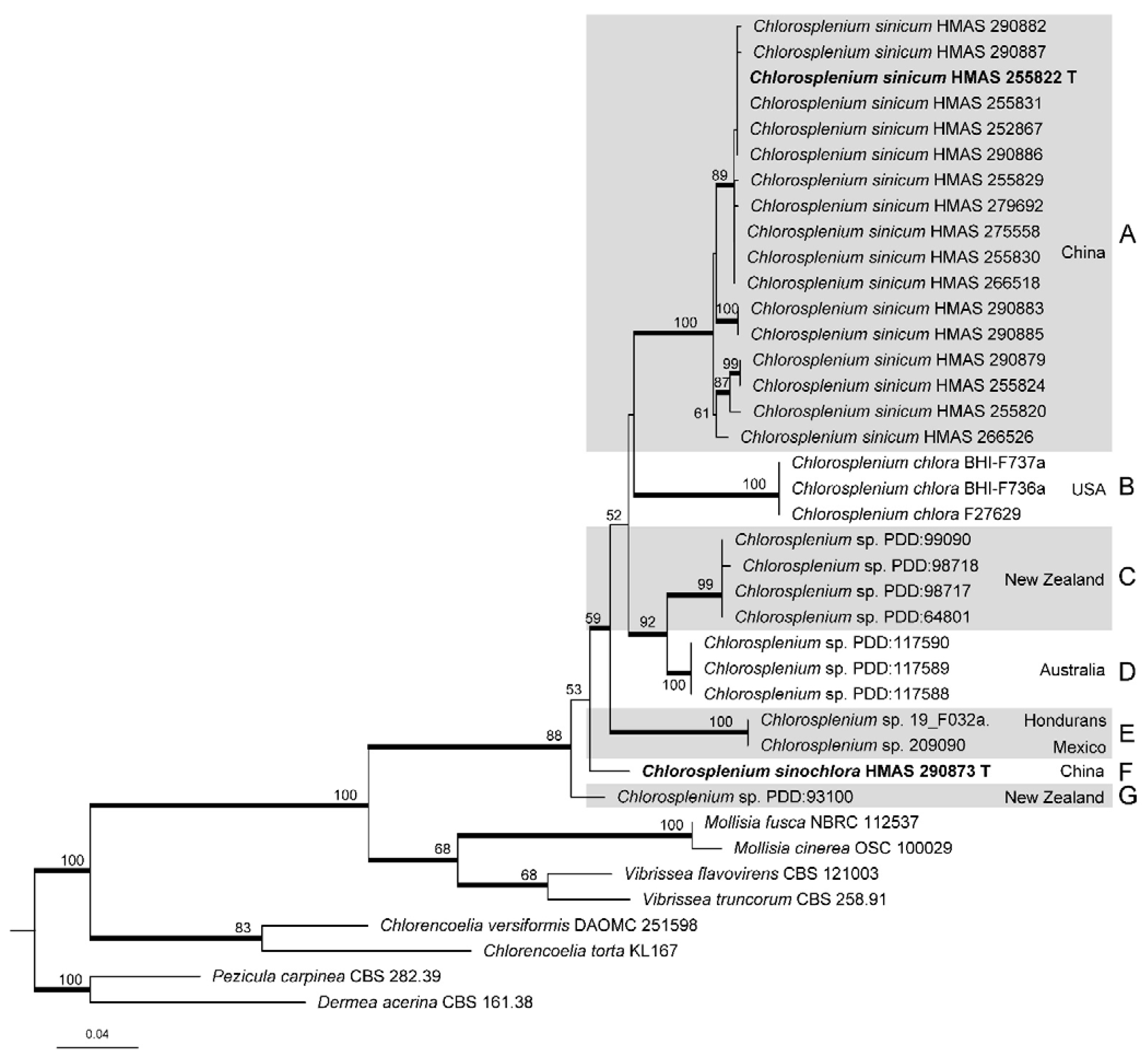
3.1. Phylogenetic Analyses

The final ITS matrix included 31 sequences of Chlorosplenium and eight of related taxa, and the alignment contained 595 nucleotide sites. GTR+I+G was selected as the best-fit model for BI analysis. The phylogenetic trees yielded by ML and BI analyses are similar in topology, and the ML tree is presented in Figure 1 with bootstrap values at nodes. All the Chlorosplenium sequences clustered together (MLBP/BIPP = 88%/1.00) that confirmed the monophyly of the genus. Seven well-supported clades/lineages (A–G) were recognized, including the two proposed new taxa from China.
Phylogenetically, all collections of C. sinicum were strongly supported in clade A which were further divided into a few subclades. Chlorosplenium sinochlora appeared as an isolated lineage F not closely related to the others. Chlorosplenium chlora from the USA materials clustered in clade B, and they were typical representatives of the species. Clades C, D, E and G consisted of unidentified samples of Chlorosplenium from New Zealand, Australia, Mexico, and Honduras, which were shown as Chlorosplenium spp. We suspect they might represent either un-named species or the existing species which have not been connected with any DNA sequences.
Due to the lack of LSU and RPB1 sequence data in most Chlorosplenium collections, the combined ITS-LSU-RPB1 dataset only included 13 sequences of the Chinese material, as well as four related inoperculate discomycete taxa. The final alignment comprised 2234 sites including gaps, of which 655 bp for ITS, 825 bp for LSU and 754 bp for RPB1. The best-fitting model for BI analysis was GTR+I for the multi-locus sequence data. Figure 2 showed the ML tree resulted from ITS-LSU-RPB1 sequence analysis with bootstrap values higher than 50%. Similar to the ITS analysis, C. sinicum is divided into several subgroups.

3.2. Taxonomy

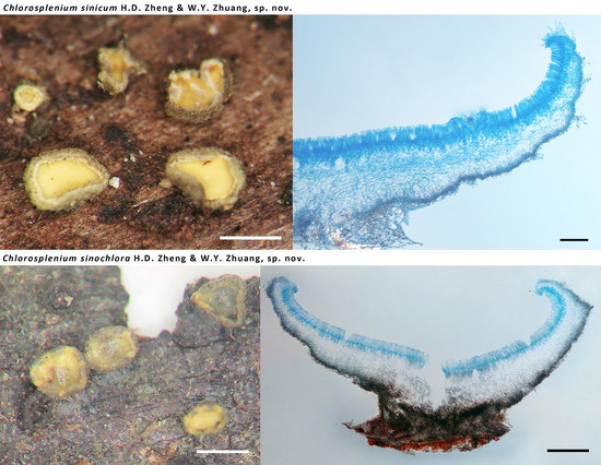
3.2.1. Chlorosplenium sinicum H.D. Zheng & W.Y. Zhuang, sp. nov.

  • Fungal Names—FN570862
  • Etymology—The specific epithet refers to the type locality of the species.
  • Holotype—CHINA, Hunan, Yizhang, Mang Mt., on rotten wood, 28 Oct. 2015, Z.Q. Zeng et al. 10345 (HMAS 255822).
Description—Apothecia scattered to gregarious, sessile, cupulate to discoid, margin slightly incurved when fresh, 0.5–4 mm in diameter; hymenium surface is greenish yellow, green tint becoming faded after drying; receptacle surface concolorous or darker, covered by hairs, whitish at margin, becoming greyish brown toward the base. Hairs on margin and upper flanks, cylindrical, round at the apex, multi-septate, smooth, straight to slightly flexuous, thin- to slightly thick-walled, hyaline at margin and light brown to brown at flanks, 2–4 μm wide, up to 90 μm long at margin and 8–45 μm long at flank, long hairs growing in groups, short hairs interspersed among those groups. Anchoring hyphae brown, thick-walled, 2.5–3.5 μm wide, walls 0.8–1 μm thick, slightly refractive, forming a layer of textura intricata at the base of apothecia, 40–140 μm thick. Ectal excipulum of two layers, less differentiated at margin; outer layer of textura angularis, 14–25 μm thick, cells slightly thick-walled, light brown to brown, nearly isodiametric, 3.5–8 μm in diam., walls 0.5–1 μm thick; inner layer of textura angularis and of textura prismatica at margin, 17–40 μm thick, cells thin-walled, subhyaline, 7–12 × 5–12 μm. Medullary excipulum of textura intricata, 20–220 μm thick, hyphae hyaline, thin- to slightly thick-walled, 2–4 μm wide. Subhymenium 15–30 μm thick. Hymenium 60–75 μm thick. Asci arising from croziers, 8-spored, uniseriate to irregularly biseriate, cylindric-clavate, J+ in Melzer’s reagent and Lugol’s solution without KOH pretreatment, visible as two blue lines, 45–66 × 4–6 μm. Ascospores subcylindrical, usually asymmetrical, aseptate, hyaline, smooth, with two or more minute guttules, 5.5–7.7 × 1.5–2.2 μm. Paraphyses filiform, slightly enlarged at apex, hyaline, 2–3 μm broad at apex and 1.5–2 μm below, slightly exceeding the asci by 5–10 μm (Figure 3).
Other specimens examined—CHINA, Anhui, Huang Mt., on rotten wood, 27 August 1957, S.C. Teng 5121 (HMAS 20093); ibid., 30 August 1957, S.C. Teng 5239 (HMAS 20500); Anhui Jiuhua Mt., on hard wood, 18 September 1933, Anhui, S.C. Teng 495 (HMAS 9326); Anhui, Jinzhai, Tiantangzhai, alt. 900−1000 m, on rotten wood, 23 August 2011, S.L. Chen et al. 7805 (HMAS 266626); Anhui, Jinzhai, Tiantangzhai, alt. 900−1000 m, on wet hard wood, 24 August 2011, S.L. Chen et al. 7879 (HMAS 252866); Chongqing, Jinyun Mt., alt. 823 m, on rotten or hard wood, 22 October 2020, H.D. Zheng et al. 12556, 12557, 12558, 12559, 12560, 12561 (HMAS 290886, 255828, 255829, 255830, 290887, 255831); Fujian, on rotten wood, S.C. Teng 1977 (HMAS 56523); Guangdong, Fengkai, alt. 300 m, on hard wood, 27 October 1998, W.Y. Zhuang & Z.H. Yu 2854 (HMAS 266519); ibid., 28 October 1998, W.Y. Zhuang & Z.H. Yu 2861 (HMAS 82038); Guangdong, Huidong, Guohua Forest Farm, alt. 800−900 m, on rotten wood, 17 October 1998, W.Y. Zhuang & Z.H. Yu 2764 (HMAS 252350); Guangdong Maoming, Dawuling, alt. 1400 m, on rotten wood, 22 October 1998, W.Y. Zhuang & Z.H. Yu 2810 (HMAS 266520); Guangdong, Shaoguan, Chebaling, on rotten wood, 31 October 2015, Z.Q. Zeng et al. 10482 (HMAS 255826); Guangdong, Shixing, Chebaling, Xianrendong, on rotten bark, 1 November 2015, Z.Q. Zeng et al. 10543 (HMAS 290882); Guangdong, Zhaoqing, Dinghu Mt., alt. 150 m, on rotten wood, 10 October 1998, W.Y. Zhuang & Z.H. Yu 2678 (HMAS 266521); Guangdong, Zhaoqing, Dinghu Mt., on rotten wood, 8 September 1958, J.H. Yu et al. 66 (HMAS 28256); Guangxi, Wuming, Daming Mt., alt. 1200 m, on rotten wood, 18 December 1997, W.Y. Zhuang & X.Q. Zhang 1796 (HMAS 74881); Guangxi, Wuming, Daming Mt., alt. 1100 m, on rotten wood, 19 December 1997, W.Y. Zhuang & X.Q. Zhang 1852 (HMAS 74882); Guangxi, Wuming, Daming Mt., alt. 900 m, 20 December 1997, W.Y. Zhuang 1863 (HMAS 266616); Henan, Jigong Mt., alt. 400 m, on hard wood, 13 November 2003, W.Y. Zhuang & Z.H. Yu 5080 (HMAS 266517); ibid., 14 November 2003, W.Y. Zhuang & Z.H. Yu 5089 (HMAS 266518); Hubei, Shennongjia, on rotten wood, 17 August 1984, H.Z. Li (HMAS 56467); ibid., 19 September 2014, H.D. Zheng et al. 9822 (HMAS 275558); Hunan, Yizhang, Mang Mt., Jiangjunzhai, on rotten wood, 26 October 2015, Z.Q. Zeng et al. 10249 (HMAS 255820); Hunan, Yizhang, Mang Mt., Jiangjunzhai, on rotten wood, 27 October 2015, Z.Q. Zeng et al. 10300 (HMAS 255821); Hunan, Yizhang, Mang Mt., on rotten wood, 28 October 2015, Z.Q. Zeng et al. 10355 (HMAS 255823); Hunan, Yizhang, Mang Mt., on rotten wood, 29 October 2015, Z.Q. Zeng et al. 10398, 10416, 10417, 10433, 10444 (HMAS 255824, 255825, 290878, 290879, 290881); Jiangxi, Lushan Botanic Garden, on wood, 19 October 1996, W.Y. Zhuang & Z. Wang 1465, 1466 (HMAS 252352, 266522); Jiangxi, Sanqing Mt., alt. 700 m, 19 September 2006, W.Y. Zhuang & J. Luo 6873 (HMAS 266523); Shaanxi, Qinling, Xiangxiyuan, on rotten wood, September 2015, P.J. Han, 8260 (HMAS 279692); Yunnan, Chuxiong, Zixi Mt., alt. 2120 m, on rotten wood, 8 Aug. 2016, X.H. Wang et al. YN16-34 (HMAS 290883); Yunnan, Hekou, Dawei Mt., alt. 1900 m, on rotten wood, 5 November 1999, W.Y. Zhuang & Z.H. Yu 3312 (HMAS 266524); Yunnan, Gaoligong Mt., Baihualing, on hard wood, 15 September 2017, H.D. Zheng et al. 11373, 11395 (HMAS 290884, 255827); Yunnan, Gaoligong Mt., Baihualing, Jinchanghe, on rotten wood, 17 September 2017, H.D. Zheng et al. 11441 (HMAS 290885); Yunnan, Menghai, Mangao, alt. 1300 m, on rotten wood, 22 October 1999, W.Y. Zhuang & Z.H. Yu 3199, 3252 (HMAS 252351, 266525); Yunnan, Pingbian, Dawei Mt., alt. 1900 m, 4 November 1999, W.Y. Zhuang & Z.H. Yu 3259 (HMAS 266526); Yunnan, Simao, 6 May 1957, L.W. Xu & G.Z. Wang 989 (HMAS 24109); Yunnan Simao, Caiyanghe, alt. 1300 m, on rotten wood, 13 October 1999, W.Y. Zhuang & Z.H. Yu 1863 (HMAS 266617).
Notes—Chlorosplenium sinicum is similar to C. chlora in gross morphology and microscopic characters, but it is distinct in receptacle surface fibrillose with conspicuous hairs and the DNA sequence data. According to Dixon (1974), the receptacle surface of C. chlora is glabrous [1]. In the phylogenetic tree (Figure 1), the Chinese collections of C. sinicum formed a well-supported clade independent from other species. The samples of the fungus further grouped into a few subclades (Figure 1 and Figure 2) and sequence variations within species were ranged 18 bp including 10 gaps for ITS, 0–3 bp for LSU, and 0–6 bp for RPB1. Morphological distinctions were not detected among the collections. We treat the above divergences as intra-specific variations.

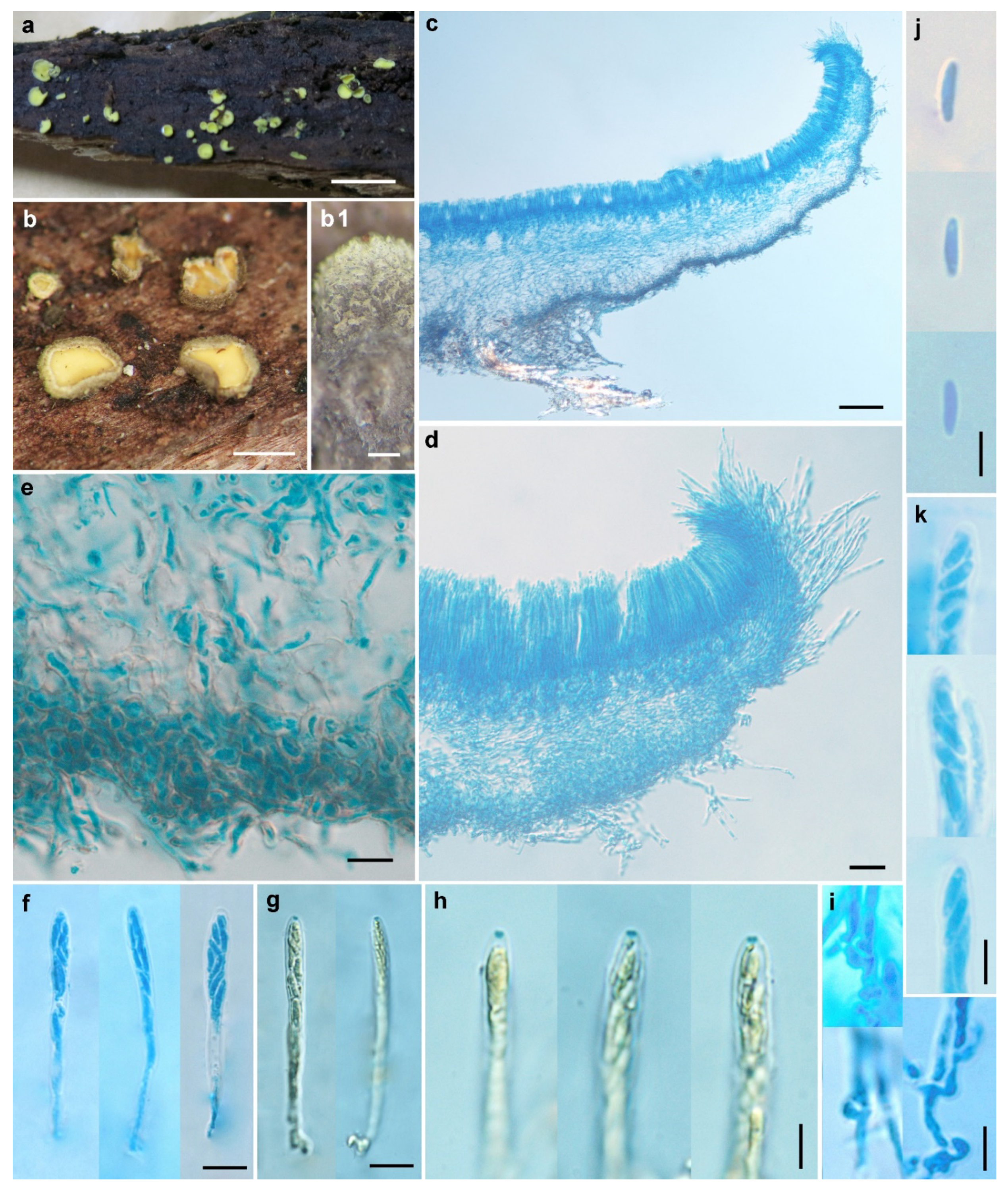
3.2.2. Chlorosplenium sinochlora H.D. Zheng & W.Y. Zhuang, sp. nov.

  • Fungal Names—FN570863
  • Etymology—The specific epithet refers to the origin of the species and its similarity with C. chlora.
  • Holotype—CHINA, Hunan, Yizhang, Mang Mt., Jiangjunzhai, on rotten bark, 27 October 2015, Z.Q. Zeng et al. 10286 (HMAS 290873).
Description—Apothecia scattered, subsessile to sessile, shallow-cupulate to discoid, margin slightly incurved when dry, 0.5–2 mm in diam; the hymenium surface is yellowish green, with the green tint becoming obscure when dry; the receptacle surface is greyish green, and greyish black when dry, nearly glabrous. Ectal excipulum of two layers, less differentiated at margin, 20–95 μm thick; cortical layer with short sparsely hyphal protrusions, scattered on the surface of the upper flanks, becoming longer and forming a covering layer from the middle flanks to the base, 5.5–14 μm thick, forming a few minute pustules, hyphae thick-walled, 2.5–3 μm wide, walls refractive, 0.7–1 μm thick; outer layer of textura globulosa-angularis, 20–70 μm thick, cells thick-walled, brown, nearly isodiametric, 3–8 μm in diam., walls 0.5–1.5 μm thick; inner layer of textura angularis, 20–40 μm thick, cells thick-walled, subhyaline to light brown, 5.5–14 × 5.5–11 μm, walls 0.5–1.5 μm thick. Medullary excipulum of textura intricata, loosely arranged, 15–125 μm thick, hyphae hyaline, thin-walled, 2–4 μm wide. Subhymenium 15–25 μm thick. Hymenium 60–70 μm thick. Asci arising from croziers, 8-spored, uniseriate or irregularly biseriate, cylindric-clavate, J+ in Melzer’s reagent and Lugol’s solution without KOH pretreatment, visible as two blue lines, 45–53 × 3.5–4.5 μm. Ascospores subcylindrical, aseptate, hyaline, smooth, with two or more minute guttules, 5–6.5 × 1.8–2.2 μm. Paraphyses filiform, slightly enlarged at apex, hyaline, 2.5–3 μm broad at apex and 1.5–2 μm below, slightly exceeding the asci by 3–8 μm (Figure 4).
Other specimens examined—CHINA, Hunan, Yizhang, Mang Mt., Jiangjunzhai, on rotten wood, 27 October 2015, Z.Q. Zeng et al. 10302, 10311 (HMAS 290875, 290876); ibid., on rotten bark, 10316 (HMAS 290877); Hunan, Yizhang, Mang Mt., on rotten bark, 29 October 2015, Z.Q. Zeng et al. 10413, 10442 (HMAS 290874, 290880).
Notes—In the ITS sequence analysis, C. sinochlora appeared as a separate lineage in the phylogenetic tree (Figure 1). Morphologically, it is similar to C. chlora but differs in solitary apothecia, narrower asci (45–53 × 3.5–4.5 μm vs. 45–55 × 5–6 μm), overlapped but slightly shorter and wider ascospores (5–6.5 × 1.8–2.2 μm vs. 6–7 × 1.5–2 μm) [1], as well as the ITS sequence variations. It is also similar to C. sinicum in ascus and ascospore morphology, but the latter differs in the hairy receptacle surface, lighter cells in outer ectal excipulum, and distinct ITS, LSU and RPB1 sequences.

3.2.3. Excluded and Doubtful Species of Chlorosplenium

Chlorosplenium fusisporum Liou & Z.C. Chen

Notes—Chlorosplenium fusisporum was originally described from Taiwan, China [7]. It is characterized by green apothecia and with a centrally placed stipe, ectal excipulum with coiling or undulate hairs encrusted by green granules, acerose ascospores with 1–3 septa, 20–35 × 2.5–3.5 μm (Figure 5). Although we did not access the voucher specimen of C. fusisporum which lacks sequence data, the combination of apothecial color, ectal excipulum structure and short and granulate hairs on the receptacle surface strongly suggests its affinity to Chlorociboria. The type material of the fungus has not been examined, and therefore formal nomenclatural treatment is not proposed for the time being.

Chlorosplenium hyperici-maculati Svrček

Notes—Chlorosplenium hyperici-maculati was originally described from Slovakia on stem of Hypericum maculatum [8]. In his original description, Svrček indicated that “the beautiful color of this discomycete is conspicuously deep green in all outer parts of apothecia, and the disc has an almost velvet appearance.” As to its similar or closely related fungus, he compared C. hyperici-maculati with C. aeruginellum (P. Karst.) P. Karst., which is now being placed in the genus Dasyscyphus Nees ex Gray [≡ Dasyscyphus aeruginellus (P. Karst.) Korf & J.R. Dixon] belonging to a different family [1]. Judged by the original description and illustration of the fungus, we conclude that C. hyperici-maculati should be excluded from Chlorosplenium in a modern sense.

3.2.4. Key to Accepted Species of Chlorosplenium

1. Asci longer than 70 μm long ................................................................................................... 2
1. Asci shorter than 70 μm long .................................................................................................. 3
 2. Ascospores 12−14 × 4−5 μm ............................................................................ C. cenangium
 2. Ascospores (8−)9−14(−15) × 2−4 μm ........................................................... C. hypochlorum
3. Receptacle surface fibrillose, hairs clearly visible at least in margin and upper flank,
 ascospores 5.5–7.7 × 1.5–2.2 μm ............................................................................ C. sinicum
3. Receptacle surface nearly glabrous ........................................................................................ 4
 4. Apothecia solitary to gregarious, asci (40−)45−55(−60) × (4−)5−6(−7) μm, ascospores
 (5−)6−7(−9) × 1.2−2(−3) μm .................................................................................... C. chlora
 4. Apothecia solitary, asci 45–53 × 3.5–4.5 μm, ascospores 5–6.5 × 1.8–2.2 μm ....................
 .............................................................................................................................. C. sinochlora

4. Discussion

Chlorosplenium is a small genus in Helotiales and contains five species. Due to the small size of the apothecia and inconspicuous appearance, members of the genus were not reported frequently. Among the accepted species of the genus, the type species, C. chlora, was the most widely reported. It was originally described from USA and reported also from Canada, China, Costa Rica, India, Indonesia, Jamaica, Japan, Korea, and Russia [1,9,21,22,23]. Chlorosplenium cenangium was initially described from Italy [24] and later recorded from Spain [25]. Chlorosplenium hypochlorum is known from its type locality in Cuba as well as Jamaica and Mexico [1]. Chlorosplenium sinicum and C. sinochlora are reported only from China in the present study. Furthermore, a few ITS sequences of un-named species of the genus deposited at GenBank were derived from Australia and New Zealand.
Morphological details provided in the literature are not enough for accurate species identification of Chlorosplenium because macro- and microscopic features are usually overlapping. Molecular study was seldom carried out in the genus and it is necessary to use molecular approaches to facilitate species recognition. Concerning the taxonomic characters, structure of excipulum and morphology of asci and ascospores are useful in species identification. According to the results of our phylogenetic analyses, inter-specific variations of ITS rDNA, the universal barcode for fungi, are higher than those of the protein-coding gene RPB1 in Chlorosplenium, which is suitable for species distinction. Based on the results of the detailed morphological comparison and phylogenetic analyses, the Chinese collections previously identified as C. chlora were proved to represent two novel species. Thus, C. chlora should be excluded from the fungal species catalogue of China [9]. Its occurrences in other Asian countries are also doubtful.
Based only upon specimens collected from China, this study has shown that the worldwide species diversity of Chlorosplenium has not been investigated using modern methods, and many hidden species are expected to be discovered. Future studies based on large-scale samplings combining with ecological, morphological, and molecular data will lead to a better understanding of the species diversity and phylogeny of the genus.

Author Contributions

Conceptualization, W.-Y.Z. and H.-D.Z.; methodology, H.-D.Z. and W.-Y.Z.; software, H.-D.Z.; validation, W.-Y.Z. and H.-D.Z.; formal analysis, H.-D.Z.; investigation, H.-D.Z.; resources, W.-Y.Z. and H.-D.Z.; data curation, W.-Y.Z. and H.-D.Z.; writing—original draft preparation, H.-D.Z.; writing—review and editing, W.-Y.Z.; visualization, H.-D.Z.; supervision, W.-Y.Z.; project administration, W.-Y.Z. and H.-D.Z.; funding acquisition, W.-Y.Z. and H.-D.Z. All authors have read and agreed to the published version of the manuscript.

Funding

This research was funded by the National Natural Science Foundation of China (31770019, 31750001).

Institutional Review Board Statement

Not applicable.

Informed Consent Statement

Not applicable.

Data Availability Statement

Names of the new species were formally registered in the database Fungal Names (http://www.fungalinfo.net/fungalname/fungalname.html (accessed on 26 August 2021)). Specimens were deposited in the Herbarium Mycologicum Academiae Sinicae (HMAS). The newly generated sequences were deposited in GenBank (https://www.ncbi.nlm.nih.gov/genbank (accessed on 28 August 2021)).

Acknowledgments

The authors would like to thank all co-collectors of specimens examined in this study for their invaluable help during the field work.

Conflicts of Interest

The authors declare no conflict of interest.

References

  1. Dixon, J.R. Chlorosplenium and its segregates. I. Introduction and the genus Chlorosplenium. Mycotaxon 1974, 1, 65–104. [Google Scholar]
  2. Dixon, J.R. Chlorosplenium and its segregates II. The genera Chlorociboria and Chlorencoelia. Mycotaxon 1975, 1, 193–237. [Google Scholar]
  3. Johnston, P.R.; Quijada, L.; Smith, C.A.; Baral, H.O.; Hosoya, T.; Baschien, C.; Partel, K.; Zhuang, W.Y.; Haelewaters, D.; Park, D.; et al. A multigene phylogeny toward a new phylogenetic classification of Leotiomycetes. IMA Fungus 2019, 10, 1. [Google Scholar] [CrossRef] [PubMed]
  4. Kirk, P.M.; Cannon, P.F.; Minter, D.W.; Stalpers, J.A. Dictionary of the Fungi, 10th ed.; CABI: Wallingford, UK, 2008; p. 771. [Google Scholar]
  5. Ekanayaka, A.; Hyde, K.; Gentekaki, E.; McKenzie, E.; Zhao, Q.; Bulgakov, T.; Camporesi, E. Preliminary classification of Leotiomycetes. Mycosphere 2019, 10, 310–489. [Google Scholar] [CrossRef]
  6. Müller, E. Zur Pilzflora der Aletschwaldreservats (Kt. Wallis, Schweiz). Beiträge zur Kryptogamenflora der Schweiz 1977, 15, 1–126. [Google Scholar]
  7. Liou, S.-C.; Chen, Z.-C. Notes on Taiwan Discomycetes I. (Pezizales and Helotiales). Taiwania 1977, 22, 29–43. [Google Scholar]
  8. Svrček, M. New or less known Discomycetes. XXII. Česká Mykologie 1992, 46, 33–40. [Google Scholar]
  9. Zhuang, W.-Y.; Zheng, H.-D.; Zeng, Z.-Q. Species Catalogue of China, Volumen 3 Fungi, Cup-Fungi; Science Press: Beijing, China, 2018; p. 142. [Google Scholar]
  10. White, T.J.; Bruns, T.; Lee, S.; Taylor, J. Amplification and direct sequencing of fungal ribosomal RNA genes for phylogenetics. In PCR Protocols: A Guide to Methods and Applications; Innis, M.A., Gelfand, D.H., Snisky, J.J., White, T.J., Eds.; Academic Press: London, UK, 1990; pp. 315–322. [Google Scholar]
  11. Moncalvo, J.-M.; Wang, H.-H.; Hseu, R.-S. Phylogenetic Relationships in Ganoderma Inferred from the Internal Transcribed Spacers and 25S Ribosomal DNA Sequences. Mycologia 1995, 87, 223–238. [Google Scholar] [CrossRef]
  12. Vilgalys, R.; Hester, M. Rapid genetic identification and mapping of enzymatically amplified ribosomal DNA from several Cryptococcus species. J. Bacteriol. 1990, 172, 4238–4246. [Google Scholar] [CrossRef] [PubMed] [Green Version]
  13. Stiller, J.W.; Hall, B.D. The origin of red algae: Implications for plastid evolution. Proc. Natl. Acad. Sci. USA 1997, 94, 4520–4525. [Google Scholar] [CrossRef] [PubMed] [Green Version]
  14. Matheny, P.B.; Liu, Y.J.; Ammirati, J.F.; Hall, B.D. Using RPB1 sequences to improve phylogenetic inference among mushrooms (Inocybe, Agaricales). Am. J. Bot. 2002, 89, 688–698. [Google Scholar] [CrossRef] [PubMed]
  15. Hall, T.A. BioEdit: A user-friendly biological sequence alignment editor and analysis program for Windows 95/98/NT. Nucleic Acids Symp. Ser. 1999, 41, 95–98. [Google Scholar]
  16. Edgar, R.C. MUSCLE: A multiple sequence alignment method with reduced time and space complexity. BMC Bioinform. 2004, 5, 113. [Google Scholar] [CrossRef] [PubMed] [Green Version]
  17. Stamatakis, A. RAxML version 8: A tool for phylogenetic analysis and post-analysis of large phylogenies. Bioinformatics 2014, 30, 1312–1313. [Google Scholar] [CrossRef]
  18. Ronquist, F.; Huelsenbeck, J.P. MrBayes 3: Bayesian phylogenetic inference under mixed models. Bioinformatics 2003, 19, 1572–1574. [Google Scholar] [CrossRef] [Green Version]
  19. Nylander, J.A.A. MrModeltest V2; Program distributed by the author; Evolutionary Biology Centre, Uppsala University: Uppsala, Sweden, 2004. [Google Scholar]
  20. Rambaut, A. FigTree, V1.4.4.; Institute of Evolutionary Biology, University of Edinburgh: Edinburgh, UK, 2018; Available online: http://tree.bio.ed.ac.uk/software/figtree/ (accessed on 22 May 2019).
  21. Chlorosplenium chlora (Schwein). M.A. Curtis in GBIF Secretariat. GBIF Backbone Taxonomy. Checklist Dataset. Available online: https://doi.org/10.15468/39omei (accessed on 27 August 2021).
  22. Arendholz, W.-R.; Sharma, R. Some new or interesting Helotiales from the eastern Himalayas. Mycotaxon 1983, 17, 473–512. [Google Scholar]
  23. Park, P.-J.; Lee, J.-Y.; Otani, Y. Taxonomical Studies on Discomycetes in Korea (I). Korean J. Mycol. 1985, 13, 27–40. [Google Scholar]
  24. Saccardo, P.A. Sylloge Fungorum; Typis Seminarii: Patavii, Italy, 1889; Volume 8, p. 1143. [Google Scholar]
  25. Vila, J.; Llistosella, J.; Llimona, I.X. Contribuciό al coneixement dels fongs de l’estatge alpí dels pirineus de Catalunya. Il. Rev. Catalana Micol. 1998, 2, 93–113. [Google Scholar]
Figure 1. Maximum likelihood phylogenetic tree inferred from ITS sequences including Chlorosplenium collections and related taxa. Bootstrap support values ≥ 50% are shown at nodes. Posterior probability values ≥ 0.90 of BI analysis are indicated as thick branches. Holotypes are shown in bold and marked with the letter “T”.
Life 11 01167 g001
Figure 2. Maximum likelihood phylogenetic tree inferred from combined ITS-LSU-RPB1 sequences of selected Chlorosplenium collections and related taxa. Bootstrap support values ≥ 50% are shown at nodes. Posterior probability values ≥ 0.90 of BI analysis are indicated as thick branches. Holotypes are shown in bold and marked with the letter “T”.
Life 11 01167 g002
Figure 3. Chlorosplenium sinicum H.D. Zheng & W.Y. Zhuang (HMAS 255822, holotype). (a) Fresh apothecia on natural substrate; (b) Dry apothecia; (b1) Receptacle surface of dry apothecium, showing groups of hairs; (c,d) Longitudinal section of apothecium; (e) Excipular structure at flank; (f,g) Asci; (h) IKI reaction of ascus apical rings; (i) Croziers at ascus base; (j) Ascospores; (k) Ascospores in asci. Scale bars: (a) = 5mm; (b) = 1 mm; (b1) = 0.2 mm; (c) = 100 μm; (d) = 20 μm; (eg) = 10 μm; (hk) = 5 μm. Mounting media: (cf,ik) lactophenol cotton blue; (g,h) Melzer’s reagent.
Life 11 01167 g003
Figure 4. Chlorosplenium sinochlora H.D. Zheng & W.Y. Zhuang (HMAS 290873, holotype). (a,b) Dry apothecia on natural substrate; (c) Longitudinal section of apothecium; (d) Structure of margin and upper flanks; (e) Structure of lower flanks; (f) Detailed excipular structure at flank; (g,h) Asci; (i,j) Ascospores; (k) IKI reaction of ascus apical rings; (l) Croziers at ascus base. Scale bars: (a) = 2 mm; (b) = 1 mm; (c) = 200 μm; (d) = 50 μm; (e,f) = 20 μm; (g,h) = 10 μm; (il) = 5 μm. Mounting media: (cg,j,l) lactophenol cotton blue; (h,i,k) Melzer’s reagent.
Life 11 01167 g004
Figure 5. Original illustrations of Chlorosplenium fusisporum (From Liou and Chen 1977 [7]).
Life 11 01167 g005
Table 1. Sequences of Chlorosplenium and related taxa used in this study.
NameVoucher/IsolateCountryGenBank Number
ITSLSURPB1
Chlorosplenium chlora (Schwein.) M.A. CurtisBHI-F736USAMG553993
C. chloraBHI-F737USAMG553994
C. chloraF27629USAMZ312166
C. sinicum H.D. Zheng & W.Y. ZhuangHMAS 279692 (8260)ChinaMK425600
C. sinicumHMAS 266518ChinaMK425599
C. sinicumHMAS 252867ChinaMZ914636
C. sinicumHMAS 266526ChinaMZ914637
C. sinicumHMAS 275558 (9822)ChinaMZ914635
C. sinicumHMAS 290882 (10543)ChinaMZ914627
C. sinicumHMAS 290886 (12556)ChinaMZ914630
C. sinicumHMAS 255830 (12559)ChinaMZ914632
C. sinicumHMAS 255831 (12561)ChinaMZ914634
C. sinicumHMAS 255822 T (10345)ChinaMZ914624MZ920115MZ945729
C. sinicumHMAS 255829 (12558)ChinaMZ914631MZ920119MZ945733
C. sinicumHMAS 290887 (12560)ChinaMZ914633MZ920120MZ945734
C. sinicumHMAS 255820 (10249)ChinaMZ914623MZ920114MZ945728
C. sinicumHMAS 255824 (10398)ChinaMZ914625MZ920116MZ945730
C. sinicumHMAS 290879 (10433)ChinaMZ914626MZ920117MZ945731
C. sinicumHMAS 290885 (11441)ChinaMZ914628MZ920118MZ945732
C. sinicumHMAS 290883 (YN16-34)ChinaMZ914629MZ920121MZ945735
C. sinochlora H.D. Zheng & W.Y. ZhuangHMAS 290873 T (10286)ChinaMZ914622MZ920122MZ945736
Chlorosplenium sp.HONDURAS19-F032HondurasMT571528
Chlorosplenium sp.209090MexicoMG976227
Chlorosplenium sp.PDD:117588AustraliaMW191764
Chlorosplenium sp.PDD:117589AustraliaMW191759
Chlorosplenium sp.PDD:117590AustraliaMW191760
Chlorosplenium sp.PDD:64801New ZealandMW191757
Chlorosplenium sp.PDD:98717New ZealandMW191758
Chlorosplenium sp.PDD:98718New ZealandMW191763
Chlorosplenium sp.PDD:99090New ZealandMW191761
Chlorosplenium sp.PDD:93100New ZealandMW191762
Chlorencoelia torta (Schwein.) J.R. DixonKL167ChinaLT158424
C. versiformis (Pers.) J.R. DixonDAOMC 251598CanadaMH457140
Dermea acerina (Peck) RehmCBS 161.38/AFTOL-ID 941CanadaMH855942MH867440DQ471164
Mollisia cinerea (Batsch) P. Karst.OSC 100029/AFTOL-ID 76UnknownDQ491498DQ470942DQ471122
M. fusca (Pers.) P. Karst.TNS:F17463/NBRC 112537JapanLC425049
Pezicula carpinea (Pers.) Tul. ex FuckelCBS 282.39/AFTOL-ID 938CanadaKR859272DQ470967DQ842032
Vibrissea flavovirens (Pers.) Korf & J.R. DixonCBS 121003GermanyMT026430
V. truncorum (Alb. & Schwein.) Fr.CBS 258.91/AFTOL-ID 1322CanadaMT026377FJ176874FJ238438
Note: Type specimens are indicated with superscript “T” and newly generated sequence data are indicated in black bold.
Publisher’s Note: MDPI stays neutral with regard to jurisdictional claims in published maps and institutional affiliations.

