Ephrin Receptors (Eph): EphA1, EphA5, and EphA7 Expression in Uveal Melanoma—Associations with Clinical Parameters and Patient Survival

 , and
, and
Abstract
:1. Introduction
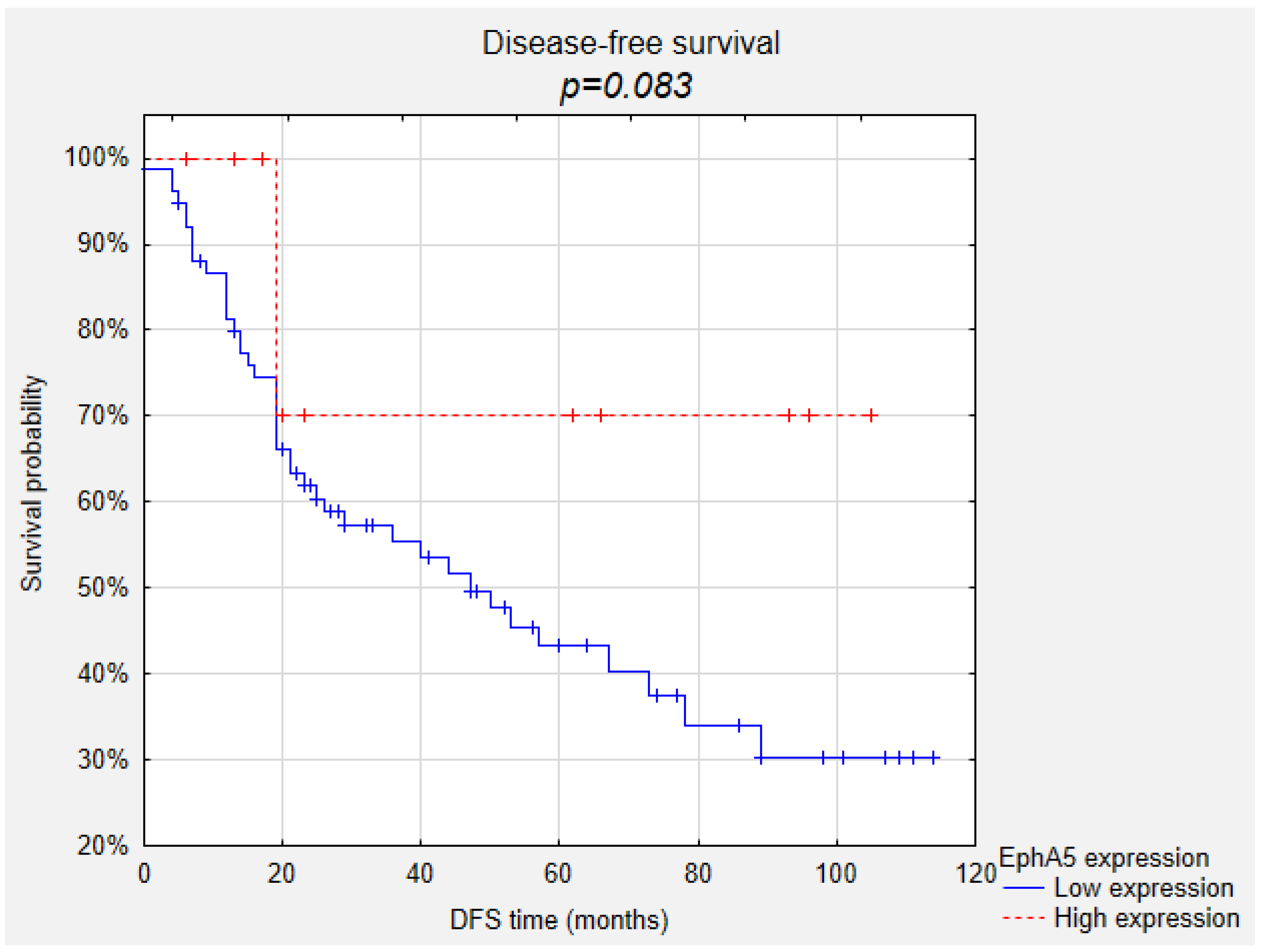
2. Results
3. Discussion
4. Materials and Methods
4.1. Patients
4.2. Immunohistochemistry
4.3. Statistical analysis
5. Conclusions
Author Contributions
Funding
Conflicts of Interest
References
- Virgili, G.; Gatta, G.; Ciccolallo, L.; Capocaccia, R.; Biggeri, A.; Crocetti, E.; Lutz, J.-M.; Paci, E. Incidence of Uveal Melanoma in Europe. Ophthalmology 2007, 114, 2309–2315.e2. [Google Scholar] [CrossRef] [PubMed]
- Krantz, B.A.; Dave, N.; Komatsubara, K.M.; Marr, B.P.; Carvajal, R.D. Uveal melanoma: Epidemiology, etiology, and treatment of primary disease. Clin. Ophthalmol. 2017, 11, 279–289. [Google Scholar] [CrossRef] [PubMed] [Green Version]
- Kivela, T.; Simpson, E.R.; Grossniklaus, H.E.; Jager, M.J.; Singh, A.D.; Caminal, J.M.; Pavlick, A.C.; Kujala, E.; Coupland, S.E.; Finger, P. Uveal melanoma. In AJCC Cancer Staging Manual; Springer: New York, NY, USA, 2017; pp. 805–817. [Google Scholar]
- Kujala, E.; Mäkitie, T.; Kivelä, T. Very long-term prognosis of patients with malignant uveal melanoma. Investig. Ophthalmol. Vis. Sci. 2003, 44, 4651–4659. [Google Scholar] [CrossRef] [Green Version]
- Yang, J.; Manson, D.K.; Marr, B.P.; Carvajal, R.D. Treatment of uveal melanoma: Where are we now? Ther. Adv. Med Oncol. 2018, 10. [Google Scholar] [CrossRef] [PubMed]
- Pasquale, E.B. Eph-ephrin bidirectional signaling in physiology and disease. Cell 2008, 133, 38–52. [Google Scholar] [CrossRef] [Green Version]
- Himanen, J.-P.; Saha, N.; Nikolov, D.B. Cell–cell signaling via Eph receptors and ephrins. Curr. Opin. Cell Biol. 2007, 19, 534–542. [Google Scholar] [CrossRef] [Green Version]
- Surawska, H.; Ma, P.C.; Salgia, R. The role of ephrins and Eph receptors in cancer. Cytokine Growth Factor Rev. 2004, 15, 419–433. [Google Scholar] [CrossRef]
- Nievergall, E.; Lackmann, M.; Janes, P.W. Eph-dependent cell-cell adhesion and segregation in development and cancer. Cell. Mol. Life Sci. 2012, 69, 1813–1842. [Google Scholar] [CrossRef]
- Pasquale, E.B. Eph receptor signalling casts a wide net on cell behaviour. Nat. Rev. Mol. Cell Biol. 2005, 6, 462–475. [Google Scholar] [CrossRef]
- Tognolini, M.; Hassan-Mohamed, I.; Giorgio, C.; Zanotti, I.; Lodola, A. Therapeutic perspectives of Eph–ephrin system modulation. Drug Discov. Today. 2014, 19, 661–669. [Google Scholar] [CrossRef]
- Brantley, D.M.; Cheng, N.; Thompson, E.J.; Lin, Q.; Brekken, R.A.; E Thorpe, P.; Muraoka, R.S.; Cerretti, D.P.; Pozzi, A.; Jackson, D.; et al. Soluble Eph A receptors inhibit tumor angiogenesis and progression in vivo. Oncogene 2002, 21, 7011–7026. [Google Scholar] [CrossRef] [PubMed] [Green Version]
- Theocharis, S.; Klijanienko, J.; Giaginis, C.; Alexandrou, P.; Patsouris, E.; Sastre-Garau, X. Ephrin receptor (Eph) -A1, -A2, -A4 and -A7 expression in mobile tongue squamous cell carcinoma: Associations with clinicopathological parameters and patients survival. Pathol Oncol Res. 2014, 20, 277–284. [Google Scholar] [CrossRef]
- Giaginis, C.; Tsourouflis, G.; Zizi-Serbetzoglou, A.; Kouraklis, G.; Chatzopoulou, E.; Dimakopoulou, K.; Theocharis, S. Clinical significance of Ephrin (Eph)-A1, -A2, -A4, -A5 and -A7 receptors in pancreatic ductal adenocarcinoma. Pathol. Oncol. Res. 2010, 16, 267–276. [Google Scholar] [CrossRef] [PubMed]
- Giaginis, C.; Tsoukalas, N.; Bournakis, E.; Alexandrou, P.; Kavantzas, N.; Patsouris, E.; Theocharis, S. Ephrin (Eph) receptor A1, A4, A5 and A7 expression in human non-small cell lung carcinoma: Associations with clinicopathological parameters, tumor proliferative capacity and patients’ survival. BMC Clin. Pathol. 2014, 14, 8. [Google Scholar] [CrossRef] [PubMed] [Green Version]
- Bai, Y.-Q.; Zhang, J.-Y.; Bai, C.-Y.; Xu, X.-E.; Wu, J.-Y.; Chen, B.; Wu, Z.-Y.; Wang, S.-H.; Shen, J.; Shen, J.-H.; et al. Low EphA7 expression correlated with lymph node metastasis and poor prognosis of patients with esophageal squamous cell carcinoma. Acta Histochem. Cytochem. 2015, 48, 75–81. [Google Scholar] [CrossRef] [Green Version]
- Wang, J.; Ma, J.; Dong, Y.; Shen, Z.; Ma, H.; Wang, X.; Shi, S.; Wu, J.; Lu, G.; Peng, L.; et al. High expression of EphA1 in esophageal squamous cell carcinoma is associated with lymph node metastasis and advanced disease. APMIS 2013, 121, 30–37. [Google Scholar] [CrossRef]
- Li, D.; Xiang, B.; Ying, X.; Ying, X.; Dong, H. Correlation analysis of EphA7 expression with clinico-pathological parameters and prognosis in tongue squamous cell carcinoma. Shanghai Kou Qiang Yi Xue 2014, 23, 575–579. Available online: http://www.ncbi.nlm.nih.gov/pubmed/25543601 (accessed on 14 August 2019).
- Hess, A.R.; Margaryan, N.V.; Seftor, E.A.; Hendrix, M.J.C. Deciphering the signaling events that promote melanoma tumor cell vasculogenic mimicry and their link to embryonic vasculogenesis: Role of the Eph receptors. Dev. Dyn. 2007, 236, 3283–3296. [Google Scholar] [CrossRef]
- Sakamoto, A.; Kato, K.; Hasegawa, T.; Ikeda, S. An agonistic antibody to EPHA2 εxhibits antitumor effects on human melanoma cells. Anticancer Res. 2018, 38, 3273–3282. [Google Scholar] [CrossRef] [Green Version]
- Wang, X.; Liu, Y.; Cao, G.; Zhang, X.; Xu, H.; Xu, H.; Wang, J. Expression of the EphA1 protein is associated with Fuhrman nuclear grade in clear cell renal cell carcinomas. Int J. Clin. Exp. Pathol. 2015, 8, 6821–6827. Available online: http://www.ncbi.nlm.nih.gov/pubmed/26261568 (accessed on 14 August 2019).
- Inokuchi, M.; Nakagawa, M.; Baogok, N.; Takagi, Y.; Tanioka, T.; Gokita, K.; Okuno, K.; Kojima, K. Prognostic significance of high EphA1-4 expression levels in locally advanced gastric cancer. Anticancer Res. 2018, 38, 1685–1693. [Google Scholar] [CrossRef]
- Nakagawa, M.; Inokuchi, M.; Takagi, Y.; Kato, K.; Sugita, H.; Otsuki, S.; Kojima, K.; Uetake, H.; Sugihara, K. Erythropoietin-producing hepatocellular A1 is an independent prognostic factor for gastric cancer. Ann Surg. Oncol. 2015, 22, 2329–2335. [Google Scholar] [CrossRef]
- Dong, Y.; Wang, J.; Sheng, Z.; Li, G.; Ma, H.; Wang, X.; Zhang, R.; Lu, G.; Hu, Q.; Sugimura, H.; et al. Downregulation of EphA1 in colorectal carcinomas correlates with invasion and metastasis. Mod. Pathol. 2009, 22, 151–160. [Google Scholar] [CrossRef] [Green Version]
- Wu, J.-C.; Sun, B.-S.; Ren, N.; Ye, Q.-H.; Qin, L.-X. Genomic aberrations in hepatocellular carcinoma related to osteopontin expression detected by array-CGH. J. Cancer Res. Clin. Oncol. 2010, 136, 595–601. [Google Scholar] [CrossRef] [PubMed]
- Chen, X.; Wang, X.; Wei, X.; Wang, J. EphA5 protein, a potential marker for distinguishing histological grade and prognosis in ovarian serous carcinoma. J. Ovarian Res. 2016, 9, 83. [Google Scholar] [CrossRef] [PubMed] [Green Version]
- Wang, X.; Xu, H.; Wu, Z.; Chen, X.; Wang, J. Decreased expression of EphA5 is associated with Fuhrman nuclear grade and pathological tumour stage in ccRCC. Int. J. Exp. Pathol. 2017, 98, 34–39. [Google Scholar] [CrossRef] [PubMed]
- Li, S.; Zhu, Y.; Ma, C.; Qiu, Z.; Zhang, X.; Kang, Z.; Wu, Z.; Wang, H.; Xu, X.; Zhang, H.; et al. Downregulation of EphA5 by promoter methylation in human prostate cancer. BMC Cancer 2015, 15, 18. [Google Scholar] [CrossRef] [PubMed] [Green Version]
- Wang, J.; Kataoka, H.; Suzuki, M.; Sato, N.; Nakamura, R.; Tao, H.; Maruyama, K.; Isogaki, J.; Kanaoka, S.; Ihara, M.; et al. Downregulation of EphA7 by hypermethylation in colorectal cancer. Oncogene 2005, 24, 5637–5647. [Google Scholar] [CrossRef] [Green Version]
- Wang, J.; Li, G.; Ma, H.; Bao, Y.; Wang, X.; Zhou, H.; Sheng, Z.; Sugimura, H.; Jin, J.; Zhou, X. Differential expression of EphA7 receptor tyrosine kinase in gastric carcinoma. Hum. Pathol. 2007, 38, 1649–1656. [Google Scholar] [CrossRef] [PubMed]
- Wang, L.-F.; Fokas, E.; Juricko, J.; You, A.; Rose, F.; Pagenstecher, A.; Engenhart-Cabillic, R.; An, H.-X. Increased expression of EphA7 correlates with adverse outcome in primary and recurrent glioblastoma multiforme patients. BMC Cancer 2008, 8, 79. [Google Scholar] [CrossRef] [PubMed] [Green Version]
- Barquilla, A.; Pasquale, E.B. Eph receptors and ephrins: Therapeutic opportunities. Annu. Rev. Pharmacol. Toxicol. 2015, 55, 465–487. [Google Scholar] [CrossRef] [PubMed] [Green Version]
- Oricchio, E.; Nanjangud, G.; Wolfe, A.L.; Schatz, J.H.; Mavrakis, K.J.; Jiang, M.; Liu, X.; Bruno, J.; Heguy, A.; Olshen, A.B.; et al. The Eph-receptor A7 is a soluble tumor suppressor for follicular lymphoma. Cell 2011, 147, 554–564. [Google Scholar] [CrossRef] [PubMed] [Green Version]
- Wang, S.; Placzek, W.J.; Stebbins, J.L.; Mitra, S.; Noberini, R.; Koolpe, M.; Zhang, Z.; Dahl, R.; Pasquale, E.B.; Pellecchia, M. Novel targeted system to deliver chemotherapeutic drugs to EphA2-expressing cancer cells. J. Med. Chem. 2012, 55, 2427–2436. [Google Scholar] [CrossRef] [Green Version]
- Cai, W.; Ebrahimnejad, A.; Chen, K.; Cao, Q.; Li, Z.; Tice, D.A.; Chen, X. Quantitative radioimmunoPET imaging of EphA2 in tumor-bearing mice. Eur. J. Nucl. Med. Mol. Imaging 2007, 34, 2024–2036. [Google Scholar] [CrossRef] [PubMed]
- Detre, S.; Saclani Jotti, G.; Dowsett, M. A “quickscore” method for immunohistochemical semiquantitation: Validation for oestrogen receptor in breast carcinomas. J. Clin. Pathol. 1995, 48, 876–878. [Google Scholar] [CrossRef] [Green Version]
- Vukoja, V.; Brandenbusch, T.; Tura, A.; Nassar, K.; Rohrbach, D.J.M.; Luke, M.; Grisanti, S. Expression of EphA2 in metastatic and non-metastatic primary uveal melanoma. Klin. Monbl. Augenheilkd. 2016, 232, 290–297. [Google Scholar] [CrossRef]
- Chen, L.-X.; Sun, B.-C.; Li, X.; He, Y.-J.; Song, G.-X. Overexpression of the receptor tyrosine kinase EphA2 in choroidal melanoma: Correlation with vesculogenic mimicry and prognosis. Chin. J. Ophthalmol. 2012, 48, 985–990. (In Chinese) [Google Scholar] [CrossRef]


| EphA1 | EphA5 | EphA7 | ||
|---|---|---|---|---|
| Reaction Intensity | Low | 69 (7.4%) | 79 (86.8%) | 82 (91.1%) |
| High | 19 (21.6%) | 12 (13.2%) | 8 (8.9%) | |
| Percentage of Positive Cells | Low | 68 (77.3%) | 83 (91.2%) | 78 (86.7%) |
| High | 20 (22.7%) | 8 (8.8%) | 12 (13.3%) | |
| Total Expression | Low | 62 (70.5%) | 78 (85.7%) | 76 (84.4%) |
| High | 26 (29.5%) | 13 (14.3%) | 14 (15.6%) | |
| Clinicopathological Parameters | EphA1 Low Expression (0–2) | EphA1 High Expression (3–6) | p-Value |
|---|---|---|---|
| Age | 0.143 | ||
| Mean 63, 63 | |||
| Gender | 0.210 | ||
| Male | 28 | 8 | |
| Female | 34 | 18 | |
| Tumor Size | 0.048 | ||
| ≤9.0 mm | 4 | 2 | |
| 9.1–12.0 mm | 8 | 8 | |
| 12.1–15.0 mm | 24 | 10 | |
| >15.0 mm | 26 | 6 | |
| Ciliary Body Involvement | 0.762 | ||
| No | 36 | 16 | |
| Yes | 26 | 10 | |
| Intrascleral Extension | 0.083 | ||
| No | 11 | 1 | |
| Yes | 51 | 25 | |
| Extra-Scleral Extension | 0.030 | ||
| No | 52 | 26 | |
| Yes | 10 | 0 | |
| Histopathological Grade | 0.690 | ||
| G1 | 17 | 8 | |
| G2 | 31 | 13 | |
| G3 | 14 | 5 | |
| Mitotic Index/40 hpf | 0.042 | ||
| 0–5 | 40 | 21 | |
| 6–10 | 13 | 3 | |
| >10 | 7 | 0 | |
| Chromosome 3 Loss | 0.064 | ||
| No | 8 | 6 | |
| Yes | 39 | 9 | |
| Metastases | 0.322 | ||
| No | 31 | 16 | |
| Yes | 31 | 10 | |
| Posterior Pole Involvement | 0.612 | ||
| No | 47 | 21 | |
| Yes | 15 | 5 | |
| Retinal Detachment | 0.487 | ||
| No | 36 | 13 | |
| Yes | 26 | 13 | |
| Vitreous Hemorrhage | 0.014 | ||
| No | 56 | 18 | |
| Yes | 6 | 8 |
| Clinicopathological Parameters | EphA5 Low Expression (0–2) | EphA5 High Expression (3–6) | p-Value |
|---|---|---|---|
| Age | 0.683 | ||
| Mean 64, 34 | |||
| Gender | 0.163 | ||
| Male | 34 | 3 | |
| Female | 44 | 10 | |
| Tumor Size | 0.269 | ||
| ≤9.0 mm | 5 | 2 | |
| 9.1–12.0 mm | 14 | 2 | |
| 12.1–15.0 mm | 28 | 6 | |
| >15.0 mm | 31 | 3 | |
| Ciliary Body Involvement | 0.341 | ||
| No | 43 | 9 | |
| Yes | 35 | 4 | |
| Intrascleral Extension | 0.463 | ||
| No | 12 | 1 | |
| Yes | 66 | 12 | |
| Extra-Scleral Extension | 0.171 | ||
| No | 68 | 13 | |
| Yes | 10 | 0 | |
| Histopathological Grade | 0.169 | ||
| G1 | 19 | 7 | |
| G2 | 42 | 3 | |
| G3 | 17 | 3 | |
| Mitotic Index/40hpf | 0.075 | ||
| 0–5 | 50 | 12 | |
| 6–10 | 16 | 1 | |
| >10 | 7 | 0 | |
| Chromosome 3 Loss | <0.001 | ||
| No | 8 | 6 | |
| Yes | 47 | 3 | |
| Metastases | 0.010 | ||
| No | 36 | 11 | |
| Yes | 42 | 2 | |
| Posterior Pole Involvement | 0.121 | ||
| No | 63 | 8 | |
| Yes | 15 | 5 | |
| Retinal Detachment | 0.606 | ||
| No | 42 | 8 | |
| Yes | 36 | 5 | |
| Vitreous Hemorrhage | 0.013 | ||
| No | 69 | 8 | |
| Yes | 9 | 5 |
| Clinicopathological Parameters | EphA7 Low Expression (0–2) | EphA7 High Expression (3–6) | p-Value |
|---|---|---|---|
| Age | 0.479 | ||
| Mean 64, 18 | |||
| Gender | 0.722 | ||
| Male | 31 | 5 | |
| Female | 45 | 9 | |
| Tumor Size | 0.425 | ||
| ≤9.0 mm | 3 | 3 | |
| 9.1–12.0 mm | 16 | 1 | |
| 12.1–15.0 mm | 27 | 7 | |
| >15.0 mm | 30 | 3 | |
| Ciliary Body Involvement | 0.969 | ||
| No | 43 | 8 | |
| Yes | 33 | 6 | |
| Intrascleral Extension | 0.094 | ||
| No | 13 | 0 | |
| Yes | 63 | 14 | |
| Extra-Scleral Extension | 0.175 | ||
| No | 67 | 14 | |
| Yes | 9 | 0 | |
| Histopathological Grade | 0.366 | ||
| G1 | 20 | 5 | |
| G2 | 38 | 7 | |
| G3 | 18 | 2 | |
| Mitotic Index/40 hpf | 1.000 | ||
| 0–5 | 50 | 11 | |
| 6–10 | 15 | 2 | |
| >10 | 6 | 1 | |
| Chromosome 3 Loss | 0.744 | ||
| No | 11 | 2 | |
| Yes | 44 | 6 | |
| Metastases | 0.283 | ||
| No | 37 | 9 | |
| Yes | 39 | 5 | |
| Posterior Pole Involvement | 0.043 | ||
| No | 62 | 8 | |
| Yes | 14 | 6 | |
| Retinal Detachment | 0.649 | ||
| No | 43 | 7 | |
| Yes | 33 | 7 | |
| Vitreous hemorrhage | 0.509 | ||
| No | 65 | 11 | |
| Yes | 11 | 3 |
© 2020 by the authors. Licensee MDPI, Basel, Switzerland. This article is an open access article distributed under the terms and conditions of the Creative Commons Attribution (CC BY) license (http://creativecommons.org/licenses/by/4.0/).
Share and Cite
Gajdzis, M.; Theocharis, S.; Gajdzis, P.; Cassoux, N.; Gardrat, S.; Donizy, P.; Klijanienko, J.; Kaczmarek, R. Ephrin Receptors (Eph): EphA1, EphA5, and EphA7 Expression in Uveal Melanoma—Associations with Clinical Parameters and Patient Survival. Life 2020, 10, 225. https://doi.org/10.3390/life10100225
Gajdzis M, Theocharis S, Gajdzis P, Cassoux N, Gardrat S, Donizy P, Klijanienko J, Kaczmarek R. Ephrin Receptors (Eph): EphA1, EphA5, and EphA7 Expression in Uveal Melanoma—Associations with Clinical Parameters and Patient Survival. Life. 2020; 10(10):225. https://doi.org/10.3390/life10100225
Chicago/Turabian StyleGajdzis, Malgorzata, Stamatios Theocharis, Pawel Gajdzis, Nathalie Cassoux, Sophie Gardrat, Piotr Donizy, Jerzy Klijanienko, and Radoslaw Kaczmarek. 2020. "Ephrin Receptors (Eph): EphA1, EphA5, and EphA7 Expression in Uveal Melanoma—Associations with Clinical Parameters and Patient Survival" Life 10, no. 10: 225. https://doi.org/10.3390/life10100225