1. Introduction
Sphalerite is not a common gemstone; it is most often used as zinc and cadmium ore and is also the main carrier of gallium, germanium, and indium [
1]. Usually, it is dark brown to black. When it is transparent or translucent and has an attractive color—yellow-brown, red-brown, green, yellow, or orange—it is suitable for faceting, especially for collectors. It is not commonly used in jewelry because it has a low hardness (3.5–4 Mohs scale) and good cleavage [
2]. Therefore, for jewelry purposes, it is set mainly to pendants or brooches, and it is not suitable for rings. Sphalerite has several varieties—the abundant black marmatite, cleiophane, which is colorless to white and translucent; red sphalerite called přibramite, a white collomorphic brunckite variety [
3]; and schalenblende— colloform, layered sphalerite [
4].
Gemological laboratories report sphalerite quality and clarity from VVS to I3 and color from greenish to dark brown-orange (according to RGB codes, the international color is expressed as 068—green, 050—yellow, 040—light orange-yellow, 025—medium orange-yellow, and 028—medium dark red-orange, strong). The large cut crystals can weigh up to 55 ct. The most common piece sizes are up to 3 ct; quite often, there are sizes of 10 to 20 ct [
5]. Sphalerites are usually cut into facet cuts and cabochons. The international color scale does not consider black sphalerites as gemstones.
Cut sphalerites are more common in Mexico (Tres Marias), USA (New York, Colorado), Spain (the Aliva mines), and Bulgaria (Madan ore field) at the gemstone market [
1,
6,
7,
8]. The most famous gem-quality sphalerite is from the Aliva mine, Santander, Spain, where large transparent red-orange gem-quality specimens are reported [
6]. Another valuable source of gemmy sphalerites is in Cananea, Sonora, Mexico, where fine green, transparent, often pale-colored and color-zoned material is observed. Exceptional sphalerite crystals up to 8 cm in size are reported [
9,
10]. Numerous localities are described in the United States, e.g., the Iron Cap mine, Aravaipa District, Graham County, or the Westinghouse mine at Duquesne, Santa Cruz County [
9]. Good specimens were also described in Colorado [
8,
9]. Transparent sphalerites are also known from Pb-Zn ore deposits of the Madan ore district, Bulgaria [
11,
12,
13]. Exceptional gem-quality sphalerites are reported from Mont Saint-Hilaire, Quebec, Canada [
5]. Sphalerites of yellow-green, brownish-green, and dark-red colors, perfectly transparent, with weight up to 55 ct in the faceted state, are reported there. Gem-quality sphalerites are documented in Kipushi, Democratic Republic of the Congo [
1]. Green sphalerite is also described from Steinperf, Germany [
14].
Sphalerites are rarely studied in detail from a gemological perspective. Gem-quality samples from the USA and Canada were characterized from a historical and geological point of view [
5,
7,
8,
9]. Most of the papers are focused on the metallogenic potential of sphalerites [
1,
11,
13,
15,
16,
17], rather than its gemological application. Major elements and trace elements composition are reported from skarn at Baisoara (Romania), Toyoha (Japan), Tres Marias (Mexico), and numerous localities from China studied with the help of LA-ICP-MS and EPMA [
1,
15,
16,
17]. The crystal morphology of fluid inclusions is thoroughly studied in sphalerites of the Madan ore district, Bulgaria [
11]. Optical absorption spectroscopy examination of sphalerite crystals is reported from Steinperf, Dill Syncline, Germany and from Santander, Spain [
14,
18,
19,
20]. Raman spectra of sphalerite of variable quality are reported by [
21,
22,
23,
24,
25]. There is a lack of modern comprehensive research on gem-quality sphalerite that combines all the methods and interprets them in order to find dependencies from each other. The current research attempts to combine the methods and provide a complex description of different colors of gem-quality sphalerites.
Sphalerite from Banská Štiavnica, mostly cleiophane variety, has optical properties (color, transparency) which make it useful as the gemological material comparable with the other famous sphalerite gem deposits. The aim of this study is the complex crystal-chemical investigation of sphalerite from the Terézia (cleiophane variety) and Rozália (marmatite variety) veins in the Banská Štiavnica ore district based on spectroscopic methods, Electron MicroProbe Analysis (EMPA), and Laser Ablation Inductively Coupled Plasma Mass Spectrometry (LA-ICP-MS).
2. History of Mining and Sphalerite Occurrences in Banská Štiavnica
Banská Štiavnica is one of the world’s most important historical mining sites, which belongs to a UNESCO cultural heritage. The first settlement dates back to 3000 BC when the Celts began to establish the first mining communities that were taken over by the Slavs in the fifth to sixth centuries. The first written mention of the “mining town” dates back to 1156, but the first real evidence was dated in 1217 when the area was called “Bana” and later, in 1228, was renamed to Strieborné Bane (Silver Mines). During the 12th and 13th centuries, the area was colonized by the Germans. In 1255, the name Schebnyzbana appeared, which was Germanized in 1275 to Schemnitz; the Slovak versions of the name—Sczawnica and Stawnicza—first appeared in 1449 and 1713. Since medieval times, it was part of the Austrian Empire. The name Banská Štiavnica finally settled in 1920 [
26].
Banská Štiavnica became famous mainly for silver and gold mining. Silver mining culminated in the 18th century when Banská Štiavnica was one of the largest cities in the monarchy. In the 20th century, mainly lead, zinc, and copper ores were mined. The main mining in Banská Štiavnica ended in 1993. At present, only the Rozália mine in the Hodruša-Hámre is open, which is the only active gold mine in Central Europe [
26].
Sphalerite is one of the most common minerals in the Banská Štiavnica district. Its crystals are usually black, brown, or yellow-brown; yellow, dark green, and green-black crystals can be also found, but pale green and colorless ones are very rare. Rounded, yellow-brown crystals up to 5 cm are characteristic of Banská Štavnica sphalerites; the abundant tetrahedral specimens from Hodruša are black [
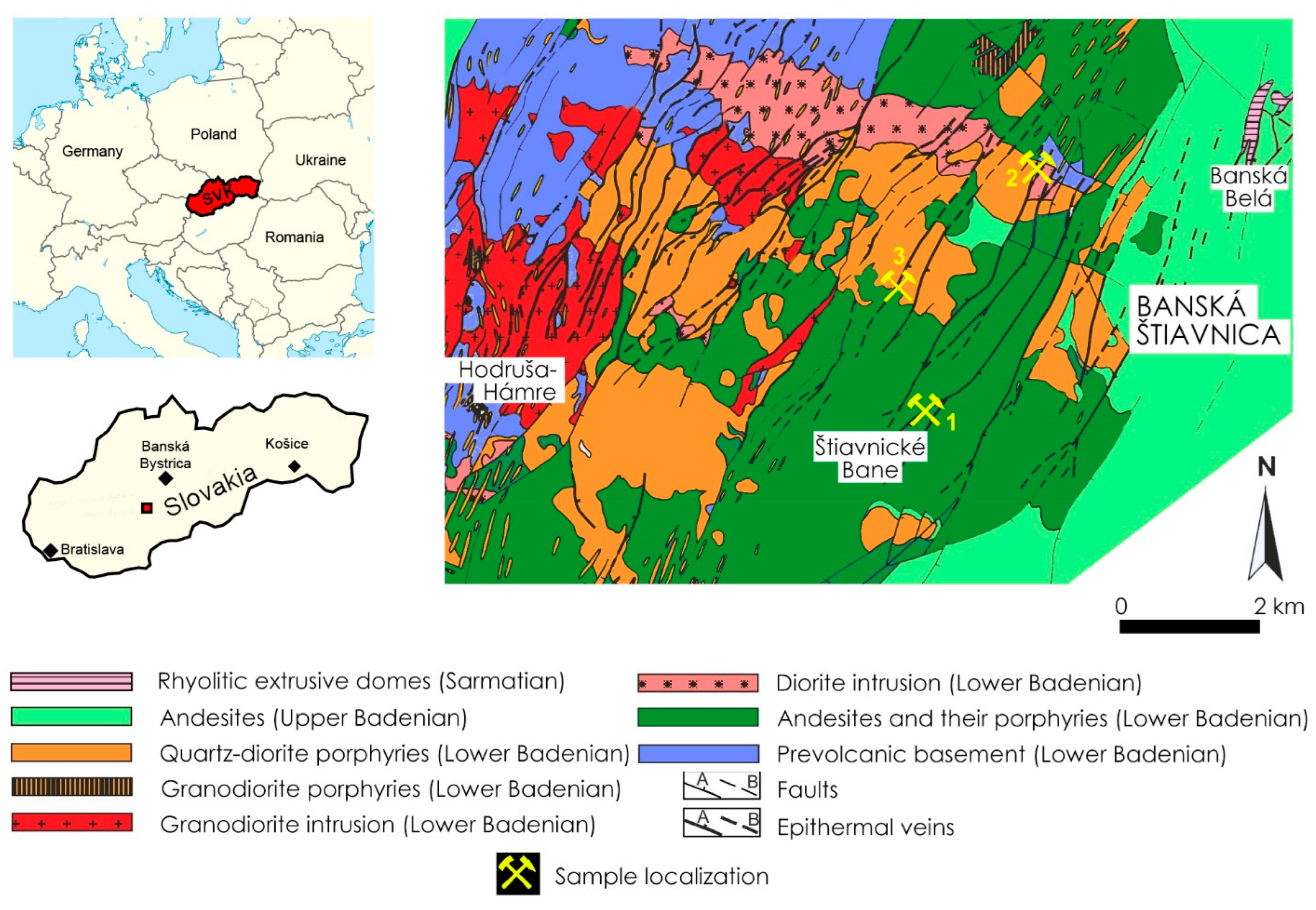
26]. During history, more sphalerite occurrences were found in the Terézia shaft (
Figure 1) [
27]. The green and brown, translucent to opaque, dodecahedral to octahedral crystals came from the Pacher adit; and the dark brown to black tetrahedral crystals were from the Michal mine, the Terézia mine, the Brenner adit, and the Pacher adit, which were associated with galena, pyrite, chalcopyrite, and quartz. Some tetrahedral to octahedral sphalerite crystals are spinel-law twins. Rounded crystals, yellow to yellow-brown, up to 2.5 cm, were discovered in 1989–1990 between the fifth and ninth Nová shaft levels. Large crystals were also located in the Amália vein. Green to dark green sphalerites were found in Štiavnické Bane and the Göllner adit, Terézia vein. The largest crystals were 4 cm in size. Attractive black glossy sphalerite druses associated with galena and chalcopyrite occurred in the Amália vein and on the 14th and the 18th level of the Rozália mine, Hodruša-Hámre, where they were discovered in 1995 and 2000 [
26,
28].
3. Geological Setting
The Banská Štiavnica ore deposits are centered on the largest stratovolcano in the Central Slovakia Neogene Volcanic Field, in the Inner Western Carpathians, with an area of over 2000 km
2 (
Figure 1). This stratovolcano began to form about 15 million years ago (15 Ma); the complex formation process was marked by intervals of igneous extrusions, subsidence, and weathering, with frequent subsurface intrusions by igneous stocks and the formation of epithermal veins. The volcano development was accompanied by metallogenetic processes that produced precious metals and polymetallic mineralization. This mineralization conditioned the development of mining cities (Banská Hodruša, Banská Štiavnica) and consolidated the mining tradition [
29].
The development of the Štiavnica stratovolcano can be divided into five stages. Stage 1: The lower stratovolcanic structure development (Early to mid-Badenian) during the explosive–effusive to intrusive activity was formed by an extensive andesite stratovolcano, which forms the lower stratovolcanic structure. Stage 2: The depression development with marsh sedimentation in the top part of a stratovolcano. The depression development was formed due to the caldera subsidization, which was not accompanied by more severe volcanism manifestations. Stage 3: The Štiavnica caldera formation was conditioned by continued subsidization of the stratovolcano top part where the explosive–effusive and extrusive biotite-amphibolic andesites to the andesites–dacites activity of the Studená formation took place. Stage 4: The stratovolcano superstructure development was renewed by several eruptive activities in Sarmatian. Stage 5: Rhyolitic volcanism and Hodruša–Štiavnica horst development (Late Sarmatian to Pannonian). At this stage, rhyolite volcanism is evident, which has allowed the rhyolite magma to reach the surface by extensive fault systems [
29].
Significant precious metal and polymetallic ores bodies and several mineralizations were created in the Štiavnica extensive stratovolcano. Sphalerite can be found at various stages of stratovolcano mineralization. Mineralization was developed in three stages: 1. The pre-ore-bearing stage formed in granodiorite with pyrite, epidote, albite, quartz, and carbonate; 2. The ore-bearing stage with dominant sphalerite and galenite (minerals dominate in this type of stratovolcano mineralization), a small amount of pyrite, chalcopyrite, linked to quartz-epidote veins; 3. The ore-bearing stage with chalcopyrite, galenite, sphalerite, accessory tetrahedrite, gold, hematite, chalcosine, and bornite. High-temperature epithermal Au mineralization occurred in the pre-caldera stage of stratovolcano development, probably in close proximity to granodiorite intrusion. The sphalerite occurrence in this mineralization can be attributed mostly to the banded veins with the mineral association: quartz, orthoclase var. adular, rhodonite, rhodochrosite, Mn-calcite, pyrite, galenite, chalcopyrite, and gold. It may also occur in the Rozália vein and other epithermal veins in the mineral association: quartz, Fe/Ca carbonates, pyrite, galenite, chalcopyrite, tetrahedrite, polybase, hessite, and gold. Polymetallic epithermal vein mineralization of the Štiavnica type has a large vertical extent, uniform periods of silicificated and sericitized zones development with a dominant polymetallic character with major minerals such as chalcopyrite, galenite, sphalerite, Ag sulfosalts, and gold in quartz-carbonate veins. Sporadic sphalerite occurrence with chalcopyrite, galenite, and marcasite can be identified in the precious metal mineralization, which is similar to the Carlin-type mineralization [
30,
31].
4. Analytical Methods
The chemical composition of sphalerite was measured using a Cameca SX100 electron microprobe (Cameca company, part of Ametek group, Gennevilliers Cedex, France) operated in wavelength-dispersion mode at the Masaryk University, Brno, Czech Republic, under the following conditions: accelerating voltage 15 kV, beam current 20 nA, and beam diameter 5 m. The samples were analyzed with the following standards: ZnS (Zn Kα), FeS2 (Fe Kα, S Kα), metallic Mn (Mn Kα), metallic Co (Co Kα), pararammelsbergite (Ni Kα, As Lβ), metallic Cu (Cu Kα), PbSe (Pb Mβ, Se Lβ), metallic Cd (Cd Lβ), InAs (In Lα), metallic Ag (Ag Lα), metallic Bi (Bi Mβ), and metallic Ge (Ge Lα). Detection limits of the measured elements varied between 150–400 ppm except for Ge (600–620 ppm), Cd (790–840 ppm), Ag (1150–1250 ppm), Zn (0.26–0.27 ppm), and Bi (2000–2200 ppm). From the measured elements, only Zn, S, Fe, and Cd were above their respective detection limits; Ag, As, Bi, Co, Cu, Ge, In, and Mn were always below the detection limit. Analytical times were 15 to 40 s during measurement, depending on the expected concentration of the element in the mineral phase. Major elements were measured using shorter times, whereas longer times were applied for elements with a low concentration. The sphalerite chemical formula was calculated based on 2 atoms.
Instrumentation for Laser Ablation Inductively Coupled Plasma Mass Spectrometry (LA-ICP-MS) at the Department of Chemistry, Masaryk University, Brno, consists of a UP 213 laser ablation system (NewWave Research, Inc., Fremont, CA, USA) and Agilent 7500 CE ICP-MS spectrometer (Agilent Technologies, Santa Clara, CA, USA). A commercial Q-switched Nd-YAG laser ablation device worked at the 5th harmonic frequency corresponding to 213 nm wavelength. The ablation device was equipped with a programmable XYZ-stage to move the sample along a programmed trajectory during ablation. Target visual inspection and photographic documentation were achieved by the built-in microscope/CCD-camera system. A sample was enclosed in the SuperCell and was ablated by the laser beam, which was focused onto the sample surface through a quartz window. The ablation cell was flushed with a helium carrier gas which transported the laser-induced aerosol to the inductively coupled plasma (1 Lmin−1). Sample argon gas flow was admixed with the helium carrier gas flow after the laser ablation cell to 1.6 Lmin−1 total gas flow. NIST SRM 610 silicate glass reference material was used to optimize the gas flow rates, the sampling depth, and MS electrostatic lens voltage in LA-ICP-MS conditions. This provided a maximum signal-to-noise ratio and minimum oxide formation (ThO+/Th+ count ratio 0.2%, U+/Th+ counts ratio 1.1%). Laser ablation required a 100 m laser spot diameter, 8 Jcm−2 laser fluence, and a 20 Hz repetition rate. The fixed sample position during laser ablation enabled 60 s hole-drilling duration for each spot. From the measured elements, Li, B, Mg, Al, Si, P, K, Ca, Cr, Ni, Rb, Sr, Y, Sn, Ba, La, Ce, Pr, Nd, Sm, Eu, Gd, Tb, Dy, Ho, Er, Tm, Yb, Lu, Hf, Hg, Th, and U were always below their detection limits.
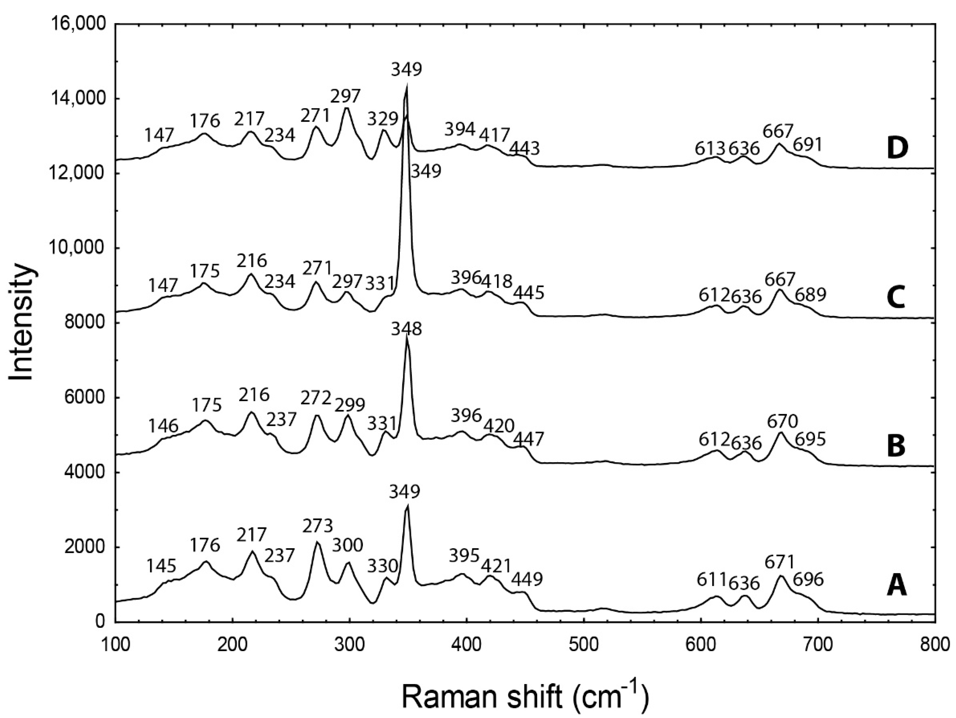
Raman analyses were performed on the LabRAM-HR Evolution (Horiba, Jobin-Yvon, Kyoto, Japan) spectrometer system with a Peltier-cooled CCD detector and Olympus BX-41 microscope (Department of Geological Sciences, Masaryk University). Raman spectra excited by blue diode laser (473 nm) in the range of 50–4000 cm−1 were collected from each stone using 50× objectives. The acquisition time of 10 s per frame and 2 accumulations were used to improve the signal-to-noise ratio.
Optical absorption spectra of cut stones in the region (500–1000 nm) were measured with the GL Gem SpectrometerTM (Gemlab Group, Vancouver, Canada) with GL Halogen light source (10 W) at room temperature at the Department of Mineralogy, Petrology, and Economic Geology, Comenius University in Bratislava.
Both Raman and UV/Vis/NIR spectra of all samples were processed in Seasolve PeakFit 4.1.12 software. Raman and absorption bands were fitted to obtain exact band positions and deconvolute overlapping bands by the Lorentz function with variable width, automatic background correction, and Savitsky–Golay smoothing. Band fitting was gradually refined until it produced reproducible results with a square regression coefficient greater than 0.995.
6. Discussion
The sphalerite crystal structure can accommodate a large variety of elements; the most significant of them are Fe and Cd. The Cd concentration can attain sufficient values for sphalerite being used as Cd; e.g., giant Zn–Pb deposits such as Red Dog (Alaska) are economically exploited for this element [
1]. Trace elements in sphalerite can use simple substitution of similar-sized ions with the same charge (Zn
2+ ↔ Fe
2+, Cd
2+, Mn
2+, Co
2+ or S
2− ↔ Se
2−) or coupled substitution of ions with different charges (e.g., 2 Zn
2+ ↔ Cu
+ + In
3+) [
1,
15]. Substitution mechanisms for other elements commonly found in sphalerite (e.g., Sn, Ag, Ga, and Ge) remain less well constrained, and it is not clear for elements such as Pb, Tl, As, Sb, and Bi whether they might enter the sphalerite structure or are almost always present as mineral inclusions [
15]. The limits for entering the sphalerite structure can be derived from the formal charge of cations (e.g., four-valent cations) and ionic radii (e.g., Fe
3+ has a too-small ionic radius of 0.49 Å [
33], Ag
+ a too-large radius with 1 Å [
33]). However, electronegativity has to be also considered; Zn has 1.65, Cd 1.69, Fe 1.83, but Ag 1.93 and Cu 1.90. Therefore, although elements possibly substituting for Zn can be abundant in the environment, they can prefer different minerals.
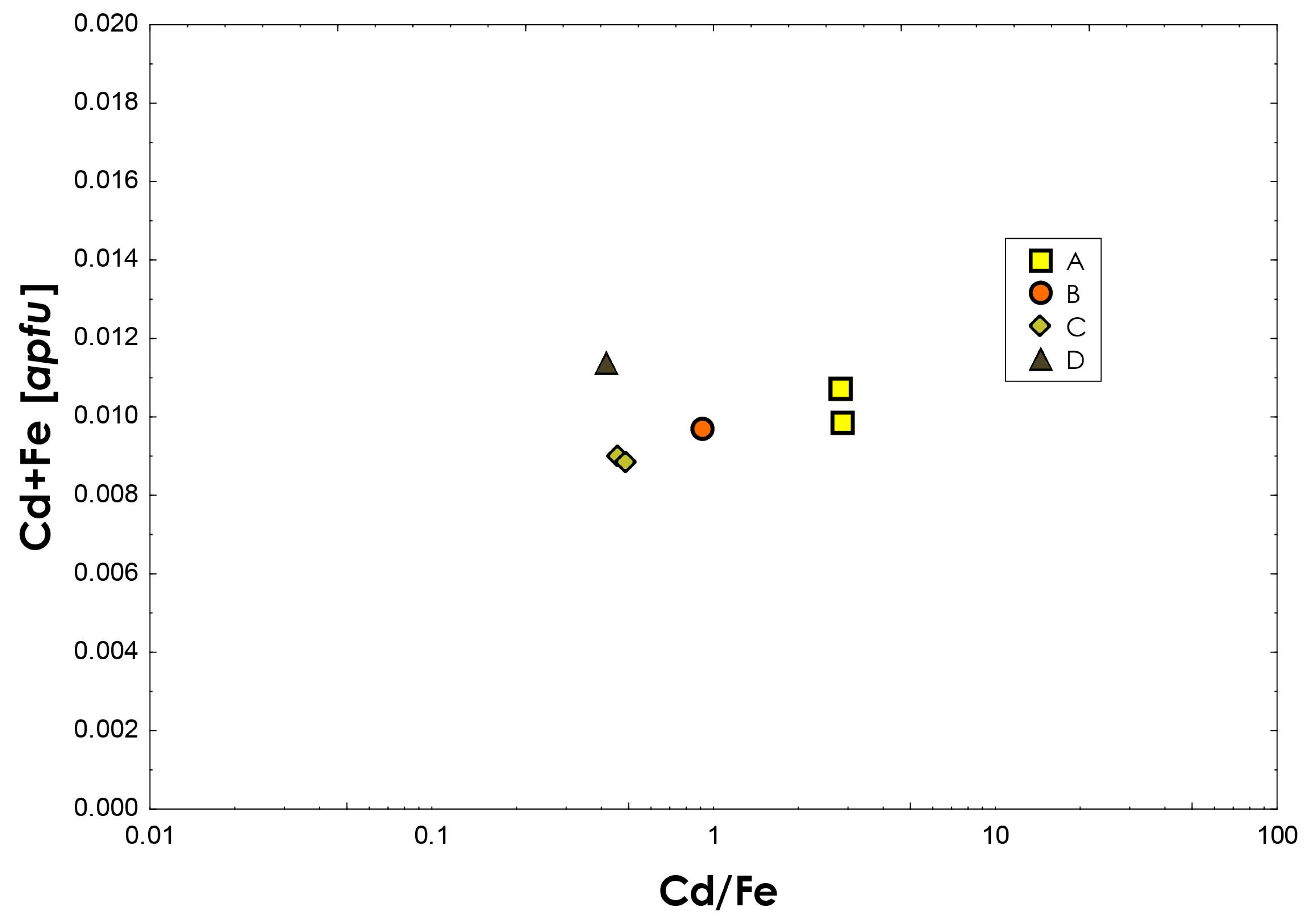
The LA-ICP-MS analysis revealed that studied samples of sphalerite from Banská Štiavnica and Hodruša-Hámre are relatively poor in trace elements except for Fe, Cd, and partly also Mn. However, Mn content is lower by two to three orders compared to Fe and Cd. Other elements (In, Sb, Co, Cu, Ga, Ag, Pb) are scarce.
The Fe content in studied sphalerite is relatively low compared to other world localities. It is significantly lower than in most of the skarn and stratabound deposits, but it shares similarities with some sphalerite from Neogene epithermal mineralization in Romania [
1]. However, Romanian sphalerite is significantly richer in Cu and Mn [
1] than the studied samples. The Fe proportion in studied samples can be correlated with the position of sphalerite within the mineralization and its spatial position. Studied sample A is from the shallower, almost surface part of the Terézia vein, and it has lower Fe than sample B from a deeper part of the same vein—the 12th horizon of the Michal shaft. The D sample from Hodruša-Hámre with the highest Fe content is also from the deeper parts of the Rozália mine.
Sphalerite from Slovakia can be considered relatively rich in cadmium. Sphalerites from Neogene epithermal mineralization in Romania and also from stratabound deposits contain 1000–7700 ppm [
1]. Sphalerites from skarns are typically uniform within the 2500–10,800 ppm range except with the exception of the Baisoara skarn deposit (Romania), where cadmium concentration shows up to 73,800 ppm [
1]. Measured concentrations of Cd in gem-quality sphalerite from Slovakia range from 5917 and 10,130 ppm. Cadmium is typically higher in lower-temperature deposits [
15,
16], which explains its opposing relationship with Fe in the studied samples.
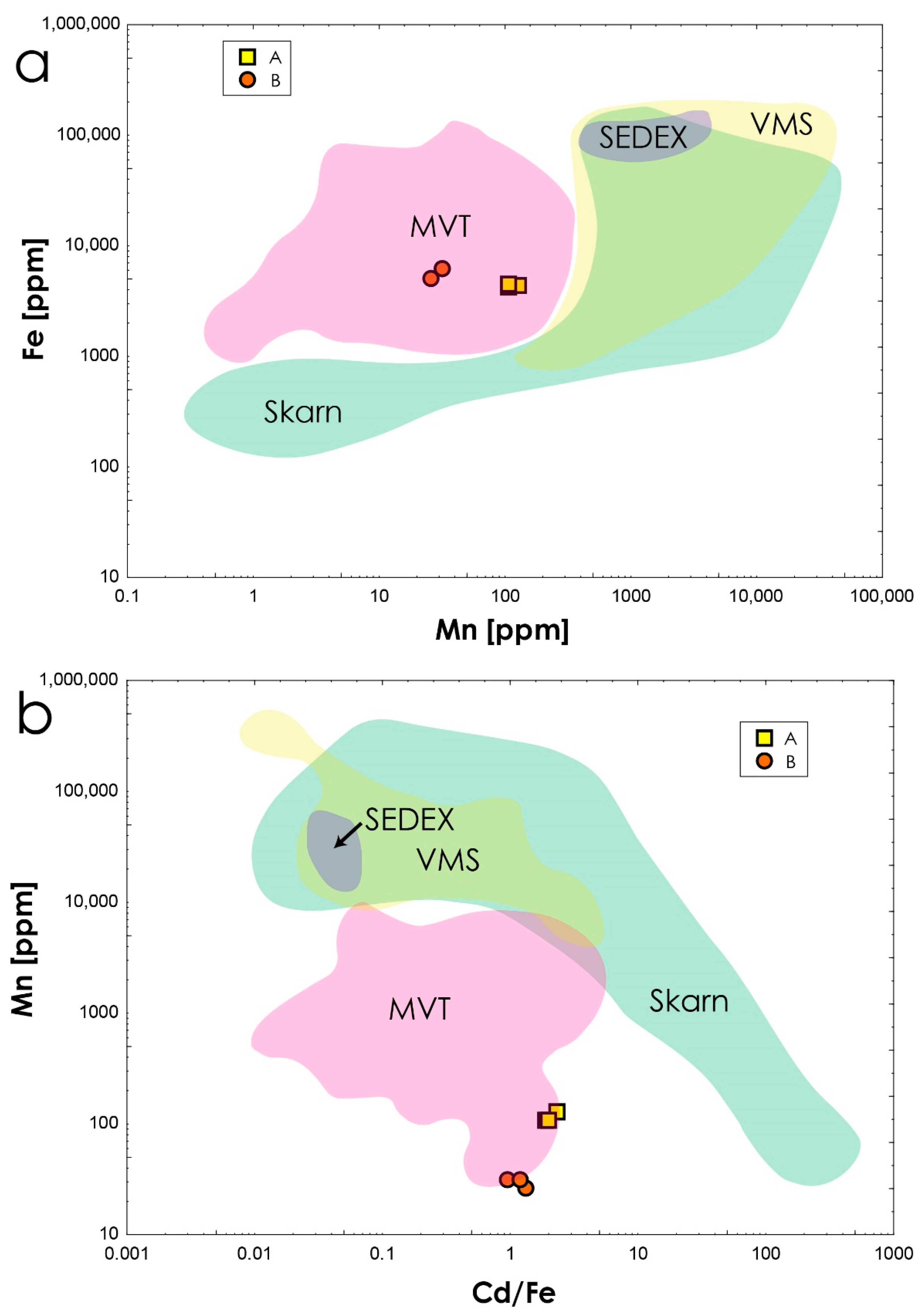
Based on the Mn vs. Fe (
Figure 6a) and Cd/Fe vs. Mn (
Figure 6b) ratios, studied sphalerites fall into the field of Mississippi Valley-Type (MVT) deposits [
17]. However, although MVT sphalerite is characterized by low Fe, Mn, In, and Co, and high Ge, Cd, and Ga contents [
1,
15,
17], studied samples are low in Ge and Ga. In contrast, sphalerite from most magmatic-related deposits is characterized by high Fe, In, Mn, and Co, but low Ge and Cd contents [
1,
15], so it also cannot be used to discriminate the genetical origin of samples and its influence on their chemical composition. The Zn/Cd ratio could also discriminate sphalerite from various genetic types of deposits. Volcano-sedimentary and Alpine (MVT)-type deposits have the highest Zn/Cd ratios of 417 to 531 [
34]. However, studied sphalerite samples have Zn/Cd ratios of 97–166, which is significantly lower. In contrast, it is typical for hydrothermal deposits, including vein magmatic-related and skarn deposits, with the Zn/Cd ratios between 104 and 214 [
34]. Consequently, we can assume that the most important factor for the generally low trace-element content, except for Cd, is the low temperature of source fluids in the hydrothermal veins. This low trace-element composition resulted in the exquisite optical properties of studied sphalerite.
Sphalerite can have quite a large variety of colors. A dark black color is commonly explained by high Fe content (>6 wt.%) [
1]. However, when iron concentration is low, other elements can also be highly influential on color. According to [
35], the yellow colors of sphalerite may be caused by Ge, Cu, and Hg; reddish by Ce, Sn, and Ag; and green colors by Co and Fe. Furthermore, gem-quality green sphalerite from Congo has Co content in the range of 20 to 820 ppm and Cu [
36].
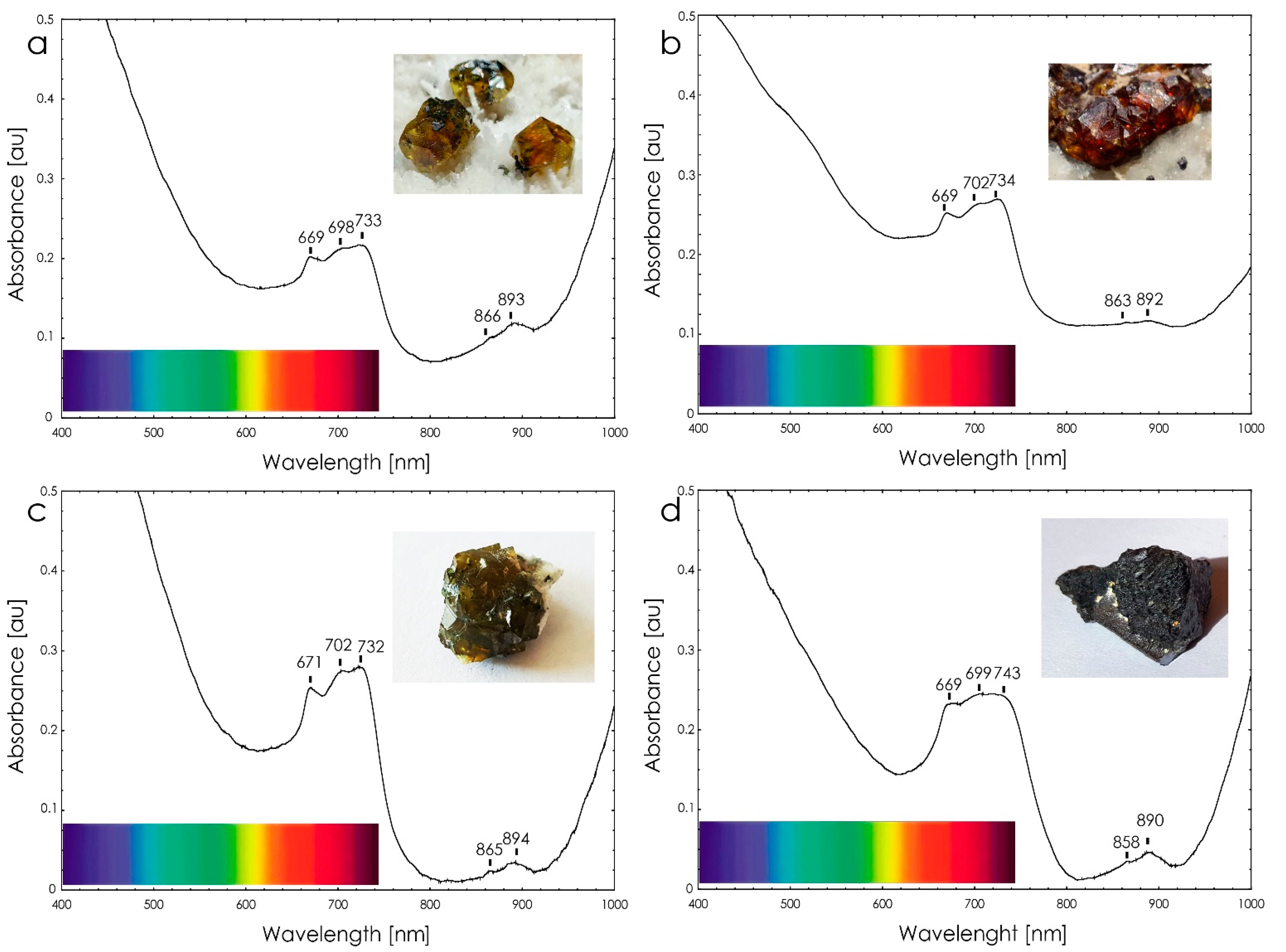
Iron and cobalt are primary elements creating absorption bands and giving rise to sphalerite’s orange, red, yellow, and green colors [
25]. Optical absorption spectra of tetrahedral Fe
2+ are reported by [
18,
19,
20]. As another principal agent of sphalerite coloring, Co
2+ in the tetrahedral Zn sites can be considered, which, even in small amounts (80 ppm), can cause the green color of sphalerite [
14]. However, studied sphalerites from Slovakia have almost negligible amounts of Co (5–17 ppm). On the other hand, Cd instead of Co has more significant concentrations, even more than Fe in some samples. However, Cd with an electronic configuration of [Kr]4
d10, similarly to Zn, has fully occupied
d orbitals; therefore, no electron transitions can be expected in the visual region of the spectrum. Consequently, the bands between 669 and 743 nm can be very likely assigned to crystal-field bands of Fe
2+ exclusively.
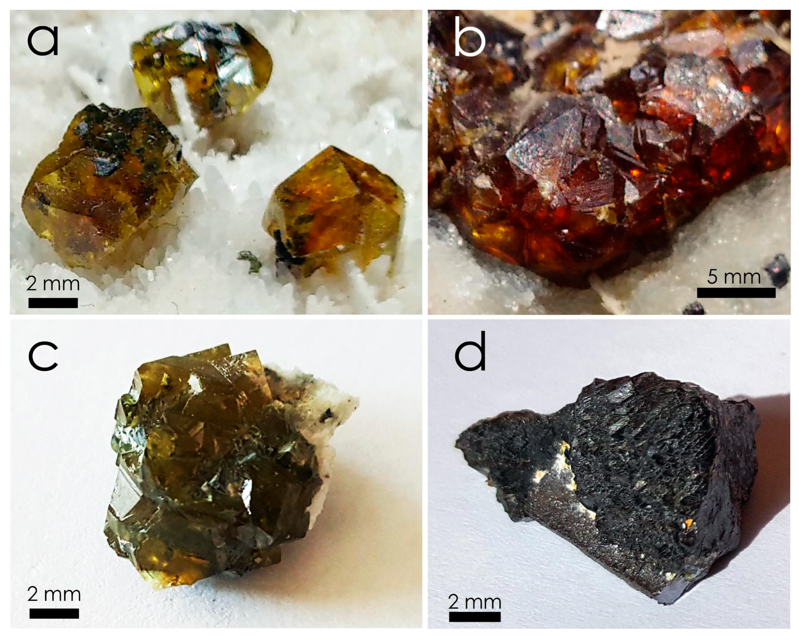
The black color of the sphalerite D sample is a matter of interest because of the similarity in absorption spectra with olive-green colored sphalerite (
Figure 3). After disassembling the black sphalerite cluster into individual crystals, it turned out that the color of these crystals is very similar to olive green (
Figure 3), which would explain the similarity of the optical spectrum of black and olive-green sphalerite. The black coloring could be caused by slightly higher concentrations of Fe (0.46 wt.%, 0.008
apfu,
Table 1), the significantly smaller size of individual crystals in the aggregate reducing macroscopic transparency, and/or the presence of submicroscopic inclusions, which, however, were not observed even in the back-scattered electron images of the studied sample.


 ,
, 


